EFFECT OF NEUROPROTECTIVE PROTOCOLS ON NGF LEVELS AND OXIDATIVE STATE IN
Somayeh Hosseinzadeh
1
Babol University
2College of Physical Education and Sport Sciences,
ARTICLE INFO ABSTRACT
The pro
brain that is related to the decline of the antioxidant defense systems. The present study investigated the protective effect of regular exercise and/or Curcumin (an active ingredient of turmeric) lead-induced neurotoxicity in rat
following groups: lead acetate, curcumin, endurance training, training + curcumin, sham groups. The exercise training protocol consisted of running on a tre
The rats in sham group received curcumin solvent (ethyl oleat) while the others received lead acetate (20 mg/kg). Moreover, the curcumin and training + curcumin groups received curcumin solution (30 mg/kg), in
and a decrease in nerve growth factor (NGF) levels in cerebral cortex and plasma TAC as compared with control. Whereas, regular exercise and/ or supplementary Curcumin
Interestingly, co
concomitant increase in NGF and TAC levels, as compared with those treated with Curcumin or lead alone. The results suggest a lifestyle
neurotoxicity.
INTRODUCTION
Environmental lead toxicity is an old but persistent public health problem throughout the world, and its toxicity has been known from ancient times and many studies have explored the mechanisms and symptoms of this toxicity through the years
(Ahamed et al., 2007a). Lead has well-characterized effects on
every organ system, including the cardiovascular, renal, immune and reproductive systems, but the nervous system is especially sensitive to the effects of Lead (White
Some of these effects include impairments in the learning process, memory consolidation, attention, visual perception, manual dexterity, response speed, nerve demyelination, encephalopathy, hyperactivity, motor skills and exploratory
behavior (Trombini et al., 2001). Acute and chr
exposure has been caused various behavioral and
neurotoxicological disorders (Ahamed et al.,
Trombini et al., 2001). Recent studies have reported lead’s potential for inducing oxidative stress and evidence is accumulating in support of the role for oxidative stress in pathophysiology of lead poisoning (Gurer and Erkal, 2000). Oxidative stress due to the loss of balance between reactive oxygen species (ROS) and antioxidant defenses affects all the vital organs, resulting in aging and the
*Corresponding author: [email protected]
ISSN: 0975-833X
Article History:
Received 28th December, 2011
Received in revised form
28th January, 2012
Accepted 15th February, 2012
Published online 31st March, 2012
Key words: Oxidative stress, NGF, Lead acetate, Cortex.
RESEARCH ARTICLE
EFFECT OF NEUROPROTECTIVE PROTOCOLS ON NGF LEVELS AND OXIDATIVE STATE IN
RAT CORTEX
Somayeh Hosseinzadeh
1and Valiollah Dabidi Roshan
Babol University of Medical Sciences, Babol, Iran
College of Physical Education and Sport Sciences, Department of Sport Physiology, University of Mazandaran,
Babolsar, Iran
ABSTRACT
The pro-oxidant effects of air pollutants can exacerbate the age-related increase in oxidative stress in brain that is related to the decline of the antioxidant defense systems. The present study investigated the protective effect of regular exercise and/or Curcumin (an active ingredient of turmeric)
induced neurotoxicity in rat cortex. Forty male Wistar rats were randomly divided into following groups: lead acetate, curcumin, endurance training, training + curcumin, sham groups. The exercise training protocol consisted of running on a treadmill at a progressive intensity for 8 weeks. The rats in sham group received curcumin solvent (ethyl oleat) while the others received lead acetate (20 mg/kg). Moreover, the curcumin and training + curcumin groups received curcumin solution (30 mg/kg), intraperitoneally. Exposure to lead acetate caused an increase in lipid peroxidation (MDA) and a decrease in nerve growth factor (NGF) levels in cerebral cortex and plasma TAC as compared with control. Whereas, regular exercise and/ or supplementary Curcumin
Interestingly, co-treatment with Curcumin and exercise caused a significant decrease in MDA with concomitant increase in NGF and TAC levels, as compared with those treated with Curcumin or lead alone. The results suggest a lifestyle-induced neuroprotective potential in ameliorating lead neurotoxicity.
Copy Right, IJCR, 2012, Academic Journals
Environmental lead toxicity is an old but persistent public health problem throughout the world, and its toxicity has been known from ancient times and many studies have explored the mechanisms and symptoms of this toxicity through the years characterized effects on every organ system, including the cardiovascular, renal, immune and reproductive systems, but the nervous system is especially sensitive to the effects of Lead (White et al., 2007). lude impairments in the learning process, memory consolidation, attention, visual perception, manual dexterity, response speed, nerve demyelination, encephalopathy, hyperactivity, motor skills and exploratory 2001). Acute and chronic lead
exposure has been caused various behavioral and
et al., 2007b and Recent studies have reported lead’s potential for inducing oxidative stress and evidence is the role for oxidative stress in pathophysiology of lead poisoning (Gurer and Erkal, 2000). Oxidative stress due to the loss of balance between reactive
oxygen species (ROS) and antioxidant defenses affects the process can be
accelerated by the damage of cellular ingredients such as proteins, lipids, and DNA as well as apoptosis (Oiae and Park, 2008). Chronic stress is known to lead to atrophy and functional impairments in several key brain areas, including the cerebral cortex and has been linked to the development of major depression. Neurodegeneration occurs with chronic stress; coupled to these changes is enhanced activation of apoptosis and decreased neuronal survival (Haack et al, 2008). These facts present a novel approach to strategies for treating environmental lead toxicity.
Physical activity and Nutritional factors are often mentioned as important modifiers of the metabolism and toxicity of lead. Studies have shown that Physical activity and moderate exercise training enhance learning and brain plasticity both in young individuals and particularly in ageing people. These effects are thought to be mediated, at least in part, through actions on growth factors (Oliff et al, 1998).
growth factors involved in the regulation of the survival, differentiation, and maintenance of neuronal functions in the brain. Amongst them, brain
(BDNF) and nerve growth factor (NGF) are known to mediate higher-order activities such as learning, memory and behavior (Oliff et al, 1998). Studies have shown NG
neurotrophic factor, particularly for the protection and neuroplasticity of cholinergic neurons that project on the
International Journal of Current Research
Vol. 4, Issue, 03, pp.019-023, March,2012
INTERNATIONAL
EFFECT OF NEUROPROTECTIVE PROTOCOLS ON NGF LEVELS AND OXIDATIVE STATE IN
Valiollah Dabidi Roshan
2,*, University of Mazandaran,
related increase in oxidative stress in brain that is related to the decline of the antioxidant defense systems. The present study investigated the protective effect of regular exercise and/or Curcumin (an active ingredient of turmeric) against . Forty male Wistar rats were randomly divided into following groups: lead acetate, curcumin, endurance training, training + curcumin, sham groups. The admill at a progressive intensity for 8 weeks. The rats in sham group received curcumin solvent (ethyl oleat) while the others received lead acetate (20 mg/kg). Moreover, the curcumin and training + curcumin groups received curcumin solution (30 traperitoneally. Exposure to lead acetate caused an increase in lipid peroxidation (MDA) and a decrease in nerve growth factor (NGF) levels in cerebral cortex and plasma TAC as compared with control. Whereas, regular exercise and/ or supplementary Curcumin reversed this process. treatment with Curcumin and exercise caused a significant decrease in MDA with concomitant increase in NGF and TAC levels, as compared with those treated with Curcumin or lead induced neuroprotective potential in ameliorating lead-induced
, Academic Journals. All rights reserved.
accelerated by the damage of cellular ingredients such as proteins, lipids, and DNA as well as apoptosis (Oiae and Park, 2008). Chronic stress is known to lead to atrophy and functional impairments in several key brain areas, including the cerebral cortex and has been linked to the development of . Neurodegeneration occurs with chronic stress; coupled to these changes is enhanced activation of apoptosis and decreased neuronal survival (Haack et al, 2008). These facts present a novel approach to strategies for treating
Physical activity and Nutritional factors are often mentioned as important modifiers of the metabolism and toxicity of lead. Studies have shown that Physical activity and moderate exercise training enhance learning and brain plasticity both in duals and particularly in ageing people. These effects are thought to be mediated, at least in part, through
actions on growth factors (Oliff et al, 1998).Neurotrophins are
growth factors involved in the regulation of the survival, intenance of neuronal functions in the brain. Amongst them, brain-derived neurotrophic factor (BDNF) and nerve growth factor (NGF) are known to mediate order activities such as learning, memory and behavior (Oliff et al, 1998). Studies have shown NGF to be a potent neurotrophic factor, particularly for the protection and neuroplasticity of cholinergic neurons that project on the
hippocampal and cerebral cortical regions associated with
cognitive performance (Xiong et al., 2010). Studies may also
indicate that an induction in the level of NGF is not sufficient to prevent age-dependent atrophy of cholinergic neurons although it may be necessary for the stimulation of a compensatory functional change in lifestyle methods where the system is undergoing progressive degeneration (Connor and Dragunow, 1998). It has been suggested that nutrition is one of the many factors that can affect brain and cognitive development, in part, by regulating the synthesis and secretion of the neurotrophins. However, it remains unclear whether supplementation of a particular nutrient in the diet can affect production of these neurotrophic factors. There has been no report on the effect of Curcumin supplementation on neurotrophin levels (Oliff et al., 1998).
Despite the knowledge that lead can induce oxidative stress, the usefulness of antioxidants alone or in conjunction with exercise has not been thoroughly investigated. Therefore, in the present study, we attempted to: (i) assess the effect of lead acetate administration on levels of cortex NGF in rats; and (ii) understand whether Curcumin supplementation and exercise regular training can affect synthesis of the brain cortex NGF and balancing oxidative/antioxidative ratio in chronically rats exposed to lead acetate.
MATERIALS AND METHODS
The experimental protocol of this study was approved by Department of Physiology, University of Mazandaran and was performed according to guiding procedures in the Care and Use of Animals, prepared by the Council of the American Physiological Society. Forty male Wistar rats (n=8 each group), 8 weeks age (initial body weight of 240 ± 20 g), were obtained from the Laboratory of Animal Bearing and Multiplying at the Pasture Institute of Iran. Each rat was housed in single standard cages of polycarbonate (20×15×15), made in Pasture Institute of Iran, in a large air-conditioned room with controlled temperature of 22±2 °C, a light- dark cycles 12:12 hours and humidity of % 50±5. According to the information from the pollution determination station of Iranian Meteorological Organization, air pollutants with consideration of pollutant standard index (PSI) were in normal range. Rats were fed with a standard rat chow provided by Pars Institute for animal and poultry factory with a daily regimen of 10/100 gr body weight for each rat. Also water was available ad libitum.
Rats were familiarized with laboratory environment and running on treadmill and then were randomly assigned into five experimental groups of 8 rats. Group1 – lead acetate (Pb) was exposed to lead at a concentration of 25 mg/kg in the form of water solution of lead acetate (for intra peritoneal injection), 3 days weekly for 8 wks; Group 2 – Curcumin (Cum) received Curcumin 30 mg/kg 5 days weekly for 8 wks (i.p.); Group 3 – endurance training (Pb + training), the rats in this group similarly received lead acetate moreover they performed the progressive running exercise of 15 to 22 m/min for 25 to 64 min, 5 times a week; Group 4 – training and Curcumin (Pb + training + Cum); the rats of this group performed physical training protocol as mentioned in above
furthermore, received lead acetate and Curcumin
supplementation as well; Group 5 – the sham-operate or
control group (sham); these rats received water and ethyl oleate, in the same manner and for the same duration of time. Lead acetate (Sigma) was solubilised in Milli-Q water, and Curcumin was solubilised in 50% ethanol. In order to intra peritoneal (i.p) injections, Curcumin was solubilised in ethyl oleate and was injected at a dose of 30 mg/kg. Curcumin was protected from light during all the time of the experiment (Sheril et al., 2004).
All groups were anesthetized with ketamine and Xaylozine and decapitated after 12-14 hours overnight fasting. Moreover, blood samples were collected 24 h after the last dose of treatment from the heart of all the groups. These blood samples were first centrifuged by a refrigerated centrifuge at 3,000 rpm for 15 minutes within 30 minutes of collection and then stored at -80°C before assay and serum was separated for biochemical estimations of TAC (Total Antioxidant Capacity) and MDA. Then, Brains were rapidly removed and the two hemispheres separated along the midline. The cortex from each hemisphere of the brain was then microdissected and
frozen in liquid nitrogen and subsequently stored at -80 ◦C for
future analysis. For protein extraction, cortex tissue was homogenized in a lysis buffer containing 137 mM NaCl, 20 mM Tris-HCl (pH 8.0), 1% NP40 (polyoxyethylene-(9)- octylphenyl ether, 161-19911; Wako Pure Chemicals, Japan), 10% glycerol, 10 μg/ml aprotinin (Sigma), 1 μg/ml leupeptin (Sigma), 0.5 mMsodium orthovanadate (Wako), and then centrifuged at 10,000 rpm at 4°C for 20 min. The supernatants were removed and after dilution with Dulbecco’s phosphate buffer saline (DPBS), aliquots were further processed at room temperature by acidification (pH < 3) for 15 min, followed by neutralization (pH 7–8) and then the NGF protein level was determined by NGF ELISA kit according to manufacturer’s recommendations and expressed as picograms of NGF per
milligram of the tissue (Kheirvari et al., 2008).Moreover, the
concentration of lead was detected by means of atomic absorption spectrophotometry (AAS) method.
Lipid peroxidation levels in the tissue homogenate were measured with the thiobarbituric-acid reaction by the method
of Samples homogenates (1 ml) were incubated at 37°C in an
oscillating water bath for one hour (Ohkawa et al.,1979). At the end of the incubation period, 0.5 ml of BHT (0.5 mg/ml in absolute ethanol) and 1 ml of TCA (25%) were added. The tubes were sealed and heated for 10 minutes in a boiling water bath to release MDA (the end product of lipid peroxidation)
from proteins. To avoid adsorption of MDA
(Malondialdehyde), to insoluble proteins, the samples were cooled to 4°C and centrifuged at 2000 x g for 20 minutes. Following centrifugation, 2 ml of the protein free supernatant was removed from each tube and 0.5 ml of TBA (butylhldroxy-toluene) (0.33%) was added to this fraction. All tubes were heated for one hour at 95°C in a water bath. After cooling, the TBA-MDA complexes were extracted with 2 ml of butanol. The light absorbance was read at 532 nm on a spectrophotometer and MDA levels were determined from
standard curve that was generated from 1,1,1,3
Tetramethoxypropan. The results are represented as n mol/mg
tissue. Serum TAC was measured using a commercially
available kit (Randox Laboratories, Crumlin, UK) as
previously described by Erel (2004).In this method, the most
sequentially produced radicals such as the brown colored dianisidinyl radical cations, produced by the hydroxyl radical, are potent radicals. Then, the antioxidative effect of the sample against the potent free radical reactions is measured. The assay has excellent precision values, which are lower than 3%. The results are expressed in μmol/ml.
Statistical analysis
Statistical analysis was performed using a commercial software package (SPSS version 16.0 for Windows). Results are expressed as means ± SE. Data for NGF and oxidant/antioxidant markers were normally distributed after log transformation. A one-way ANOVA was used to detect statistical difference between groups. Furthermore, a Post-Hoc test (Tukey test) was performed to establish changes differences in the mentioned markers between groups. The differences were considered significant at p<0.05.
RESULTS
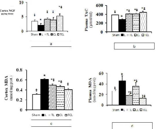
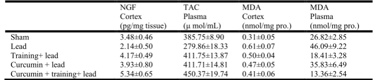
The administration of lead at the concentration of 20 mg/kg for the period of 8 weeks resulted in changes in NGF levels in cortex, plasma TAC as beneficial biomarkers and MDA in cortex and plasma as a harmful biomarker in lead-induced rats in each group (table). Exposure to lead (20 mg/kg) resulted in a decrease in the TAC and NGF levels by about 27/45% and 38/50%, respectively in comparison to the sham group. While, Curcumin and/or exercise training significantly increased the TAC (47.11%, 47.13%) and NGF (83.64%, 94.86%) levels of lead-induced reduced. However, Curcumin+ training+ lead treatment was more effective than Curcumin+ lead and training+ lead alone treatment (figure 1( a,b)). Lead acetate also, caused an increase in the concentration of cortex and plasma MDA by about 96/77% and 71.81%, respectively in comparison to the sham group. Whereas, Curcumin and/or exercise training significantly reversed the MDA both in cortex (22.95%, 18.03%) and plasma (22.24%, 60.05%). However, Curcumin+ training+ lead treatment was more effective than Curcumin+ lead and training+ lead alone treatment (figure 2(c,d)).
DISCUSSION
The present study demonstrates the protective effects of regular exercise and Curcumin supplement against lead neurotoxicity. Enhancement of MDA associated with reduced TAC and NGF may deteriorate neurodegeneration diseases result in lead toxicity via disrupting oxidant/antioxidant balance, whereas lifestyle factors such as endurance exercise and Curcumin supplement reverse this process through increasing NGF and improving anti oxidative defense systems. The production of ROS and the associated oxidative damage is greatly enhanced under the influence of various environmental and life style factors such as pollution (Agarwal and Prabakaran, 2005). Several epidemiological studies among workers with high occupational exposure to lead have reported associations between lead exposure and levels of MDA (malondialdehyde), a product of oxidative stress (Adonaylo and Oteiza. 1999). Lipid peroxidation appears to be markedly enhanced in the brain of lead-treated
rats (Antonio-Garcia and Masso-Gonzalez, 2008), which
concurs with our findings. Lead causes significant changes in oxidative stress in different brain regions, particularly in the hippocampus and cerebral cortex which are found to be more
vulnerable to Lead-induced neurotoxicity (Prasanthi et al.,
2010). The preponderance of results from the recently published studies demonstrate that exposure to quantities of lead typically present in the environment have adverse health effects (Ahamed and Siddiqui, 2007). The detection and prevention of lead toxicity have been a major international public health priority (Ahamed and Siddiqui, 2007). For several decades neurotrophic theory has guided thinking regarding the role of neurotrophic factors in the developing
and adult nervous system (Al-shawi et al., 2007).
Concurrently, neurotrophins are master regulators of cortical and subcortical cell growth, expression of neuropeptides, glutamic acid decarboxylase and parvalbumin, plasticity (Patz and Wahle, 2004) and increased resistance to oxidative stress
(Radak et al., 2006). The induction of NGF could be strongly
[image:3.595.162.432.541.763.2]dependent on the activation of cyclic AMP response element binding protein (CREB), which has been shown to be very
Fig 1. Concentration of a) Nerve Growth Factor (NGF) in cerebral cortex, b) Total Antioxidant Capacity (TAC) in plasma,
c)Malondialdehyde (MDA) in cerebral cortex and d) MDA in plasma in experimental animals.
Statistical significance p < 0.05: * significantly than sham group: ‡ significantly than lead group L: Lead /TL: Training + Lead / CL: Curcumin + Lead / TCL: Training + Curcumin + Lead groups
Cortex NGF
sensitive to redox state (Radak et al., 2006). If this neurotrophic factor fall below a certain level it is generally believed that neurons are more vulnerable to damage, or conversely, if levels are maintained or enhanced this may provide neurons with a margin of protection (Oliff et al.,
1998). A great variety of factors and situations have been shown to up regulate precursor cell proliferation. Physical activity is the quintessential type of this stimulus. Physical exercise simply leads to an increased circulation and release of growth factors, leading to the activation of neurogenesis (Fabel and Kempermann, 2008). The molecular mechanism for beneficial effects of exercise is unknown, though several hypotheses have been proposed. Moderate exercise is associated with enhanced neurogenesis, a reduction in markers of aging, including oxidative stress and enhanced trophic
influences (Oliff et al., 1998). Our experimental evidence
indicates that exercise concurrent with chronic stress prevent the increased concentration of MDA that set the stage for cellular apoptosis in the cerebral cortex during chronic stress and also promote brain survival via increasing NGF and TAC which decreased by lead acetate. These findings are coherent with previous results that speculated that for long term, regular endurance training induced an adaptive response in the
antioxidant defense system and improved the TAC (Radak et
al., 2006; Fabel and Kempermann, 2008 and Amani et al.,
2010). Also, available information from separate studies suggests that exercise has a capability to alter the level of NGF, memory and the rate of oxidative damage in brain of
animals (Radak et al., 2006). Changes in rodent behavior due
to chronic stress, such as decreased sucrose consumption, decreased motor activity and impaired learning have been prevented when animals were allowed voluntary access to a running wheel during or immediately following the period of stress (Haack et al., 2008). Also, neurochemical changes due to chronic stress, such as altered glucocorticoid sensitivity, were prevented with concurrent wheel running. Voluntary exercise, on the other hand, robustly increases growth factor and growth-associated molecule expression in the cerebral cortex and hippocampus and has recently been shown to enhance the activation of intracellular signal transduction
pathways promoting neuronal survival (Haack et al., 2008).
The learning and memory deficits associated with chronic stress may be alleviated using novel therapeutic strategies involving dietary and medicinal phyto-antioxidants. One such nutraceutical is turmeric which has been used throughout Asia as a food additive and a traditional herbal medicine. Turmeric’s pharmacologically active substance is Curcumin, the yellow pigment extracted from the rhizoma of Curcuma
longa (Xu et al., 2009). Curcumin has been discovered to
have a variety of pharmacological activities, including anti-inflammatory, antioxidant, anti-proliferative, and
neuroprotective effects (Wang et al., 2008 and Gomez-Pinilla,
2008a). Curcumin is as the strong antioxidant supplement which is decreases the lipid peroxidation and improves TAC
in the human and animal body (Amani et al., 2010). This
study demonstrated for the first time that administration of Curcumin improve oxidative balance in cerebral cortical through NGF increased in the Wistar rats. Accordingly, managed dietary manipulations and exercise have strong therapeutic potential and can contribute to the ability of the brain to counteract neurologic disorders (Gomez-Pinilla, 2008b). To summarize, our present study showed that long-term environmental factors such as exercise and/or Curcumin, increased NGF in cerebral cortex and also improved redox equilibrium against lead-induced neurotoxicity. However, when diet and exercise are combined, the success of regeneration and healing seems more pronounced than when either option is implemented by itself.
REFERENCES
Adonaylo VN, Oteiza PI.1999. Lead intoxication: antioxidant
defenses and oxidative damage in rat brain. Toxicol., 135:
77–85.
Agarwal A, Prabakaran SA. 2005. Oxidative stress and antioxidants in male infertility: a difficult balance. Iran j Reprod Med., 3(1): 1-8.
Ahamed M, Kaleem M, Siddiqui J. 2007. Environmental lead toxicity and nutritional factors. Clin Nutr., 26: 400–408. Ahamed M, Siddiqui MKJ. 2007. Low level lead exposure and
oxidative stress: Current opinions. Clin Chim Acta, 383: 57–64.
Al-shawi R, Hafner A, Chun S, Raza S, Crutcher K, Thrasivoulou CH, Simons P, Cowen T. 2007. ProNGF,
Sortilin, and Age-related Neurodegeneration. Ann. N.Y.
Acad. Sci., 1119: 208–215.
Amani AR, Somchit MN, Konting MM B, Kok LY. 2010. Vitamin E and Curcumin Intervention on
Lipid-Peroxidation and A ِ◌ntioxidant Defense System. Am J Sci.,
6(3).
Antonio-Garcia MT, Masso-Gonzalez EL. 2008. Toxic effects of perinatal lead exposure on the brain of rats: Involvement of oxidative stress and the beneficial role of antioxidants.
Food Chem Toxicol., 46: 2089–2095.
Connor B, Dragunow M. 1998. The role of neuronal growth factors in neurodegenerative disorders of the human brain.
Brain Res Rev., 27: 1–39.
Erel O. 2004. A novel automated direct measurement method for total antioxidant capacity using a new generation, more
stable ABTS radical cation. Clin Biochem., 37: 277-285.
Fabel K, Kempermann G. 2008. Physical Activity and the Regulation of Neurogenesis in the Adult and Aging Brain.
[image:4.595.111.482.86.166.2]Neuromol Med., 10: 59–66.
Table 1. Effect of exercise training and Curcumin on NGF, TAC and MDA levels in plasma and cerebral cortex in lead-induced rats (mean ± SEM for eight rats)
NGF Cortex (pg/mg tissue)
TAC Plasma (μ mol/mL)
MDA Cortex (nmol/mg pro.)
MDA Plasma (nmol/mg pro.)
Sham 3.48±0.46 385.75±8.90 0.31±0.05 26.82±2.85
Lead 2.14±0.50 279.86±18.33 0.61±0.07 46.09±9.22
Training+ lead 4.17±0.49 411.75±13.87 0.50±0.04 18.41±3.28
Curcumin + lead 3.93±0.80 411.71±14.81 0.47±0.05 35.83±6.49
Curcumin + training+ lead 5.34±0.65 450.37±19.74 0.41±0.06 13.36±2.54
Gomez-Pinilla F. 2008. The influences of diet and exercise on mental health through hormesis. Ageing Res Rev., 7: 49– 62.
Gomez-Pinilla F. 2008. The influence of diet and physical activity on brain repair and neurosurgical outcome. Surg Neurol., 70: 333–336.
Gurer H, Erkal N. 2000. Can antioxidants be beneficial in the treatment of lead poisoning? Free Radic Bio & Med.,
29(10): 927–945.
Haack D, Luu H, Cho J, Chen M J, Russo-Neustadt A. 2008. Exercise reverses chronic stress-induced Bax oligomer
formation in the cerebral cortex. Neurosci Lett., 438: 290–
294.
Kheirvari S, Uezu K, Yamamoto Sh, Nakaya Y. 2008. High-dose dietary supplementation of vitamin A induces brain-derived neurotrophic factor and nerve growth factor production in mice with simultaneous deficiency of
vitamin A and zinc.Nutr Neurosci., 11(5): 228-234.
Ohkawa H, Ohishi N, Yagi K. 1979. Assay for lipid peroxides
in animal tissues by thiobarbituric acid reaction. Anal
Biochem., 95: 351–358.
Oiae Ch-H, Park S. 2008. Effect of Regular Exercise and Dl-a-Iipoic Acid Supplementation on BDNF, Caspase-3
Proteins and Apoptosis in Aging-Induced Rat
Hippocampus. Int J App Sports Sci., 20(2): 78-95.
Oliff HS, Berchtold NC, Isackson P, Cotman CW. 1998.
Exercise-induced regulation of brain-derived neurotrophic
factor_BDNF/ transcripts in the rat hippocampus.. Mol
Brain Res., 61:147–153.
Patz S, Wahle P. 2004. Neurotrophins induce short-term and long-term changes of cortical neurotrophin expression.
European Journal of Neurosci., 20: 701–708.
Prasanthi RP, Devi CB, Basha DC, Reddy NS, Reddy GR. 2010. Calcium and zinc supplementation protects lead (Pb)-induced perturbations in antioxidant enzymes and lipid peroxidation in developing mouse brain. Int. J. Devl Neurosci., 28(2): 161-167.
Radak Z, Toldy A, Szabo Z, Siamilis S, Nyakas C, Silye G, et al. 2006. The effects of training and detraining on memory, neurotrophins and oxidative stress markers in rat brain.
Neurochem. Int., 49: 387–392.
Sheril D, Janice LL, Amichand D, Gareth MW, Santy D. 2004. Through mental binding, Curcumin protects against lead- and cadmium- induced lipid peroxidation in rat brain homogenates and against lead-induced tissue damage in rat brain. Biochem., 98: 266-275.
Trombini TV, Carina G. Pedroso, Daniela P, Alaor AA, Antonio FG. 2001. Developmental lead exposure in rats: is
a behavioral sequel extended at F2 generation? Pharmacol
Biochem Behav., 68: 743-751.
Wang R, Li YB, Li YH, Xu Y, Wu Hl, Li XJ. 2008. Curcumin protects against glutamate excitotoxicity in rat cerebral cortical neurons by increasing brain-derived neurotrophic
factor level and activating TrkB. Brain Res., 1210: 84-91.
White LD, Cory-Slechta DA, Gilbert ME, Tiffany-Castiglioni E, Zawia NH., Virgolini M. , Rossi-George A., Lasley SM. , Qian YC. , Riyaz Basha MD. 2007. New and evolving
concepts in the neurotoxicology of lead. Toxicol Appl
Pharmacol., 225:1-27.
Xiong P, Zeng Y, Zhu Z, Tan D, Xu F, Lu J, et al. 2010. Reduced NGF serum levels and abnormal P300 event-related potential in first episode schizophrenia. Schizophr Res., 119: 34–39.
Xu Y, Lin D, Li Sh, Li G, Shyamala S G., Barish Ph A,
Vernon M M, Pan J, Ogle W O.2009. Curcumin reverses
impaired cognition and neuronal plasticity induced by
chronic stress. Neuropharmacol., 1–9