Overexpression of the death-promoting gene
bax-alpha which is downregulated in breast
cancer restores sensitivity to different apoptotic
stimuli and reduces tumor growth in SCID mice.
R C Bargou, … , H D Royer, B Dörken
J Clin Invest.
1996;
97(11)
:2651-2659.
https://doi.org/10.1172/JCI118715
.
We have studied the expression of members of the bcl-2 family in human breast cancer. The
expression pattern of these genes in breast cancer tissue samples was compared with the
expression pattern in normal breast epithelium. No marked difference with regard to bcl-2
and bcl-xL expression was observed between normal breast epithelium and cancer tissue.
In contrast, bax-alpha, a splice variant of bax, which promotes apoptosis, is expressed in
high amounts in normal breast epithelium, whereas only weak or no expression could be
detected in 39 out of 40 cancer tissue samples examined so far. Of interest, downregulation
of alpha was found in different histological subtypes. Furthermore, we transfected
bax-alpha into breast cancer cell lines under the control of a tetracycline-dependent expression
system. We were able to demonstrate for the first time that induction of bax expression in
breast cancer cell lines restores sensitivity towards both serum starvation and
APO-I/Fas-triggered apoptosis and significantly reduces tumor growth in SCID mice. Therefore, we
propose that dysregulation of apoptosis might contribute to the pathogenesis of breast
cancer at least in part due to an imbalance between members of the bcl-2 gene family.
Research Article
Find the latest version:
J. Clin. Invest.
© The American Society for Clinical Investigation, Inc. 0021-9738/96/06/2651/09 $2.00
Volume 97, Number 11, June 1996, 2651–2659
Overexpression of the Death-promoting Gene bax-
a
Which Is Downregulated in
Breast Cancer Restores Sensitivity to Different Apoptotic Stimuli and Reduces
Tumor Growth in SCID Mice
Ralf C. Bargou,*‡ Christian Wagener,* Kurt Bommert,* Markus Y. Mapara,*‡ Peter T. Daniel,*‡ Wolfgang Arnold,*
Manfred Dietel,§ Hans Guski,§ Alfred Feller,i Hans D. Royer,* and Bernd Dörken*‡
*Max Delbrück Center for Molecular Medicine, Berlin-Buch, Germany; ‡Humboldt University of Berlin, Universitätsklinikum Rudolf
Virchow, Robert Rössle Klinik, Berlin-Buch, Germany; §Humboldt University of Berlin, Universitätsklinikum Charite, Institute of
Pathology; and iUniversity of Lübeck, Institute of Pathology, Lübeck, Germany
Abstract
We have studied the expression of members of the bcl-2 family in human breast cancer. The expression pattern of these genes in breast cancer tissue samples was compared with the expression pattern in normal breast epithelium. No marked difference with regard to bcl-2 and bcl-xL expres-sion was observed between normal breast epithelium and cancer tissue. In contrast, bax-a, a splice variant of bax, which promotes apoptosis, is expressed in high amounts in normal breast epithelium, whereas only weak or no expres-sion could be detected in 39 out of 40 cancer tissue samples examined so far. Of interest, downregulation of bax-a was found in different histological subtypes. Furthermore, we transfected bax-a into breast cancer cell lines under the con-trol of a tetracycline-dependent expression system. We were able to demonstrate for the first time that induction of bax expression in breast cancer cell lines restores sensitivity to-wards both serum starvation and APO-I/Fas–triggered apop-tosis and significantly reduces tumor growth in SCID mice. Therefore, we propose that dysregulation of apoptosis might contribute to the pathogenesis of breast cancer at least in part due to an imbalance between members of the bcl-2 gene family. (J. Clin. Invest. 1996. 97:2651–2659.) Key words: APO-I/Fas • bcl-2 family • epithelium • mammary gland • malignancies
Introduction
Apoptosis or programmed cell death plays an important role in the regulation of tissue development, differentiation, and homeostasis (1). Therefore, it is possible that dysregulation of apoptosis contributes to the pathogenesis of cancer. This has been shown in the case of malignant lymphomas where resis-tance towards apoptosis is an important factor for tumor de-velopment (2–6). Apoptosis can be distinguished
biochemi-cally and morphologibiochemi-cally from necrosis by the following criteria: (a) chromatin condensation, (b) membrane blebbing, (c) the appearance of apoptotic bodies, and (d) fragmentation of the genomic DNA (1).
The role of apoptosis has been studied extensively in the neuronal and lymphoid system during the past few years. It is well documented that apoptosis occurs during antigen-medi-ated selection processes within B and T cell ontogeny (7). In epithelia, apoptotic mechanisms are not as precisely under-stood as in the lymphoid system, although it has been reported that apoptosis is involved in the regulation of epithelial devel-opment and differentiation. For example, in hormone-depen-dent tissues like mammary and prostate gland, apoptosis is readily induced by hormone ablation and by treatment with antiestrogens or antiandrogens (8, 9). Programmed cell death is also observed during involution of the lactating breast (10, 11). Onset of cell death in these epithelia is accompanied by the induction of multiple genes, e.g., TRMP-2, stromelysin, and
c-fos/jun D (9, 11, 12). Although little is known about the mechanisms which regulate apoptosis in epithelia it is conceiv-able, however, that defects in apoptosis-regulating genes are involved in the pathogenesis of human carcinomas. This hy-pothesis is supported by the fact that the tumor suppressor gene product p53, which is frequently mutated or deleted in different neoplasias including breast cancer, is involved in reg-ulating apoptosis (13–17).
Apart from these genes, a well characterized molecule in-volved in apoptosis is the APO-1/Fas (CD95) antigen (18, 19). The CD95 antigen belongs to the TNF/NGF receptor super-family (20, 21) and agonistic antibodies directed against this antigen have been shown to induce apoptosis in normal and malignant hematopoietic cells (18, 19, 22, 23). Recently, the CD95 ligand has been cloned and was shown to induce apop-tosis. CD95 expression has also been observed in a wide vari-ety of epithelial tissues (24). In the normal mammary gland CD95 is easily detected by immunohistochemistry on the aci-nar and ductular epithelial cells. In breast carcinoma most tu-mors are CD95 positive (24).
Another important regulator of apoptosis is the bcl-2 onco-gene which was identified at t(14;18) chromosomal transloca-tion breakpoints in the majority of follicular B cell lymphomas (4). These translocations lead to juxtaposition of the bcl-2 gene on chromosome 18 with the immunoglobulin heavy chain gene (IgH) on chromosome 14 (5). Subsequent overexpression of the bcl-2 gene renders the lymphoma cells resistant to apopto-sis. Thus, dysregulation of apoptosis may be a causative event in the evolution of B cell malignancies. In addition to lym-phoid cells the bcl-2 gene is also expressed in other tissues, such as mammary gland, prostate, complex differentiating epi-thelia with long lived stem cells (intestine, skin), and fully
dif-Ralf C. Bargou and Christian Wagener contributed equally to this work.
Address correspondence to Ralf C. Bargou, Max Delbrück Cen-ter for Molecular Medicine, Robert Rössle Str. 10, 13122 Berlin-Buch, Germany. Phone: 30-9406-3817; FAX: 30-9406-3124; E-mail: [email protected]
ferentiated noncycling cells like neurons (25). However, not all long-lived cell types express the bcl-2 gene. In muscle cells and in other tissues of mesodermal origin bcl-2 is not expressed. Thus, bcl-2 gene expression seems to be restricted to tissues which are able to undergo apoptosis.
Recently, two bcl-2–related genes were identified: bcl-x and bax, both revealing significant sequence homology to bcl-2 (26, 27). Differential splicing of the bcl-x mRNA gives rise to a short (bcl-xs) and to a long (bcl-xL) splice variant with differ-ent functions: bcl-xL has an antiapoptotic effect whereas bcl-xs suppresses 2 function. In vivo, the long splice variant bcl-xL is predominantly expressed in postmitotic quiescent cells, whereas bcl-xs is expressed in tissues with a high cell turnover. bax mRNA is also differentially spliced leading to bax-a with a length of 1 kb, bax-b with a length of 1.5 kb, and bax-g (27). bax-g lacks the small exon 2 (53 bp) resulting in a g form of bax-a or bax-b. Recently, another splice variant of bax has been cloned, bax-d, lacking exon 3 (28). The function of the b
and d variants as well as the two g forms is still unclear. The bax-a mRNA encodes a bax protein that promotes apoptosis due to its ability to form heterodimers with bcl-2 (27). Thus, bcl-2, bcl-x, and bax are closely related factors which regulate apoptosis.
It has not yet been determined whether bcl-2 or bcl-2 re-lated genes play any role in the development of breast cancer. It was reported that the bcl-2 gene is expressed in breast can-cer specimens. However, similar levels of expression were identified in normal tissues (29, 30). In other cases loss of bcl-2 gene expression was somewhat linked to markers of poor prognosis (29). Others have reported that the predictive role of bcl-2 expression in lymph node–negative patients was mainly dependent on p53 expression (31).
Recently, we have found that bax-a mRNA is expressed in high amounts in normal breast tissue, whereas only weak or no expression could be detected in breast cancer cell lines and in 10 different breast cancer tissue samples, which were all of the ductal invasive type (32). Furthermore, we showed that in con-trast to breast cancer–derived cell lines, which expressed low levels of bax-a, normal epithelial cell lines displaying high amounts of bax-a were highly sensitive to induction of pro-grammed cell death by both serum starvation and APO-1/Fas (CD95) triggering. Therefore, in this study we were interested whether our preliminary data on mRNA level can be con-firmed by immunohistological and Northern blot analysis of a larger number of histologically different breast cancer sam-ples. To analyze the role of the bcl-2 gene family and of the APO-1/Fas system in regulating apoptosis in epithelial cells and to define its potential function in the development of breast cancer we transfected bax-a into breast cancer cell lines. We were interested in whether induction of bax-a expression could restore sensitivity of serum starvation or APO-1/Fas– mediated apoptosis in breast cancer cells and if so, whether this has consequences for tumor growth in vivo.
Methods
Cells and tissues. The following human cell lines were analyzed in this study. The human breast cancer cell lines MCF-7 (estrogen re-ceptor [ER]1 positive) and R30C (ER negative) (a generous gift from
Prof. Scherneck and Dr. Theile, Berlin, Germany). Breast cancer
samples and breast tissue samples from patients with fibrocystic dis-ease were collected at surgery and processed as described previously (33). Tumor-free (normal) breast tissue as determined by histology was obtained from the same patients.
Culture conditions and reagents. Cells were maintained in RPMI 1640 (Seromed-Biochrom, Hamburg, Germany), 10% heat-inacti-vated FCS, 2 mM l-glutamine (Gibco, Karlsruhe, Germany), and
penicillin-streptomycin (Seromed-Biochrom). Where indicated, cells were cultured in the presence of tetracycline at a final concentration of 2 mg/ml.
[3H]Thymidine incorporation assay.5 3 104 cells/well were
cul-tured in medium in 96-well round-bottom plates at 378C for 48 h. Where indicated, cells were cultured with culture medium in the pres-ence of 1 mg/ml mAbs FII23c (22) or anti–APO-1 (22). The cells were pulsed with 1 mCi of [3H]thymidine (Amersham, Braunschweig,
Ger-many) after 2 d of culture and DNA synthesis was measured during the last 16 h of culture. The cells were harvested onto glass filters and the incorporated radioactivity was measured using a TopCount counter (Canberra Packard, Frankfurt, Germany).
Determination of apoptotic cells by acridine orange staining. After serum depletion cultures were seeded at 3 3 105 cells per 1 ml tissue
culture well in RPMI 1640, stained with acridine orange (5 mg/ml), and observed by fluorescence microscopy. The number of frag-mented nuclei, which reliably indicate apoptosis, was determined.
DNA fragmentation assay. For DNA fragmentation analysis, cell lysates were obtained by incubating a cell pellet containing 106 cells in
20 ml lysis buffer (10 mM EDTA, 50 mM Tris, pH 8, 0.5% sarcosyl, 0.5 mg/ml proteinase K) for 1 h at 508C. After addition of 5 ml of RNase (1 mg/ml) and another incubation for 1 h at 508C, lysates were electrophoresed in a 2% agarose gel containing ethidium bromide. The gel was run in a solution containing 26 mM Na2HPO4, 33 mM
NaH2PO4, 10 mM EDTA, and photographed under ultraviolet
illumi-nation.
Stable breast cancer cell lines R30C and MCF-7 expressing induc-ible bax-a. In the first step the plasmid pUHD 15-1 (34) containing the tetracycline repressor gene fused with the viral VP16 coding re-gion was linearized with ScaI, cotransfected with the resistance plas-mid pUC18 (puromycin resistance) (Boehringer Mannheim, Mann-heim, Germany), and selected for stable R30C and MCF-7 cell lines.
The bax-a full-length cDNA was derived from RT-PCR (see be-low). PCR fragments were sequenced and cloned into the expression plasmid pUHD 10-3 (34). This plasmid was cotransfected with the re-sistance plasmid pKEX (hygromycin rere-sistance) (35) in a second step into the stable cell lines.
Modulation of bax-a expression was induced by incubating the cells in medium with (2 mg/ml) or without tetracycline. Additionally, using the same protocol, mock transfectants containing no insert in pUHD 10-3 were generated.
mRNA preparation and Northern blotting. RNA preparation was performed using the guanidium isothiocyanate (GITC)/CsCl method. Briefly, cells or tissue were lysed with GITC solution (4 M GITC, 20 mM sodium acetate, pH 5.2, 0.1 mM DTT, 0.5% sarcosyl [Sigma Chemicals, Grünwald, Germany]). The resulting GITC cell lysate was layered on top of a CsCl cushion (5.7 M, 100 mM EDTA). After ul-tracentrifugation at 150,000 g for 23 h at 188C the RNA pellet was re-suspended in Tris-EDTA (Tris 10 mM, EDTA 5 mM, pH 7.4) and precipitated by 3 M sodium acetate and ethanol at 2808C for 30 min. 10 mg of RNA was loaded onto a 1.2% agarose/1.1% formaldehyde gel and separated electrophoretically. Northern transfer of RNA to nylon membrane was performed by vacuum blotting (Appligene). After prehybridization (50% deionized formamide, 0.6 M NaCl, 0.04 M NaH2PO4, pH 7.4, 4 mM EDTA, 1% SDS, 100 mg/ml herring
sperm DNA) at 428C for 4–16 h, blots were hybridized for 20 h with a
32P-random prime labeled probe.
RT-PCR analysis and human cDNA probes. Total RNA was ex-tracted from cells and tissues as described above. Purification of poly(A)1 RNA from 10 mg of total RNA was performed using the
of mRNA by PCR was performed as described (36) using a Geneamp RNA PCR kit (Perkin Elmer-Cetus, Überlingen, Germany) and a thermocycler (Bachhofer, Reutlingen, Germany) according to the PCR protocol. In brief, 10 ng of poly(A)1 RNA was used per
experi-ment and 30 cycles were performed (conditions per cycle: 40 s at 948C, 60 s at 608C, 40 s at 728C). bcl-x, bcl-2, and bax PCR products were cloned, sequenced, and subsequently used as cDNA probes for Northern blot hybridization as described before (32). Full-length bax cDNA was generated for expression experiments (see above).
To distinguish the bcl-x splice variants, poly(A)1 RNA from the
various cells and tissues was analyzed by RT-PCR. Up- and down-stream primers were used to amplify bcl-xL and bcl-xs simultaneously (upstream: 59-TTGGACAATGGACTGGTTGA-39; downstream: 59-GTAGAGTGGATGGTCAGTG-39) resulting in bands of 780 and 591 bp, respectively. bcl-2 was amplified using the following primers: upstream: 59-GGTGCCACCTGTGGTCCACCTG-39, downstream: 59-CTTCACTTGTGGCCCAGATAGG-39 (resulting fragment length: 459 bp). bax-a was amplified using the following primers: upstream: 59-CCGGAATTCCGGATGGACGGGTCCGGGGAGCAG-39, downstream: 59 -TGCTCTAGAGCATCAGCCCATCTTCTTCCAG-39 (resulting fragment length: 579 bp). b-actin was amplified using the following primers: upstream: 59 -GAGCTGCGTGTGGCTCCCG-AGG-39 (resulting fragment length: 579 bp), downstream: 59 -CG-CAGGATGGCATGGGGGAGGGCATACCCC-39 (resulting frag-ment length: 246 bp).
Protein preparation and immunoblotting. Cell lysates were ob-tained by incubating 107 cells in lysis buffer (20 mM Tris-acetate, pH
7, 10 mM sodium-glycerophosphate, 50 mM sodium-fluoride, 5 mM sodium-pyrophosphate, 1% Triton X-100, 0.1 mM EDTA, 1 mM EGTA, 0.2 mM PMSF, 0.27 mM sucrose, 2 mg/ml leupeptin) for 30 min at 48C. Thereafter lysates were centrifuged for 10 min (1,000 g). The supernatant containing the membrane and cytoplasmic proteins was precipitated using methanol/chloroform. 20 mg protein per lane was electrophoretically separated using 12% SDS-PAGE. Detection of bcl-2, bax, or tubulin protein by immunoblotting or immunohistol-ogy (see below) was performed using the following antibodies: anti-bax: polyclonal rabbit antiserum purchased from Santa Cruz
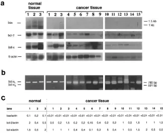
[image:4.612.56.380.86.350.2]Biotech-Figure 1. mRNA expression of bcl-2, bcl-xL, bcl-xs, and bax in breast cancer tissue. (a) Shown is Northern blot hybridization of three different normal tissue samples (lanes 1–3) and 15 different cancer tissue samples (lanes 1–15). Normal tissue (lanes 1–3) was derived from nonmalignant areas (as defined by histology) of the same breast as the malig-nant tissue (lanes 1–3). Cancer-derived cell lines show low bax expression. In cancer tis-sue samples no bax signal can be detected. In contrast, strong expression is found in all normal cell lines and tissues. bcl-2 and bcl-x are expressed in all tissue samples, b-actin served as a control. Note the shorter exposi-tion time of the right b-actin blot (lanes 10– 15). (b) RT-PCR analysis of bcl-x. PCR primers were designed to detect the long (bcl-xL) and short (bcl-xs) splice variant si-multaneously generating PCR bands of 780 and 591 bp, respectively. The same tissue samples (lanes 1–15) as in a were analyzed. bcl-xs mRNA could only be detected in R30C cells. (c) Northern data on normal (lanes 1–3) and malignant (lanes 1–15) pri-mary tissue were quantified by using an In-stant Imager (Canberra Packard). The ratio of bax/actin, bcl-2/actin, and bcl-x/actin was determined.
Table I. Expression of Members of the bcl-2 Family in Normal and Malignant Breast Tissue
Histology bcl-2 bcl-xL bcl-xs bax-a
Ductal invasive 31/33 33/33 0/33 1/33 Lobular 2/2 2/2 0/2 0/2 Intraductal 4/4 4/4 0/4 0/4 Mucinous 1/1 1/1 0/1 0/1 Fibrocystic 4/4 4/4 0/4 4/4 Normal 6/6 6/6 0/6 6/6
[image:4.612.57.298.386.482.2]Tween 20) for 40 min at room temperature. Bands were visualized us-ing chromogenic substrates NBT/BCIP (Roth, Karlsruhe, Germany).
Immunohistology. From the quick-frozen tissues frozen sections of z 4–6 mm in thickness were air dried, fixed in acetone for 10 min at
room temperature, and immunostained immediately or stored at 2208C. Antibodies against bax or bcl-2 are described above and were used for immunohistology. For antibody staining, an alkaline phosphatase/ anti–alkaline phosphatase staining procedure was applied as described before (38). The sections were counterstained with hematoxylin. nology, Inc. (Santa Cruz, CA); anti–bcl-2: monoclonal mouse
[image:5.612.59.550.170.675.2]anti-body purchased from Sigma Chemicals; anti-tubulin: monoclonal rat antibody purchased from Serotec (Wiesbaden, Germany). After transfer to nitrocellulose and blocking (3% BSA 13 PBS, overnight 48C), filters were incubated with antiserum (dilution 1:200 [anti-bax], 1:1,000 [anti–bcl-2 or anti–bcl-x] in 13 PBS, 1% BSA, 0.5% Tween 20). Thereafter filters were incubated with alkaline phosphatase–con-jugated goat anti–rabbit or goat anti–mouse antibody (Dianova, Hamburg, Germany; dilution 1:7,500 in 13 PBS, 1% BSA, 0.5%
Expression of p53 was evaluated according to previously de-scribed methods (39) using a monoclonal anti-p53 antibody (PAb1801; Cambridge Research Biochemicals, Wilmington, DE).
Xenotransplantation of cells into SCID mice. R30C and MCF-7 cells (106) were injected subcutaneously into C.B.-17 scid/scid mice.
The mice were obtained from our own breeding colony and were kept in isolators under stringent conditions in the central animal labo-ratory of the Max Delbrück Center for Molecular Medicine. Microbi-ological controls were performed regularly by addition of sterile sen-tinel animals to the colony. Tumors were measured in two dimensions, length (a) and width (b), by means of calipers in millime-ters. Tumor volume (V) was calculated according to V5 ab2/2, where
a is the longer of the two measurements.
Results
bcl-x and bcl-2 are expressed in normal and malignant breast epithelium. Expression of bcl-x and bcl-2 was studied in 40 dif-ferent breast cancer tissue samples and 10 nonmalignant breast tissue samples by Northern blot analysis. Nonmalignant tissue samples included four cases of fibrocystic disease and tumor-free tissue of six different cancer samples. To discriminate be-tween the two bcl-x splice variants we additionally performed RT-PCR analysis. No major difference was observed with
[image:6.612.57.556.83.198.2]re-gard to bcl-2 or bcl-xL expression between normal and malig-nant breast tissue (Fig. 1 and Table I). No or only weak bcl-2 expression could be detected in two cancer samples of the duc-tal invasive type (Table I). bcl-xs expression could not be de-tected, neither in normal nor in malignant breast epithelia.
Table II. Comparison between mRNA Expression Levels of Members of the bcl-2 Family in Breast Cancer Tissue and Other Tumor/Patient Characteristics
Lane 1 2 3 4 5 6 7 8 9 10 11 12 13 14 15
bax/actin , 0.01 , 0.01 , 0.01 , 0.01 , 0.01 , 0.01 , 0.01 , 0.01 , 0.01 , 0.01 , 0.01 , 0.01 , 0.01 , 0.01 , 0.01 bcl-2/actin 0.5 0.2 1.5 0.2 0.2 0.15 0.4 0.2 0.2 1 0.5 1.5 1 1 1.5 bcl-x/actin 1 1 1 0.4 0.4 0.1 0.3 5 0.4 1 0.5 1.5 2 0.5 0.5 Postmenopausal 1 2 1 1 2 1 2 1 2 2 2 2 2 1 1
ER 2 2 1 2 1 2 2 1 1 2 1 2 2 2 1
PR 1 1 2 1 2 2 2 2 1 2 2 2 2 1 1
p53 2 2 2 1 2 2 2 2 1 1 2 2 1 2 2
Quantitative mRNA analysis of bcl-2, bcl-x, and bax expression of 15 patients is also shown in Fig. 1 c. PR, progesterone receptor. p53 status was de-termined by immunohistology as described in Methods.
[image:6.612.314.556.325.663.2]Figure 3. Inducible expression of bax-a in breast cancer cell lines. Shown is Western blot analysis of bax-a protein expression in MCF-7 and R30C breast cancer cells. Mock transfectants and stable bax clones, which are under transcriptional control of a tetracycline-dependent repressor, were generated as described in Methods. Cells were cultured in the presence or absence of tetracycline. Protein was extracted 24 h after tetracycline withdrawal. Equal amounts of pro-tein (20 mg) were loaded on each lane. Strong induction of bax-a pro-tein can be detected after tetracycline withdrawal, whereas in the presence of tetracycline bax expression is completely repressed. Tu-bulin served as a control.
[image:6.612.58.274.532.622.2]Additionally, we quantified Northern data of 15 patients using an Instant Imager. The ratio of bax/actin, bcl-2/actin, and bcl-x/actin was determined (Fig. 1 c). Quantification revealed differences in bcl-2 and bcl-x mRNA expression levels be-tween different tumor samples. However, different bcl-2 and bcl-x expression levels could also be observed in normal breast tissue in a similar range as in tumor tissue. There are some tu-mors which showed bcl-2 and bcl-x expression levels beneath the range of normal tissue. One tumor showed elevated bcl-x expression compared with normal tissue. However, the major-ity of tumor tissue samples analyzed so far revealed similar amounts of bcl-2 or bcl-xL compared with normal mammary tissue.
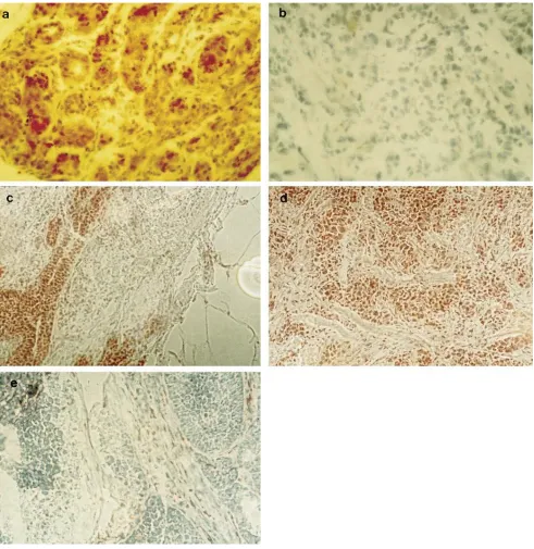
bax-a is strongly expressed in normal breast epithelium but is downregulated in malignant epithelia.Expression of bax mRNA and protein was studied in normal and malignant breast epithelium (see above) by Northern blot hybridization and immunohistology. Nonmalignant tissue samples included four cases of fibrocystic disease and tumor-free tissue of six different cancer samples. bax-a was expressed in high amounts in normal breast epithelium, whereas only partial, weak, or no expression could be detected in the vast majority of cancer tis-sue samples examined so far (Figs. 1 and 2, all tistis-sue data are summarized in Table I). Thus, data on downregulation of bax expression on mRNA level in breast cancer tissue could be confirmed by immunohistological analysis.
In addition to the 1-kb splice variant which encodes the functional death-promoting bax-a protein, a longer 1.5-kb mRNA which is consistent with bax-b could be detected in normal tissue. The function of this variant which is probably not translated into protein is unknown.
Only 1 out of 40 cancer tissue samples examined so far re-vealed a bax expression level comparable with that found in normal epithelium (Table I). Interestingly, downregulation of bax-a was found in different histological subtypes, including
ductal invasive, lobular, mucinous, and even intraductal (carci-noma in situ) carci(carci-nomas (Table I and Fig. 2).
It has been reported that p53 might be involved in the tran-scriptional regulation of bax expression (15). Furthermore, it has been published that estrogens may modulate bcl-2 expres-sion in breast tissue (40). Therefore, we provided data on the hormonal status (pre- versus postmenopausal, ER, progester-one receptor) and the p53 status and compared these data with bcl-2, bcl-x, and bax mRNA expression levels in different breast cancer tissues (Table II). Differences in bcl-2 and bcl-x mRNA expression levels as well as the strong downregulation of bax in breast cancer tissue did not correlate with the hor-monal status or the p53 status. The lack of a correlation be-tween p53 status and bax expression was also reported by Kra-jewski et al. (41).
Induction of bax-a expression in breast cancer cell lines can restore sensitivity of serum starvation or APO-I/Fas–mediated apoptosis. Recently, we have shown that low bax-a expression in breast cancer cell lines correlates with resistance towards apoptosis (32). In contrast to malignant cell lines, which ex-press low levels of bax-a, nonmalignant epithelial cell lines dis-playing high amounts of bax-a were highly sensitive to induc-tion of programmed cell death by both serum starvainduc-tion and APO-I/Fas (CD95) triggering (32). To test whether low bax-a
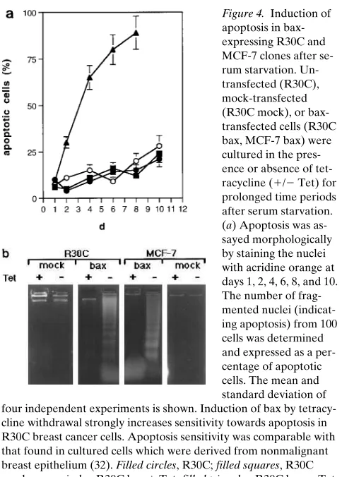
expression in breast cancer cell lines is due to apoptosis resis-tance we transfected bax cDNA under the control of a tetracy-cline-regulated expression system into these cells. As shown in Fig. 3, withdrawal of tetracycline led to strong induction of bax expression in R30C and MCF-7 cells. In the presence of tetra-cycline bax expression was completely repressed. As depicted in Fig. 4, a and b, induction of bax-a expression strongly in-creases sensitivity towards apoptosis induced by serum starva-tion. bax-expressing cells started to die within 2 d, whereas clones which were devoid of bax expression showed prolonged resistance to apoptosis at least until day 10 after serum
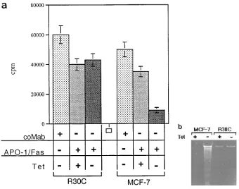
starva-Figure 5. Induction of apoptosis in bax expressing R30C and MCF-7 clones after APO-1/Fas triggering. (a) Antiprolifera-tive effect of anti–APO-1 on R30C and MCF-7 breast cancer cells. Inducible bax transfectants of R30C or MCF-7 were cultured in the presence (1) or absence (2) of 2 mg/ml tetracycline. 24 h after tet-racycline withdrawal 1 mg/ml anti–APO-1 or FII23c was added to the culture me-dium. [3H]Thymidine incorporation assay
[image:7.612.56.396.475.743.2]ated apoptosis in R30C cells, indicating that bax expression is important but not sufficient to confer APO-1/Fas sensitivity.
Thus, we showed that induction of bax-a can restore sensi-tivity towards different apoptotic stimuli in breast cancer cell lines.
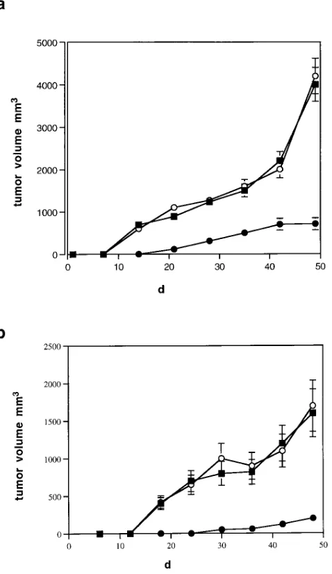
Induction of bax-a expression reduces tumor growth in SCID mice. To test the effects of bax expression on tumor growth in vivo, we transplanted bax-expressing clones of the breast cancer cell lines R30C and MCF-7 into SCID mice. As shown in Fig. 6, bax-expressing breast cancer cells are still tum-origenic. However, tumor size and velocity of tumor growth are significantly reduced compared with mock transfected or untransfected clones.
Discussion
Apoptosis is a physiological process that contributes to the reg-ulation of development, differentiation, and growth in various tissues. Therefore, dysregulation of apoptosis might be involved in pathogenesis of neoplasias. This is particularly well docu-mented for malignant lymphomas where bcl-2 overexpression was shown to be involved in malignant transformation (42).
In carcinomas, the role of bcl-2 in tumorigenesis is still un-clear. We and others (29, 32) have observed bcl-2 expression in breast cancer without a marked difference to normal epithe-lium. Some tumors failed to express bcl-2, which surprisingly correlated with markers of poor prognosis (29). Similar results were obtained for non–small cell lung carcinoma (43). bcl-xL expression appeared to be elevated in some tumors and re-duced in others. However, in accordance with the data con-cerning bcl-2, the majority of tumor tissue samples analyzed so far revealed similar amounts of bcl-xL compared with normal mammary tissue. bcl-xs expression could not be detected, nei-ther in normal nor in malignant breast epithelia.
In contrast, analysis of bax expression revealed a marked difference between normal and cancer epithelia: strong ex-pression of bax-a was observed in normal epithelial cells and breast tissue, whereas only weak or partial expression was found in their malignant counterparts. Low levels of bax in conjunction with normal bcl-2 and bcl-x levels might disrupt the cellular homeostasis leading to an accumulation of cells, which might thus become susceptible for secondary mutagenic events resulting in malignant transformation. Since differences in bcl-x and bcl-2 expression between normal and some malig-nant tissue samples could be found, we cannot exclude that in some cases deregulation of bcl-2 or bcl-x expression might also modulate apoptosis in breast cancer cells. However, compared with the striking change in bax expression, these differences are presumably of minor significance.
As low bax expression is still found in some tumors and cell lines, weak bax expression is probably not due to gross genetic alterations of the bax locus itself. It is conceivable that other genes involved in malignant transformation might cause down-regulation of bax. p53 itself or p53-regulated genes could be candidates, because p53 has also been shown to be a regulator of apoptosis and a direct transcriptional activator of the human bax gene (13–15, 44). Thus, constitutive expression of bcl-2 and bcl-xL and low expression of bax might at least in part be due to a loss of function of p53. However, since we found no correlation between bax downregulation and p53 status, we as-sume that besides p53 further genes are involved in the down-regulation of bax expression in malignant breast epithelium. tion. Apoptosis sensitivity in bax-expressing cancer cell lines
was comparable with that found in HMEC and HBL-100 cells (32), which were derived from normal (nonmalignant) human breast epithelium.
Furthermore, we could show that induction of bax-a in the breast cancer cell line MCF-7 strongly increases sensitivity to-wards APO-1/Fas–mediated apoptosis (Fig. 5, a and b). A weak apoptotic effect of APO-1/Fas triggering (30% growth inhibition) could also be observed in cells which were devoid of detectable bax expression levels. In contrast, induction of bax expression did not affect resistance to
APO-1/Fas–medi-Figure 6. Tumor growth of bax-expressing R30C and MCF-7 clones in SCID mice. Shown is tumor growth of five different bax-expressing and five different mock clones of R30C and MCF-7 breast cancer cells. Untransfected clones were included. Each cell clone was trans-planted on six different animals. Animals were challenged with equal cell numbers (106) at day 0. Tumor size was determined at different
[image:8.612.60.299.201.614.2]The CD95 antigen and its role in apoptosis have been stud-ied mainly in normal and malignant lymphoid cells. However, CD95 can also be detected in many epithelial tissues (24). In these experiments we provide evidence that, as in lymphoid cells, expression of CD95 does not necessarily correlate with sensitivity towards apoptosis (45). In B-CLL cells an inverse correlation between bcl-2 expression and sensitivity to CD95-mediated apoptosis was found (45). In contrast, sensitivity to CD95-mediated apoptosis does not correlate with bcl-2 ex-pression in prostatic adenocarcinoma or colon carcinoma cell lines (46) and overexpression of bcl-2 is able to confer only partial protection against CD95-mediated apoptosis (47). Thus, additional genes have to be involved in regulation of CD95-triggered apoptosis. Recently, it has been shown that coexpression of bcl-2 and bag-1, another apoptosis-regulating gene, which does not belong to the bcl-2 gene family, com-pletely protects from CD95-mediated apoptosis (48).
Recently, we have shown that low bax-a expression in breast cancer cell lines correlates with resistance towards apop-tosis (32). In contrast to malignant cell lines, which express low levels of bax-a, nonmalignant epithelial cell lines displaying high amounts of bax-a were highly sensitive to induction of programmed cell death by both serum starvation and APO-I/ Fas (CD95) triggering. To test whether low bax-a expression in breast cancer cell lines is due to apoptosis resistance, we transfected bax cDNA under the control of a tetracycline-reg-ulated expression system into these cells. We were able to show that induction of bax expression in breast cancer cell lines restores sensitivity towards both serum starvation or CD95-induced apoptosis. Thus, we were able to demonstrate for the first time that bax can confer sensitivity to different apoptotic stimuli, e.g., serum starvation or CD95 triggering. Therefore, it appears that the bcl-2 family constitutes a major death checkpoint integrating different apoptotic pathways. The decreased sensitivity of breast carcinoma cell lines to-wards CD95-mediated apoptosis furthermore indicates a po-tential role of an intact CD95 apoptosis signaling pathway in the homeostasis of normal breast epithelium.
Consistent with our observations, another recent paper demonstrated reduced bax immunostaining in breast cancers and found correlations with poor responses to chemotherapy and patient survival (41). The authors arbitrarily defined bax-negative cases as those in which the vast majority of the tumor cells were unstained (, 10%), with bax-positive cases repre-senting all others (. 10%), and found that complete loss (, 10%) of bax expression is correlated with poor response rates to chemotherapy and shorter survival in women with metastatic breast adenocarcinoma. Since antineoplastic drugs can kill at least in part tumor cells via apoptosis, our functional data, showing that induction of the bax gene can restore sensi-tivity to different apoptotic stimuli in cultured breast cancer cells, might serve as an explanation for this correlation. In ad-dition, the authors report that they could not detect reduced bax expression in in situ carcinomas, which is in contrast to our data. This contradiction might be due to different immunohis-tological staining techniques.
To test whether induction of bax-a affects tumor growth in vivo, we transplanted a bax-expressing breast cancer cell line into SCID mice. Although tumor growth could not be com-pletely prevented, we were able to show that bax expression led to a significant reduction of tumor growth. This finding provides further evidence that dysregulation of apoptosis, at
least in part due to downregulation of bax-a expression, con-tributes to the pathogenesis of breast cancer.
In summary, we could show the following: (a) bax-a ex-pression is downregulated in malignant breast epithelium in comparison to normal epithelium; (b) induction of bax-a re-stores sensitivity towards serum starvation or APO-1/Fas– mediated apoptosis in breast cancer cell lines; and (c) induc-tion of bax-a reduces tumor growth in SCID mice. Therefore, we propose that the bcl-2 family and APO-1/Fas are involved in the regulation of tissue homeostasis not only of the lym-phoid system but also of epithelial cells. An imbalance within the bcl-2 family members leads to dysregulation of apoptosis and therefore might contribute to the pathogenesis of breast cancer.
Acknowledgment
We wish to thank Dr. P. Krammer (German Cancer Research Center, Heidelberg, Germany) for kindly providing anti–APO-1 IgG3 mAb.
References
1. Ellis, R.E., J.Y. Yuan, and H.R. Horvitz. 1991. Mechanisms and functions of cell death. Annu. Rev. Cell Biol. 7:663–698.
2. Vaux, D.L., S. Cory, and J.M. Adams. 1988. Bcl-2 gene promotes hae-mopoietic cell survival and cooperates with c-myc to immortalize pre-B cells. Nature (Lond.). 335:440–442.
3. Tsujimoto, Y., L.R. Finger, J. Yunis, P.C. Nowell, and C.M. Croce. 1984. Cloning of the chromosome breakpoint of neoplastic B cells with the t(14;18) chromosome translocation. Science (Wash. DC). 226:1097–1099.
4. Tsujimoto, Y., J. Gorham, J. Cossman, E. Jaffe, and C.M. Croce. 1985. The t(14;18) chromosome translocations involved in B-cell neoplasms result from mistakes in VDJ joining. Science (Wash. DC). 299:1390–1393.
5. Tsujimoto, Y., and C.M. Croce. 1986. Analysis of the structure, tran-scripts, and protein products of bcl-2, the gene involved in human follicular lymphoma. Proc. Natl. Acad. Sci. USA. 83:5214–5218.
6. Tsujimoto, Y., N. Ikegaki, and C.M. Croce. 1987. Characterization of the protein product of bcl-2, the gene involved in human follicular lymphoma. On-cogene. 2:3–7.
7. Krammer, P.H., I. Behrmann, P.T. Daniel, J. Dhein, and K.M. Debatin. 1994. Regulation of apoptosis in the immune system. Curr. Opin. Immunol. 6: 279–289.
8. Sandford, N.L., J.W. Searle, and J.F. Kerr. 1984. Successive waves of apop-tosis in the rat prostate after repeated withdrawal of testosterone stimulation. Pathology. 16:406–410.
9. Tenniswood, M.P., R.S. Guenette, J. Lakins, M. Mooibroek, P. Wong, and J.E. Welsh. 1992. Active cell death (ACD) in hormone-dependent tissues. Cancer Metastasis Rev. 11:197–220.
10. Walker, N.I., R.E. Bennett, and J.F. Kerr. 1989. Cell death by apoptosis during involution of the lactating breast in mice and rats. Am. J. Anat. 185:19– 32.
11. Marti, A., B. Jehn, E. Costello, N. Keon, G. Ke, F. Martin, and R. Jaggi. 1994. Protein kinase A and AP-1 (c-Fos/JunD) are induced during apoptosis of mouse mammary epithelial cells. Oncogene. 9:1213–1223.
12. Lefebvre, O., C. Wolf, J.M. Limacher, P. Hutin, C. Wendling, M. LeMeur, P. Basset, and M.C. Rio. 1992. The breast cancer-associated strome-lysin-3 gene is expressed during mouse mammary gland apoptosis. J. Cell Biol. 119:997–1002.
13. Shaw, P., R. Bovery, S. Tardy, R. Sahli, B. Sordat, and J. Costa. 1992. In-duction of apoptosis by wild-type p53 in a human colon tumor derived cell line. Proc. Natl. Acad. Sci. USA. 89:5214–5218.
14. Yonish-Rouach, E., D. Resnitzky, J. Lotem, L. Sachs, A. Kimchi, and M. Oren. 1991. Wild-type p53 induces apoptosis of myeloid leukaemic cells that is inhibited by interleukin-6. Nature (Lond.). 352:345–347.
15. Miyashita, T., and J. Reed. 1995. Tumor suppressor p53 is direct tran-scriptional activator of the human bax gene. Cell. 80:293–299.
16. Miyashita, T., S. Krajewski, M. Krajewski, H.G. Wang, H.K. Lin, D.A. Liebermann, B. Hoffmann, and J.C. Reed. 1994. Tumor suppressor p53 is a reg-ulator of bcl-2 and bax gene expression in vitro and in vivo. Oncogene. 9:1799– 1805.
17. Miyashita, T., M. Harigai, M. Hanada, and J.C. Reed. 1994. Identifica-tion of a p53-dependent negative response element in the bcl-2 gene. Cancer Res. 12:3133–3135.
re-gression by induction of apoptosis. Science (Wash. DC). 245:301–305. 19. Yonehara, S., A. Ishii, and M. Yonehara. 1989. A cell-killing mono-clonal antibody (anti-Fas) to a cell surface antigen co-downregulated with the receptor of tumor necrosis factor. J. Exp. Med. 169:1747–1756.
20. Oehm, A., I. Behrmann, W. Falk, M. Pawlita, G. Maier, C. Klas, M. Li-Weber, S. Richards, J. Dhein, B.C. Trauth, et al. 1992. Purification and molecu-lar cloning of the APO-1 cell surface antigen, a member of the TNF/NGF re-ceptor superfamily. J. Biol. Chem. 267:10709–10716.
21. Itoh, N., S. Yonehara, A. Ishii, M. Yonehara, S. Mizushima, M. Sameshima, A. Hase, Y. Seto, and S. Nagata. 1991. The polypeptide encoded by the cDNA for human cell surface antigen Fas can mediate apoptosis. Cell. 66: 233–243.
22. Dhein, J., P.T. Daniel, B.C. Trauth, A. Oehm, P. Möller, and P.H. Krammer. 1992. Induction of apoptosis by monoclonal antibody anti-APO-1 class switch variants is dependent on cross-linking of APO-1 cell surface anti-gens. J. Immunol. 149:3166–3173.
23. Daniel, P.T., and P.H. Krammer. 1994. Activation induces sensitivity to-wards APO-1 (CD95) mediated apoptosis in human B cells. J. Immunol. 153: 800–810.
24. Leithäuser, F., J. Dhein, G. Mechtersheimer, K. Koretz, K. Brüderlein, C. Henne, A. Schmidt, K.M. Debatin, P.H. Krammer, and P.H. Möller. 1993. Constitutive and induced expression of APO-1, a new member of the nerve growth factor/tumor necrosis factor receptor superfamily, in normal and neo-plastic cells. Lab. Invest. 69:415–429.
25. Nunez, G., L. London, D. Hockenbery, M. Alexander, J.P. McKearn, and S.J. Korsmeyer. 1990. Deregulated Bcl-2 gene expression selectively pro-longs survival of growth factor-deprived hemopoietic cell lines. J. Immunol. 144:3602–3610.
26. Boise, L.H., M. Gonzalez-Garcia, C.E. Postema, L. Ding, T. Lindsten, L.A. Turka, X. Mao, G. Nunez, and C.B. Thompson. 1993. bcl-x, a bcl-2 related gene that functions as a dominant regulator of apoptotic cell death. Cell. 74: 597–608.
27. Oltvai, Z.N., C.L. Milliman, and S.J. Korsmeyer. 1993. Bcl-2 het-erodimerizes in vivo with a conserved homolog, Bax, that accelerates pro-grammed cell death. Cell. 74:609–619.
28. Apte, S.S., M.G. Mattei, and B.R. Olsen. 1995. Mapping of the human bax gene to chromosome 19q13.3-q13.4 and isolation of a novel alternatively spliced transcript, bax-delta. Genomics. 26:592–594.
29. Russel, D.L., L. Kaklamanis, F. Pezzella, K.C. Gatter, and A.L. Harris. 1994. Bcl-2 in normal human breast and carcinoma, association with oestrogen receptor-positive, epidermal growth factor receptor-negative tumours and in situ cancer. Br. J. Cancer. 69:135–139.
30. Gasparini, G., M. Barbareschi, C. Doglioni, P.D. Palm, F.A. Mauri, P. Boracchi, P. Bevilacqua, O. Caffo, L. Morelli, P. Verderio, et al. 1995. Expres-sion of Bcl-2 protein predicts efficacy of adjuvant treatments in operable node-positive breast cancer. Clin. Cancer Res. 1:189–198.
31. Silvestrini, R., S. Veneroni, M.G. Daidone, E. Benini, P. Boracchi, M. Mezzetti, G. Di-Fronzo, F. Rilke, and U. Veronesi. 1994. The bcl-2 protein: a prognostic indicator strongly related to p53 protein in lymph node-negative breast cancer patients. J. Natl. Cancer Inst. 86:499–504.
32. Bargou, R.C., P.T. Daniel, M.Y. Mapara, K. Bommert, C. Wagener, B.
Kallinich, H.D. Royer, and B. Dörken. 1995. Expression of the bcl-2 gene fam-ily in normal and malignant breast tissue: low bax-a expression in tumor cells correlates with resistance towards apoptosis. Int. J. Cancer. 60:854–859.
33. Walker, K.J., N. Bouzubar, J.F.R. Robertson, I.O. Ellis, C.W. Elston, R.W. Blamey, D.W. Wilson, K. Griffiths, and R.I. Nicholson. 1988. Immunocy-tochemical localization of estrogen receptor in human breast tissue. Cancer Res. 48:6517–6522.
34. Gossen, M., and H. Bujard. 1992. Tight control of gene expression in mammalian cells by tetracycline-responsive promoters. Proc. Natl. Acad. Sci. USA. 89:5547–5551.
35. Ritter, K., H. Stöpfer, M. Pawlita, and G. Sczakiel. 1991. Versatile eu-caryotic vectors for strong and constitutive transient and stable gene expres-sion. Methods Mol. Cell. Biol. 2:176–181.
36. Wang, A.M., M.V. Doyle, and D.F. Mark. 1989. Quantitation of mRNA by the polymerase chain reaction. Proc. Natl. Acad. Sci. USA. 86:9717–9720.
37. Deleted in proof.
38. Miltenyi, S., W. Müller, W. Weichel, and A. Radbruch. 1990. High gra-dient magnetic cell separation with MACS. Cytometry. 11:231–238.
39. Thor, A.D., D.H. Moore, and S.M. Edgerton. 1992. Accumulation of p53 tumor suppressor gene protein: an independent marker of prognosis in breast cancers. J. Natl. Cancer Inst. 330:1260–1266.
40. Sabourin, J.C., A. Martin, J. Baruch, J.B. Truc, A. Gompel, and P. Poitout. 1994. bcl-2 expression in normal tissue during the menstrual cycle. Int. J. Cancer. 59:1–6.
41. Krajewski, S., C. Blomqvist, K. Franssila, M. Krajewski, V.-M. Wase-nius, E. Niskanen, S. Nordling, and J.C. Reed. 1995. Reduced expression of proapoptotic gene bax is associated with poor response rates to combination chemotherapy and shorter survival in women with metastatic breast adenocar-cinoma. Cancer Res. 55:4471–4478.
42. McDonnell, T.J., and S.J. Korsmeyer. 1991. Progression from lymphoid hyperplasia to high-grade malignant lymphoma in mice transgenic for the t(14; 18). Nature (Lond.). 349:254–256.
43. Pezzella, F., H. Turley, I. Kuzu, M.F. Tungekar, M.S. Dunnill, C.B. Pierce, A. Harris, K.C. Gatter, and D.Y. Mason. 1993. Bcl-2 protein in non-small-cell lung carcinoma. N. Engl. J. Med. 329:690–694.
44. Wafik, S.E.-D., J.W. Harper, P.M. O’Connor, V.E. Velculescu, C.E. Canman, J. Jackman, J.A. Pietenpol, M. Burrell, D.E. Hill, Y. Wang, et al. 1994. WAF/CIP1 is induced in p53-mediated G1 arrest and apoptosis. Cancer Res. 54: 1169–1174.
45. Mapara, M., R. Bargou, C. Zugck, H. Döhner, F. Ustaoglu, P.H. Kram-mer, and B. Dörken. 1993. APO-1 mediated apoptosis or proliferation in hu-man chronic B lymphocytic leukemia: correlation with bcl-2 oncogene expres-sion. Eur. J. Immunol. 23:702–708.
46. Owen-Schaub, L., R. Radinsky, E. Kruzel, K. Berry, and S. Yonehara. 1994. Anti-Fas on nonhematopoetic tumors: levels of Fas/APO-1 and bcl-2 are not predictive of biological responsiveness. Cancer Res. 54:1580–1586.
47. Itoh, N., Y. Tsujimoto, and S. Nagata. 1993. Effect of bcl-2 on Fas anti-gen-mediated cell death. J. Immunol. 151:621–627.