Angiotensin II inhibits insulin signaling in aortic
smooth muscle cells at multiple levels. A
potential role for serine phosphorylation in
insulin/angiotensin II crosstalk.
F Folli, … , J L Bouchie, E P Feener
J Clin Invest.
1997;
100(9)
:2158-2169.
https://doi.org/10.1172/JCI119752
.
To investigate potential interactions between angiotensin II (AII) and the insulin signaling
system in the vasculature, insulin and AII regulation of insulin receptor substrate-1 (IRS-1)
phosphorylation and phosphatidylinositol (PI) 3-kinase activation were examined in rat
aortic smooth muscle cells. Pretreatment of cells with AII inhibited insulin-stimulated PI
3-kinase activity associated with IRS-1 by 60%. While AII did not impair insulin-stimulated
tyrosine phosphorylation of the insulin receptor (IR) beta-subunit, it decreased
insulin-stimulated tyrosine phosphorylation of IRS-1 by 50%. AII inhibited the insulin-insulin-stimulated
association between IRS-1 and the p85 subunit of PI 3-kinase by 30-50% in a
dose-dependent manner. This inhibitory effect of AII on IRS-1/PI 3-kinase association was
blocked by the AII receptor antagonist saralasin, but not by AT1 antagonist losartan or AT2
antagonist PD123319. AII increased the serine phosphorylation of both the IR beta-subunit
and IRS-1. In vitro binding experiments showed that autophosphorylation increased IR
binding to IRS-1 from control cells by 2.5-fold versus 1.2-fold for IRS-1 from AII-stimulated
cells, suggesting that AII stimulation reduces IRS-1's ability to associate with activated IR. In
addition, AII increased p85 serine phosphorylation, inhibited the total pool of p85 associated
PI 3-kinase activity, and decreased levels of the p50/p55 regulatory subunit of PI 3-kinase.
These results suggest that activation of the renin-angiotensin system may lead to insulin
resistance in the vasculature.
Research Article
Find the latest version:
J. Clin. Invest.
© The American Society for Clinical Investigation, Inc. 0021-9738/97/11/2158/12 $2.00
Volume 100, Number 9, November 1997, 2158–2169 http://www.jci.org
Angiotensin II Inhibits Insulin Signaling in Aortic Smooth Muscle Cells at
Multiple Levels
A Potential Role for Serine Phosphorylation in Insulin/Angiotensin II Crosstalk
Franco Folli,*‡ C. Ronald Kahn,‡ Hans Hansen,‡ Julie L. Bouchie,‡ and Edward P. Feener‡
*Department of Medicine I and Unitá di Malattie Metaboliche, I.R.C.C.S.H. San Raffaele, 20132 Milano, Italy; and ‡Research Division, Joslin Diabetes Center, Harvard Medical School, Boston, Massachusetts 02215
Abstract
To investigate potential interactions between angiotensin II (AII) and the insulin signaling system in the vasculature, in-sulin and AII regulation of inin-sulin receptor substrate-1 (IRS-1) phosphorylation and phosphatidylinositol (PI) 3–kinase activation were examined in rat aortic smooth muscle cells. Pretreatment of cells with AII inhibited insu-lin-stimulated PI 3–kinase activity associated with IRS-1 by 60%. While AII did not impair insulin-stimulated tyrosine phosphorylation of the insulin receptor (IR) b-subunit, it decreased insulin-stimulated tyrosine phosphorylation of IRS-1 by 50%. AII inhibited the insulin-stimulated associa-tion between IRS-1 and the p85 subunit of PI 3–kinase by 30–50% in a dose-dependent manner. This inhibitory effect of AII on IRS-1/PI 3–kinase association was blocked by the AII receptor antagonist saralasin, but not by AT1 antagonist
losartan or AT2 antagonist PD123319. AII increased the
serine phosphorylation of both the IR b-subunit and IRS-1. In vitro binding experiments showed that autophosphoryla-tion increased IR binding to IRS-1 from control cells by 2.5-fold versus 1.2-2.5-fold for IRS-1 from AII-stimulated cells, suggesting that AII stimulation reduces IRS-1’s ability to associate with activated IR. In addition, AII increased p85 serine phosphorylation, inhibited the total pool of p85 asso-ciated PI 3–kinase activity, and decreased levels of the p50/ p55 regulatory subunit of PI 3–kinase. These results suggest that activation of the renin-angiotensin system may lead to insulin resistance in the vasculature. (J. Clin. Invest. 1997.
100:2158–2169.) Key words: angiotensin II• insulin receptor
substrate-1 • phosphatidylinositol 3-kinase • vascular smooth muscle cells • insulin resistance
Introduction
Angiotensin II (AII)1 is the main effector peptide of the renin– angiotensin system (RAS) and plays an important role in the regulation of vascular homeostasis (1, 2). Increased AII action
in the vasculature via its local induction within the vascular wall (3–5) has been strongly implicated in the pathogenesis of macrovascular disease by, for example, stimulating neointimal thickening and altering vascular gene expression (6–9). Benefi-cial effects of angiotensin converting enzyme (ACE) inhibition on cardiovascular disease has been reported in many (for view see reference 10) but not all (11) clinical trials. Recent re-ports have also shown that inhibition of the RAS can increase insulin sensitivity (12–14), suggesting a role for the RAS in the regulation of insulin action. As hypertension and insulin resis-tance often coexist and are leading risk factors for cardiovascu-lar disease (15–18), interactions between AII and insulin sig-naling systems may have an important role in the regulation of vascular physiology and the development of atherosclerosis.
While several isoforms of the AII receptor have been de-scribed, the predominant isoform expressed in macrovascular smooth muscle cells is the AT1 receptor (2, 19). This receptor is G protein–coupled to a variety of intercellular signaling path-ways, including the phospholipase C/protein kinase C (PKC) pathway, cytosolic calcium, and tyrosine kinases including JAK and FAK (2, 20–22). In addition to its direct ability to regulate growth, migration and gene expression in vascular cells (23– 25), AII may exhibit additional actions in vivo by modulating the signaling of other hormones. Crosstalk between G pro-tein–coupled receptors and cell surface tyrosine kinase recep-tors (26, 27) may provide a mechanism for such indirect ac-tions of AII on vascular cell physiology. Recently, in in vivo studies we have shown that AII infused into the heart can both stimulate insulin receptor substrate-1 and -2 (IRS-1, -2) ty-rosine phosphorylation and inhibit PI 3-kinase activity associ-ated with IRS proteins (28, 29).
The insulin receptor is a heterotetrameric tyrosine kinase and after insulin binding undergoes a rapid tyrosine autophos-phorylation that both activates the receptor kinase and allows transient interaction with intracellular protein substrates in-cluding IRS-1 and -2 via their phosphotyrosine binding do-mains (PTB dodo-mains) (30–33). This allows tyrosine phosphor-ylation of IRS-1 and IRS-2 on YMXM/YXXM motifs that induces their binding to Src homology 2-domain (SH-2) con-taining molecules, including phosphatidylinositol 3-kinase (PI 3–kinase), Grb2, SHPTP2, NCK, and Fyn (30, 32, 34, 35). The interaction between the IRS proteins and PI 3–kinase occurs through the p85 regulatory subunit of the enzyme resulting in an increase in catalytic activity of the p110 subunit (34, 36, 37). While alterations in insulin action in the vasculature, mediated via both the insulin and IGF-1 receptors, have been proposed Address correspondence to Edward P. Feener, Ph.D., Research
Divi-sion, Joslin Diabetes Center, One Joslin Place, Boston, MA 02215. Phone: 617-732-2599; FAX: 617-732-2637; E-mail: feenere@joslab. harvard.edu
Received for publication 22 January 1997 and accepted in revised form 28 August 1997.
to contribute to atherosclerosis and the regulation of vascular tone (38, 39), little is known regarding the pathways of insulin signaling, and their regulation, in vascular cells.
In this report, we have used cultured rat aortic smooth muscle cells (RASMC) as a model to examine the potential ef-fects of AII on insulin signaling to IRS-1 and PI 3–kinase in macrovascular cells. We show that AII impairs insulin stimula-tion of IRS-1 tyrosine phosphorylastimula-tion and coupling of the in-sulin receptor pathway to PI 3–kinase. These studies suggest that activation of the RAS may contribute to insulin resistance in AII-responsive tissues such as the vasculature.
Methods
Cell culture.RASMC were isolated from Sprague-Dawley rats as de-scribed previously (23), cultured in Dulbecco’s minimal essential me-dium (DMEM) containing 10% FCS, and used between passage 7 and 13. Confluent monolayers of cells were serum deprived in DMEM containing 0.1% (wt/vol) bovine serum albumin (BSA) for 18 h be-fore the experiment. Cells were stimulated with 100 nM AII, insulin, or insulin-like growth factor-1 (IGF-1), 25 ng/ml platelet-derived growth factor (PDGF), or 160 nM phorbol 12-myristate 13-acetate (PMA) (Sigma Chemical Co., St. Louis, MO; Boehringer Mannheim Biochemicals, Indianapolis, IN; UBI, Lake Placid, NY). AII receptor antagonist studies were performed on cells that were pretreated for 15 min with 1 mM Sar1, Ile8-angiotensin (Sigma Chemical Co.),
losar-tan (provided by Dr. Ronald D. Smith, DuPont Merck Pharmaceuti-cal Co., Wilmington, DE), or PD123319 (provided by Dr. D.G. Tay-lor, Parke-Davis, Ann Arbor, MI) followed by stimulation with AII and/or insulin. The effects of PI 3–kinase inhibition were examined in cells pretreated with 50 mM LY294002 (BioMol) for 15 min.
PI 3–kinase assay. Confluent 10-cm dishes of serum-deprived RASMC were stimulated for 7 min with 100 nM AII, 5 min with 100 nM insulin, or for 2 min with AII followed by an additional 5 min with in-sulin. Cells were then washed twice with ice-cold PBS containing 2 mM Na2VO4 and lysed in ice-cold freshly prepared lysis buffer containing
50 mM Hepes, pH 7.4, 137 mM NaCl, 1 mM MgCl2, 1 mM CaCl2, 2 mM
Na2VO4, 10 mM sodium pyrophosphate, 100 mM sodium fluoride, 2 mM
EDTA, 2 mM phenylmethylsulfonylfluoride, 10 mg/ml aprotinin, 10 mg/ml leupeptin, 10 mM benzamidine, 10% (vol/vol) glycerol, and 1% (vol/vol) Triton X-100 for 1 h. Insoluble material was removed by centrifugation for 15 min at 12,000 rpm at 48C. 1-ml aliquots of super-natants, normalized to protein, were incubated overnight at 48C with rabbit polyclonal antibodies against IRS-1 (29), p85 subunit of PI 3–kinase, and PDGF receptor (UBI) and the immunocomplexes were precipitated with a 50% solution of protein A–Sepharose 6MB (Pharmacia Biotech AB, Uppsala, Sweden). In vitro PI 3–kinase assays were performed as described previously (34). The 32P-labeled
3-P-phosphatidylinositol was visualized using a PhosphorImager and normalized using IMAGEQUANT software (Molecular Dynamics, Sunnyvale, CA).
Immunoprecipitation and Western blotting. Immunoprecipitations of cell lysates, normalized to protein, were performed overnight at 48C. Tyrosine phosphorylation of immunoprecipitated IRS-1 and IR was detected by Western blotting using antiphosphotyrosine antibody (4G10) followed by rabbit anti–mouse IgG (DAKO Corp., Carpente-ria, CA) and 125I-protein A (Amersham Corp., Arlington Heights,
IL), as described previously (29, 34). Western blotting was performed using antibodies against p85 (UBI) and p110a or IR b-subunit (Santa Cruz Biotechnology, Santa Cruz, CA) followed by 125I-protein-A.
In vivo phosphorylation. Confluent monolayers of RASMC in 15-cm plates were serum-starved for 18 h and then incubated for 4 h in phosphate-free Eagle’s MEM (GIBCO BRL, Gaithersburg, MD) containing 0.5 mCi/ml [32P]orthophosphate (DuPont-New England
Nuclear, Boston, MA). Cells were then stimulated as described above. Labeling was terminated with liquid N2 and cells were thawed
in lysis buffer and scraped from the dish. Insoluble material was re-moved by centrifugation for 15 min at 12,000 rpm at 48C. Superna-tants were incubated overnight at 48C with antibodies directed against IRS-1 or p85 subunit of PI 3–kinase and the immunocomplexes were precipitated with a 50% solution of protein A–Sepharose. For in vivo phosphorylation studies of the insulin receptor, cell lysates were cen-trifuged at 100,000 g for 30 min, the soluble fraction was applied to a wheat germ agglutinin-Sepharose column (Sigma Chemical Co.) and eluted with 0.3 M N-acetyl-D-glucosamine (Sigma Chemical Co.). The insulin receptor enriched eluates were incubated with 20 mg of polyclonal anti-insulin receptor antibodies (Santa Cruz Biotechnol-ogy) and the immunocomplexes were precipitated with a 50% solu-tion of protein A–Sepharose 6MB. The washed immunocomplexes were eluted with Laemmli sample buffer and separated on 6% SDS-PAGE for the IRS-1 protein and 7.5% SDS-SDS-PAGE for the insulin re-ceptor b-subunit and p85 regulatory subunit of PI 3–kinase. Proteins were transferred to nitrocellulose paper (0.2 mm pore size, Schleicher & Schuell Inc., Keene, NH) and visualized by autoradiography. The
32P-labeled protein bands were quantitated using IMAGEQUANT
software of the PhosphorImager.
Phosphoamino acid analysis. The nitrocellulose containing 32
P-labeled IRS-1, p85 and b-subunit was excised and treated with 0.5% (wt/vol) PVP-40 (Sigma Chemical Co.) in 100 mM acetic acid for 30 min at 378C. The paper was then washed five times with water and di-gested with 10 mg of TPCK-trypsin (Worthington Biochemical Corp., Freehold, NJ) in NaHCO3 (pH 8.2) containing 5% acetonitrile at
378C for 24 h. An additional 10 mg of TPCK-trypsin was added, and the digestion was continued for 24 h. This technique consistently eluted 90–95% of the phosphopeptides. The peptides were then evap-orated in a Speed Vac, partially hydrolyzed in 100 ml of 6 N HCl at 1108C for 2 h, diluted in 1 ml of water and dried. Phosphoamino acid analysis was performed by electrophoresis on cellulose thin layer plates as described previously (40).
In vitro insulin receptor/IRS binding. Control or AII-stimulated (100 nM, 7 min) RASMC were harvested in ice-cold lysis buffer and fractionated in a column using wheat germ agglutinin (WGA) Sepha-rose. The fraction eluted with 0.3 mM N-acetyl-glucosamine (NAGA) was autophosphorylated in vitro for 30 min (40) and allowed to inter-act with a glutathione S–transferase fusion protein containing the PTB domain of IRS-1 (GST-IRS-1-PTB) (kindly provided by Dr. S. Shoelson, Joslin Diabetes Center, Boston, MA). After a 2-h incuba-tion the samples were precipitated using glutathione Sepharose 4B (Pharmacia Biotech), separated by SDS-PAGE, and the insulin re-ceptor associated with the GST-IRS-1-PTB was quantitated by im-munoblotting using an antibody against the b-subunit of the insulin receptor. The flow through from the WGA column was immunopre-cipitated with anti–IRS-1 antibody and protein A–Agarose. Immuno-precipitates were washed three times and divided into two equal ali-quots. WGA purified human insulin receptor (HIR) was isolated from Chinese hamster ovary cells expressing HIR (CHO-HIRc). Equal aliquots of HIR, autophosphorylated for 30 min at 228C as described previously (40) or used in its basal phosphorylation state, were incu-bated with IRS-1 immunoprecipitates for 2 h at 48C and washed once, as described by Wolf et al. (41). Immunoprecipitates were separated by SDS-PAGE and immunoblotted with antibody against the b -sub-unit of the insulin receptor followed by 125I-protein A.
Statistics. Comparison of data presented as mean6SEM was per-formed using the one way ANOVA or the unpaired Student’s t test as indicated. Values of P, 0.05 were considered significantly different.
Results
activation of the PI 3–kinase enzymatic activity (34, 36, 37). To investigate the interactions between AII and insulin on the IRS-1 signaling pathway in RASMC, cells were stimulated for 5 min with 100 nM insulin in the absence or presence of a 2 min preincubation with 100 nM AII. PI 3–kinase activity asso-ciated with IRS-1 precipitates was measured using an in vitro assay, and results from a typical experiment are shown in Fig. 1
A. A low level of IRS-1–associated PI 3–kinase activity was present in the basal state and this was stimulated by insulin fivefold (P, 0.05, ANOVA). Pretreatment of cells with AII caused a small, nonsignificant decrease in basal IRS-1 associ-ated PI 3–kinase activity and decreased insulin stimulassoci-ated IRS-1 associated PI 3–kinase activity by 60% (P, 0.05, Fig. 1 B). A time-course study of the AII effect demonstrated that simulta-neous addition of AII and insulin, or the addition of AII 2 min after insulin, also reduced insulin stimulation of PI 3–kinase activity, although to a lesser extent (data not shown). A signifi-cant IGF-1 stimulation of IRS-1 associated PI 3–kinase activity compared with control (P, 0.05, ANOVA) was also inhibited by AII (Fig. 1 C), indicating that AII’s effect was not unique to the insulin receptor.
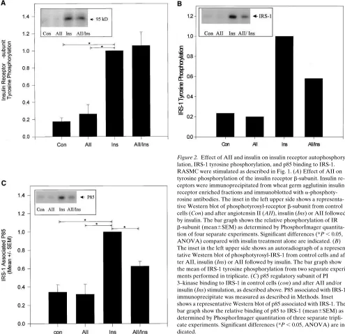
To investigate the potential mechanisms responsible for the AII-induced decrease in insulin-stimulated PI 3–kinase as-sociated with IRS-1, effects of AII on insulin receptor and IRS-1 tyrosine phosphorylation and IRS-1/p85 docking were examined. RASMC were stimulated with AII and/or insulin as described above, insulin receptor was immunoprecipitated from WGA purified cell lysates, and tyrosine phosphorylation
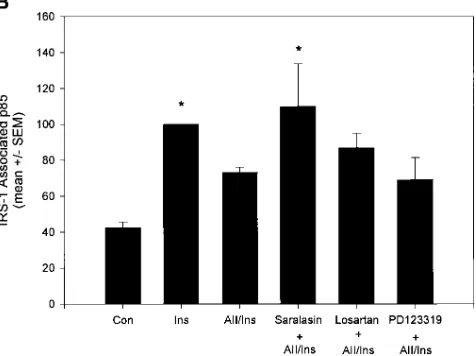
of the insulin receptor b-subunit was assessed by immunoblot-ting using antiphosphotyrosine antibodies. Insulin stimulated a fivefold increase (P, 0.05) in tyrosine phosphorylation of the insulin receptor in these cells (Fig. 2 A). Immunoprecipitation of cell extracts with anti–IRS-1 antibodies followed by West-ern blotting with antiphosphotyrosine antibodies revealed that insulin also stimulated a fivefold increase in IRS-1 tyrosine phosphorylation (Fig. 2 B). AII (100 nM) did not significantly alter tyrosine phosphorylation of the insulin receptor in either the absence or presence of insulin (Fig. 2 A). AII alone also had no effect on basal IRS-1 tyrosine phosphorylation, but AII pretreatment did inhibit insulin stimulated IRS-1 phosphoryla-tion by 50% (Fig. 2 B). The AII-induced decrease in insulin-stimulated tyrosine phosphorylation of IRS-1 was accompa-nied by a parallel decrease in p85 binding to IRS-1 (Fig. 2 C). The level of IRS-1/p85 association in the presence of AII and insulin was significantly reduced compared with insulin stimu-lation alone (P, 0.05) and was not significantly different than that in the basal state or in the presence of AII alone. The inhi-bition of insulin-stimulated IRS-1/p85 association by AII oc-curred in a dose-dependent manner (Fig. 3 A) and significant inhibition of insulin-stimulated IRS-1/p85 docking was ob-served in cells pretreated with 0.1 nM AII, suggesting that this modulatory effect of AII on insulin signaling occurs at physio-logical levels.
[image:4.612.304.551.403.568.2]In order to determine if this AII effect on insulin stimu-lated IRS-1/PI 3–kinase association was a receptor mediated phenomenon, cells were preincubated with AII receptor
Figure 1. Effects of angiotensin II (AII), insulin, and IGF-1 on IRS-1–associated PI 3–kinase activity in RASMC. Cells were stimu-lated with 100 nM AII (7 min), and/or 100 nM insulin (5 min) and IRS-1 immunoprecipitable PI 3–kinase was measured. (A) Autoradiog-raphy of a representative experiment in triplicate. PI 3-P indicates the migration position of phosphatidylinositol 3-phosphate. Origin indi-cates the migration origin of PI 3-P. (B) Bar graph shows the relative incorporation of 32P into PI 3-P (mean6SEM) from three separate
ex-periments. Significant differences (*P , 0.05, ANOVA) are indicated. (C) IRS-1–associated PI 3–kinase activity in control cells and after AII (7 min), IGF-1 (5 min), or AII followed by IGF-1. The bar graph shows the relative incorporation of 32P into PI 3-P (mean6SEM) from one
antagonists Sar1, Ile8-angiotensin (saralasin), losartan, or PD123319 followed by stimulation with the combination AII and insulin. Saralasin, a general AII receptor antagonist, com-pletely blocked the inhibitory effect of AII on insulin-stimu-lated IRS-1/PI 3–kinase docking (Fig. 3 B). Losartan, a specific AT1 receptor antagonist, and PD123319, an AT2 antagonist, did not significantly alter this AII action. Similarly, saralasin blocked AII’s inhibitory effect on IRS-1 associated PI 3–kinase activity whereas losartan had no significant effect (Fig. 3 C). These results reinforce the concept that reduction of IRS-1–asso-ciated PI 3–kinase activity after stimulation with AII was due to a reduction in PI 3–kinase binding to IRS-1 and was medi-ated via a saralasin-sensitive angiotensin II receptor. Previ-ously, we and others (23, 42) have shown that losartan inhibits AII-stimulated PAI-1 expression and MAP kinase activation,
demonstrating the effectiveness of this antagonist on other AII actions in this cell type.
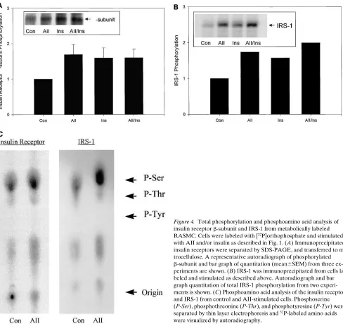
[image:5.612.56.551.60.539.2]increase in insulin receptor phosphorylation was due to an in-crease in phosphoserine (Fig. 4 C). Stimulation of RASMC with AII also increased the phosphorylation of IRS-1 by 75% (Fig. 4 B). Again this phosphorylation was associated with an increase in phosphoserine (Fig. 4 C). Interestingly, the magni-tudes of AII-induced increases in total phosphorylation of both the insulin receptor and IRS-1 were similar to that in-duced by insulin.
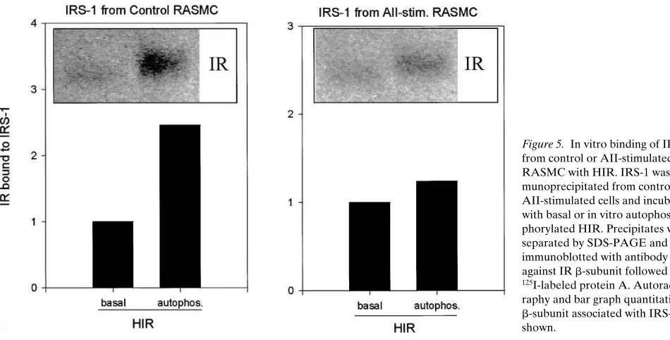
The PTB domain of IRS-1 plays a key role in the docking of IRS-1 to tyrosine 960 in the juxtamembrane region of the insulin receptor b-subunit that allows for subsequent substrate tyrosine phosphorylation (31, 40, 48). Thus, it can be hypothe-sized that serine phosphorylation of the insulin receptor or IRS-1 might influence their ability to interact with each other for subsequent tyrosine phosphorylation events. In order to in-vestigate this hypothesis, a glutathione S–transferase fusion protein containing the PTB domain of IRS-1 (GST-IRS-1-PTB) was allowed to interact in vitro with autophosphorylated WGA purified insulin receptors from control RASMC or cells that had been preincubated with AII for 7 min. The insulin re-ceptor bound to GST-IRS-1-PTB was detected by Western
blotting employing anti-insulin receptor antibodies (41). Using this assay, AII did not decrease the ability of the insulin recep-tor to bind the PTB-domain of IRS-1 (not shown). In another set of experiments, immunopurified IRS-1 from control RASMC or cells treated with AII were incubated with fully autophosphorylated WGA purified insulin receptors from CHO cells over-expressing the HIR. In this case, AII caused a reduction in the binding of IRS-1 to in vitro tyrosine phosphor-ylated insulin receptors when compared to control IRS-1 (Fig. 5). This suggests that AII-induced modifications of IRS-1, pos-sibly because of serine phosphorylation, decreases IRS-1’s ability to associate with the tyrosine phosphorylated b-subunit of the insulin receptor.
[image:6.612.310.548.64.242.2]Since AII increased serine phosphorylation correlated with the inhibition of insulin stimulated IRS-1/PI 3–kinase associa-tion (Figs. 1 B and 2 C), the effect of other agents that may reg-ulate serine phosphorylation were examined. Okadaic acid, a serine/threonine phosphatase inhibitor has been shown to in-duce a state of postreceptor insulin resistance by inhibiting IRS-1 tyrosine phosphorylation and PI 3–kinase activation in 3T3-L1 adipocytes and 293 cells overexpressing IRS-1 (43, 44). Figure 3. AII dose response and receptor antagonist effects on IRS-1/ PI 3–kinase association. (A) Dose response of AII on insulin-stimu-lated IRS-1/p85 association. Cells were pretreated with 0.1–100 nM AII for 2 min followed by a 5 min stimulation with 100 nM insulin (Ins) as indicated. IRS-1 associated p85 was determined as described in Fig. 2
C. Bar graph quantitation on p85 from three experiments in triplicate is shown. Significant differences (**P , 0.01, ANOVA) compared with insulin stimulation alone are indicated. (B) Effect of AII receptor an-tagonists of IRS-1/p85 docking. Cells were pretreated for 15 min with AII receptor antagonist saralasin (1 mM), AT1 specific receptor
antago-nist losartan (1 mM), and AT2 antagonist PD123319 (1 mM) followed
In RASMC, okadaic acid (300 nM) inhibited insulin-stimu-lated IRS-1 tyrosine phosphorylation and p85 binding by 30– 40%. These effects of okadaic acid are similar to those pro-duced by AII on insulin signaling (Fig. 6 A and data not shown). Altogether these data suggest that AII-mediated inhi-bition of the early steps of insulin signaling could be at least partially mediated by serine-phosphorylation, as in the case of okadaic acid (this study and reference 43). Pretreatment of cells with 160 nM PMA, to activate PKC also mimicked AII’s effect, inhibiting IRS-1-associated PI 3–kinase activity by 40%. However, PKC inhibition, by pretreatment of cells with 10 mM bisindolylmaleimide I (GF 109203X) for 15 min, did not block AII’s inhibitory effects on IRS-1 associated PI 3–kinase activ-ity (not shown), suggesting that the common PKC isoforms were not mediating this AII action.
To further investigate potential serine kinases that could be mediating AII’s effects on IRS-1, we examined the possibility
of feedback inhibition via PI 3–kinase itself. PI 3–kinase is a dual-specificity kinase (lipid/protein kinase) and activation of its serine kinase has been shown to both stimulate the serine phosphorylation of the p85 regulatory subunit and inhibit the lipid-phosphorylating enzymatic activity of its catalytic subunit (49, 50). Since the serine kinase activity of PI 3–kinase has been shown to phosphorylate IRS-1 in vitro (51, 52), the effect of PI 3–kinase inhibition on IRS-1/p85 binding was examined. Pretreatment of RASMC with LY294002 increased p85 associ-ation with IRS-1 induced by insulin (P, 0.05, Fig. 6 B), an ef-fect similar to that recently reported by Rameh et al. (53). However, PI 3–kinase inhibition did not block AII’s ability to reduce p85 association with IRS-1, suggesting that the p85-associated serine kinase was not responsible for AII’s effect on p85/IRS-1 association (Fig. 6 B).
[image:7.612.61.549.57.517.2]Effects of AII and PMA on p85 associated PI 3–kinase ac-tivity and p85 phosphorylation. While the decrease in IRS-1 Figure 4. Total phosphorylation and phosphoamino acid analysis of insulin receptor b-subunit and IRS-1 from metabolically labeled RASMC. Cells were labeled with [32P]orthophosphate and stimulated
with AII and/or insulin as described in Fig. 1. (A) Immunoprecipitated insulin receptors were separated by SDS-PAGE, and transferred to ni-trocellulose. A representative autoradiograph of phosphorylated b-subunit and bar graph of quantitation (mean6SEM) from three ex-periments are shown. (B) IRS-1 was immunoprecipitated from cells la-beled and stimulated as described above. Autoradiograph and bar graph quantitation of total IRS-1 phosphorylation from two experi-ments is shown. (C) Phosphoamino acid analysis of the insulin receptor and IRS-1 from control and AII-stimulated cells. Phosphoserine (P-Ser), phosphothreonine (P-Thr), and phosphotyrosine (P-Tyr) were separated by thin layer electrophoresis and 32P-labeled amino acids
associated PI 3–kinase activity in RASMC correlates with a decrease in IRS-1/p85 docking, in vivo studies of AII/insulin crosstalk from cardiac tissue showed that AII could decrease PI 3–kinase activity relative to the amount of p85 associated with IRS-1 (29). To determine whether AII may elicit direct effects on the p85/p110 PI 3–kinase complex, RASMC were stimulated with 100 nM AII for 7 and 15 min or 160 nM PMA for 15 min and p85 immunoprecipitable PI 3–kinase activity was measured as described in the Methods. Incubation with AII for 7 min had no effect on the p85 associated PI 3–kinase
activity, while prolonged incubation (15 min) with either AII or PMA decreased the p85 pool of PI 3–kinase activity by 35% (P, 0.05) and 42% (P, 0.05, ANOVA), respectively (Fig. 7
[image:8.612.58.533.58.299.2]A). Immunoprecipitation of p85 from cells incubated with AII or PMA followed by Western blotting with antibodies directed against the p85 and the p110 catalytic subunit of PI 3–kinase demonstrated that the amount of the two proteins was unal-tered as compared to the basal condition. An additional pro-tein of 50–55 kD was identified in p85 immunoblots of p85 im-munoprecipitates with an intensity of approximately one tenth Figure 5. In vitro binding of IRS-1 from control or AII-stimulated RASMC with HIR. IRS-1 was im-munoprecipitated from control or AII-stimulated cells and incubated with basal or in vitro autophos-phorylated HIR. Precipitates were separated by SDS-PAGE and immunoblotted with antibody against IR b-subunit followed by
125I-labeled protein A.
Autoradiog-raphy and bar graph quantitation of b-subunit associated with IRS-1 are shown.
Figure 6. Effect of okadaic acid and PI 3–kinase inhibition on IRS-1 tyrosine phosphorylation and p85 association in RASMC. (A) RASMC were preincubated in the presence or absence of 300 nM okadaic acid for 40 min (as indicated) and then incubated with no addition (Con), insu-lin (Ins), or AII followed by insulin as described above. IRS-1 proteins were immunoprecipitated from cleared lysates, separated by SDS-PAGE, transferred to nitrocellulose and revealed by monoclonal antiphosphotyrosine antibodies and 125I-protein A. A representative autoradiograph of
tyrosine-phosphorylated IRS-1 and bar graph of quantitation (mean6SEM) from one experiment performed in triplicate are shown. Significant differences (*P , 0.05, ANOVA) are indicated. (B) Cells were pretreated with 50 mM LY294002 for 15 min and stimulated with insulin and AII as described above. IRS-1 immunoprecipitates were separated by SDS-PAGE and immunoblotted with antibody against p85 followed by
125I-labeled protein A. An autoradiograph and bar graph quantitation of one experiment in triplicate are shown. *Significant differences
[image:8.612.64.551.463.641.2]that of p85. Interestingly, the level of this protein was reduced by 50% (P, 0.05, ANOVA) after AII or PMA stimulation versus the basal conditions (Fig. 7 B). This protein was recog-nized by antibodies against the entire p85 sequence, but not by an antibody that recognizes the SH3 domain of p85, suggesting that these cells express one or more of the alternatively spliced forms of the regulatory subunit of PI 3–kinase that have been described in other cells and tissues and termed p55a and p85/ AS53 (54, 55).
Since these results suggest that AII has an effect on p85 as-sociated PI 3–kinase above and beyond the effect of docking to IRS-1, we examined the effect of AII on PI 3–kinase cou-pled to tyrosine phosphorylated PDGF receptor (56). RASMC were stimulated with AII, PDGF, or a combination of the two hormones and PI 3–kinase activity associated with the PDGF receptor was measured as described above. PDGF stimulated a 30-fold increase in the PDGF receptor associated PI 3–kinase activity. While AII alone had no significant effect on the PDGF receptor associated PI 3–kinase activity as compared to control, as with insulin and IGF-1, AII was able to inhibit the
PDGF stimulated PI 3–kinase activity by 30–40% (P, 0.05, ANOVA, Fig. 7 C).
[image:9.612.56.556.58.421.2]To examine the possibility that the inhibitory effects of AII and PMA were related to changes in the phosphorylation state of PI 3–kinase, RASMC were metabolically labeled with [32P]orthophosphate, stimulated with AII or PMA, and the p85 subunit of PI 3–kinase was immunoprecipitated and processed for phosphoamino acid analysis. Incubation of cells with AII or PMA increased phosphorylation of the p85 subunit of PI 3–kinase by 50 and 150%, respectively (Fig. 8 A). Phospho-amino acid analysis revealed that the increase in total phos-phorylation can be accounted for by an increase in serine phosphorylation (Fig. 8 B). A low level of phosphothreonine labeling remained unchanged and no labeling of phosphoty-rosine was detected in the basal state or after stimulation with AII or PMA. Thus, AII directly alters PI 3–kinase by decreas-ing p85 associated PI 3–kinase activity and increasdecreas-ing the serine phosphorylation of p85. AII also decreases the levels of a 50–55 kD protein which is likely an alternatively spliced form of p85.
Figure 7. Effects of AII and PMA on p85-associated PI 3–kinase activ-ity. (A) RASMC were incubated with no addition (Con); AII, 7 min; AII, 15 min; and PMA, 15 min. Cell lysates were immunoprecipitated with a-p85 antibodies and PI 3–kinase assays performed as de-scribed in Methods. The bar graph shows the relative incorporation of
32P into PI 3-P (mean6SEM) from
four to five separate triplicate ex-periments. Significant differences (*P , 0.05, ANOVA) are indicated. (B) p85 immunoprecipitates from cells treated with AII or PMA for 15 min were separated and immuno-blotted with antibody against p85 or p110. A representative autoradiograph and bar graph quantitation from three experiments in triplicate are shown. (C) PDGF receptor–associ-ated PI 3–kinase activity in RASMC control cells and after AII (7 min), PDGF (5 min) or AII followed by PDGF. Cell lysates were immunopre-cipitated with anti-PDGF receptor antibodies and PI 3–kinase assays were performed as described in Methods. The bar graph shows the relative incorporation of 32P into PI 3-P (mean6SEM) from one triplicate experiment. Similar results were obtained in another experiment performed in
Discussion
Intracellular crosstalk between different signaling systems is one of the mechanisms by which cells can integrate multiple hormonal signals for survival and growth. Recent studies have demonstrated that typical G protein–coupled receptors may activate or inhibit the signaling systems classically employed by tyrosine kinase coupled receptors, such as mitogen acti-vated protein kinase, PI 3–kinase, Src, FAK, and JAK2 (21, 22, 57, 58). This signal integration is particularly important to vas-cular tissues which are in intimal contact with a variety of cir-culating hormones. Moreover, a milieu of increased hormone action within the vascular wall, involving the combination of AII, insulin and PDGF, have been strongly implicated in the neointimal formation after balloon injury and atherosclerosis (9, 59, 60). In this report, we examined the signaling crosstalk between these hormones in cultured aortic smooth muscle cells.
We found that AII inhibited insulin-, IGF-1–, and PDGF-tyrosine kinase receptor activation of the PI 3–kinase pathway. Further analysis of AII’s effects on the insulin signaling path-way revealed that AII inhibited insulin-stimulated tyrosine phosphorylation of IRS-1 and its corresponding association with the p85 subunit of PI 3–kinase. This occurred via a sarala-sin-sensitive AII receptor–stimulated pathway that was not blocked by the AT1 isoform antagonist losartan nor the AT2 antagonist PD123319. Previously, we and others have shown that AII-stimulation of plasminogen activator-1 and -2 expres-sion, and the activation of MAP kinase in these cells are AII signaling pathways which signal via the AT1 receptor (23, 42). A role for the AT2 receptor in AII’s inhibitory effect on insulin signaling appears unlikely since the AT2 antagonist PD123319
had no effect on IRS-1/p85 association. Since AII’s inhibitor effect on IRS-1/p85 docking occurs rapidly, with only a 2 min preincubation, and at low concentrations of AII (0.1 nM), it is unlikely that AII’s effects are mediated via biologically active peptides (such as angiotensin III or IV) generated from the limited proteolysis of AII. Therefore, we hypothesize that AII’s inhibitory effects on the insulin signaling pathway in these cells are primarily mediated via an AII receptor other than AT1 or AT2.
Recent in vivo studies of AII/insulin crosstalk in rat heart have shown that AII is capable of stimulating tyrosyl-phos-phorylation of IRS-1 and IRS-2, docking with PI 3–kinase and inhibition of its enzymatic activity, both in the basal and insu-lin stimulated states, without affecting tyrosine phosphoryla-tion of IRS-1 and of the insulin receptor b-subunit. These lat-ter phenomena could be blocked by AT1 receptor antagonists (28, 29). Thus, both in vivo and in vitro studies demonstrate that AII inhibits insulin signaling via the PI 3–kinase pathway, although AII exerts opposite effects on IRS-1 phosphorylation and IRS-1/PI 3–kinase docking in heart and RASMC.
[image:10.612.59.544.56.326.2]Reduced tyrosyl-phosphorylation as well as increased ser-ine phosphorylation of the b-subunit of the insulin receptor and IRS-1 have been proposed as mechanisms for the inhibi-tion of insulin signaling (43–47, 61, 62). In this report, we dem-onstrate that AII is capable of stimulating serine-phosphoryla-tion of three crucial components of the insulin signaling network, namely the insulin receptor itself, IRS-1 and the p85 subunit of PI 3–kinase (Fig. 9). Pretreatment of cells with AII did not alter insulin receptor autophosphorylation, suggesting that the decrease in IRS-1 tyrosine phosphorylation was not due to a decrease in insulin receptor kinase activity. The ef-fects of AII on IRS-1 in RASMC, increased IRS-1 serine Figure 8. AII stimulates total phosphorylation of the p85 regulatory sub-unit of PI 3–kinase. RASMC were in vivo labeled with [32
phosphorylation in association with decreased tyrosine phorylation, are reminiscent of those reported with phos-phatase 2A inhibitor, okadaic acid (43, 44). In this study, we also found that okadaic acid as well as phorbol ester mimicked AII’s inhibition of PI 3–kinase associated with IRS-1 in RASMC. Since AII increased insulin receptor and IRS-1 serine phosphorylation without impairing the insulin receptor tyrosine kinase, we examined the possibility that AII may in-hibit insulin-stimulated IRS-1 tyrosine phosphorylation by re-ducing IR/IRS-1 association. Consistent with this hypothesis, we found that IRS-1 from AII-stimulated RASMC exhibited a decreased ability to bind autophosphorylated insulin receptor in an in vitro assay. These results suggest that AII can reduce insulin stimulated IRS-1 tyrosine phosphorylation by decreas-ing the association between the receptor b-subunit and IRS-1. Thus it is tempting to hypothesize that serine-phosphorylation of IRS-1 impairs its docking to the autophosphorylated insulin receptor. Alternatively, AII may decrease IRS-1/PI 3–kinase association by rapidly inducing the tyrosine dephosphorylation of IRS-1 via the activation or upregulation of a protein-tyro-sine phosphatase (63).
While in RASMC the decrease in IRS-1 associated PI 3–kinase is in part due to a decrease in IRS-1/p85 docking, in vivo studies of AII/insulin crosstalk from cardiac tissue have shown that AII can decrease PI 3–kinase, even in the presence of an increased amount of p85 associated with IRS-1 (29). As these results suggested additional effects of AII on the PI 3–kinase system, the effect of AII on p85 associated PI 3–kinase activity and p85 phosphorylation was measured. We now find that both AII and phorbol ester significantly reduced the total pool of PI 3–kinase activity associated with p85. The AII- or PMA-stimulated decreases in PI 3–kinase activity could not be explained by decreases in the total amount of immunoprecipi-tated p85 or p110 subunits, showing that these agonists altered the intrinsic activity rather than the amount of PI 3–kinase.
In-terestingly, IRS-1 and PI 3–kinase are also intracellular targets for receptors other than the insulin receptor, such as growth hormone and interferon g (64), suggesting that these inhibi-tory effects of AII may crosstalk with other receptor pathways. In order to determine if AII could interfere with the coupling of PI 3–kinase to other receptor tyrosine kinase systems, we examined the effect of AII on PDGF-receptor associated PI 3–kinase. Pretreatment of cells with AII decreased PDGF-stimulated increases in both PI 3–kinase activity and p85 bind-ing to the PDGF receptor. Since PI 3–kinase autophosphoryla-tion, via a serine kinase intrinsic to the enzyme, has been reported to act as an inhibitory feed back loop on PI 3–kinase activity (49, 50), the effect of AII on p85 phosphorylation was measured. These experiments demonstrated that both AII and PMA were capable of increasing serine phosphorylation of p85. To our knowledge this is the first evidence of a hormone-induced serine phosphorylation and inhibition of the p85/p110 PI 3–kinase. Interestingly, both AII and PMA also decreased the amount of a p85 isoform in these precipitates with molecu-lar masses of z 50–55 kD. This protein(s) was recognized by a polyclonal antibody against the entire p85 sequence but not by a monoclonal antibody specific to the SH3 domain of p85, sug-gesting that this protein is either the p50a or AS53 alterna-tively spliced version of the regulatory protein of PI 3–kinase that has been reported recently (54, 55). These results further reinforce the finding that AII can modulate the activity of the PI 3–kinase system.
[image:11.612.58.385.59.300.2]and IGF-1 receptors, has been proposed to contribute to ath-erosclerosis and abnormalities in blood flow. While the spe-cific role of insulin-stimulated PI 3–kinase in the vasculature has not yet been established, a recent study suggests that this pathway is important for insulin-stimulated nitric oxide pro-duction in endothelial cells (66). Reports from other cell types have shown that activation of PI 3–kinase by growth factors is a crucial step for DNA synthesis and cell division (67, 68) and, interestingly, IRS-1 has been shown to be an important re-quirement for cell cycle progression, cell division, and growth (69–71). Thus, crosstalk between AII and insulin on the IRS-1/ PI 3–kinase pathway may have an important role in the regula-tion of hemodynamics and vascular growth. The results from this study suggest that AII, and other agents that are able to in-duce serine phosphorylation of the IR, IRS-1, and/or PI 3–kinase, can contribute to insulin resistance in the vasculature.
Acknowledgments
This work was supported in part by National Institutes of Health Grants DK 48358 (to E.P. Feener), DK 33201 (to C.R. Kahn), DK 36836 (Joslin’s Diabetes and Endocrinology Research Center Grant), and Grants from Istituto Scientifico San Raffaele, Milano, Italy and Ministero della Sanita, Italy (to F. Folli) and the Markey Charitable Trust (E.P. Feener).
References
1. Dzau, V.J. 1994. Cell biology and genetics of angiotensin in cardiovascu-lar disease. J. Hypertens. 12(suppl):S3–S10.
2. Regitz-Zagrosek, V., M. Neub, and J. Holzmeister. 1996. Molecular biol-ogy of angiotensin receptors and their role in human cardiovascular disease. J. Mol. Med. 74:233–251.
3. Diet, F., R.E. Pratt, G.J. Berry, N. Momose, G.H. Gibbons, and V.J. Dzau. 1996. Increased accumulation of tissue ACE in human atherosclerotic coronary artery disease. Circulation. 94:2756–2767.
4. Rakugi, H., H.J. Jacob, J.E. Krieger, J.R. Ingelfinger, and R.E. Pratt. 1993. Vascular injury induces angiotensinogen gene expression in the media and neointima. Circulation. 87:283–290.
5. Rakugi, H., D.K. Kim, J.E. Krieger, D.S. Wang, V.J. Dzau, and R.E. Pratt. 1994. Induction of angiotensin converting enzyme in the neointima after vascular injury. Possible role in restenosis. J. Clin. Invest. 93:339–346.
6. Daemen, M.J.A.P., D.M. Lombardi, F.T. Bosman, and S.M. Schwartz. 1991. Angiotensin II induces smooth muscle cell proliferation in the normal and injured rat arterial wall. Circ. Res. 68:450–456.
7. Hamdan, A.D., W.C. Quist, J.B. Gagne, and E.P. Feener. 1996. Angio-tensin-converting enzyme inhibition suppresses plasminogen activator inhibi-tor-1 expression in the neointima of balloon-injured rat aorta. Circulation. 93: 1073–1078.
8. Kim, S., M. Kawamura, H. Wanibuchi, K. Ohta, A. Hamaguchi, T. Omura, T. Yukimura, K. Miura, and H. Iwao. 1995. Angiotensin II type 1 re-ceptor blockade inhibits the expression of immediate-early genes and fibronec-tin in rat injured artery. Circulation. 92:88–95.
9. Powell, J.S., J.-P. Clozel, R.K.M. Muller, H. Kuhn, F. Hefti, M. Hosang, and H.R. Baumgartner. 1989. Inhibitors of angiotensin converting enzyme pre-vent myointimal proliferation after vascular injury. Science (Wash. DC). 245: 186–188.
10. Garg, R., and S. Yusef. 1995. Overview of randomized trials of angio-tensin-converting enzyme inhibitors on mortality and morbidity in patients with heart failure. JAMA (J. Am. Med. Assoc.). 273:1450–1456.
11. MERCATOR Study Group. 1992. Does the new angiotensin-convert-ing-enzyme inhibitor cilazapril prevent restenosis after percutaneous translumi-nal coronary angioplasty? Results of the MERCATOR study: a multicenter, randomized, double blind placebo-controlled trial. Circulation. 86:100–110.
12. Torlone, E., M. Britta, A.M. Rambotti, G. Perriello, F. Santeusanio, P. Brunetti, and G.B. Bolli. 1993. Improved insulin action and glycemic control af-ter long-af-term angiotensin-converting enzyme inhibition in subjects with araf-terial hypertension and type II diabetes. Diabetes Care. 16:1347–1355.
13. Shieh, S., D. Shen, W.H.H. Sheu, M. Fuh, C.Y. Jeng, J.R. Jeng, Y.I. Chen, and G.M. Reaven. 1992. Improvement in metabolic risk factors for coro-nary heart disease associated with cilazapril treatment. Am. J. Hypertens. 5: 506–510.
14. Raccah, D., M. Pettenuzzo-Mollo, O. Provendier, L. Boucher, J.A.
Co-zic, R. Gorlier, P. Huin, J. Sicard, and P. Vague. 1994. Comparison of the effects of captopril and nicardipine on insulin sensitivity and thrombotic profile in pa-tients with hypertension and android obesity. Am. J. Hypertens. 7:731–738.
15. Howard, G., D.H. O’Leary, D. Zaccaro, S. Haffner, M. Rewers, R. Hamman, J.V. Selby, M.F. Saad, P. Savage, R. Bergman, and IRAS Investiga-tors. 1996. Insulin sensitivity and atherosclerosis. Circulation. 93:1809–1817.
16. Haffner, S.M., M.P. Stern, H.P. Hazuda, B.D. Mitchell, and J.K. Patter-son. 1990. Cardiovascular risk factors in confirmed prediabetic individuals. JAMA (J. Am. Med. Assoc.). 263:2893–2898.
17. Ferrannini, E., G. Buzzigoli, R. Bonadonna, M.A. Giorico, M. Oleggini, L. Graziadei, R. Pedrinelli, L. Brandi, and S. Bevilacqua. 1987. Insulin resis-tance in essential hypertension. N. Engl. J. Med. 317:350–357.
18. Zavaroni, I., E. Bonora, M. Pagliara, E. Dall’Aglio, L. Luchetti, G. Buo-nanno, P.A. Bonati, M. Bergonzani, L. Gnudi, M. Passeri, et al. 1989. Risk fac-tors for coronary artery diease in healthy persons with hyperinsulinemia and normal glucose tolerance. N. Engl. J. Med. 320:702–706.
19. Murphy, T.J., R.W. Alexander, K.K. Griendling, M.S. Runge, and K.E. Bernstein. 1991. Isolation of a cDNA encoding the vascular type-1 angiotensin II receptor. Nature (Lond.). 351:233–236.
20. Dixon, B.S., R.V. Sharma, T. Dickerson, and J. Fortune. 1994. Bradyki-nin and angiotensin II: activation of protein kinase C in arterial smooth muscle. Am. J. Physiol. 266:C1406–C1420.
21. Marrero, M.B., B. Schieffer, W.G. Paxton, I. Heerdt, B.C. Berk, P. Delafontaine, and K.E. Bernstein. 1995. Direct stimulation of Jak/STAT path-way by the angiotensin II AT1 receptor. Nature (Lond.). 375:247–250.
22. Schorb, W., T.C. Peeler, N.N. Madigan, K.M. Conrad, and K.M. Baker. 1994. Angiotensin II-induced protein tyrosine phosphorylation in neonatal rat cardiac fibroblasts. J. Biol. Chem. 269:19626–19632.
23. Feener, E.P., J.M. Northrup, L.P. Aiello, and G.L. King. 1995. Angio-tensin II induces plasminogen activator inhibitor-1 and -2 expression in vascular endothelial and smooth muscle cells. J. Clin. Invest. 95:1353–1362.
24. Bell, L., D.J. Luthringer, J.A. Madri, and S.L. Warren. 1992. Autocrine angiotensin system regulation of bovine aortic endothelial cell migration and plasminogen activator involves modulation of proto-oncogene pp60c-src ex-pression. J. Clin. Invest. 89:315–320.
25. Weber, H., D.S. Taylor, and C.J. Molloy. 1994. Angiotensin II induces delayed mitogenesis and cellular proliferation in rat aortic smooth muscle cells. J. Clin. Invest. 93:788–798.
26. Daub, H., F.U. Weiss, C. Wallasch, and A. Ullrich. 1996. Role of trans-activation of the EGF receptor in signalling by G protein–coupled receptors. Nature (Lond.). 379:557–560.
27. Linseman, D.A., C.W. Benjamin, and D.A. Jones. 1995. Convergence of angiotensin II and platelet-derived growth factor receptor signaling cascades in vascular smooth muscle cells. J. Biol. Chem. 270:12563–12568.
28. Saad, M.J.A., L.A. Velloso, and C.R.O. Carvalho. 1995. Angiotensin II induces tyrosine phosphorylation of insulin receptor substrate 1 and its associa-tion with phosphatidylinositol 3–kinase in rat heart. Biochem. J. 310:741–744.
29. Velloso, L.A., F. Folli, X.J. Sun, M.F. White, M.J.A. Saad, and C.R. Kahn. 1996. Cross-talk between the insulin and angiotensin signaling systems. Proc. Natl. Acad. Sci. USA. 93:12490–12495.
30. White, M.F., and C.R. Kahn. 1994. The insulin signaling system. J. Biol. Chem. 269:1–4.
31. Eck, M.J., S. Dhe-Paganon, T. Trub, R.T. Nolte, and S.E. Shoelson. 1996. Structure of the IRS-1 PTB domain bound to the juxtamembrane region of the insulin receptor. Cell. 85:695–705.
32. Sun, X.J., L. Wang, Y. Zhang, L. Yenush, M.G. Myers, Jr., E. Glasheen, W.S. Lane, J.H. Pierce, and M.F. White. 1995. Role of IRS-2 in insulin and cy-tokine signalling. Nature (Lond.). 377:173–177.
33. Sun, X.J., P. Rotheenberg, C.R. Kahn, J.M. Backer, E. Araki, P.A. Wilden, D.A. Cahill, B.J. Goldstein, and M.F. White. 1991. Structure of the in-sulin receptor substrate IRS-1 defines a unique signal transduction protein. Na-ture (Lond.). 352:73–77.
34. Folli, F., M.J. Saad, J.M. Backer, and C.R. Kahn. 1992. Insulin stimula-tion of phosphatidylinositol 3–kinase activity and associastimula-tion with insulin recep-tor substrate 1 in liver and muscle on the intact rat. J. Biol. Chem. 267:22171– 22177.
35. Sun, X.J., S. Pons, T. Asano, M.G. Myers, Jr., E. Glasheen, and M.F. White. 1996. The Fyn tyrosine kinase binds IRS-1 and forms a distinct signaling complex during insulin stimulation. J. Biol. Chem. 271:10583–10587.
36. Kelly, K.L., and N.B. Ruderman. 1993. Insulin-stimulated phosphati-dylinositol 3–kinase. J. Biol. Chem. 268:4391–4398.
37. Backer, J.M., M.G. Myers, Jr., S.E. Shoelson, D.J. Chin, X.-J. Sun, M. Miralpeix, P. Hu, B. Margolis, E.Y. Skolnik, J. Schlessinger, and M.F. White. 1992. Phosphatidylinositol 39–kinase is activated by association with IRS-1 dur-ing insulin stimulation. EMBO (Eur. Mol. Biol. Organ.) J. 11:3469–3479.
38. Steinberg, H.O., G. Brechtel, A. Johnson, N. Fineberg, and A.D. Baron. 1994. Insulin-mediated skeletal muscle vasodilation is nitric oxide dependent. A novel action of insulin to increase nitric oxide release. J. Clin. Invest. 94:1172– 1179.
40. Feener, E.P., J.M. Backer, G.L. King, P.A. Wilden, X.J. Sun, C.R. Kahn, and M.F. White. 1993. Insulin stimulates serine and tyrosine phosphorylation in the juxtamembrane region of the insulin receptor. J. Biol. Chem. 268:11256– 11264.
41. Wolf, G., T. Trub, E. Ottinger, L. Groninga, A. Lynch, M.F. White, M. Miyazaki, J. Lee, and S.E. Shoelson. 1995. PTB domains of IRS-1 and shc have distinct but overlapping binding specificities. J. Biol. Chem. 270:27407–27410.
42. Eguchi, S., T. Matsumoto, E.D. Motley, H. Utsunomiya, and T. Ina-gami. 1996. Identification of an essential signaling cascade for mitogen-activated protein kinase activation by angiotensin II in cultured rat vascular smooth mus-cle cells. J. Biol. Chem. 274:14169–14175.
43. Tanti, J., T. Gremeaux, E. Van Obberghen, and Y. Le Marchand-Brus-tel. 1994. Serine/threonine phosphorylation of insulin receptor substrate 1 mod-ulates insulin receptor signaling. J. Biol. Chem. 269:6051–6057.
44. Mothe, I., and E. Van Obberghen. 1996. Phosphorylation of insulin re-ceptor substrate-1 on multiple serine residues, 612, 632, 662, and 731, modulates insulin action. J. Biol. Chem. 271:11222–11227.
45. Takayama, S., M.F. White, and C.R. Kahn. 1988. Phorbol ester–induced serine phosphorylation of the insulin receptor decreases its tyrosine kinase ac-tivity. J. Biol. Chem. 263:3440–3447.
46. Hotamisligil, G.S., P. Peraldi, A. Budavari, R. Ellis, M.F. White, and B.M. Spiegelman. 1996. IRS-1–mediated inhibition of insulin receptor tyrosine kinase activity in TNF-a and obesity-induced insulin resistance. Science (Wash. DC). 271:665–668.
47. Pillay, T.S., S. Xiao, and J.M. Olefsky. 1996. Glucose-induced phos-phorylation of the insulin receptor. J. Clin. Invest. 97:613–620.
48. White, M.F., J.N. Livingston, J.M. Backer, V. Lauris, T.J. Dull, A. Ull-rich, and C.R. Kahn. 1988. Mutation of the insulin receptor at tyrosine 960 in-hibits signal transmission but does not affect its tyrosine kinase activity. Cell. 54: 641–649.
49. Dhand, R., I. Hiles, G. Panayotou, S. Roche, M.J. Fry, I. Gout, N.F. Totty, O. Truong, P. Vicendo, K. Yonezawa, et al. 1994. PI 3–kinase is a dual specificity enzyme: autoregulation by an intrinsic protein-serine kinase activity. EMBO (Eur. Mol. Biol. Organ.) J. 13:522–533.
50. Carpenter, C.L., K.R. Auger, B.C. Duckworth, W.M. Hou, B. Schaff-hausen, and L.C. Cantley. 1993. A tightly associated serine/threonine protein kinase regulates phosphoinositide 3–kinase activity. Mol. Cell. Biol. 13:1657– 1665.
51. Tanti, J., T. Gremeaux, E. Van Obbergen, and Y. Le Marchand-Brustel. 1994. Insulin receptor substrate 1 is phosphorylated by the serine kinase activity of phosphatidylinositol 3–kinase. Biochem. J. 304:17–21.
52. Lam, K., C.L. Carpenter, N.B. Ruderman, J.C. Friel, and K.L. Kelley. 1994. The phosphatidylinositol 3–kinase serine kinase phosphorylates IRS-1. Stimulation by insulin and inhibition by Wortmannin. J. Biol. Chem. 269:20648– 20652.
53. Rameh, L.E., C. Chen, and L.C. Cantley. 1995. Phosphatidylinositol (3,4,5)P3 interacts with SH2 domains and modulates PI 3–kinase association with tyrosine-phosphorylated proteins. Cell. 83:821–830.
54. Antonetti, D.A., P. Algenstaedt, and C.R. Kahn. 1996. Insulin receptor substrate 1 binds two novel splice variants of the regulatory subunit of phos-phatidylinositol 3–kinase in muscle and brain. Mol. Cell. Biol. 16:2195–2203.
55. Inukai, K., M. Anai, E. Van Breda, T. Hosaka, H. Katagiri, M. Funaki, Y. Fukushima, T. Ogihara, Y. Yazaki, M. Kikuchi, et al. 1996. A novel 55-kDa regulatory subunit for phosphatidylinositol 3–kinase structurally similar to p55PIK is generated by alternative splicing of the p85a gene. J. Biol. Chem. 271:5317–5320.
56. Klippel, A., J.A. Escobedo, W.J. Fantl, and L.T. Williams. 1992. The C-terminal SH2 domain of p85 accounts for the high affinity and specificity of the binding of phosphatidyinositol 3–kinase to phosphorylated platelet-derived growth factor b receptor. Mol. Cell. Biol. 12:1451–1459.
57. Stoyanov, B., S. Volinia, T. Hanck, I. Rubio, M. Loubtchenkov, D. Malek, S. Stoyanova, B. Vanhaesebroeck, R. Dhand, B. Nurnberg, et al. 1995. Cloning and characterization of a G protein–activated human phosphoinosi-tide–3 kinase. Science (Wash. DC). 269:690–693.
58. Wan, Y., T. Kurosaki, and X.-Y. Huang. 1996. Tyrosine kinases in acti-vation of the MAP kinase cascade by G protein–coupled receptors. Nature (Lond.). 380:541–544.
59. Jawien, A., D.F. Bowen-Pope, V. Lindner, S.M. Schwartz, and A.W. Clowes. 1992. Platelet-derived growth factor promotes smooth muscle migra-tion and intimal thickening in a rat model of balloon angioplasty. J. Clin. Invest. 89:507–511.
60. Ridray, S., D. Heudes, O. Michel, L. Penicaud, and A. Ktorza. 1994. In-creased SMC proliferation after endothelial injury in hyperinsulinemic obese Zucker rats. Am. J. Physiol. 267:H1976–H1983.
61. Caro, J.F., O. Ittoop, W.J. Pories, D. Meelheim, E.G. Flickinger, F. Tho-mas, M. Jenquin, J.F. Silverman, P.G. Khazanie, and M.K. Sinha. 1986. Studies on the mechanism of insulin resistance in the liver from humans with nonlin-dependent diabetes. Insulin action and binding in isolated hepatocytes, insu-lin receptor structure, and kinase activity. J. Clin. Invest. 78:249–258.
62. Folli, F., M.J. Saad, J.M. Backer, and C.R. Kahn. 1993. Regulation of the phosphatidylinositol 3–kinase activity in liver and muscle of animal models of insulin-resistant and insulin-deficient diabetes mellitus. J. Clin. Invest. 92: 1787–1794.
63. Duff, J.L., M.B. Marrero, W.G. Paxton, C.H. Charles, L.F. Lau, K.E. Bernstein, and B.C. Berk. 1993. Angiotensin II induces 3CH134, a protein-tyrosine phosphatase, in vascular smooth muscle cells. J. Biol. Chem. 268: 26037–26040.
64. Myers, M.G., Jr., and M.F. White. 1996. Insulin signal transduction and the IRS proteins. Annu. Rev. Pharmacol. Toxicol. 36:615–658.
65. Banskota, N.K., R. Taub, K. Zellner, P. Olsen, and G.L. King. 1989. Characterization of the induction of proto-oncogene c-myc and cellular growth in human vascular smooth muscle cells by insulin and IGF-1. Diabetes. 38:123– 129.
66. Zeng, G., and M.J. Quon. 1996. Insulin-stimulated production of nitric oxide is inhibited by wortmannin. J. Clin. Invest. 98:894–898.
67. Cheatham, B., C.J. Vlahos, L. Cheatham, L. Wang, J. Blenis, and C.R. Kahn. 1994. Phosphatidylinositol 3-kinase activation is required for insulin stimulation of pp70 S6 kinase, DNA synthesis, and glucose transporter translo-cation. Mol. Cell. Biol. 14:4902–4911.
68. Chung, J., T.C. Grammer, K.P. Lemon, A. Kazlauskas, and J. Blenis. 1994. PDGF- and insulin-dependent pp70S6k activation mediated by phos-phatidylinositol-3-OH kinase. Nature (Lond.). 370:71–75.
69. Araki, E., M.A. Lipes, M.E. Patti, J.C. Bruning, B. Haag 3rd, R.S. Johnson, and C.R. Kahn. 1994. Alternative pathway of insulin signalling in mice with targeted disruption of the IRS-1 gene. Nature (Lond.). 372:186–190.
70. Tamemoto, H., T. Kadowaki, K. Tobe, T. Yagi, H. Sakura, et al. 1994. Insulin resistance and growth retardation in mice lacking insulin receptor sub-strate-1. Nature (Lond.). 372:182–186.