0095-1137/96/$04.0010
Copyrightq1996, American Society for Microbiology
Epidemiology of
Burkholderia cepacia
Infection in Patients with
Cystic Fibrosis: Analysis by Randomly Amplified
Polymorphic DNA Fingerprinting
ESHWAR MAHENTHIRALINGAM,1* MAUREEN E. CAMPBELL,1DEBORAH A. HENRY,1
ANDDAVID P. SPEERT1,2
Division of Infectious and Immunological Diseases, Department of Paediatrics and the Canadian Bacterial Diseases Network,1and Department of Microbiology and Immunology,2
University of British Columbia, Vancouver, Canada
Received 23 May 1996/Returned for modification 5 August 1996/Accepted 4 September 1996
We fingerprinted a collection of 627Burkholderia cepaciaisolates from 255 patients with cystic fibrosis (CF)
and 43 patients without CF and from the environment, by a PCR-based randomly amplified polymorphic DNA (RAPD) method with primers selected for their ability to produce discriminatory polymorphisms. The RAPD typing method was found to be reproducible and discriminatory, more sensitive than PCR ribotyping, and able
to group epidemiologically relatedB. cepaciastrains previously typed by both pulsed-field gel electrophoresis
and conventional ribotyping. Seven strain types infecting multiple CF patients were found at several different CF treatment centers in Canada, the United States, the United Kingdom, France, and Australia, indicating the presence of epidemic strain types. Most CF patients were each colonized with a single strain type, and several
patients harbored the same strain type for 5 or more years.B. cepaciaisolates recovered from other clinical
sources (44 isolates examined) and from the environment (58 isolates examined) possessed RAPD fingerprints that were generally distinct from CF-associated strain types (525 isolates examined). RAPD is a versatile
fingerprinting method for studying the epidemiology ofB. cepacia.
Cystic fibrosis (CF) is the most common potentially fatal autosomal recessive disease in North America, afflicting ap-proximately 1 in 2,000 live births among Caucasians (5). Al-though Pseudomonas aeruginosais the predominant respira-tory pathogen in patients with CF (26),Burkholderia(formerly Pseudomonas)cepaciahas emerged as a particularly problem-atic pulmonary pathogen in these patients. The organism is highly virulent in certain patients with CF (8, 30). It is intrin-sically resistant to a wide range of antimicrobial agents (22), and there is considerable evidence that B. cepacia, unlike P. aeruginosa, can spread from one CF patient to another both within and outside the hospital (2, 7, 9, 13, 15). However, investigations finding no evidence of patient-to-patient trans-mission (27) or documenting only the transtrans-mission of one epidemic clone (29) suggest that the transmissibility ofB. ce-pacia may vary depending on a number of factors including strain, treatment center, and CF patient population. It has become critically important to determine the risk factors for patient-to-patient spread ofB. cepaciaand to identify strains that are prevalent and pose the greatest risk of infection at CF treatment centers.
Several techniques have been employed for typingB. cepa-cia. Phenotypic methods such as serology, biochemical profile, or pigment production have been widely used (23) but are subject to instability because the phenotype ofB. cepaciaCF isolates may vary markedly (12). Multilocus enzyme electro-phoresis, a phenotypic method, has also been applied toB. ce-pacia (9); multilocus enzyme electrophoresis demonstrated that CF isolates are clustered clonally and are generally distinct from nosocomial and environmental isolates; however, the
number of strains examined from non-CF sources was small. Genetic typing methods, such as ribotyping (28), have been shown to provide good specificity and sensitivity for epidemi-ological study ofB. cepacia(23). Ribotyping analysis has been used to demonstrate patient-to-patient transmission (7, 13, 25), clustering of B. cepacia types at treatment centers (15), and transatlantic spread of one transmissible lineage (9, 29). Typ-ing by pulsed-field gel electrophoresis (PFGE) has also dem-onstrated the spread of certain strains among CF patients (7, 25, 29); however, Steinbach et al. (27) reported no PFGE typing evidence for person-to-person transmission ofB. cepa-ciaat one CF treatment center. Three PCR-based methods for typing B. cepacia have also been reported: PCR ribotyping (11), randomly amplified polymorphic DNA (AP-PCR or RAPD) (2), and enterobacterial repetitive intergenic consen-sus sequence PCR (17).
Over the last 10 years, our laboratory has established a large collection of B. cepaciaisolates from pediatric and adult CF treatment centers in Vancouver, British Columbia, Canada. This collection has recently been expanded to include isolates from other CF centers in Canada, the United States, the United Kingdom, France, and Australia, as well as many iso-lates from other clinical sources and from the environment. We sought to type this collection of isolates to establish which (if any) strains ofB. cepaciaare prevalent in patients with CF at various treatment centers and to identify strains that might be transmitted from patient to patient. Because of the large numbers of isolates involved, a PCR-based assay was utilized to enable high sample throughput (10, 18). We developed a RAPD typing method, which, unlike that reported previously (2), utilized several different arbitrary primers screened and selected for their ability to produce stable and discriminatory polymorphisms. The same RAPD method has been success-fully applied to differentiate a large collection ofP. aeruginosa isolates from CF patients (18). The RAPD typing results of 627
* Corresponding author. Mailing address: Rm. 304, The Research Center, 950 W. 28th Avenue, Vancouver, British Columbia, Canada V5Z 4H4. Phone: (604) 875-2466. Fax: (604) 875-2226. Electronic mail address: speert2@cbdn.ca.
2914
on May 15, 2020 by guest
http://jcm.asm.org/
B. cepaciaisolates from patients at several CF treatment cen-ters and from various other clinical and environmental sources form the basis of this report.
MATERIALS AND METHODS
Collection ofB. cepacia isolates and microbiological methods.Isolates of
B. cepaciawere received from CF clinics and clinical and research laboratories across Canada and from the United States and the United Kingdom. A total of 627 isolates were examined and included 525 isolates recovered from 255 pa-tients with CF, 44 isolates recovered from 43 papa-tients without CF, and 58 isolates recovered from the environment. Canadian contributors were as follows: P. Zu-berbuhler and N. Brown (University of Alberta Hospitals, Edmonton, Alberta), E. Sheperd (Janeway Child Health Center, St. John’s, Newfoundland), M. Ruel and L. Cote (University of Laval Hospital Center, Sainte Foy, Quebec), L. Wil-cox (McMaster University Medical Centre, Hamilton, Ontario), A. Matlow (Hospital for Sick Children, Toronto, Ontario), E. Tullis (Wellesley Hospital, Toronto, Ontario), J. Hillsdon-Smith (Laurentian Hospital, Sudbury, Ontario), D. Hughes (Izaak Walton Killam Children’s Hospital, Halifax, Nova Scotia), N. Cimolai (British Columbia’s Children’s Hospital, Vancouver, British Columbia), A. Clarke (St. Paul’s Hospital, Vancouver, British Columbia), M. Li (Victoria General Hospital, Victoria, British Columbia), and W. Johnson (Laboratory Center for Disease Control, Ottawa, Ontario). U.S. contributors were as follows: J. Burns (Children’s Hospital and Medical Center, Seattle, Wash.), T. Stull (Medical College of Philadelphia, Philadelphia, Pa.), M. Roy (Genentech, Inc., San Francisco, Calif.), and P. Ferrieri (University of Minnesota, Minneapolis). A collection of isolates from a variety of different sources, including CF patents in the United Kingdom, was provided by J. Govan (University Medical School, Edinburgh, Scotland, United Kingdom). Isolates from France were provided by E. Bingen (Hoˆpital Robert Debre´, Paris, France), and isolates from Australia were provided by P. Taylor (Prince of Wales Hospital, Randwick, New South Wales, Australia).
Culture and confirmation of identification of isolates were carried out as described previously (3). After confirmation of species, epidemiological (if known) and biochemical data were entered into a computer database. All iso-lates were assigned a code and, after DNA extraction, were assigned a separate arbitrary number (with the prefix “B”) to allow for blinding and unbiased inter-pretation of results. The repository code for one isolate representative of each RAPD type is shown in Tables 1, 2, and 3.
RAPD typing ofB. cepacia.Genomic DNA was isolated fromB. cepaciaby mechanical disruption with glass beads exactly as described forP. aeruginosa
(18). One hundred random 10-base RAPD primers were screened for their ability to amplify polymorphisms fromB. cepaciaATCC 25416 DNA with the PCR conditions described previously (1, 18). The eight primers able to amplify polymorphisms fromP. aeruginosa(18) were also found to produce fingerprint-ing profiles fromB. cepacia(see Fig. 1A); their sequences are as follows (59to 39): 208, ACGGCCGACC; 228, GCTGGGCCGA; 241, GCCCGAGCGG; 270, TGCGCGCGGG; 272, AGCGGGCCAA; 275, CCGGGCAAGC; 277, AGG AAGGTGC; and 287, CGAACGGCGG. Primary typing of all theB. cepacia
isolates described in this study was performed with primer 270, and confirmation of the strain types established with this primer was provided with primers 208 and 272. RAPD fingerprint profiles were compared visually and with the aid of computer analysis (GelManager for Windows; Biosystematica, Prague, Czech Republic) (18). Similarity coefficients were calculated across the entire
absor-bance profile of each fingerprint by a Pearson product moment coefficient (GelManager for Windows).
PCR ribotyping.The PCR primers and conditions described by Kostman et al. (11) were used to amplify the polymorphisms present in the 16S–23S spacer regions of the rRNA operon ofB. cepacia. DNA polymorphisms were then visualized after separation of fragments on 2% agarose gels as described previ-ously (24). Comparison of the PCR ribotyping polymorphisms was made by eye.
RESULTS
Identification of primers for RAPD analysis.The
polymor-phisms amplified by the eight functional RAPD primers from DNA extracted from two subcultures of B. cepacia ATCC 25416 made from the same freezer vial on two separate occa-sions are shown in Fig. 1A. Each primer amplified a DNA fingerprint ranging from 5 to 20 bands, over a size range of 100 bp to 5 kb, which was reproducible for the two independent preparations of DNA. Two conditions were found to affect the reproducibility of the RAPD fingerprint: template DNA con-centration and primer concon-centration. The effect of the tem-plate DNA concentration on fingerprint profile is shown in Fig. 1B. The RAPD polymorphisms remained stable with 10 to 100 ng of template DNA; if less than 10 ng ofB. cepaciaDNA was present in the PCR, no or partial fingerprints were obtained, and with greater than 100 ng of DNA, there was loss of band-ing (Fig. 1B). RAPD fband-ingerprints were also stable when be-tween 10 and 160 pmol of primer was added to reaction mix-tures containing 40 ng of template DNA (data not shown). Reaction mixtures containing 40 ng of template DNA and 40 pmol of RAPD primer were found to be optimal for amplifi-cation of reproducible fingerprints from B. cepaciaand were applied throughout the study. Failure to generate a reproduc-ible fingerprint from a sample of DNA was rare; such problems were corrected by preparation of a fresh DNA stock, and reproducible fingerprints were generated for all 627 isolates examined in the study. Although all eight primers were found to discriminate among unrelatedB. cepacia strains (data not shown), primer 270 (the first primer to be evaluated) was used to type all the isolates examined in this study (see below). Primers 208 and 272 were used to confirm RAPD types found with primer 270; further evaluation of the remaining five prim-ers was not carried out.
Identification of genetically relatedB. cepacia strains. All
B. cepaciaisolates were typed without prior knowledge of their source by RAPD with primer 270. Nine groups of related RAPD fingerprints and five unique fingerprints were found in
FIG. 1. (A) The polymorphisms amplified by eight RAPD primers fromB. cepaciaATCC 25416 DNA prepared on two separate occasions (a and b) from the same frozen culture stock. Primer numbers are indicated above each lane. (B) The effect of template DNA concentration on the RAPD fingerprint profile. The amount of DNA (nanograms) added to each reaction mixture is shown above each lane; DNA extracted fromB. cepaciaFC0001 (RAPD type 4) was used and amplified with primer 208 under the conditions described in Materials and Methods. Molecular size markers were run in lane M, and their size (in kilobases) is indicated.
on May 15, 2020 by guest
http://jcm.asm.org/
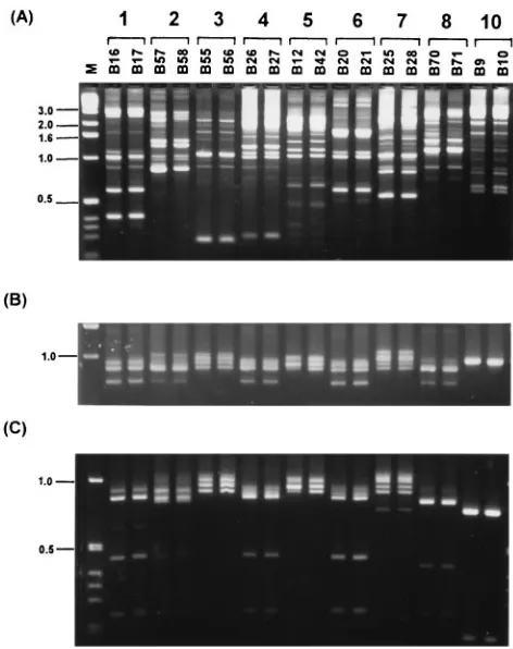
[image:2.612.71.548.72.220.2]the first 60 isolates examined. The polymorphisms generated with primer 270 of these nine RAPD types are shown in Fig. 2A. The fingerprints generated by primer 270 enabled good pri-mary discrimination ofB. cepaciastrain types. Within RAPD types, the amplified polymorphisms were conserved and the similarity coefficients of the banding profile were above 0.7 as determined by computer-assisted analysis. Between RAPD types, differences in the number of amplified markers, their mo-lecular size, and intensity were considerable and enabled good interstrain discrimination.
The RAPD typing criteria applied to the first 60 isolates also enabled epidemiologically related strains to be grouped, in-cluding sequential isolates from individual CF patients and isolates of published epidemiologic and ribotype similarity (see below and Table 1). For example, RAPD types 3, 5, 6, and 7 were representative of sequential isolates each recovered from individual CF patients attending the pediatric clinic in Van-couver (type 6 was later recovered from additional CF patients [see below]). Types 8 and 10 were each representative of se-quential isolates recovered from individual CF patents in Seattle, Wash. (12), and Philadelphia, Pa. (14); the published ribotype of each strain type was also distinct (12, 14). Type 1, 2, and 4 strains each infected multiple CF patients but were center related in epidemiology (see below). Isolates belonging to each epidemiologically related cluster possessed RAPD fin-gerprints with similarity coefficients above 0.7 and differed by no more than three bands; these criteria formed the
experi-mental basis upon which all further isolates were typed by RAPD.
To validate the strain types assigned by RAPD against an-other PCR typing method, the first 60 isolates were also eval-uated by PCR-mediated ribotyping (11). The polymorphisms generated before and after restriction digestion with the en-donuclease HaeIII are shown in Fig. 2B and C, respectively. Within eachB. cepaciastrain type designated by RAPD, iso-lates also possessed a conserved PCR-ribotype fingerprint (Fig. 2B); however, PCR ribotyping did not easily distinguish among some of the RAPD-assigned types (e.g., groups 1, 4, and 6). The restriction fragment length polymorphisms obtained after digestion of the ribotype products with the enzymeHaeIII are shown in Fig. 2C. The endonuclease-digested PCR-ribotype profiles of members of an RAPD group were also identical. The HaeIII restriction fragment length polymorphism of the amplified PCR-ribotype polymorphisms enabled 8 of the 10 groups to be differentiated, but the polymorphisms generated for groups 4 and 6 remained similar in profile and indistin-guishable. All furtherB. cepaciaisolates described in this study were typed by RAPD fingerprinting.
RAPD analysis ofB. cepaciaisolates recovered from patients
with CF.The results of the RAPD analysis ofB. cepacia
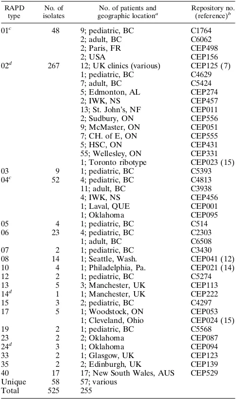
iso-lates recovered from CF patients are summarized in Table 1. A total of 525 CF isolates were analyzed, and 20 fingerprint types in which two or more isolates shared the same pattern were identified; 58 isolates possessed fingerprint profiles which were unique. Ten isolate types, 1, 2, 4, 6, 13, 15, 17, 23, 35, and 40, were recovered from two or more CF patients (Table 1). Type 15 B. cepacia was recovered from two pediatric patients in Vancouver (Table 1); however, the strain was subsequently not cultured from one of the patients, who became stably colonized with type 6B. cepacia. Type 23B. cepaciaisolates were recov-ered from two CF patients in Oklahoma, and the type 35 isolates were recovered from two CF patents in Edinburgh; no other strains of each type were present in our collection to support their transmissibility.
Of the remaining B. cepacia types infecting multiple pa-tients, each was recovered from three or more patients. Type 1 B. cepaciawas the predominant strain type in the Vancouver pediatric CF clinic and also infected CF patients in the United States and France. Type 2 isolates were recovered from mul-tiple CF patients in the United Kingdom and across Canada; this RAPD type included strains of published ribotype and PFGE fingerprint which belong to the epidemic B. cepacia lineage (7, 15, 29). Type 4 B. cepacia was the predominant strain among adult CF patients in Vancouver and also infected patients in Quebec and Nova Scotia. B. cepacia type 6 was recovered from five CF patients in Vancouver. Type 17 organ-isms were recovered from a CF patient in Ontario and in-cluded strains representative of the predominant B. cepacia ribotype infecting multiple patients at a CF treatment center in Cleveland (15). Finally, type 40 B. cepacia was an epidemic strain type which was recovered from 17 CF patients in an Australian treatment center.
[image:3.612.60.296.67.365.2]RAPD group 2 was the most common CF strain type in our collection (267 of 525 CF isolates tested). RAPD analysis dem-onstrated that this strain type was widespread in Canadian CF clinics, infecting more than 100 CF patients residing in Nova Scotia, Newfoundland, Ontario, Alberta, and British Columbia (Table 1). The polymorphisms amplified by primer 270 from 38 members of this typing group are shown in Fig. 3. Minor variations in fingerprint patterns within RAPD type are illus-trated in Fig. 3. The similarity coefficients of the polymor-phisms were greater than 0.8, designating them as a single RAPD type. However, the isolates from Newfoundland were
FIG. 2. RAPD fingerprint and PCR ribotype of typedB. cepaciaisolates. (A) The RAPD fingerprints, amplified with primer 270, of nine types ofB. cepacia. (B) The corresponding PCR-ribotype amplification patterns. (C) TheHaeIII digestion products of the PCR-ribotype polymorphisms. Isolate numbers are indicated above each lane, and the designated RAPD group number is above each pair of isolates in panel A. Molecular size markers were run in lane M, and
their size in kilobases is indicated.
on May 15, 2020 by guest
http://jcm.asm.org/
consistently different from the rest, having one extra band of approximately 0.7 kb (Fig. 3). Isolate B491 from the Toronto center was the only other member ofB. cepaciatype 2 to have this band. There was no difference in the PCR-ribotype poly-morphisms of these isolates (Fig. 2B and C).
Epidemiology of B. cepacia colonization in Vancouver. A
total of 58 CF patients attending clinics in Vancouver were colonized withB. cepacia. The pediatric and adult treatment centers were on the same hospital site separated by about 200 m until September 1993, at which time the adult CF clinic was moved to another hospital. Although the clinics were in proximity, the patients were cared for by different staff, there was little interaction between the clinics, and the patients were hospitalized in separate facilities. The prevalent B. cepacia
strain types at each CF clinic were different (Fig. 4).B. cepacia type 1 predominated among patients attending the pediatric clinic (9 of 30 patients); two adult patients were colonized with this strain type.B. cepaciatype 4 was the predominant strain among patients attending the adult CF clinic (11 of 18 pa-tients). Two CF patients who were colonized as children with types 5 and 7 subsequently lost these strains and became col-onized with type 4 after attending the adult clinic. The epide-miology of RAPD type 2, the epidemicB. cepaciastrain (7, 29), in Vancouver was investigated. Although this type was
preva-TABLE 1. Summary of RAPD analysis ofB. cepaciaisolates recovered from patients with CF
RAPD type
No. of isolates
No. of patients and geographic locationa
Repository no. (reference)b
01c 48 9; pediatric, BC C1764
2; adult, BC C6062
2; Paris, FR CEP498
2; USA CEP156
02d 267 12; UK clinics (various) CEP125 (7)
1; pediatric, BC C4629
7; adult, BC C5424
5; Edmonton, AL CEP274
2; IWK, NS CEP457
13; St. John’s, NF CEP011
2; Sudbury, ON CEP556
9; McMaster, ON CEP051
7; CH. of E, ON CEP555
5; HSC, ON CEP431
55; Wellesley, ON CEP331 1; Toronto ribotype CEP023 (15)
03 9 1; pediatric, BC C5393
04c 52 4; pediatric, BC C4813
11; adult, BC C3938
4; IWK, NS CEP456
1; Laval, QUE CEP001
1; Oklahoma CEP095
05 4 1; pediatric, BC C514
06 23 4; pediatric, BC C2303
1; adult, BC C6508
07 2 1; pediatric, BC C3430
08 14 1; Seattle, Wash. CEP041 (12)
10 4 1; Philadelphia, Pa. CEP021 (14)
12 2 1; pediatric, BC C5274
13 5 3; Manchester, UK CEP113
14d 1 1; Manchester, UK CEP222
15 3 2; pediatric, BC C4297
17 5 1; Woodstock, ON CEP053
1; Cleveland, Ohio CEP024 (15)
19 2 1; pediatric, BC C5568
23 2 2; Oklahoma CEP087
24d 3 1; Oklahoma CEP094
33 2 1; Glasgow, UK CEP123
35 2 2; Edinburgh, UK CEP139
40 17 17; New South Wales, AUS CEP529 Unique 58 57; various
Total 525 255
aAbbreviations: pediatric, pediatric clinic; BC, British Columbia; adult, adult
clinic; FR, France; USA, United States; UK, United Kingdom; AL, Alberta; IWK, Izaak Walton Killam Children’s Hospital; NS, Nova Scotia; NF, New-foundland; ON, Ontario; CH of E, Ottawa; HSC, Hospital for Sick Children; QUE, Quebec; AUS, Australia.
bRepository code of a representative isolate for the RAPD type. cAlso a clinical isolate type (see Table 2).
[image:4.612.315.553.69.316.2]dAlso an environmental isolate type (see Table 3).
FIG. 3. The polymorphisms amplified by primer 270 from members ofB. ce-paciaRAPD type 2. Isolates recovered from individual patients are shown; strain numbers and their geographic source are indicated above each lane. Isolates from Toronto were recovered from patients attending the Wellesley Hospital; Ontario 1 isolates were from the McMaster University Medical Center, and Ontario 2 isolates were from the Laurentian Hospital. Molecular size markers were run in lane M, and their size in kilobases is indicated.
FIG. 4.B. cepaciaRAPD types recovered from CF patients in Vancouver. The number of colonized patients at each treatment center is shown on theyaxis, and the RAPD type is indicated in the key.
on May 15, 2020 by guest
http://jcm.asm.org/
[image:4.612.57.299.90.499.2] [image:4.612.317.558.527.700.2]lent at other centers (Table 1), the number of patients colo-nized with this strain type attending the Vancouver treatment centers was low (8 of 58), and strains of other RAPD geno-types predominated (Fig. 4).
RAPD analysis of B. cepacia isolates from other clinical
sources or from the environment.The fingerprinting results of
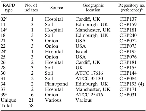
clinical and environmentalB. cepaciaisolates are summarized in Tables 2 and 3, respectively. Of 44 clinical isolates, 28 fell into one of eight RAPD types and 16 were unique.B. cepacia types 1 and 4, major CF RAPD types, were also recovered from patients without CF (Table 2). Two type 1 strains were isolated from patients in an intensive care unit in the United Kingdom. Type 4 was also found in collections of non-CF isolates from the Centers for Disease Control and Prevention, Atlanta, Ga., and from the United Kingdom. Three isolates from separate patients without CF attending British Colum-bia’s Children’s Hospital were RAPD type 4 (Table 2);B. ce-paciaof this genotype was present among CF patients attend-ing the pediatric clinic in the hospital and was also the predominant strain type in the adult CF clinic (Table 1 and Fig. 4). One clinical isolate was type 39, the same RAPD group as B. cepaciaATCC 25416 isolated from rotten onions (Table 3). Of the 58 environmental isolates analyzed by RAPD, each of 37 fell into 1 of 15 RAPD strain types and 21 possessed unique fingerprints (Table 3). Several of the environmental isolates provided by different investigators were the same RAPD type and were obtained from the American Type Culture Collec-tion. For example, RAPD types 30 and 39 were of replicate isolates of two American Type Culture Collection strains, 17616 and 25416, respectively, deposited in our collection by separate investigators, and matched the fingerprint of the re-spective type strains obtained from the American Type Culture Collection for our repository. Since typing was performed with-out knowledge of isolate source, matching of these identical type strains confirmed the specificity of the RAPD typing sys-tem.B. cepaciaisolates recovered from hospital environments in Cardiff and Manchester, RAPD types 2 and 14, respectively (Table 3), were also isolated from CF patients attending these centers (Table 1). One other hospital environmental isolate
from Israel, type 24, matched the fingerprint of threeB. cepa-ciaisolates recovered from a CF patient in Oklahoma.
DISCUSSION
The RAPD technique we have developed enabled large numbers ofB. cepaciaisolates from our strain repository to be compared at the genetic level. RAPD was able to distinguish B. cepaciaisolates more effectively than PCR ribotyping, con-sistently type serial isolates from individual CF patients, group isolates that were related epidemiologically, and appropriately distinguish isolates with known ribotype and PFGE genomic fingerprints without prior knowledge of epidemiology. Our study also provides further evidence that CF treatment centers may harbor one or more predominant strains ofB. cepaciaand that these CF isolates are, in general, genetically distinct from other clinical isolates and environmental strains.
[image:5.612.57.299.90.247.2]RAPD fingerprinting was able to produce a discriminatory and reproducible genetic fingerprint from all B. cepacia iso-lates tested. We used this method to examine P. aeruginosa isolates recovered from CF patients and found it to be as sensitive as PFGE for typing this species once discriminatory primers were identified (18). ForP. aeruginosa(18) andB. ce-pacia, the primer-to-template ratio was optimized; however, the PCR cycle conditions described by Akopyanz et al. (1) were unaltered. These data suggest that the original parame-ters described by Akopyanz et al. (1) for typingHelicobacter pyloriare a versatile RAPD thermal cycle that may be applied to many bacterial species. Indeed, reproducible and discrimi-natory polymorphisms were amplified from the following bac-terial species that were tested as part of our collection because they had been originally misidentified asB. cepacia(3): Alcali-genes faecalis, Alcaligenes xylosoxidans, Burkholderia gladioli, Comamonas acidovorans,Enterobacter agglomerans, and Steno-trophomonas(Xanthomonas)maltophilia(17a). Successful typ-ing ofB. cepacia,P. aeruginosa(10, 18), andH. pylori(1) with the same basic RAPD technique illustrates that such tech-niques are transferable from one laboratory to another and that reports of unreliable RAPD typing schemes (9) are mis-leading.
TABLE 3. RAPD analysis ofB. cepaciaisolates from environmental sourcesa
RAPD type
No. of
isolates Source
Geographic location
Repository no. (reference)b
02c 1 Hospital Cardiff, UK CEP137
11 3 Soil Edinburgh, UK CEP159
14c 1 Hospital Manchester, UK CEP181
18 3 Soil Edinburgh, UK CEP240
21 3 Onion USA CEP072
22 3 Onion USA CEP073
24c 1 Hospital Israel CEP195
25 3 Onion USA CEP076
26 2 Hospital Cardiff, UK CEP181
28 3 Soil UK CEP155
30 2 Soil ATCC 17616 CEP144
31 2 Soil ATCC 35130 CEP084
34 2 Plant/pond Edinburgh, UK CEP193 (4)
37 2 Hospital Manchester, UK CEP171
39d 6 Onion ATCC 25416 CEP031
Unique 21 Various Various
Total 58
aAbbreviations: UK, United Kingdom; USA, United States. bStrain number of representative isolate.
[image:5.612.315.557.509.694.2]cAlso a CF RAPD type (see Table 1). dAlso a clinical RAPD type (see Table 2).
TABLE 2. RAPD analysis ofB. cepaciaisolates from patients without CFa
RAPD type
No. of isolates
No. of patients
Source of infection
Geographic location
Reposi-tory no.b
01c
2 2 ICU outbreak UK CEP237 04c 5 5 Non-CFd CDC CEP150
3 3 Non-CF UK CEP211 3 3 Blood/ICU Children’s Hospital,
BC
CEP048
09 2 1 CGD Sacramento, Calif. CEP108 29 3 3 Non-CF UK CEP231 32 4 4 Non-CF CDC CEP191 36 2 2 Non-CF CDC CEP192 38 3 3 Non-CF UK CEP209 39e
1 1 Non-CF USA CEP232 Unique 16 16 Non-CF Various
CGD Total 44 43
aAbbreviations: ICU, intensive care unit; UK, United Kingdom; CDC,
Cen-ters for Disease Control and Prevention; BC, British Columbia; CGD, chronic granulomatous disease. USA, United States.
bStrain number of representative isolate for RAPD type. cAlso a CF isolate type (see Table 2).
dNon-CF, culture site not known.
eAlso an environmental isolate type (see Table 3).
on May 15, 2020 by guest
http://jcm.asm.org/
RAPD analysis correctly clustered isolates of known ribo-type and PFGE profile (7, 15), although direct comparison with these methods was not performed. Comparison of RAPD fingerprints with PCR-ribotyping profiles demonstrated, as others have described (17), that PCR ribotyping is not as dis-criminatory as RAPD for epidemiological analysis ofB. cepa-cia. Kostman et al. (11) suggested that restriction digestion of the amplified products of PCR ribotyping may improve the discriminatory power of PCR ribotyping. Digestion of PCR-ribotyping products with the enzyme HaeIII did not signifi-cantly improve the discrimination of the B. cepacia isolates examined in this study. RAPD has been shown to be as dis-criminatory as PFGE (2, 17) and enterobacterial repetitive intergenic consensus sequence PCR (17) for distinguishing B. cepacia; however, day-to-day variation in the fingerprints of the RAPD method evaluated (2) was found (17). Such varia-tion was not apparent with our method, and the good repro-ducibility was also found with RAPD analysis ofP. aeruginosa (18). The large amount of template DNA required for RAPD (a minimum of 10 ng [Fig. 1B]) may also be advantageous for a PCR-based assay, since contaminating DNA would not pro-duce conflicting polymorphisms unless it represented more than 25% of the sample (40 ng was used in each PCR). In contrast, because of the specific nature of ribotyping primers (11), PCR ribotyping was able to amplify polymorphisms from as little as 10 pg of DNA (data not shown), indicating that trace amounts of contaminating DNA may interfere with the band-ing patterns produced by this PCR method.
Our study provides further evidence that CF centers may harbor one or more predominantB. cepaciastrains. Steinbach et al. (27) found no evidence of strain transmission among 17 CF patients at a single treatment center and stated that pre-vious reports (7, 15) had incorrectly suggested that CF centers generally harbor one or more transmissibleB. cepaciastrains. This statement was contested by other investigators (6, 16, 20). The broad study of CF centers presented in this study, in which several patients at each center were found to harbor the same strain type, is in agreement with previous reports (7, 15, 25) indicating the presence of center-specific predominant B. ce-paciastrains. RAPD type 2 was the most prevalent CF strain type found (Table 2). The transatlantic spread of this epidemic clone from the United Kingdom where it was originally de-scribed (7), to centers in Ontario, Canada, has been docu-mented by others (9, 29), and a possible route of transmission via patient contact at summer camps has been reported (21). We have also shown that this type is widespread among CF patients attending treatment centers outside Ontario, suggest-ing that this strain type, as others have stated (29), is a hyper-transmissibleB. cepacialineage. However, our study ofB. ce-pacia isolates from Vancouver treatment centers illustrated that, despite the presence of patients colonized with the epi-demic strain (RAPD type 2), other strain types clustered among the patents studied (Fig. 4). These data suggest that the inabil-ity of Steinbach et al. (27) to detect evidence of patient-to-patient spread may have been due to the nature of their patient-to-patient population, which was largely referred from other centers.
The apparent spread of type 6B. cepaciafrom one CF pa-tient to other CF papa-tients attending the Vancouver pediatric clinic (Table 1; Fig. 4) further demonstrates the potential risk of patient-to-patient transmission of this organism. One pa-tient alone was chronically colonized with this type 6 strain for 6 years, suggesting low transmissibility by the criteria stated by Steinbach et al. (27). However, in year 7, type 6 was recovered from three other patients, suggesting that this strain type might have spread from one patient to another. Prolonged social contact is an identified risk factor for transmission ofB. cepacia
(7) and may have accounted for the spread of type 6B. cepacia in the pediatric clinic. Furthermore, in Vancouver, each prev-alentB. cepaciastrain type was center specific (Fig. 4), despite the proximity of the pediatric and adult clinics. This suggests that transmission ofB. cepaciamay have occurred as a result of interaction among patients attending each clinic, although a common source at each center cannot be ruled out.
After blinded typing of the isolates described in this study, the majority of strains were grouped as clonal by RAPD. Five hundred thirty-two isolates were found to belong to 37 distinct fingerprint types, and 95 isolates were unique in their RAPD profile (Tables 1, 2, and 3). CF-associated strain types were generally different from those obtained from other clinical sources and from the environment, and only 6 of the 132 strain types found (types 1, 2, 4, 14, 24, and 39) were recovered from more than one of these sources. The difference between B. cepaciastrains recovered from CF patents and those recovered from the environment is in agreement with previous reports (4, 9). We have preliminary data which suggest that B. cepacia strains that are transmissible and infect multiple CF patients (types 1, 2, 4, 6, 13, 17, and 40) harbor a region of the genome, identified by RAPD with primer 272, which is generally absent from isolates colonizing single CF patients and those recovered from other sources (reference 19 and unpublished data).
In conclusion, the RAPD method reported herein is a robust fingerprinting technique which is able to amplify discrimina-tory polymorphisms from the genome ofB. cepacia. The PCR-based technique was suitably versatile to enable a large collec-tion of B. cepacia isolates to be screened without prior knowledge of epidemiology. The method has enabled us to establish the prevalence of variousB. cepaciastrain types col-onizing CF patients treated in Vancouver and monitor the spread of problematic transmissible strain types at other treat-ment centers. We have also identified two further epidemic B. cepaciastrains (types 1 and 4) which infect multiple patients in North America and Europe. These data suggest that other B. cepacialineages apart from the epidemic strain type (29) may infect the multiple patients within the global CF commu-nity. This method should permit important epidemiological questions to be answered regarding the risk of patient-to-pa-tient spread ofB. cepaciain different environments.
ACKNOWLEDGMENTS
We thank Nicole Glenham for excellent technical assistance and Robert Shukin for helpful suggestions concerning DNA extraction and PCR. We acknowledge the Clinic Directors and Microbiologists at Canadian CF treatment centers and Jane Burns, Patricia Ferrieri, John Govan, Margaret Roy, and Terrence Stull for providing additional
B. cepaciaisolates.
E.M. was supported by a fellowship from the Canadian Cystic Fi-brosis Foundation. This work was supported with funds from the Canadian Cystic Fibrosis Foundation.
REFERENCES
1.Akopyanz, N., N. O. Bukanov, T. U. Westblom, S. Kresovich, and D. E. Berg.
1992. DNA diversity among clinical isolates ofHelicobacter pyloridetected by PCR-based RAPD fingerprinting. Nucleic Acids Res.20:5137–5142. 2.Bingen, E. D., M. O. Weber, J. Derelle, N. Brahimi, N. Y.
Lambert-Zechoovsky, M. Vidailhet, J. Navarro, and J. Elion.1993. Arbitrarily primed polymerase chain reaction as a rapid method to differentiate crossed from independent Pseudomonas cepacia infections in cystic fibrosis patients. J. Clin. Microbiol.31:2589–2593.
3.Burdge, D. R., M. A. Noble, M. E. Campbell, V. L. Krell, and D. P. Speert.
1995.Xanthomonas maltophilia misidentified asPseudomonas cepacia in cultures of sputum from patients with cystic fibrosis: a diagnostic pitfall with major clinical implications. Clin. Infect. Dis.20:445–448.
4. Butler, S. L., C. J. Doherty, J. E. Hughes, J. W. Nelson, and J. R. W. Govan.
1995.Burkholderia cepacia and cystic fibrosis: do natural environments present a potential hazard? J. Clin. Microbiol.33:1001–1004.
on May 15, 2020 by guest
http://jcm.asm.org/
5.di Sant’Agnese, P. A., and P. B. Davis.1976. Research in cystic fibrosis. N. Engl. J. Med.295:597–602.
6.Govan, J. R. W.1995.Burkholderia cepaciain cystic fibrosis. N. Engl. J. Med.
332:819–820.
7.Govan, J. R. W., P. H. Brown, J. Maddison, C. J. Doherty, J. W. Nelson, M. Dodd, A. P. Greening, and A. K. Webb.1993. Evidence for transmission of
Pseudomonas cepaciaby social contact in cystic fibrosis. Lancet342:15–9. 8.Isles, A., I. Maclusky, M. Corey, R. Gold, C. Prober, P. Fleming, and H.
Levison.1984.Pseudomonas cepaciainfection in cystic fibrosis: an emerging problem. J. Pediatr.104:206–210.
9.Johnson, W. M., S. D. Tyler, and K. R. Rozee.1994. Linkage analysis of geographic and clinical clusters inPseudomonas cepaciainfections by mul-tilocus enzyme electrophoresis and ribotyping. J. Clin. Microbiol.32:924–930. 10. Kersulyte, D., M. J. Struelens, A. Deplano, and D. E. Berg.1995.
Compar-ison of arbitrarily primed PCR and macrorestriction (pulsed-field gel elec-trophoresis) typing ofPseudomonas aeruginosastrains from cystic fibrosis patients. J. Clin. Microbiol.33:2216–2219.
11. Kostman, J. R., T. D. Edlind, J. J. LiPuma, and T. L. Stull.1992. Molecular epidemiology ofPseudomonas cepaciadetermined by polymerase chain re-action ribotyping. J. Clin. Microbiol.30:2084–2087.
12. Larsen, G. Y., T. L. Stull, and J. L. Burns.1993. Marked phenotypic vari-ability inPseudomonas cepaciaisolated from a patient with cystic fibrosis. J. Clin. Microbiol.31:788–792.
13. LiPuma, J. J., S. E. Dasen, D. W. Nielson, R. C. Stern, and T. L. Stull.1990. Person-to-person transmission ofPseudomonas cepacia between patients with cystic fibrosis. Lancet336:1094–1096.
14. LiPuma, J. J., M. C. Fischer, S. E. Dasen, J. E. Mortenson, and T. L. Stull.
1991. Ribotype stability of isolates ofPseudomonas cepacia. J. Infect. Dis.
164:133–136.
15. LiPuma, J. J., J. E. Mortensen, S. E. Dasen, T. D. Edlind, D. V. Schidlow, J. L. Burns, and T. L. Stull.1988. Ribotype analysis ofPseudomonas cepacia
from cystic fibrosis centers. J. Pediatr.113:859–862.
16. LiPuma, J. J., and T. L. Stull.1995.Burkholderia cepaciain cystic fibrosis. N. Engl. J. Med.332:820–821.
17. Liu, P. Y.-K., Z.-Y. Shi, Y.-J. Lau, B.-S. Hu, J.-M. Shyr, W.-S. Tsai, Y.-H. Lin, and C.-Y. Tseng.1995. Comparison of different PCR approaches for the characterization ofBurkholderia(Pseudomonas)cepaciaisolates. J. Clin. Microbiol.33:3304–3307.
17a.Mahenthiralingam, E.Unpublished data.
18. Mahenthiralingam, E., M. E. Campbell, J. Foster, J. S. Lam, and D. P. Speert.1996. Random amplified polymorphic DNA typing ofPseudomonas aeruginosaisolates recovered from patients with cystic fibrosis. J. Clin. Mi-crobiol.34:1129–1135.
19. Mahenthiralingam, E., M. E. Campbell, D. A. Henry, and D. P. Speert.1995. Molecular epidemiology ofBurkholderia cepaciausing random amplified polymorphic DNA (RAPD) analysis, abstr. S17.4, p. 164–165.InProgram and abstracts of the Ninth Annual North American Cystic Fibrosis Confer-ence. Pediatric pulmonology, suppl. 12. Willey-Liss, New York.
20. Mahenthiralingam, E., M. E. Campbell, and D. P. Speert.1995.Burkholderia cepaciain cystic fibrosis. N. Engl. J. Med.332:819.
21. Pegues, D. A., L. A. Carson, O. C. Tablan, S. C. FitzSimmons, S. B. Roman, J. M. Miller, W. R. Jarvis, and the Summer Camp Study Group.1994. Acquisition ofPseudomonas cepaciaat summer camps for patients with cystic fibrosis. J. Pediatr.124:694–702.
22. Prince, A.1986. Antibiotic resistance ofPseudomonas species. J. Pediatr.
108:830–834.
23. Rabkin, C. S., W. R. Jarvis, R. L. Anderson, J. Govan, J. Klinger, J. J. LiPuma, W. J. Martone, H. Montell, C. Richard, S. Shigeta, A. Sosa, T. Stull, J. Swenson, and D. Woods.1989.Pseudomonas cepaciatyping systems: col-laborative study to assess their potential in epidemiologic investigations. Rev. Infect. Dis.11:600–607.
24. Sambrook, J., E. F. Fritsch, and T. Maniatis.1989. Molecular cloning: a laboratory manual, 2nd ed. Cold Spring Harbor Laboratory Press, Cold Spring Harbor, N.Y.
25. Smith, D. L., L. B. Gumery, E. G. Smith, D. E. Stableforth, M. E. Kaufmann, and T. L. Pitt.1993. Epidemic ofPseudomonas cepaciain an adult cystic fibrosis unit: evidence of person-to-person spread. J. Clin. Microbiol.31:
3017–3022.
26. Speert, D. P.1994.Pseudomonas aeruginosainfections in patients with cystic fibrosis, p. 183–235.InA. L. Baltch and R. P. Smith (ed.),Pseudomonas aeruginosainfections and treatment. Marcel Dekker, Inc., New York. 27. Steinbach, S., L. Sun, R.-Z. Jiang, P. Flume, P. Gilligan, T. M. Egan, and R.
Goldstein.1994.Pseudomonas cepaciain cystic fibrosis lung transplant re-cipients and clinic patients. N. Engl. J. Med.331:981–987.
28. Stull, T. L., J. J. LiPuma, and T. D. Edlind.1988. A broad-spectrum probe for molecular epidemiology of bacteria: ribosomal RNA. J. Infect. Dis.157:
280–285.
29. Sun, L., R.-Z. Jiang, S. Steinbach, A. Holmes, C. Campanelli, J. Forstner, U. Sajjan, Y. Tan, M. Riley, and R. Goldstein.1995. The emergence of a highly transmissible lineage ofcbl1Pseudomonas(Burkholderia)cepaciacausing epidemics in North America and Britain. Nat. Med.1:661–666.
30. Tablan, O. C., T. L. Chorba, D. V. Schidlow, J. W. White, K. A. Hardy, P. H. Gilligan, W. M. Morgan, L. A. Chow, W. J. Martone, and W. R. Jarvis.1985.
Pseudomonas cepaciacolonization in patients with cystic fibrosis: risk factors and clinical outcome. J. Pediatr.107:382–387.


