Gerald I. Shulman
J Clin Invest.
2000;
106(2)
:171-176.
https://doi.org/10.1172/JCI10583
.
It is estimated that by the year 2020 there will be approximately 250 million people affected
by type 2 diabetes mellitus worldwide (1). Although the primary factors causing this disease
are unknown, it is clear that insulin resistance plays a major role in its development.
Evidence for this comes from (a) the presence of insulin resistance 10–20 years before the
onset of the disease (2, 3); (b) cross-sectional studies demonstrating that insulin resistance
is a consistent finding in patients with type 2 diabetes (3–6); and (c) prospective studies
demonstrating that insulin resistance is the best predictor of whether or not an individual will
later become diabetic (2, 3). Here, I focus on some recent advances in our understanding of
human insulin resistance that have been made using nuclear magnetic resonance
spectroscopy (NMR). This technique takes advantage of the spin properties of the nuclei of
certain isotopes, such as 1H, 13C, and 31P, which endow the isotopes with a magnetic
component that can be used to measure the concentration of intracellular metabolites
noninvasively and to assess biochemical differences between normal and diabetic subjects.
Drawing on NMR studies from my laboratory and others, I first consider the control of
glucose phosphorylation and transport in regulating muscle responses to insulin. I then turn
to the effects of fatty acids on insulin responses, showing […]
Perspective
It is estimated that by the year 2020 there will be approx-imately 250 million people affected by type 2 diabetes mellitus worldwide (1). Although the primary factors causing this disease are unknown, it is clear that insulin resistance plays a major role in its development. Evidence for this comes from (a) the presence of insulin resistance 10–20 years before the onset of the disease (2, 3); (b) cross-sectional studies demonstrating that insulin resist-ance is a consistent finding in patients with type 2 dia-betes (3–6); and (c) prospective studies demonstrating that insulin resistance is the best predictor of whether or not an individual will later become diabetic (2, 3). Here, I focus on some recent advances in our understanding of human insulin resistance that have been made using nuclear magnetic resonance spectroscopy (NMR). This technique takes advantage of the spin properties of the nuclei of certain isotopes, such as 1H, 13C, and 31P, which
endow the isotopes with a magnetic component that can be used to measure the concentration of intracellular metabolites noninvasively and to assess biochemical dif-ferences between normal and diabetic subjects. Drawing on NMR studies from my laboratory and others, I first consider the control of glucose phosphorylation and transport in regulating muscle responses to insulin. I then turn to the effects of fatty acids on insulin respons-es, showing that commonly accepted models that attempt to explain the association of insulin resistance and obesity are incompatible with recent findings. Final-ly, I propose an alternative model that appears to fit these and other available data.
Contributions of muscle glycogen synthesis to whole-body insulin-stimulated glucose metabolism
Our initial studies addressed two questions. First, what is the contribution of insulin-stimulated muscle glyco-gen synthesis to whole-body insulin-stimulated glucose metabolism in normal individuals? Second, to what extent is this process defective in patients with type 2 dia-betes (7)? We have measured rates of muscle glycogen synthesis using 13C NMR spectroscopy to monitor the
rate of [1-13C]glucose incorporation into muscle
glyco-gen. Under steady-state plasma concentrations of insulin and glucose that mimic postprandial conditions, we found that muscle glycogen synthesis was
approximate-ly 50% lower in diabetic subjects than in normal volun-teers. When the mean rate of muscle glycogen synthesis was extrapolated to the whole body, the synthesis of muscle glycogen accounted for most of the whole-body glucose uptake, and virtually all of the nonoxidative glu-cose metabolism in both normal and diabetic subjects. These studies demonstrate that under hyperglycemic, hyperinsulinemic conditions, muscle glycogen synthesis is the major pathway for glucose metabolism in both normal and diabetic individuals, and that defective mus-cle glycogen synthesis plays a major role in causing insulin resistance in patients with type 2 diabetes.
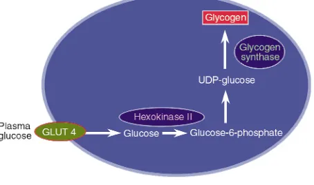
The next major task is to identify the rate-control-ling step in this process. Defects in glycogen synthase (8–10), hexokinase II (11–15), and glucose transport (14–17) have all been implicated in the loss of muscle glycogen synthesis in type 2 diabetics (Figure 1), mak-ing each of the correspondmak-ing biochemical events a potential target for antidiabetic therapy. To determine the relative importance of these steps to insulin-stim-ulated muscle glucose metabolism, we performed 13C
and 31P NMR studies to measure intracellular
con-centrations of glucose, glucose-6-phosphate, and glycogen in muscle of patients with type 2 diabetes and muscle of age- and weight-matched control sub-jects (18). Intracellular glucose-6-phosphate is an intermediary metabolite between glucose transport and glycogen synthesis, so its intracellular concentra-tion will respond to the relative activities of these two steps. In the event of decreased activity of glycogen synthase in diabetes, glucose-6-phosphate concentra-tions in diabetic patients would be expected to increase relative to that of normal individuals. Using
31P NMR to assess intracellular glucose-6-phosphate
concentrations under similar conditions of hyper-glycemia and hyperinsulinemia as in the study dis-cussed above, we found an increase of approximately 0.1 mM in intracellular glucose-6-phosphate in nor-mal individuals, and no change in patients with type 2 diabetes (18). The blunted incremental changes in glucose-6-phosphate in the type 2 diabetic patients in response to insulin stimulation can therefore be ascribed to either decreased glucose transport activi-ty or decreased hexokinase II activiactivi-ty.
Cellular mechanisms of insulin resistance
Gerald I. Shulman
Howard Hughes Medical Institute, Departments of Internal Medicine and Cellular and Molecular Physiology, Yale University School of Medicine, New Haven, Connecticut, USA
To examine whether this defect in glucose transport or hexokinase II activity was a primary defect or an acquired defect secondary to other factors, such as glu-cose toxicity (19), we studied insulin-resistant offspring of parents with type 2 diabetes, examining the rate of muscle glycogen synthesis and the muscle glucose-6-phosphate concentration under the same clamp con-ditions (20). Although these individuals were in all cases lean and normoglycemic, they were known to be at an approximately 40% increased risk for developing diabetes (2). Compared to age- and weight-matched control subjects, the children of diabetics had a 50% reduction in the rate of insulin-stimulated whole-body glucose metabolism, mainly due to a decrease in rates of muscle glycogen synthesis (20). Furthermore, their insulin-stimulated increment of intramuscular glu-cose-6-phosphate was severely reduced. This is consis-tent with impaired muscle glucose transport or reduced hexokinase II activity, and is similar to changes seen in patients with fully developed type 2 diabetes. When control subjects were studied at similar insulin levels but at euglycemia, both the rate of glycogen syn-thesis and the glucose-6-phosphate concentration decreased to values similar to that of the type 2 diabet-ic offspring. Therefore, we found that even before the onset of diabetes, insulin-resistant offspring of patients with type 2 diabetes have reduced rates of muscle glyco-gen synthesis that are secondary to a defect in either muscle glucose transport or hexokinase II activity. Clearly, defects in one or both of these activities occur early in the pathogenesis of type 2 diabetes.
To determine whether glucose transport or hexokinase II activity is rate controlling for insulin-stimulated mus-cle glycogen synthesis in patients with type 2 diabetes, we used a novel 13C NMR method to assess intracellular
glu-cose concentrations in muscle under similar hyper-glycemic, hyperinsulinemic conditions as those used in the previous studies (21). Intracellular glucose is an intermediary metabolite between glucose transport and glucose phosphorylation, and its concentration reflects
the relative activities of glucose transporters (particular-ly GLUT4) and of hexokinase II. Unlike the standard biopsy method, this approach is noninvasive and is not subject to the errors caused by contamination of biopsy tissue with plasma glucose or incomplete removal of non-muscle constituents. If hexokinase II activity is reduced relative to glucose transport activity in diabetes, one would predict a substantial increase in intracellular glu-cose (21), whereas if gluglu-cose transport is primarily responsible for maintaining intracellular glucose metab-olism, intracellular glucose and glucose-6-phosphate should change proportionately. We found that the intra-cellular glucose concentration was far lower in the dia-betic subjects than the concentration expected if hexoki-nase II was the primary rate-controlling enzyme for glycogen synthesis. When the rates of muscle glycogen synthesis in the diabetic subjects were increased by infus-ing greater amounts of insulin, the changes in the con-centrations of intracellular glucose and glucose-6-phos-phate indicated that the rates of glucose transport were matched by increases in the rates of glucose phosphory-lation and glycogen synthesis. These data suggest a pre-dominant role for glucose transport control of insulin-stimulated muscle glycogen synthesis in patients with type 2 diabetes, but they do not rule out the possibility that there are other downstream abnormalities in the pathway of glycogen synthesis that do not exert rate-con-trolling effects under these conditions.
It has also been hypothesized that decreased delivery of substrate or insulin to the tissue bed might be respon-sible for the insulin resistance in type 2 diabetes (22). With regard to substrate delivery, we found no difference in the 13C NMR–measured ratio of extra- to intracellular
water space between the normal subjects and diabetic patients, implying that there were no significant differ-ences between the groups that might be attributed to insulin-mediated vasodilatation. We also found no dif-ferences between the two groups in the interstitial insulin concentrations during the hyperinsulinemic clamps, suggesting that the delivery of insulin is not responsible for the insulin resistance in patients with type 2 diabetes. Overall, these data are consistent with the hypothesis that glucose transport is the rate-con-trolling step for insulin-stimulated muscle glycogen syn-thesis in patients with type 2 diabetes (see Pessin and Saltiel, this Perspective series, ref. 23), and that muscle glucose transport represents an important therapeutic target for this disease. These results also suggest that agents that enhance hexokinase II or glycogen synthase activity will not be as effective in improving insulin sen-sitivity in skeletal muscle of patients with type 2 diabetes as those that enhance glucose transport activity.
Fatty acid–induced insulin resistance
[image:3.612.61.288.555.689.2]Increased plasma free fatty acid concentrations are typ-ically associated with many insulin-resistant states, including obesity and type 2 diabetes mellitus (24–27).
Figure 1
In a cross-sectional study of young, normal-weight off-spring of type 2 diabetic patients, we found an inverse relationship between fasting plasma fatty acid concen-trations and insulin sensitivity, consistent with the hypothesis that altered fatty acid metabolism con-tributes to insulin resistance in patients with type 2 dia-betes (28). Furthermore, recent studies measuring intramuscular triglyceride content by muscle biopsy (29) or intramyocellular triglyceride content by 1H
NMR (30–32) have shown an even stronger relation-ship between accumulation of intramyocellular triglyc-eride and insulin resistance. In a classic series of stud-ies, Randle et al. demonstrated that fatty acids compete with glucose for substrate oxidation in isolated rat heart muscle and rat diaphragm muscle. They specu-lated that increased fat oxidation causes the insulin resistance associated with obesity (33–35). The mecha-nism they proposed to explain
the insulin resistance was that an increase in fatty acids caused an increase in the intramito-chondrial acetyl CoA/CoA and NADH/NAD+ratios, with
sub-sequent inactivation of pyruvate dehydrogenase (Fig-ure 2, top). This in turn would cause intracellular cit-rate concentrations to increase, leading to inhibition of phosphofructokinase, a key rate-controlling enzyme in glycolysis. Subsequent accumulation of glucose-6-phosphate would inhibit hexokinase II activity, result-ing in an increase in intracellular glucose concentra-tions and decreased glucose uptake.
A recent series of studies by our group has challenged this conventional hypothesis (36–38). In the first study, we used 13C and 31P NMR spectroscopy to measure
[image:4.612.227.538.320.737.2]skeletal muscle glycogen and glucose-6-phosphate con-centrations in healthy subjects. The subjects were main-tained in euglycemic, hyperinsulinemic conditions with either low or high levels of plasma fatty acids (36). Increasing the plasma fatty acid concentration for 5 hours caused a reduction of approximately 50% in
Figure 2
Top: Mechanism of fatty acid–induced insulin resistance in skeletal muscle as pro-posed by Randle et al. An increase in fatty acid concentration results in an elevation of the intramitochondrial acetyl CoA/CoA and NADH/NAD+ratios, with subsequent
insulin-stimulated rates of muscle glycogen synthesis and whole-body glucose oxidation compared to con-trols. In contrast to the results from the model of Ran-dle and coworkers, which predicted that fat-induced insulin resistance would result in an increase in intra-muscular glucose-6-phosphate (34), we found that the drop in muscle glycogen synthesis was preceded by a fall in intramuscular glucose-6-phosphate. These data suggest that increases in plasma fatty acid concentra-tions initially induce insulin resistance by inhibiting glucose transport or phosphorylation activity, and that the reduction in muscle glycogen synthesis and glucose oxidation follows. The reduction in insulin-activated glucose transport and phosphorylation activity in nor-mal subjects maintained at high plasma fatty acid lev-els is similar to that seen in obese individuals (39), patients with type 2 diabetes (18), and lean, normo-glycemic insulin-resistant offspring of type 2 diabetic individuals (20). Hence, accumulation of intramuscu-lar fatty acids (or fatty acid metabolites) appears to play an important role in the pathogenesis of insulin resist-ance seen in obese patients and patients with type 2 dia-betes. Moreover, fatty acids seem to interfere with a very early step in insulin stimulation of GLUT4 transporter activity or hexokinase II activity. This conclusion is at odds with the mechanism proposed by Randle et al. (33–35), which predicts an increase in intramuscular glucose-6-phosphate concentrations resulting from inhibitory effects of fatty acid on phosphofructokinase activity (due to an increase in intracellular citrate con-centration).
To distinguish between possible effects of fatty acids on glucose transport activity and on hexokinase II activity, we measured intracellular concentrations of glucose in muscle using 13C NMR (37). The logic of this
experiment was similar to that described above, in which we followed concentrations of glucose-6-phos-phate to determine the relative activity of glucose trans-port and glycogen synthesis. Because intracellular glu-cose is an intermediary metabolite between gluglu-cose transport and hexokinase II, its concentration reflects the relative activities of these two steps. If a decrease in hexokinase II activity was responsible for the lower rate of insulin-stimulated muscle glycogen synthesis, intra-cellular glucose concentrations should increase. How-ever, if the impairment was at the level of glucose trans-port, there should be no difference or a decrease in the intracellular glucose concentration. We found that ele-vated plasma fatty acid concentrations caused a signif-icant reduction in intracellular glucose concentration in the lipid infusion studies compared to control stud-ies in which glycerol (the other metabolite released by lipolysis) was infused in the absence of any exogenous fatty acid. These data imply that the rate-controlling step for fatty acid–induced insulin resistance in humans is glucose transport, and offer further evidence against the Randle mechanism, which predicts an
increase in both intracellular glucose-6-phosphate and glucose concentrations.
This reduced glucose transport activity could be the result of fatty acid effects on the GLUT4 transporter directly — alterations in the trafficking, budding, fusion, or activity of GLUT4 (40) — or it could result from fatty acid–induced alterations in upstream insulin signaling events, resulting in decreased GLUT4 translocation to the plasma membrane (Figure 2, bot-tom). To explore the latter possibility, we examined IRS-1–associated phosphatidylinositol kinase (PI 3-kinase) activity in muscle biopsy samples, using the identical lipid infusion protocol described above for study of fatty acid effects. We found that elevations in plasma fatty acid concentrations similar to the previ-ous NMR studies (36, 37) abolished insulin-stimulat-ed, IRS-1–associated PI 3-kinase activity compared with a fourfold insulin stimulation observed in the glycerol-control infusion studies (37). The reduced insulin-stimulated PI 3-kinase activity may be due to a direct effect of intracellular free fatty acids (or some fatty acid metabolite) on PI 3-kinase, or may be secondary to alterations in upstream insulin signaling events. Con-sistent with an indirect effect, we found that a similar lipid infusion protocol in rats resulted in a reduction of insulin-stimulated IRS-1 tyrosine phosphorylation, which was associated with activation of protein kinase Cθ(38), a known serine kinase that has been shown to be activated by diacylglycerol (41). High-fat feeding has also been shown to both increase the content of long-chain fatty acyl CoA in muscle and alter the protein kinase C isoenzymes θand ε(42).
A unifying hypothesis for common forms of human insulin resistance
action, particularly IRS-1/IRS-2–dependent activation of PI 3-kinase, in muscle and liver (44). Interestingly, these abnormalities were associated with a twofold increase in muscle and liver triglyceride content, and upon transplantation of fat tissue into these mice, triglyceride content in muscle and liver returned to nor-mal, as did insulin signaling and action. These findings are consistent with the hypothesis that insulin resist-ance develops in obesity, type 2 diabetes, and lipodys-trophy because of alterations in the partitioning of fat between the adipocyte and muscle or liver. This change leads to the intracellular accumulation of triglycerides, and, probably more importantly, of intracellular fatty acid metabolites (fatty acyl CoA’s, diacylglycerol, and ceramides, among others) in these insulin-responsive tissues, which leads to acquired insulin signaling defects and insulin resistance (Figure 2, bottom).
This hypothesis might also explain how thiazo-lidinediones improve insulin sensitivity in muscle and liver tissue. By activating PPAR-γ receptors in adipocytes and promoting adipocyte differentiation, these agents might promote a redistribution of fat from liver and muscle into the adipocytes, much as fat transplantation does in fat-deficient mice (44). This hypothesis is supported by some recent thiazolidine-dione studies in rats fed high-fat diets (45, 46). It might also be expected that any alteration in the ability of muscle and liver to metabolize fatty acids, such as inherited or acquired defects in mitochondria function, would also lead to intracellular accumulation of fatty acid metabolites and subsequent defects in insulin sig-naling and action. Given the polygenic nature of type 2 diabetes, it is likely that examples of both of these pos-sibilities will be identified. This mechanism, if it proves to be correct, offers many new therapeutic targets for novel insulin-sensitizing agents.
Acknowledgments
Space limitations preclude this from being a compre-hensive review, and this unfortunately limits appropri-ate recognition of many of my colleagues worldwide who have contributed immeasurably to the develop-ment of this field. However, I specifically wish to thank Kitt Petersen, Gary Cline, and Douglas Rothman for their scientific contributions, advice, and editorial assistance, and Dennis McGarry for many stimulating discussions. The studies summarized in this review were supported in part by grants from the NIH (R01 DK-49230, R01 DK-40936, and P30 DK-45735). G.I. Shulman is an investigator of the Howard Hughes Medical Institute.
1. O’Rahilly, S. 1997. Science, medicine, and the future. Non-insulin dependent diabetes mellitus: the gathering storm. BMJ.314:955–959. 2. Warram, J.H., Martin, B.C., Krolewski, A.S., Soeldner, J.S., and Kahn, C.R. 1990. Slow glucose removal rate and hyperinsulinemia precede the devel-opment of type II diabetes in the offspring of diabetic patients. Ann. Intern. Med.113:909–915.
3. Lillioja, S., et al. 1988. Impaired glucose tolerance as a disorder of insulin action. Longitudinal and cross-sectional studies in Pima Indians. N. Engl. J. Med.318:1217–1225.
4. Haffner, S.M., et al. 1990. Diminished insulin sensitivity and increased insulin response in nonobese, nondiabetic Mexican Americans. Metabo-lism.39:842–847.
5. Reaven, G.M., Bernstein, R., Davis, B., and Olefsky, J.M. 1976. Nonke-totic diabetes mellitus: insulin deficiency or insulin resistance? Am. J. Med.60:80–88.
6. DeFronzo, R.A. 1988. The triumvirate: beta-cell, muscle, liver: a collu-sion responsible for NIDDM. Diabetes.37:667–687.
7. Shulman, G.I., et al. 1990. Quantitation of muscle glycogen synthesis in normal subjects and subjects with non-insulin-dependent diabetes by
13C nuclear magnetic resonance spectroscopy. N. Engl. J. Med.
322:223–228.
8. Bogardus, C., Lillioja, S., Stone, K., and Mott, D. 1984. Correlation between muscle glycogen synthase activity and in vivo insulin action in man. J. Clin. Invest.73:1185–1190.
9. Damsbo, P., Vaag, A., Hother-Nielsen, O., and Beck-Nielsen, H. 1991. Reduced glycogen synthase activity in skeletal muscle from obese patients with and without type 2 (non-insulin-dependent) diabetes mel-litus. Diabetologia.34:239–245.
10. Wright, K.S., Beck, N.H., Kolterman, O.G., and Mandarino, L.J. 1988. Decreased activation of skeletal muscle glycogen synthase by mixed-meal ingestion in NIDDM. Diabetes.37:436–440.
11. Kelley, D.E., et al. 1996. The effect of non-insulin-dependent diabetes mellitus and obesity on glucose transport and phosphorylation in skele-tal muscle. J. Clin. Invest.97:2705–2713.
12. Braithwaite, S.S., Palazuk, B., Colca, J.R., Edwards, C.W., III, and Hof-mann, C. 1995. Reduced expression of hexokinase II in insulin-resistant diabetes. Diabetes.44:43–48.
13. Kruszynska, Y.T., Mulford, M.I., Baloga, J., Yu, J.G., and Olefsky, J.M. 1998. Regulation of skeletal muscle hexokinase II by insulin in nondia-betic and NIDDM subjects. Diabetes.47:1107–1113.
14. Rothman, D.L., Shulman, R.G., and Shulman, G.I. 1992. 31P nuclear magnetic resonance measurements of muscle glucose-6-phosphate. Evi-dence for reduced insulin-dependent muscle glucose transport or phos-phorylation activity in non-insulin-dependent diabetes mellitus. J. Clin. Invest.89:1069–1075.
15. Bonadonna, R.C., et al. 1996. Roles of glucose transport and glucose phophorylation in muscle insulin resistance of NIDDM. Diabetes.45:915–925. 16. Zierath, J.R., et al. 1996. Insulin action on glucose transport and plas-ma membrane GLUT4 content in skeletal muscle from patients with NIDDM. Diabetologia.39:1180–1189.
17. Dohm, G.L., et al. 1988. An in vitro human muscle preparation suitable for metabolic studies. Decreased insulin stimulation of glucose trans-port in muscle from morbidly obese and diabetic subjects. J. Clin. Invest.
82:486–494.
18. Rothman, D.L., Shulman, R.G., and Shulman, G.I. 1992. 31P nuclear
magnetic resonance measurements of muscle glucose-6-phosphate: evi-dence for reduced insulin-dependent muscle glucose transport or phos-phorylation activity in non-insulin-dependent diabetes mellitus. J. Clin. Invest.89:1069–1075.
19. Rossetti, L., Giaccari, A., and DeFronzo, R.A. 1990. Glucose toxicity. Dia-betes Care.13:610–630.
20. Rothman, D.L., et al. 1995. Decreased muscle glucose transport/phos-phorylation is an early defect in the pathogenesis of non-insulin-dependent diabetes mellitus. Proc. Natl. Acad. Sci. USA.92:983–987. 21. Cline, G., et al. 1999. Glucose transport is rate controlling for insulin
stimulated muscle glycogen synthesis in type 2 diabetes. N. Engl. J. Med.
341:240–246.
22. Yang, Y.J., Hope, I.D., Ader, M., and Bergman, R.N. 1989. Insulin trans-port across capillaries is rate limiting for insulin action in dogs. J. Clin. Invest.84:1620–1628.
23. Pessin, J.E., and Saltiel, A.R. 2000. Signaling pathways in insulin action: molecular targets of insulin resistance. J. Clin. Invest.106:165–169. 24. Reaven, G.M., Hollenbeck, C., Jeng, C.-Y., Wu, M.S., and Chen, Y.-D.
1988. Measurement of plasma glucose, free fatty acid, lactate, and insulin for 24 h in patients with NIDDM. Diabetes.37:1020–1024. 25. Frayne, K.N. 1993. Insulin resistance and lipid metabolism. Curr. Opin.
Lipidol.4:197–204.
26. McGarry, J.D. 1992. What if Minkowski had been ageusic? An alterna-tive angle on diabetes. Science. 258:766–770.
93:2438–2446.
28. Perseghin, G., Ghosh, S., Gerow, K., and Shulman, G.I. 1997. Metabol-ic defects in lean nondiabetMetabol-ic offspring of NIDDM parents: a cross-sec-tional study. Diabetes.46:1001–1009.
29. Pan, D.A., et al. 1997. Skeletal muscle triglyceride levels are inversely related to insulin action. Diabetes.46:983–988.
30. Krssak, M., et al. 1999. Intramyocellular lipid concentrations are corre-lated with insulin sensitivity in humans: a 1H NMR spectroscopy study. Diabetologia.42:113–116.
31. Perseghin, G., et al. 1999. Intramyocellular triglyceride content is a determinant of in vivo insulin resistance in humans: a 1H-13C nuclear magnetic resonance spectroscopy assessment in offspring of type 2 dia-betic parents. Diabetes.48:1600–1606.
32. Stein, D.T., Szczepaniak, L.S., Dobbins, R.L., Snell, P., and McGarry, J.D. 1998. Skeletal muscle triglycerides stores are increased in insulin resist-ant states. Proceedings of the 6th Scientific Meeting for the International Soci-ety for Magnetic Resonance in Medicine (Sydney).388. (Abstr.).
33. Randle, P.J., Garland, P.B., Hales, C.N., and Newsholme, E.A. 1963. The glucose fatty-acid cycle: its role in insulin sensitivity and the metabolic disturbances of diabetes mellitus. Lancet.i:785–789.
34. Randle, P.J., Garland, P.B., Newsholme, E.A., and Hales, C.N. 1965. The glucose fatty-acid cycle in obesity and maturity onset diabetes mellitus.
Ann. NY Acad. Sci.131:324–333.
35. Randle, P.J., Newsholme, E.A., and Garland, P.B. 1964. Regulation of glu-cose uptake by muscle. Effects of fatty acids, ketone bodies and pyruvate, and of alloxan, diabetes and starvation, on the uptake and metabolic fate of glucose in rat heart and diaphragm muscles. Biochem. J.93:652–665. 36. Roden, M., et al. 1996. Mechanism of free fatty acid-induced insulin
resistance in humans. J. Clin. Invest. 97:2859–2865.
37. Dresner, A., et al. 1999. Effects of free fatty acids on glucose transport and IRS-1-associated phosphatidylinositol 3-kinase activity. J. Clin. Invest.103:253–259.
38. Griffin, M.E., et al. 1999. Free fatty acid-induced insulin resistance is associated with activity of protein kinase C theta and alterations in the insulin signaling cascade. Diabetes.48:1270–1274.
39. Petersen, K.F., et al. 1998. 13C/31P NMR studies on the mechanism of insulin resistance in obesity. Diabetes.47:381–386.
40. Kahn, B.B. 1992. Facilitative glucose transporters: regulatory mecha-nisms and dysregulation in diabetes. J. Clin. Invest.89:1367–1374. 41. Chalkley, S.M., Hettiarachchi, N., Chisholm, D.J., and Kraegen, E.W.
1998. Five-hour fatty acid elevation increases muscle lipids and impairs glycogen synthesis in the rat. Metabolism.47:1121–1126.
42. Schmitz-Peiffer, C., et al. 1997. Alterations in the expression and cellu-lar localization of protein kinase C isozymes epsilon and theta are asso-ciated with insulin resistance in skeletal muscle of the high-fat-fed rat.
Diabetes. 46:169–178.
43. Gavrilova, O., et al. 2000. Surgical implantation of adipose tissue revers-es diabetrevers-es in lipoatrophic mice. J. Clin. Invest.105:271–278. 44. Kim, J., Gavrilova, O., Chen, Y., Reitman, M., Shulman, G.I. 2000.
Mech-anisms of insulin resistance in A-ZIP/F-1 fatless mice. J. Biol. Chem.
275:8456–8460.
45. Oakes, N.D., et al. 1994. A new antidiabetic agent, BRL 49653, reduces lipid availability and improves insulin action and glucoregulation in the rat. Diabetes.43:1203–1210.