PRIMER
How to make a cardiomyocyte
Daniela Später1,2,*, Emil M. Hansson1,3, Lior Zangi1,4,5and Kenneth R. Chien1,3,*
ABSTRACT
During development, cardiogenesis is orchestrated by a family of heart progenitors that build distinct regions of the heart. Each region contains diverse cell types that assemble to form the complex structures of the individual cardiac compartments. Cardiomyocytes are the main cell type found in the heart and ensure contraction of the chambers and efficient blood flow throughout the body. Injury to the cardiac muscle often leads to heart failure due to the loss of a large number of cardiomyocytes and its limited intrinsic capacity to regenerate the damaged tissue, making it one of the leading causes of morbidity and mortality worldwide. In this Primer we discuss how insights into the molecular and cellular framework underlying cardiac development can be used to guide thein vitrospecification of cardiomyocytes, whether by directed differentiation of pluripotent stem cells or via direct lineage conversion. Additional strategies to generate cardiomyocytesin situ, such as reactivation of endogenous cardiac progenitors and induction of cardiomyocyte proliferation, will also be discussed.
KEY WORDS: Cardiac development, Cardiomyocyte, Heart, Progenitor, Stem cell
Introduction
The heart is one of the first organs formed in the embryo, highlighting its vital function in supplying oxygen and nutrients to the various tissues of the organism both during development and in later life. In humans the primordial heart begins to beat at around 22 days after fertilization, whereas in mice this occurs around day 8. The early beating of the heart is due to the presence of cardiomyocytes, which are coordinated to contract en masse in order to pump blood and nutrients around the developing embryo. The crucial role of the cardiomyocytes in ensuring proper heart function is maintained throughout life. Thus, manipulating cardiomyocyte number and function has emerged as a focus for cardiac disease prevention and treatment.
Given the crucial role of the heart, and of the cardiomyocytes in particular, it is not surprising that diseases or injuries compromising heart function are often severe or fatal (see Box 1). Acquired heart diseases, such as myocardial infarction, often result in a massive loss of cardiomyocytes due to an ischemic event. Even though the heart has a low intrinsic cardiomyocyte turnover rate (Ali et al., 2014; Bergmann et al., 2009; Kajstura et al., 2010a,b; Senyo et al., 2013), it is clearly inadequate to reverse the decreased cardiac function following injury, spurring research into alternative modes of heart
repair. There are multiple different approaches for increasing cardiomyocyte numbers in the adult heart, including stimulation of proliferation in existing cardiomyocyte populations, direct lineage conversion of fibroblasts into cardiomyocytes, activation of endogenous cardiac progenitor cells, or transplantation ofin vitro derived cardiomyocytes into the injured area. Over the past decade, further efforts have been made to enhance cardiac function following injury, particularly via cell therapy using non-cardiac and cardiac adult stem cell sources. However, the benefits from such treatments have remained controversial and, generally, at best modest (reviewed by Garbern and Lee, 2013; Hansson et al., 2009; Nowbar et al., 2014). Given the staggering amount of cells that are lost during a myocardial infarction [∼109 (Laflamme and Murry, 2005)], it is clear that a very large number of cardiomyocytes will be required to achieve a clinically meaningful regenerative response in the heart.
In this Primer, we review the fundamental principles that govern cardiogenesis and, in particular, the important processes involved in cardiomyocyte specification and proliferation. We then provide an update on current progress in generating cardiomyocytesde novo, both in vitro and in vivo, which depends on an in-depth understanding of cardiomyocyte development and homeostasis.
Cardiac specification and development
The formation of the mammalian heart is regulated by the interplay between major developmental signaling pathways, an
Box 1. Heart disease and its impact on world health
Generally, one can distinguish between congenital heart defects (CHDs) and acquired heart disease. CHDs are usually apparent at birth and are characterized by structural abnormalities of the heart and/or the great vessels, such as atrial or ventricular septation defects and transposition of the arteries, and by functional abnormalities, such as electrical conduction abnormalities or cardiomyopathies. These abnormalities are often linked to mutations affecting the transcriptional circuits that control normal heart development, such as in Holt-Oram syndrome (TBX5) and DiGeorge syndrome (TBX1) (McCulley and Black, 2012). Acquired heart diseases usually manifest during adulthood and increase in incidence with age, and include coronary heart disease, congestive heart failure and arrhythmias. Coronary heart disease is generally due to atherosclerosis (occlusion of one or several coronary arteries) and results in myocardial infarction, or heart attack. Depending on the severity of the attack, the magnitude of cardiomyocyte loss and formation of a fibrotic scar, the pumping capacity of the heart becomes diminished and may lead to heart failure if it falls below a critical threshold. Congestive heart failure includes conditions other than artery disease that impair the pumping capacity of the heart, such as a weakened or stiffened heart muscle or valve defects. The number of deaths due to cardiovascular disease (CVD) totals 17.3 million a year according to data published by the WHO in 2008, thereby representing 30% of all global deaths. Of these deaths, an estimated 7.3 million were due to coronary heart disease. The number of people who die from CVD, mainly from heart disease and stroke, is estimated to increase to 23.3 million by 2030 (Lopez et al., 2006; Mathers and Loncar, 2006; Pagidipati and Gaziano, 2013).
1
Department of Stem Cell and Regenerative Biology, Harvard University and Harvard Medical School, 7 Divinity Avenue, Cambridge, MA 02138, USA. 2
Department of Bioscience, CVMD iMED, AstraZeneca, Pepparedsleden 1, Mölndal 43150, Sweden.3Department of Cell and Molecular Biology and Medicine, Karolinska Institutet, 35 Berzelius Vag, Stockholm 171 77, Sweden.4Department of Cardiology, Children’s Hospital, 300 Longwood Avenue, Boston, MA 02115, USA. 5
Cardiovascular Research Center, Mount Sinai School of Medicine, One Gustave L. Levy Place, New York, NY 10029, USA.
*Authors for correspondence (daniela.spaeter@astrazeneca.com;
kenneth.chien@ki.se)
DEVEL
O
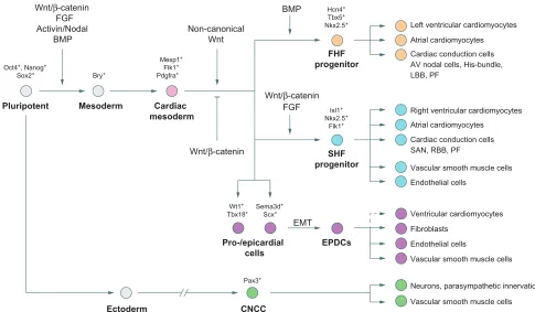
increasingly cardiac-specific array of transcription factors, as well as other transcriptional regulators. The four-chambered heart consists of the left and right atria, left and right ventricles (the myocardial components), the epicardium (the outer epithelial layer of the heart) and the endocardium (inner endothelial layer of the heart) (Fig. 1). The major cell types of the heart include cardiomyocytes, cardiac conduction cells, cardiac fibroblasts and vascular smooth muscle cells ( predominantly within the myocardium) and endothelial cells ( predominantly within the myocardium and endocardium) (Fig. 2). The cardiomyocyte lineage is highly specialized, consisting of several different subtypes defined both by their location and function (see Box 2). The first step in understanding how to generate functional and mature cardiomyocytesin vitro is to unravel the complexity that is intrinsic to their formation in vivo.
Specification of the cardiac mesoderm
Most of the cardiac cell lineages arise from the mesoderm, which is one of three distinct germ layers specified from the inner cell mass following gastrulation at embryonic day (E) 6 during mouse development (Fig. 1) (reviewed by Abu-Issa and Kirby, 2007; Rossant and Tam, 2009). The induction of mesoderm formation and its further specification into cardiac mesoderm and distinct populations of cardiac progenitor cells is primarily controlled by three families of extracellular signaling molecules: wingless integrated (Wnt), fibroblast growth factor (FGF) and transforming growth factor-beta (TGFβ) superfamily ligands, which include Wnt3a, bone morphogenetic protein 4 (BMP4), Nodal and activin A (Fig. 2) (reviewed by Noseda et al., 2011). These ligands are expressed in gradients, thereby patterning the developing embryo and sending either activating or inhibitory instructive cues to the underlying cells, depending on the spatiotemporal context. The future cardiac field is specified in response to this combination of signaling factors and downstream transcriptional events, which eventually lead to the expression of cardiac-specific factors in a defined population of mesodermal cells (reviewed by Noseda et al., 2011).
The next step: cardiac tissue formation
Several cardiac progenitor pools contribute to the different cell types and structures of the nascent heart, primarily derivatives of mesoderm posterior 1 (Mesp1)+mesodermal cells identified as the first (FHF) and second (SHF) heart fields (Figs 1 and 2) (Brade et al., 2013; Vincent and Buckingham, 2010). Of these, the FHF acts as the initiator of heart formation and is exclusively committed to a cardiomyogenic cell fate (Liang et al., 2013; Später et al., 2013). The FHF forms via the migration ofMesp1+ mesodermal cells towards the anterior of the embryo where they coalesce to form a crescent-shaped structure that constitutes the first morphological sign of heart development (Fig. 1). The FHF will develop into the primitive heart tube that rapidly begins to contract in order to pump blood and nutrients throughout the embryo. The SHF progenitors are located posteriorly and medially to the FHF-derived cardiac crescent and later, behind the primitive and looping heart tube in an area also known as the pharyngeal mesoderm. From there, SHF progenitors migrate towards the primitive and looping heart tube, which serves as a scaffold for the arriving cells, allowing subsequent growth of the heart (Fig. 1).
The fine tuning of canonical Wnt signaling in a spatiotemporal manner is essential for these early steps in cardiac development. Active Wnt/β-catenin signaling is required for primitive streak and pre-cardiac mesoderm formation (Barrow et al., 2007; Haegel et al., 1995; Liu et al., 1999; Rivera-Pérez and Magnuson, 2005), whereas its inhibition via Crescent [also known as frizzled-related protein 2 (FRZB2)] and Dickkopf 1 (DKK1), expressed in the adjacent endoderm, is required to allow further specification into cardiac progenitor cells (Foley and Mercola, 2005; Schneider and Mercola, 2001). At later stages, active Wnt signaling is important for the specification and expansion of SHF progenitors; the inactivation of β-cateninin vivoresults in a partial loss of SHF progenitors, whereas its constitutive activation leads to their expansion (Ai et al., 2007; Bu et al., 2009; Cohen et al., 2007; Klaus et al., 2007; Kwon et al., 2007, 2009; Lin et al., 2007; Qyang et al., 2007). FGF, BMP, Hedgehog (Hh), Notch and non-canonical Wnt signaling are also involved at various stages during FHF and SHF progenitor specification, expansion and differentiation (Cohen et al., 2008; High et al., 2009; Hoffmann et al., 2009; Klaus et al., 2007; Nagy et al., 2010;
CM PS
ML FHF
SHF
OFT OFT
RV LV PHT
PM PM
VP PEO
CNCCs
RV RA
LV LA SVC
IVC EPC
AO
PT ICM
Anterior
Anterior
Posterior
Posterior
E6.5
[image:2.612.57.560.66.198.2]Blastocyst E7.0 E8.0 E9.0 Adult heart
Fig. 1. Cardiac development in the mouse embryo.Following gastrulation, the cells of the inner cell mass (ICM) are specified into three distinct germ layers: endoderm, ectoderm and mesoderm. The first signs of cardiac development can be detected at∼E6.5 with the formation of the cardiac mesoderm (CM, yellow) at the posterior side of the embryo, along the primitive streak (PS). At E7, the cardiac mesodermal cells migrate towards the anterior side of the embryo to form the two major cardiac progenitor pools: the first heart field (FHF, orange) or cardiac crescent; and the second heart field (SHF, blue), located posterior to the crescent. The FHF gives rise to the beating primitive heart tube (PHT) and eventually to the left ventricle (LV) and parts of the right and left atria (RA and LA, respectively). The SHF progenitors, located behind the PHT within the pharyngeal mesoderm (PM, light blue) by E8, migrate towards the primitive and looping heart tube to contribute to the right ventricle (RV), parts of the atria and outflow tract (OFT), and later to the base of the aorta (AO) and pulmonary trunk (PT). At∼E9.0 the transient proepicardial organ (PEO, purple), which eventually will form the outer lining of the heart known as the epicardium (EPC), becomes apparent. In addition, cardiac neural crest cells (CNCCs, green) migrate in from the dorsal neural tube through the pharyngeal arches, contributing to smooth muscle cells within the aortic and pulmonary arteries. Cells of the venous poles (VP, red), apparent at E8, contribute to the base of the superior and inferior vena cava (SVC and IVC, respectively). ML, midline.
DEVEL
O
Park et al., 2008; Zhang et al., 2008; Zhou et al., 2007; reviewed in detail by Noseda et al., 2011; Vincent and Buckingham, 2010).
FHF and SHF derivatives give rise to distinct compartments of the adult heart (Fig. 1). The FHF-derived primitive heart tube has mostly a left ventricular identity, but there is also a FHF contribution to the atria, whereas SHF progenitors mostly contribute to the right ventricle (RV), parts of the atria and the outflow tract (OFT) (Buckingham et al., 2005; Vincent and Buckingham, 2010). Apart from these two major cell sources, there is also a contribution from the proepicardial organ (PEO) and cardiac neural crest cells (CNCCs) (Fig. 1). The PEO is a transient structure that starts to develop at∼E8.5 from a cluster of mesothelial cells near the venous pole/inflow tract of the heart tube and will eventually form the epicardium, which is the outer epithelial lining of the embryonic heart (Männer et al., 2001; Schlueter and Brand, 2012). The CNCCs arise from the dorsal neural tube and migrate through the pharyngeal arches towards the heart, starting from ∼E10.5. Derivatives of the CNCCs form the parasympathetic innervation of the heart, contribute to the valves, and play a pivotal role in OFT patterning and septation (Hildreth et al., 2008; Hutson and Kirby, 2007; Waldo et al., 2005).
The cardiomyocyte lineage
The mesoderm-derived FHF and SHF are the predominant sources of cardiomyocytes in the heart, with a minor contribution from the proepicardium.In vivoandin vitrolineage-tracing experiments and clonal analysis using an Islet1 (Isl1)-based reporter system have suggested that Isl1+ SHF progenitors in mouse and human can contribute to various cell lineages within the heart, such as cardiomyocytes of the RV and atria as well as vascular smooth muscle and endothelial cells (Fig. 2) (Bu et al., 2009; Moretti et al., 2006).In vivolineage tracing and single-cell clonal analysis have
demonstrated that, unlike SHF progenitors, FHF cells are marked by the expression of the ion channel hyperpolarization-activated cyclic nucleotide-gated channel 4 (HCN4) and are committed very early on towards a cardiomyogenic cell lineage. These cells contribute to left ventricular cardiomyocytes, as well as to cardiomyocytes of the atria (Fig. 2, Box 2) (Liang et al., 2013; Später et al., 2013). There is a minor mutual contribution of FHF cells to the RV and of SHF cells to the left ventricle (LV) and they also both contribute to distinct parts of the cardiac conduction system (CCS), which is thought to arise through maturation of immature cardiomyocytes (Box 2) (Mikawa and Hurtado, 2007; Miquerol et al., 2011). Although under discussion, recent studies have suggested that cells of the epicardium, which undergo an epithelial-to-mesenchymal transition (EMT) and migrate into the myocardium as epicardium-derived cells (EPDCs), also contribute to a small number of cardiomyocytes (Fig. 2) (Cai et al., 2008; Christoffels et al., 2009; Katz et al., 2012; Zhou et al., 2008). The PEO has been shown to be compartmentalized into subpopulations, marked either by Wilms tumor 1/T-box18 (Wt1/Tbx18), or semaphorin 3D (Sema3d) and scleraxis (Scx) (Katz et al., 2012). Lineage-tracing studies in mouse revealed a predominant contribution of both PEO populations to vascular smooth muscle cells and fibroblasts, whereas theSema3d/ Scx-positive populations also contributed to endothelial cells (Cai et al., 2008; Katz et al., 2012; Zhou et al., 2008).
Following cardiomyocyte lineage specification, newly formed cardiomyocytes must undergo proliferation and terminal differentiation in order to produce sufficient numbers of fully functional cells. Paracrine signals from the epicardium and endocardium play a crucial role in orchestrating these processes. Secreted insulin growth factor (IGF) from the epicardium as well as canonical Wnt signaling are involved in controlling cardiomyocyte
Wnt/β-catenin Non-canonical
Wnt
Oct4+, Nanog+ Sox2+
Pluripotent Mesoderm
Ectoderm CNCC
Cardiac mesoderm
FHF progenitor
SHF progenitor
Wnt/β-catenin FGF Activin/Nodal
BMP
Bry+
Mesp1+ Flk1+ Pdgfra+
Pax3+
Left ventricular cardiomyocytes
Atrial cardiomyocytes
Cardiac conduction cells AV nodal cells, His-bundle, LBB, PF
Right ventricular cardiomyocytes
Atrial cardiomyocytes
Cardiac conduction cells SAN, RBB, PF
Vascular smooth muscle cells
Endothelial cells
Hcn4+ Tbx5+ Nkx2.5+
Isl1+ Nkx2.5+
Flk1+
Ventricular cardiomyocytes
Fibroblasts
Endothelial cells
Vascular smooth muscle cells
Vascular smooth muscle cells Neurons, parasympathetic innervation
Pro-/epicardial cells
EPDCs
EMT
Wt1+ Tbx18+
Sema3d+ Scx+
BMP
[image:3.612.60.545.56.339.2]FGF Wnt/β-catenin
Fig. 2. Specification and progression of the cardiac cell lineage during development.The stepwise commitment of pluripotent cells via various intermediate stages towards mature cardiac cell types within the heart during development. The intermediate stages can be characterized by specific molecular signatures and the progression of differentiation is influenced by various signaling pathways. EPDCs, epicardium-derived cells; EMT, endothelial-to-mesenchymal transition; SAN, sinoatrial node; RBB, right bundle branch; LBB, left bundle branch; PF, Purkinje fibers; AV, atrioventricular.
DEVEL
O
numbers and thereby also the size of the mouse embryonic and postnatal heart (Buikema et al., 2013; Kerkela et al., 2008; Li et al., 2011). Interestingly, both Wnt and IGF pathways synergistically act via glycogen synthase kinase 3 beta (GSK3β) to stabilizeβ-catenin and are connected to the Hippo signaling pathway via the transcriptional co-activator Yes-associated protein (YAP), an important mediator of cardiac growth and size (Heallen et al., 2013; von Gise et al., 2012; Xin et al., 2011). FGFs and BMPs were also shown to have a proliferative effect on cardiomyocytes during development, as was Notch signaling from the endocardium (Collesi et al., 2008; Hotta et al., 2008; Qi et al., 2007). After birth, most cardiomyocytes become acytokinetic and undergo terminal differentiation, which coincides with their increased
polyploidy and multinucleation (Brodsky et al., 1992; Li et al., 1996; Olivetti et al., 1996; Schmid and Pfitzer, 1985; Bergmann et al., 2011). For many years it was believed that cardiomyocytes undergo terminal differentiation and permanently exit the cell cycle shortly after birth. Subsequent growth of the heart, which occurs in humans until the age of 20, is thought to be mostly achieved by hypertrophy of cardiomyocytes. However, a recent study discovered a preadolescent proliferation burst at postnatal day (P) 15 in mouse, initiated by a surge in the concentration of thyroid hormone (T3), which increased the cardiomyocyte number by∼40% or 500,000 cells through the activation of the IGF1/IGF1R/AKT pathway, before subsequent growth is achieved via hypertrophy (Naqvi et al., 2014).
Box 2. Cardiac myocyte subtypes within the heart
Cardiac myocytes of the adult mammalian heart display significant heterogeneity in their function with regard to anatomical location. Specialized cardiac myocyte subtypes include atrial and ventricular cardiomyocytes, and cardiac conduction cells such as those of the sinoatrial node (SAN), the atrioventricular node (AVN), the HIS bundle, the left and right bundle branches (LBB and RBB), and Purkinje fibers. The frequency at which the heart contracts under normal circumstances is determined by the pacemaker cells of the SAN that generate spontaneous action potentials leading to the depolarization and contraction of the atria. The electrical impulses converge on the AVN and spread through the ventricular conduction system (LBB, RBB, Purkinje cells), leading to a depolarization of the syncytium of cardiomyocytes, thereby ensuring orchestrated contraction of the chambers. Nodal, atrial and ventricular cardiomyocytes can be defined based on their location as well as their functional, molecular and electrophysiological properties (see table); the characteristic action potentials (APs) of cardiac myocyte subtypes are illustrated (Atkinson et al., 2011; Bird et al., 2003; Bootman et al., 2011; Chandler et al., 2009; Gourdie et al., 1998; Greener et al., 2011; Hoogaars et al., 2007; Mikawa and Hurtado, 2007; Miquerol et al., 2011; Ng et al., 2010).
Cardiac
myocyte Location Morphology Molecular markers Electrophysiology
Atrial Right and left atria Central body, flat, radial projections, rod-shaped, prominent contractile apparatus, sarcomeric striation, myofibrils, lack of transverse tubules (T-tubules)
cMyBP-C, Mlc-2a, Cx40, Sarcolipin, Nppa, Cx43, Smpx, Hrt1
No distinct plateau during repolarization, see AP #1
Ventricular Right and left ventricle Central body, flat, radial projections, rod-shaped, prominent contractile apparatus, sarcomeric striation, distinct myofibrils, T-tubules
cMyBP-C, Mlc-2v, Irx4, Cx43, Hrt2, Serca
Distinct plateau during repolarization, see AP #2
SA nodal Right atrial wall, entrance of superior vena cava
Spindle-like cells, primitive contractile apparatus, no T-tubules
Hcn4, Tbx18, Tbx3, Shox2, Lbh
‘Funny’(If) or‘pacemaker’current, automaticity (spontaneous depolarization), see AP #3 AV nodal Base of interatrial
septum, right side
Primitive contractile apparatus, spindle-like or rod-shaped, no T-tubules
Hcn4, Cx30.2, Tbx3 Slow conduction velocity, see AP #4
HIS bundle, LBB, RBB
Top, left and right of ventricular septum
Primitive contractile apparatus, no T-tubules Hcn4, Cx40, Tbx3 (proximal part of LBB+RBB)
Fast conduction velocity
Purkinje fiber cell
Subendocardial, ventricular wall
Primitive contractile apparatus, no T-tubules, dendritic or neuronal-like shape
Hcn4, Cx40, sMHC, Nav1.1, NF-M, Nppa, cMyBP-C–
Fast conduction velocity
Molecular markers are cross-species. For some markers, the expression is enriched but not specific. cMyBP-C, cardiac myosin binding protein C; Mlc-2a, myosin light chain 2a; Cx40/30.2/43, connexin 40/30.2/43; Nppa, natriuretic peptide A; Smpx, small muscle protein, X-linked; Hrt1/2, Hairy-related transcription factor 1/2; Mlc-2v, myosin light chain 2v; Irx4, Iroquois homeobox gene 4; Serca, sarco/endoplasmic reticulum Ca2+ATPase; Hcn4, hyperpolarization-activated cyclic nucleotide-gated channel 4; Tbx18/3, T-box transcription factor 18/3; Shox2, short stature homeobox 2; Lbh, limb bud and heart development transcription factor; sMHC, slow tonic myosin heavy chain; Nav1.1, voltage-gated sodium channel; NF-M, neurofilament-M.
SA nodal cell Atrial cardiomyocyte
AV nodal cell Ventricular cardiomyocyte
0 mV 30 mV
0 mV 30 mV
–80 mV
–60 mV –70 mV
–60 mV
2
3
4 1
DEVEL
O
Establishing a cardiomyocyte gene regulatory network
The specification of the cardiomyogenic lineage is a complex process governed by extracellular signaling pathways and a network of transcription factors. Most of the heart is of mesodermal origin, as characterized by the expression of the T-box transcription factor brachyury (Bry), a direct target of canonical Wnt signaling (Yamaguchi et al., 1999). Bry+cells will give rise toBry+/Flk1+
[fetal liver kinase 1; also known as vascular endothelial growth factor receptor 2 (VEGFR2) and kinase insert domain receptor (KDR)] subpopulations, which further develop into hematopoietic and vascular progenitors, as well as cardiovascular progenitor cells, (Huber et al., 2004; Kattman et al., 2006; Kouskoff et al., 2005; Yang et al., 2008). Downregulation of Bry coincides with the upregulation of the T-box transcription factor eomesodermin [Eomes; also known as T-box brain protein (Tbr2)] and the long noncoding RNA Braveheart (Bvht), which in turn induce expression of the transcription factor Mesp1 along the primitive streak (Costello et al., 2011; Klattenhoff et al., 2013). Bvht further mediates epigenetic changes that facilitate cardiac lineage specification.Mesp1 is not specific to the cardiogenic mesoderm, asMesp1-Cre lineage tracing also demonstrated its contribution to other mesodermal-derived populations in the embryo, but it is crucial for cardiac development (Chan et al., 2013). AnMesp1mutation results in cardia bifida andMesp1andMesp2double-mutant cells fail to contribute to the forming heart (Kitajima et al., 2000). Further, Mesp1 was shown to act upstream of a set of key cardiac-specific transcription factors, including GATA binding protein 4 (Gata4), Isl1, myocyte enhancer factor 2c (Mef2c) and NK2 homeobox 5 (Nkx2.5), which are indispensable for cardiac development (Bondue et al., 2008; Kitajima et al., 2000; Saga et al., 1999).
InDrosophila, genome-wide ChIP-seq experiments revealed a transcription factor collective composed of Pannier (Pnr; a member of the GATA transcription factor family), Tinman (Tin; the homolog of vertebrate Nkx2.5) and Dorsocross1 (Doc1; a T-box transcription factor), which, together with DNA-binding effectors of the Wingless (Wnt) and Decapentaplegic (Dpp; also known as BMP) signaling pathways, cooperatively bind downstream target genes that define the genetic program for cardiac differentiation (Junion et al., 2012). Experiments in mammalian systems support the requirement for these factors in cardiogenesis, and work over past decades has established a complex network of interactions between growth factors and transcriptional regulators involved in the development of the heart fields and their specification into the various cell lineages (reviewed by Vincent and Buckingham, 2010). Transcription factors, such as the zinc finger factorsGata4andGata5, the homeodomain factor Nkx2.5, T-box factors such asTbx5, the MADS-box factorMef2c, and the basic helix-loop-helix factors heart and neural crest derivatives expressed 1 (Hand1) and Hand2, are expressed at the cardiac crescent stage, when myocardial differentiation is initiated. In addition, mutations inGATA4,NKX2.5 and inTBX1,TBX5andTBX20all cause congenital heart disease in humans, further emphasizing their crucial role in human cardiogenesis (Bruneau, 2008; McCulley and Black, 2012; Srivastava, 2006).
Recent findings in mouse identified chromatin remodeling as a key event responsible for the onset of the cardiac master program. It was shown that, in the presence of the chromatin remodeling component BRG1-associated factor 60c [Baf60c; also known as SWI/SNF related, matrix associated, actin dependent regulator of chromatin, subfamily d, member 3 (Smarcd3)],Gata4andTbx5are sufficient to induce beating myocardial tissue when ectopically expressed in mesoderm (Takeuchi and Bruneau, 2009).Gata4and Baf60calso trigger Nkx2.5expression, which acts withGata4 to
initiate the cardiac program, whereasTbx5is required for further differentiation. Gata4 and Nkx2.5 also interact with the MADS-box transcription factor serum response factor (Srf ), which, together with myocardin (Myocd), plays an important role in establishing and maintaining the contractile apparatus of cardiomyocytes (Balza and Misra, 2006; Niu et al., 2007; further reviewed by Evans et al., 2010; Noseda et al., 2011).
De novocardiomyocyte generationin vivo
For many years, the prevailing dogma of cardiac biology was that there is no renewal of cardiomyocytes during adult life. However, this has been challenged in recent years, and today there is a growing consensus that the mammalian heart exhibits a capacity, albeit limited, to generate new cardiomyocytes (Bergmann et al., 2009, 2012; Kajstura et al., 2010a,b; Senyo et al., 2013). Huge variations in cardiomyocyte turnover rates varying from 0.5% to 40% have been reported: carbon-14 birth-dating studies suggested that fewer than 50% of cardiomyocytes are replaced over an entire life span (Bergmann et al., 2009), whereas iododeoxyuridine labeling, as used in cancer therapy, has suggested that the human heart replaces its entire myocyte population several times during life (Kajstura et al., 2010a). Although it is difficult to reconcile this variation between studies, evidence continues to mount for a low (∼1% per annum) turnover rate in the adult mammalian heart, which declines with age (Ali et al., 2014; Garbern and Lee, 2013; Malliaras et al., 2013; Mollova et al., 2013; Senyo et al., 2013; Walsh et al., 2010). Given the fact thatde novo cardiomyocyte generation has been observed in the adult mammalian heart, it is tempting to postulate that this phenomenon could be harnessed in order to stimulate de novo cardiomyocyte generation following cardiac injury or in disease.
Considerable debate exists as to the source of the newly generated cardiomyocytes: some studies attribute cardiomyocyte turnover mainly to proliferation, whereas others suggest a requirement for an endogenous cardiac stem or progenitor population. Importantly, these two possibilities are not mutually exclusive, and both represent possible avenues for increasingde novocardiomyocyte generation for cardiac regenerative therapies.
Cardiomyocyte proliferation as a strategy to enhance regeneration
During early postnatal stages, the mouse heart possesses a transient robust regenerative capacity in response to injury (Naqvi et al., 2014; Porrello et al., 2011; Strungs et al., 2013) that is comparable to that of certain fish and amphibians, which can regenerate their heart throughout life (Garbern et al., 2013; González-Rosa et al., 2011; Jopling et al., 2011; Wang et al., 2011). Unfortunately, as shown using several different injury methods, this regenerative capacity is rapidly lost (Mahmoud et al., 2013; Porrello et al., 2011; Strungs et al., 2013). During the regenerative process, the highly organized sarcomeric structure disassembles in order to allow cell division. Although postnatal regeneration has been reported by several labs (Sadek et al., 2014), a recent study questioned the neonatal regenerative capacity of mice (Andersen et al., 2014a,b). In this study, injured hearts healed after apex resection, but regeneration was incomplete and accompanied by scar formation. Further follow-up studies are required in order to determine the reason for the differences seen in cardiac regenerative capacity of postnatal mice. Nevertheless, it is important to note that in all studies neomyogenesis occurred in neonatal mice, whether complete or not, suggesting that this model constitutes a valuable tool to provide further important insights in the search for repair strategies for the
adult mammalian heart (Kotlikoff et al., 2014).
DEVEL
O
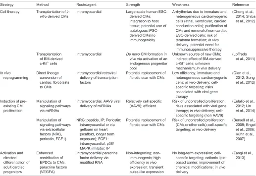
Many research groups have focused their efforts on identifying the mechanisms that govern the eventual postnatal loss of cardiomyocyte proliferation, with the aim of manipulating these pathways to reactivate cardiomyocyte proliferation in the injured adult mammalian heart. The mitogen-activated protein kinase (MAPK) p38α(Mapk14) signaling pathway is a key target for such studies (Engel et al., 2005, 2006; Jopling et al., 2012). In neonatal mice, the miR-15 family and the transcription factor Meis homeobox 1 (MEIS1) play a role in the postnatal loss of cardiomyocyte proliferation. MEIS1 was identified as a transcriptional activator of the cyclin-dependent kinase (CDK) inhibitors p15 (Cdkn2b), p16 (Cdkn2a) and p21 (Cdkn1a) that synergistically inhibit cell cycle progression (Mahmoud et al., 2013; Porrello et al., 2013). A high-throughput screen using a human whole-genome miRNA library identified the additional miRNAs, miR-590 and miR-199a, as proliferation inducers of neonatal mouse and rat cardiomyocytes (Eulalio et al., 2012). After myocardial infarction in mice, these two miRNAs stimulated robust cardiac regeneration and substantially improved cardiac function (Table 1). Recent studies in mice revealed that inactivation of Hippo signaling, which is normally involved in heart size control, was sufficient for cardiomyocyte re-entry into mitosis, cytokinesis and regeneration after injury (Heallen et al., 2013; Lin et al., 2014) (Table 1). The signaling pathways that act through neuregulin 1 (NRG1)/epidermal growth factor receptor 4 (ERBB4), periostin (POSTN)/integrin (ITG) and FGF1 have also been shown to be positive regulators of cardiomyocyte proliferation via activation of
phosphoinositide 3-kinase (PI3K) and to improve cardiac function after myocardial infarction (Fig. 3) (Bersell et al., 2009; Engel et al., 2006; Kühn et al., 2007). Postnatal metabolic changes, from a glycolytic to an oxidative metabolism, and a coinciding DNA damage response have also been uncovered as a major mechanism to mediate permanent cell cycle exit in the majority of cardiomyocytes. Interference with these processes has been shown to prolong the postnatal proliferative window (Puente et al., 2014).
Endogenous cardiac progenitor cells as a source for new cardiomyocytes
To date, numerous different cardiac stem or progenitor cell populations have been reported as a source of new cardiomyocytes in the adult heart. Hsieh and colleagues used an inducible lineage-tracing system permitting irreversible labeling of cardiomyocytes to trace the origin of newly formed cardiomyocytes after myocardial injury in adult mice and documented a significant increase in the number of unlabeled cardiomyocytes at the expense of labeled cardiomyocytes upon myocardial injury, suggesting a contribution from a stem cell or precursor pool (Hsieh et al., 2007) (Table 1). The molecular identity or location of the progenitor pool could not be defined; however, transplantation of bone marrow-derived c-Kit+
cells further diluted the pool of labeled cardiomyocytes, accompanied by an augmentation ofNkx2.5+ and/orGata4+ cardiac progenitor
[image:6.612.48.565.67.422.2]cells. These c-Kit+cells were found not to be the source of the newly formed cardiomyocytes, which led to the speculation that the
Table 1. Strategies to generate cardiomyocytesin vivo
Strategy Method Route/agent Strength Weakness Reference
Cell therapy Transplantation ofin vitroderived CMs
Intramyocardial Large-scale human ESC-derived CMs; integration to host tissue; potential use of autologous iPSC-derived CMs/no immunogenicity
Arrhythmias due to immature and heterogeneous cardiomyogenic cells (atrial, ventricular, cardiac conduction cells); purification of CMs and removal of non-cardiac ESC-derived cells; risk of teratoma formation;in vivo delivery; potential need for immunosuppressive therapy
(Chong et al., 2014; Shiba et al., 2012)
Transplantation of BM-derived c-Kit+cells
Intramyocardial De novoCM formationin vivovia activation of an endogenous progenitor pool
Unknown source of new CMs; indirect effect of BM-derived c-Kit+cells; unknown mechanism;in vivodelivery
(Loffredo et al., 2011)
In vivo
reprogramming
Direct lineage conversion of cardiac fibroblasts to CMs
Intramyocardial retroviral delivery of transcription factors
Potential replacement of fibrotic scar with CMs
Low efficiency; immature and heterogeneous cardiomyogenic cells;in vivodelivery; cell-specific targeting; risks associated with viral gene therapy
(Qian et al., 2012; Song et al., 2012)
Induction of pre-existing CM proliferation
Manipulation of signaling pathways via miRNAs
Intramyocardial, AAV9 viral delivery of miRNAs
Relatively cell specific (AAV9); efficient
Risk of uncontrolled proliferation; risks associated with viral gene therapy;in vivodelivery; cell-specific targeting (non AAV9)
(Eulalio et al., 2012; Lin et al., 2014)
Manipulation of signaling pathways via extracellular factors (NRG, periostin, FGF1)
NRG: peptide, IP; Periostin: intramyocardial or via gelfoam on heart (scaffold, longer term exposure); FGF1: intramyocardial; p38/ MAPK inhibitor: IP
Potential replacement of fibrotic scar with CMs
Risk of uncontrolled proliferation (CMs or other cells); cell-specific targeting;in vivodelivery
(Bersell et al., 2009; Engel et al., 2006; Kühn et al., 2007)
Activation and directed differentiation of adult cardiac progenitors
Enhanced contribution of EPDCs to CMs, paracrine factors (VEGFA)
Intramyocardial paracrine factor delivery via modified RNA
Non-integrating; non-immunogenic; high efficiencyin vivo expression; transient pulse-like expression
No long-term expression; cell-specific targeting; cationic lipid-based carrier; improvement of chemical modifications;in vivo delivery
(Zangi et al., 2013)
CM, cardiomyocyte; BM, bone marrow; EPDCs, epicardium-derived cells; ESC, embryonic stem cell; iPSC, induced pluripotent stem cell; IP, intraperitoneal.
DEVEL
O
secretion of paracrine factors by the c-Kit+cells was required for the
generation of these new cells (Loffredo et al., 2011). There is an ongoing debate fuelled by contradictory studies as to whether c-Kit+ cardiac stem cells (CSC) exist and whether they can give rise to cardiomyocytes (Beltrami et al., 2003; Ellison et al., 2013; Jesty et al., 2012; van Berlo et al., 2014; Zaruba et al., 2010). The observed discrepancies could be due to different experimental methodologies (for further discussion see Molkentin and Houser, 2013; Molkentin and Houser, 2014; Nadal-Ginard et al., 2014; Torella et al., 2014). As c-Kit is a relatively ubiquitous marker (including endothelial, hematopoietic, tissue monocytes), its expression alone is insufficient to define a specific c-Kit+ cell population (Smith et al., 2014). Therefore, cell surface marker signatures will be needed to define such c-Kit+subtypes. c-Kit+cells further progressed towards their first therapeutic use by the initiation of a first clinical trial [stem cell infusion in patients with ischemic cardiomyopathy (SCIPIO)] in 2009 (Bolli et al., 2011). Apart from c-Kit, other markers have also been suggested to label endogenous cardiac progenitor cells, which may or may not reside in the heart (Bu et al., 2009; Chong et al., 2011; Laugwitz et al., 2005; Leobon et al., 2003; Léobon et al., 2009; Makino et al., 1999; Makkar et al., 2012; Martin et al., 2004; Messina et al., 2004; Oh et al., 2003; Sampaolesi et al., 2005; Smith et al., 2007; Tseliou et al., 2014; Yamada et al., 2006). To this date, putative adult endogenous cardiac progenitor cells have generally been shown to contribute to the cardiomyogenic cell lineage either in vitroor in vivo when delivered after injury; however, so far, there is no
convincing evidence that they can be reactivated in situto induce cardiac regeneration following cardiac injury.
Although not traditionally considered cardiac progenitor cells, cells of the epicardium constitute another population of interest with regard to cardiac regeneration (Table 1). EPDCs are activated during injury by the transcription factor C/EBP, which functions together with HOX, MEIS and Grainyhead (GRH) transcription factors to reactivate an embryonic gene program in the epicardium (Huang et al., 2012). Such injury-mediated activation results in an intrinsic cell fate plasticity of EPDCs that can be influenced via extrinsic factors. There is controversial data as to whether priming of Wt1+ EPDCs by the peptide Thymosin-beta 4 (TMSB4) results in their contribution to new cardiomyocytes in the adult heart in response to injury, but if so, then it is likely to be a very minimal contribution (Smart et al., 2011; Zhou et al., 2012). The observed positive effects on cardiac function and scar size (Smart et al., 2011) appear more likely to be due to enhanced neovascularization, given that EPDCs activated by injury have also been identified as a source of pro-angiogenic paracrine factors, which may enhance cardiomyocyte survival (Smart et al., 2007; Zhou et al., 2011).
The injury-induced plasticity of EPDCs can also be exploited to modify the cardiac response to injury, as shown by a recent study. Here, delivery of vascular endothelial growth factor A (VEGFA) through injection of synthetic chemically modified mRNA (modRNA) to the site of injury after myocardial infarction in mice reactivated, expanded, and directed Wt1+epicardial cells away
Reprogramming Directed differentiation
Oct4, Sox2, Klf4, cMyc
Fibroblasts iPSC ESC
Direct lineage conversion Bmp4,
Nodal/activin A Wnt/β-catenin
Cardiac progenitors
Expansion Transplantation/
cell therapy FGF
Wnt/β-catenin inhibition
Cardiomyocyte Cardiac
mesoderm Wnt/β-catenin inhibition Nodal/activin inhibition
Wnt/β-catenin, IGF, NRG FGF1, Notch1, periostin
Expansion Transplantation/cell therapy
Notch activation
Pacemaker cells Purkinje fibers Tbx18
GMT +/– H (mouse)
GMTH + miR-1, miR-133 (human) miR-1,-133,-208,-499, JAK inhibitor Oct4, SB431542, CHIR99021, parnate, Forskolin, Bmp4
[image:7.612.57.558.57.309.2]MESP1, ETS2 (human)
Fig. 3. Strategies to generate cardiomyocytesin vitro.Two major strategies exist for the generation of cardiomyocytesin vitro: directed differentiation from pluripotent stem cells (PSCs) and direct lineage conversion of somatic cells, usually fibroblasts. Various protocols have been established to guide the stepwise differentiation of PSCs, namely embryonic stem cells (ESCs) or induced pluripotent stem cells (iPSCs), into cardiomyocytes. These protocols closely recapitulate the different stages of cardiac development, and so success depends on the fine tuning of the addition of growth factors and small molecules in a time- and dose-dependent manner. In contrast to directed differentiation, generation of cardiomyocytes via direct lineage conversion does not involve PSCs. Instead, the cells transit directly from one somatic cell type, in this case a fibroblast, to the cardiac lineage. This can be achieved by forced expression of the transcription factors Gata4, Mef2c, Tbx5 (collectively GMT) and Hand2 (H), which have all been shown to be important in the specification and development of the cardiac lineagein vivo. Direct lineage conversion to cardiomyocytes can also be achieved by a combination of microRNAs (miRs) or a mix of the single transcription factor Oct4 with a specific set of small molecules. In addition, it has also been shown that direct lineage conversion can produce cardiac progenitor cells, which can then be differentiated into cardiomyocytes via a directed differentiation protocol. Once cardiomyocytes have been generatedin vitro, they can potentially be used for cell therapy approaches to treat cardiac diseases. However, cardiomyocyte expansion is required in order to produce the quantity of cardiomyocytes needed to replace the huge number of muscle cells lost after myocardial infarction.
DEVEL
O
from a fibroblast cell fate towards an endothelial and myocardial cell fate. This approach improved cardiac function, reduced scar size and prolonged survival (Zangi et al., 2013). The identification of further paracrine factors that may activate repair mechanisms, or activate and recruit endogenous progenitors towards the cardiomyogenic lineage, is now experimentally and clinically feasible with this new technological platform (Lui et al., 2013, 2014).
De novogeneration of cardiomyocytesin vitro
In addition toin vivoapproaches, another method to overcome the massive loss of cardiomyocytes from a myocardial infarction or other cardiac insultin vivois to generate cardiomyocytes or cardiac progenitor cells in vitroand then deliver these cells back to the injured heart. This approach relies on recent advances in stem cell and reprogramming technologies, whereby both pluripotent stem cells (PSCs) and somatic cells can be forced to adopt a cardiomyocyte fatein vitro (Ieda et al., 2010; Kehat et al., 2001; Maltsev et al., 1994; Metzger et al., 1995; Song et al., 2012). Such an approach has a distinct advantage over endogenous approaches in that a vast number of starting cells is available for manipulation and eventual transplantation to the injured myocardium. This is made possible by the theoretically limitless self-renewal capacity of PSCs, as well as the expansion potential of somatic cells such as fibroblasts, which can also be converted to induced pluripotent stem cells (iPSCs) via direct reprogramming (Takahashi and Yamanaka, 2006) (Fig. 3, Table 1). The prospect of generating large numbers of cardiomyocytes has implications not only for cellular therapies but also for drug screening. Patient-specific iPSCs can be generated and converted into cardiomyocytes, in order to model particular diseases and to screen for candidate drug treatments.
In order to induce a cardiomyocyte fate from PSCs or somatic cells, a detailed understanding of cardiac muscle development in vivois required. Directed differentiation approaches of PSCs into cardiomyocytes mimic the steps of embryonic development in the dish, starting from cardiac mesoderm formation all the way to cardiomyocyte specification and maturation. During in vitro directed differentiation, PSCs are exquisitely sensitive to their immediate environment, and much emphasis is placed on the particular signaling pathways and growth factors that drive commitment to the cardiomyogenic lineage. By contrast, direct lineage conversion of other somatic cells into cardiomyocytes typically involves the delivery of transcription factors as opposed to growth factors, and the focus is more directed at understanding the distinct gene regulatory network that instructs the cardiomyocyte phenotype. Despite these conceptual differences, both approaches rely on a detailed understanding of the fundamental principles that govern cardiac developmentin vivo in order to recapitulate these eventsin vitro.
Directed differentiation of PSCs to cardiomyocytes
Key signaling pathways, including the Wnt, Activin/Nodal/TGFβ, BMP and FGF signaling pathways, that coordinate cardiogenesisin vivoalso play a pivotal role in controlling the differentiation of PSCs to cardiovascular progenitor cells and cardiomyocytesin vitro. They are used reiteratively during cardiac differentiation in the dish and must be activated and inhibited as the differentiating culture of cells passes through specific developmental stages (Figs 2 and 3) (Kattman et al., 2011; Laflamme et al., 2007; Lian et al., 2012; Noseda et al., 2011; Paige et al., 2010; Yang et al., 2008).
Initial efforts to differentiate human embryonic stem cells (ESCs) into cardiomyocytes employed the embryoid body (EB) technique
in medium containing fetal calf serum, where spheres of pluripotent cells spontaneously recapitulate early steps of embryogenesis in an uncontrolled manner (Kehat et al., 2001). This method proved to be inefficient, only yielding a low percentage (∼1-5%) of cardiomyocytes. Further studies revealed that the timed addition of defined extracellular molecules, either inhibiting or activating specific intracellular signaling pathways, improved the differentiation efficiency into cardiomyocytes (Fig. 3) (Noseda et al., 2011). The temporary and dose-controlled addition of activin A and BMP4 efficiently induced cardiac mesoderm formation in mouse and human EBs characterized by co-expression ofFlk1and platelet-derived growth factor receptor alpha (Pdgfra) or by the generation of a cardiovascular progenitor characterized by co-expression ofKDRandc-KIT(Kattman et al., 2011; Yang et al., 2008). Addition of defined factors, such as FGF2, VEGFA and DKK1, and inhibition of Nodal or TGFβ receptor 2 signaling, enhanced the efficiency of cardiomyocyte differentiation to greater than 50% (Cai et al., 2013; Kattman et al., 2011; Laflamme et al., 2007; Willems et al., 2012; Yang et al., 2008). However, optimization is required for individual cell lines and the differentiation process needs to be monitored for the timely addition of factors to achieve optimal results. In order to generate a relatively pure cardiomyocyte culture (up to 98%), further enrichment steps were included, such as cell sorting with an antibody against signal-regulatory proteinα(SIRPA) (Dubois et al., 2011), cell purification based on biochemical differences in glucose and lactate metabolism between cardiomyocytes and non-cardiomyocytes (Tohyama et al., 2013), or cell purification based on the relatively high content of mitochondria in cardiomyocytes using mitochondria-specific fluorescent dyes (Hattori et al., 2010). As an alternative to EB differentiation, monolayer-based directed differentiation techniques using sequential exposure to growth factors in a defined RPMI/B-27 medium have been established, generating∼30% cardiomyocytes, although significant variability was observed between cell lines and experiments (Laflamme et al., 2007; Melkoumian et al., 2010; Paige et al., 2010). This variability could be due to differences in the sensitivity to growth factors, passage number and, in the case of iPSCs, also the organ of origin, which appears to affect the response to extracellular signaling molecules (Kim et al., 2010; Ohi et al., 2011; Paige et al., 2010; Xu et al., 2012). Finally, endogenous canonical Wnt signaling was shown to be required for human ESCs to differentiate into cardiomyocytes (Paige et al., 2010). Temporal modulation of canonical Wnt signaling in an otherwise fully defined culture setting, in which canonical Wnt signaling was first enhanced and then inhibited, was sufficient to produce a high yield of 80-98% cardiomyocytes from multiple human ESC lines (Lian et al., 2012, 2013). Such a biphasic Wnt regulation mechanism is also required during cardiogenesis in vivo (Naito et al., 2006; Ueno et al., 2007) (Figs 2 and 3), indicating the importance of following developmental progression in thein vitrosetting.
For the development of cell replacement therapy, it will be important to not only obtain pure cardiomyocyte cultures in a large-scale format, but also to further select for the appropriate subtype of cardiomyocytes. Different subtypes of cardiomyocytes can be distinguished by their gene expression profile and electrophysiological properties (Box 2). Myocardial infarction and heart failure predominantly affect the ventricles of the heart, and it is therefore important to be able to selectively purify the ventricular subtype of cardiomyocytes to prevent arrhythmias after transplantation to the cardiac ventricles (Table 1). Recent studies have reported some success in biasing cardiomyogenic differentiation towards an atrial versus
DEVEL
O
ventricular cardiomyocyte subtype by manipulation of retinoic acid signaling (Jiang et al., 2012; Weng et al., 2014; Zhang et al., 2011), or towards a conduction system-like phenotype via activation of the Notch signaling pathway (Rentschler et al., 2012). Despite this progress, differentiation protocols need to be optimized further to obtain pure cultures of cardiomyocyte subtypes for transplantation.
Direct lineage conversion of somatic cells into cardiomyocytes
The successful reprogramming of fully differentiated fibroblasts to pluripotent cells by forced expression of the pluripotency transcription factors octamer-binding transcription factor 4 [Oct4; also known as POU domain, class 5, transcription factor 1 (Pou5f1)], SRY (sex determining region Y)-box 2 (Sox2), Kruppel-like factor 4 (Klf4) and myelocytomatosis oncogene (Myc) (collectively OSKM factors) has fuelled subsequent attempts to directly convert fully differentiated cells to those of another cell lineage, without going through the pluripotent state. By complementing the original iPSC reprogramming protocol, using only OSK factors, with a chemical cocktail and modifications of cell culture conditions, it has been shown that partial dedifferentiation of fibroblasts followed by cardiomyocyte differentiation can be achieved without reaching the pluripotent stage (Efe et al., 2011). Transdifferentiation through a cardiac progenitor stage instead of a pluripotent stage was achieved by the induced expression of the transcription factors Mesp1 and Ets2in fibroblasts (Islas et al., 2012) or ofOct4in combination with a mix of small molecules (Wang et al., 2014). Recent studies have demonstrated the feasibility of direct lineage conversion from fibroblasts to cardiomyocyte-like cells without passing through a stem cell-like stage (Fu et al., 2013; Ieda et al., 2010; Nam et al., 2013; Qian et al., 2012; Song et al., 2012). This was achieved by the forced expression of key cardiac lineage transcription factors (see below for details) leading to partial dedifferentiation of the existing cellular phenotype and instructing a cardiomyocyte-like phenotype (Fig. 3). These elegant studies were inspired by the original observation by Weintraub and colleagues that overexpression of the myogenic master geneMyodcan convert fibroblasts to muscle cells (Davis et al., 1987), followed by successful cell lineage conversion studies in the hematopoietic system (Laiosa et al., 2006; Xie et al., 2004). An important advantage of this direct lineage conversion approach is that, by bypassing pluripotency, the risk of tumor formation is reduced, which is a concern when using PSC-derived cells for therapy purposes.
The first direct lineage conversion of postnatal cardiac and dermal fibroblasts to cardiomyocyte-like cells in vitro was achieved by retroviral delivery of the transcription factors Gata4, Mef2c and Tbx5 (collectively known as GMT), and later also by GMT plus Hand2 (collectively GMTH) to cardiac and tail tip murine fibroblasts. These transcription factors are all well known for their role in controlling normal cardiac development during embryogenesis (Fig. 2) (Ieda et al., 2010; Song et al., 2012). The Srivastava and Olson laboratories then independently used the GMT and GMTH cocktails, respectively, to directly reprogram resident cardiac fibroblasts in 2-month-old mice by intramyocardial injection of retroviruses encoding these transcription factors. Interestingly, this resulted in improved cardiac function and reduced scar size, pointing to a potential therapeutic approach for heart repair (Ieda et al., 2010; Qian et al., 2012; Song et al., 2012). However, the direct reprogramming efficiency was rather limited (5-7% with GMT and∼20% with GMTH) and transdifferentiated cells did not adapt a mature cardiomyocyte phenotype but one that resembled that of neonatal ventricular cardiomyocytes. Given the rather low number of cardiomyocytes generated, the functional
improvement is greater than expected and it will be important to clarify whether the observed improvements were due to other effects, such as the secretion of paracrine factors, new blood vessel formation, inhibition of fibrosis, or improvement of cardiomyocyte survival (Fig. 3, Table 1). The forced expression of the miRNAs miR-1, -133, -208 and -499 was also sufficient to directly convert mouse fibroblasts into cardiomyocytesin vitroandin vivo, which was enhanced by inhibition of Janus kinase 1 (JAK1) (Jayawardena et al., 2012) (Fig. 3). Importantly, the ability to generate functional cardiac-like myocytes via direct lineage conversion is not limited to the mouse, but has also been accomplished using human adult cardiac and dermal fibroblasts, and neonatal foreskin fibroblasts, where reprogramming was achieved within 4-11 weeks by a different combination of factors, including GATA4, HAND2, TBX5,MYOCD, as well as two microRNAs, miR-1 and miR-133 (Nam et al., 2013). In another study, expression of GMT plus estrogen-related receptor gamma (ESRRG),MESP1,MYOCDand zinc finger protein, FOG family member 2 (ZFPM2) induced a cardiomyocyte-like phenotype in human fibroblasts derived from ESCs, fetal heart, and neonatal skin (Fu et al., 2013).
Other examples of induced conversions of cell fate within the cardiac lineage include demonstrations that cardiomyocytes can be forced to adopt the identity of other cardiomyogenic subtypes, such as cells of the CCS. A sinoatrial-like phenotype was induced by forced expression of Tbx18 or Tbx3, as verified by pacemaker characteristics in vitro and in vivo, whereas activation of Notch signaling in cardiomyocytes resulted in Purkinje-like cells (Fig. 3) (Bakker et al., 2012; Kapoor et al., 2013; Rentschler et al., 2012).
Much work remains to optimize direct reprogramming approaches and to fully understand the mechanistic aspects of directed reprogramming to cardiac lineages. A major problem remains the incomplete reprogramming into fully mature cardiomyocytes (Chen et al., 2012) and the rather heterologous population of predominantly atrial- and ventricular-like cardiomyocytes generated through reprogramming, which might increase the risk of arrhythmic events following transplantation. To further translate this method toward clinical application, many other issues need to be addressed, particularly with regard to efficacy and safety. Examples include the open chest surgical procedure currently used for cardiac delivery of molecules and/or cells that perhaps can be replaced by catheter-based delivery during a coronary artery intervention after myocardial infarction, and the need to move to non-integrative methods for delivery of reprogramming factors instead of viruses that integrate into the genome.
From bench to bedside: functional maturation and delivery ofin vitro derived cardiomyocytes
In order to movein vitroderived cardiomyocytes into the clinical setting, it is important to directly compare the maturity, function and durability of these cells with the resident cardiomyocytes found in the adult heart. Characterization of PSC-derived cardiomyocytes has revealed that they share molecular, electrical, mechanical and ultrastructural features with primary cardiomyocytes, but also exhibit diverse characteristics of fetal rather than adult cardiomyocytes. A recent study has shown that long-term cultures of differentiated PSCs can result in structural and functional maturation to adult-like cardiomyocytes (Lundy et al., 2013), but achieving full maturation to an adult phenotype in vitro appears to remain elusive. However, it is important to recognize that this might not be a problem for transplantation purposes, as it has been shown that immature cardiomyocytes exhibit a markedly higher engraftment rate than adult
DEVEL
O
cardiomyocytes when transplanted to the injured heart, and immature cardiomyocytes are likely to mature as they integrate into the recipient myocardium (Reinecke et al., 1999). Two elegant studies have documented the feasibility of transplanting PSC-derived cardiomyocytes to injured hearts in model organisms. In the first study, ESC-derived cardiomyocytes were transplanted into rodent hearts and the results indicate that, at least to some extent, such transplants can integrate functionally with the recipient myocardium, couple electrically with cardiomyocytes from the host, suppress arrhythmias, and improve cardiac output by contributing to the pumping capacity of the heart (Shiba et al., 2012). The second study highlighted the feasibility of producing clinical-scale human ESC-derived cardiomyocytes in vast numbers, as well as their successful intra-myocardial delivery to a non-human primate model of myocardial ischemia followed by reperfusion, moving this approach a substantial step closer towards clinical application (Chong et al., 2014). Here, the human ESC-derived cardiomyocytes formed new muscle grafts that were perfused by host vasculature and electromechanically coupled to the host myocytes; however, non-lethal ventricular arrhythmias were observed and these need to be overcome before further translation to the clinical setting is possible.
Recent advances also allow for the large-scale generation of human PSC-derived endothelial progenitors, which constitutes an attractive cell population for therapeutic regenerative vascularization (Sahara et al., 2014). The capacity to produce scalable cultures of purified human PSC-derived cardiomyocytes and endothelial cells will also be of importance for tissue engineering attempts to produce more organized cardiac tissue. Early experiments have shown that co-culture of PSC-derived cardiac patches with fibroblasts and/or endothelial cells lead to vascularization and better survival of the graft (Sekine et al., 2008; Stevens et al., 2009). With the enormous progress made in large-scale derivation of cardiomyocytes from human PSCs, additional progress in tissue engineering is required to improve the function, engrafting, survival, electromechanical integration and delivery of engineered cardiac tissue for use in transplantation therapies. At the same time, more advanced engineered cardiac tissue would provide for better drug screening and patient-specific disease modelingin vitro(further reviewed by Cimetta et al., 2013; Hirt et al., 2014; Vunjak-Novakovic et al., 2011).
Future directions
Over the past decade, a host of novel therapeutic strategies have been developed that are aimed at capitalizing on recent advances in our understanding of the pathways and principles that guide cardiogenesis itself. These include both cell-based and paracrine factor-based therapeutic approaches, each employing different technological platforms ( peptide, recombinant protein, plasmid, adenoviral, adeno-associated viruses, retroviruses, miRNA, RNAi) in attempting to drive the generation of new cardiomyocytes, additional vascular components (both endothelial and vascular smooth muscle) and/or to inhibit fibrosis and accompanying scar formation (reviewed by Ptaszek et al., 2012). More recent studies have suggested that direct reprogramming of non-myocytes in the intact heart to cardiomyocyte-like cells could be an alternative approach (Qian et al., 2012; Song et al., 2012). All these strategies have inherent advantages and limitations (regarding such as safety, scalability, efficiency and duration of expression,in vivodelivery, target specificity, off-target effects, other organ side effects), necessitating additional studies to determine which, if any of these approaches, could eventually be promoted to clinical practice.
We believe that the new technological platform of modRNAs provides a promising route for the short-term, localized expression of paracrine factors [such as VEGFA (Zangi et al., 2013)] in diseased hearts, but future studies will need to determine their safety and efficacy compared with alternative therapeutic approaches. In any case, the key to understanding how to remake a human heart might lie in unraveling the pathways that make the heart during cardiogenesis itself. A more refined map of human cardiogenesis and the development of novel human and non-human primate model systems are on the horizon, which should lay the foundation for the next generation of strategies for regenerative cardiology.
Acknowledgements
We apologize to our colleagues whose work could not be cited due to limitations to the length of this Primer.
Competing interests
The authors declare no competing financial interests.
Funding
Financial support from the Swedish Research Council [VR 539-2013-7002, VR 541-2013-8351, SFO], Karolinska Institutet/AstraZeneca ICMC [ECHO Project ID:10023735], AstraZeneca [Dealz ID SEML-935CA8] and the Knut and Alice Wallenbergs Stiftelse [KAW 2013.0028] is gratefully acknowledged. E.M.H. is a Fellow of the Wenner-Gren Foundation and supported by the Swedish Heart-Lung foundation. L.Z. is supported by an American Heart Association Postdoctoral Fellowship.
References
Abu-Issa, R. and Kirby, M. L.(2007). Heart field: from mesoderm to heart tube.
Annu. Rev. Cell Dev. Biol.23, 45-68.
Ai, D., Fu, X., Wang, J., Lu, M.-F., Chen, L., Baldini, A., Klein, W. H. and Martin, J. F.(2007). Canonical Wnt signaling functions in second heart field to promote right ventricular growth.Proc. Natl. Acad. Sci. USA104, 9319-9324.
Ali, S. R., Hippenmeyer, S., Saadat, L. V., Luo, L., Weissman, I. L. and Ardehali, R.(2014). Existing cardiomyocytes generate cardiomyocytes at a low rate after birth in mice.Proc. Natl. Acad. Sci. USA111, 8850-8855.
Andersen, D. C., Ganesalingam, S., Jensen, C. H. and Sheikh, S. P.(2014a). Do
neonatal mouse hearts regenerate following heart apex resection?Stem Cell Rep. 2, 406-413.
Andersen, D. C., Jensen, C. H. and Sheikh, S. P.(2014b). Response to Sadek
et al. and Kotlikoff et al.Stem Cell Rep.3, 3-4.
Atkinson, A., Inada, S., Li, J., Tellez, J. O., Yanni, J., Sleiman, R., Allah, E. A.,
Anderson, R. H., Zhang, H., Boyett, M. R. et al.(2011). Anatomical and
molecular mapping of the left and right ventricular His-Purkinje conduction networks.J. Mol. Cell. Cardiol.51, 689-701.
Bakker, M. L., Boink, G. J. J., Boukens, B. J., Verkerk, A. O., van den Boogaard, M., den Haan, A. D., Hoogaars, W. M. H., Buermans, H. P., de Bakker, J. M. T.,
Seppen, J. et al.(2012). T-box transcription factor TBX3 reprogrammes mature
cardiac myocytes into pacemaker-like cells.Cardiovasc. Res.94, 439-449. Balza, R. O. and Misra, R. P.(2006). Role of the serum response factor in regulating
contractile apparatus gene expression and sarcomeric integrity in cardiomyocytes.J. Biol. Chem.281, 6498-6510.
Barrow, J. R., Howell, W. D., Rule, M., Hayashi, S., Thomas, K. R., Capecchi,
M. R. and McMahon, A. P.(2007). Wnt3 signaling in the epiblast is required for
proper orientation of the anteroposterior axis.Dev. Biol.312, 312-320. Beltrami, A. P., Barlucchi, L., Torella, D., Baker, M., Limana, F., Chimenti, S.,
Kasahara, H., Rota, M., Musso, E., Urbanek, K. et al.(2003). Adult cardiac stem cells are multipotent and support myocardial regeneration.Cell114, 763-776. Bergmann, O., Bhardwaj, R. D., Bernard, S., Zdunek, S., Barnabé-Heider, F.,
Walsh, S., Zupicich, J., Alkass, K., Buchholz, B. A., Druid, H. et al.(2009).
Evidence for cardiomyocyte renewal in humans.Science324, 98-102.
Bergmann, O., Zdunek, S., Alkass, K., Druid, H., Bernard, S. and Frisén, J.
(2011). Identification of cardiomyocyte nuclei and assessment of ploidy for the analysis of cell turnover.Exp. Cell Res.317, 188-194.
Bergmann, O., Zdunek, S., Frisén, J., Bernard, S., Druid, H. and Jovinge, S. (2012). Cardiomyocyte renewal in humans.Circ. Res.110, e17-e18; author reply e19-e21.
Bersell, K., Arab, S., Haring, B. and Kühn, B.(2009). Neuregulin1/ErbB4 signaling induces cardiomyocyte proliferation and repair of heart injury.Cell138, 257-270. Bird, S. D., Doevendans, P. A., van Rooijen, M. A., Brutel de la Riviere, A.,
Hassink, R. J., Passier, R. and Mummery, C. L.(2003). The human adult
cardiomyocyte phenotype.Cardiovasc. Res.58, 423-434.
Bolli, R., Chugh, A. R., D’Amario, D., Loughran, J. H., Stoddard, M. F., Ikram, S.,
Beache, G. M., Wagner, S. G., Leri, A., Hosoda, T. et al.(2011). Cardiac stem