Abstract
The vasculature plays a crucial role in inflammation, angiogenesis, and atherosclerosis associated with the pathogenesis of inflam-matory rheumatic diseases, hence the term ‘vascular rheumatology’. The endothelium lining the blood vessels becomes activated during the inflammatory process, resulting in the production of several mediators, the expression of endothelial adhesion molecules, and increased vascular permeability (leakage). All of this enables the extravasation of inflammatory cells into the interstitial matrix. The endothelial adhesion and transendothelial migration of leukocytes is a well-regulated sequence of events that involves many adhesion molecules and chemokines. Primarily selectins, integrins, and members of the immunoglobulin family of adhesion receptors are involved in leukocyte ‘tethering’, ‘rolling’, activation, and trans-migration. There is a perpetuation of angiogenesis, the formation of new capillaries from pre-existing vessels, as well as that of vasculogenesis, the generation of new blood vessels in arthritis and connective tissue diseases. Several soluble and cell-bound angiogenic mediators produced mainly by monocytes/macro-phages and endothelial cells stimulate neovascularization. On the other hand, endogenous angiogenesis inhibitors and exogenously administered angiostatic compounds may downregulate the process of capillary formation. Rheumatoid arthritis as well as systemic lupus erythematosus, scleroderma, the antiphospholipid syndrome, and systemic vasculitides have been associated with accelerated atherosclerosis and high cardiovascular risk leading to increased mortality. Apart from traditional risk factors such as smoking, obesity, hypertension, dyslipidemia, and diabetes, inflam-matory risk factors, including C-reactive protein, homocysteine,
folate deficiency, lipoprotein (a), phospholipid antibodies, anti-bodies to oxidized low-density lipoprotein, and heat shock proteins, are all involved in atherosclerosis underlying inflammatory rheu-matic diseases. Targeting of adhesion molecules, chemokines, and angiogenesis by administering nonspecific immunosuppressive drugs as well as monoclonal antibodies or small molecular compounds inhibiting the action of a single mediator may control inflammation and prevent tissue destruction. Vasoprotective agents may help to prevent premature atherosclerosis and cardiovascular disease.
Introduction
Vessels and the vascular endothelium are involved in the pathogenesis of inflammatory rheumatic diseases. Rheuma-toid arthritis (RA) serves as a prototype of these diseases as it is the most common type of arthritis and a great body of data is available regarding leukocyte recruitment into the synovium, angiogenesis, and accelerated atherosclerosis. The term ‘vascular rheumatology’ has been accepted by many investigators and includes both microvascular and macrovascular involvement in rheumatic diseases. Apart from RA, systemic lupus erythematosus (SLE), systemic sclerosis (SSc), the antiphospholipid syndrome (APS), and systemic vasculitides have been associated with vascular inflammation, altered angiogenesis, and increased cardiovascular morbidity and mortality. In this review, we will discuss the most relevant
Review
Vascular involvement in rheumatic diseases:
‘vascular rheumatology’
Zoltán Szekanecz
1and Alisa E Koch
2,31University of Debrecen Medical Center, Institute of Medicine, Department of Rheumatology, 22 Móricz street, Debrecen, H-4032, Hungary 2Veterans’ Administration Ann Arbor Healthcare System, 2215 Fuller Road, Ann Arbor, MI 48105, USA
3University of Michigan Health System, Division of Rheumatology, Department of Internal Medicine, University of Michigan Medical School, 109 Zina Pitcher Place, Ann Arbor, MI 48109-2200, USA
Corresponding author: Zoltán Szekanecz, szekanecz@gmail.com
Published: 10 October 2008 Arthritis Research & Therapy2008, 10:224 (doi:10.1186/ar2515) This article is online at http://arthritis-research.com/content/10/5/224
© 2008 BioMed Central Ltd
information on arthritis-related vascular inflammation, inclu-ding the role of endothelial cells (ECs), endothelial adhesion molecules (CAMs) and chemokines, as well as the involve-ment of neovascularization and some aspects of accelerated atherosclerosis in rheumatic diseases. We will discuss RA in more detail, and other connective tissue diseases described above will also be mentioned. Finally, some aspects of vascular targeting in rheumatology will also be briefly summarized.
Endothelial biology and leukocyte trafficking
through the vessel wall
Vascular permeability and vascular damage underlying inflammation
In arthritis, leukocyte ingress into the synovium occurs by leukocyte adhesion to ECs and then by transendothelial migration [1-8]. The chemotaxis of these leukocytes is regu-lated mainly by various chemokines [1,8,9-14]. Several CAMs have been implicated in leukocyte-EC interactions [1-4,7,8]. ECs play an active role in inflammation. Synovitis is associated with vasodilation and increased endothelial per-meability (leakage) and vascular injury followed by endothelial regeneration [4-6]. ECs secrete several vasodilatory mediators, including nitric oxide, prostacyclin (PGI2), platelet-activating factor, histamine, and others [4-6]. Increased vascular permeability associated with EC retraction and contraction may be a physiological process, while in inflammation, pro-inflammatory mediators trigger vascular damage [4-6]. Increased vascular permeability is induced primarily by vasoactive agents such as histamine, serotonin, bradykinin, and others [4-6,15]. Vascular injury is caused primarily by activated neutrophils, inflammatory mediators released by these cells, including reactive oxygen intermediates and matrix metalloproteinases (MMPs). Anti-EC antibodies (AECAs), tumor necrosis factor-alpha (TNF-α), interleukin-1 (IL-1), or interferon-gamma (IFN-γ) stimulates EC injury [4-6,15]. The abundant production of AECAs, markers of vascular damage, has been reported in RA, SLE, systemic vasculitis, and other rheumatic diseases [15] (Table 1). Injury is followed by endothelial regeneration, which may be associated with angiogenesis or may occur without the formation of new blood vessels [5,6,16].
Intercellular adhesion molecules in arthritis
The cascade of leukocyte transendothelial migration begins with the adhesion of leukocytes, including neutrophils, lymphocytes, and monocytes, to postcapillary venules. Leukocyte recruitment occurs through the wall of these venules. In some RA patients, specialized ECs resembling high endothelial venules (HEVs) are found in the synovium. These HEVs are surrounded by lymphoid aggregates composed of T cells [1,2,8]. Inflammatory leukocyte recruit-ment into inflamed tissue is very similar to the ‘homing’ associated with physiological immune surveillance [1-3]. Leukocyte adhesion to ECs or to extracellular matrix (ECM) constituents is mediated by endothelial CAMs and their counter-receptors on infiltrating white blood cells. Primarily
selectins, integrins, and some members of the immuno-globulin superfamily of adhesion molecules (CAMs) have been implicated in leukocyte extravasation, but some other CAMs may also play a role in this process [2,3,7]. These CAMs are summarized in Table 2. During leukocyte trans-endothelial migration, selectins mediate the initial ‘tethering’ and ‘rolling’ of leukocytes whereas integrins and other CAMs are involved in firm adhesion and migration of leukocytes [1,3,8]. All selectins, most integrins, and members of the immunoglobulin superfamily are abundantly expressed in arthritic synovial tissues [2,3]. Other CAMs involved in leuko-cyte-EC adhesion underlying inflammation include intra-cellular adhesion molecule-3 (ICAM-3), the lymphocyte function-associated antigen-3 (LFA-3)-CD2 counter-receptors, various alternative forms of CD44, vascular adhesion proteins (VAP-1 and VAP-2), endoglin (CD105), E-cadherin, N-cadherin, cadherin-11, platelet-endothelial cell adhesion molecule-1 (PECAM-1) (CD31), junctional adhesion molecules (JAMs), CD99, and others [1-3,7]. All of these CAMs have been detected in arthritic synovial tissues [1-3].
Chemokines and chemokine receptors in synovial inflammation
Chemokines are small proteins exerting chemotactic activity toward leukocytes [9-12,14,17,18]. Chemokines have been classified into supergene families according to the location of cysteine (C) in their molecular structure. These families are designated as CXC, CC, C, and CX3C chemokines; the particular chemokine ligand members are CXCL, CCL, CL, and CX3CL, and the four chemokine receptor groups are
[image:2.612.313.557.128.286.2]CXCR, CCR, CR, and CX3CR, respectively [9,10,12]. To date, more than 50 chemokines and 19 chemokine receptors have been identified [9,10,12] (Table 3). Most CXC chemo-kines chemoattract neutrophils, but platelet factor-4 (PF4)/ CXCL4 and IFN-γ-inducible 10-kDa protein (IP-10)/CXCL10 recruit lymphocytes and monocytes [9]. Among CXC chemokines, IL-8/CXCL8, epithelial neutrophil-activating
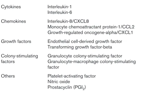
Table 1
Some important inflammatory mediators released by vascular endothelial cells
Cytokines Interleukin-1 Interleukin-6 Chemokines Interleukin-8/CXCL8
Monocyte chemoattractant protein-1/CCL2 Growth-regulated oncogene-alpha/CXCL1 Growth factors Endothelial cell-derived growth factor
Transforming growth factor-beta Colony-stimulating Granulocyte colony-stimulating factor factors Granulocyte-macrophage colony-stimulating
factor
Others Platelet-activating factor Nitric oxide
protein-78 (ENA-78)/CXCL5, growth-regulated oncogene-alpha (groα)/CXCL1, connective tissue-activating peptide-III (CTAP-III)/CXCL7, granulocyte chemotactic protein-2/ CXCL6, IP-10/CXCL10, PF4/CXCL4, monokine induced by IFN-γ (Mig)/CXCL9, stromal cell-derived factor-1 (SDF-1)/ CXCL12, B cell-activating chemokine-1/CXCL13, and CXCL16 have been implicated in the pathogenesis of synovial inflammation [14,17,18]. CC chemokines stimulate monocyte chemotaxis and some of them also chemoattract lymphocytes [10]. Monocyte chemoattractant protein-1 (MCP-1)/CCL2, macrophage inflammatory protein-1-alpha (MIP-1α)/CCL3, MIP-3α/CCL20, RANTES (Regulated upon Activation, Normal T-cell Expressed and Secreted)/CCL5, Epstein-Barr virus-induced gene-1 ligand chemokine (ELC)/ CCL19, secondary lymphoid tissue chemokine (SLC)/CCL21, and chemokine-like factor-1 (CKLF1) have been implicated in inflammatory mechanisms underlying synovitis [14,17,18]. The C chemokine family contains two members: lympho-tactin/XCL1 and single C motif-1-beta (SCM-1β)/XCL2 [12]. Lymphotactin/XCL1 has been detected on synovial T cells in RA [14,18]. The CX3C chemokine subfamily contains
frac-talkine/CX3CL1 [12,19]. This chemokine chemoattracts mononuclear cells and also serves as a CAM [17,19]. Fractalkine/CX3CL1 has also been detected in RA synovial
tissues [19]. Fractalkine/CX3CL1 has also been implicated in the development of accelerated atherosclerosis [18], a topic discussed later. Chemokines bind to their seven-transmembrane domain receptors expressed on the target
cells [12,18]. Some of these receptors have numerous chemokine ligands whereas others are specific receptors for single ligands [14]. Chemokine receptors have also been associated with various histological subtypes of inflammation. For example, whereas CXCR3 and CCR5 may be involved primarily in Th1 type diseases (such as RA), CCR3, CCR4, and CCR8 may play a role in leukocyte migration underlying Th2 type inflammation (such as asthma) [11]. Most CXC and CC chemokine receptors mentioned above as well as XCR1 and CX3CR1 are expressed in the arthritic synovium [14,17,18].
The process of leukocyte recruitment into inflamed tissues
Leukocyte adhesion to ECs occurs following a cascade of events. White blood cells in the bloodstream weakly adhere to the endothelium lining the inner vessel wall (tethering) followed by rolling of leukocytes on the endothelial layer. Tethering and rolling are mediated primarily by selectins and their ligands. These events are followed by leukocyte activation, which is dependent upon interactions between chemokine receptors expressed on leukocytes and proteo-glycans on ECs. Activation-dependent firm adhesion occurs next, involving α4β1integrin/VCAM-1 (vascular cell adhesion
molecule-1), β2 integrin/ICAM-1, and JAM/integrin inter-actions. This is associated with the secretion of chemokines. These chemokines may also upregulate integrin expression on the adhering cells via PI3K (phosphatidylinositol
3-kinase)-Table 2
Relevant members of the selectin, integrin, and immunoglobulin adhesion molecule superfamilies
Adhesion receptors Ligands
Selectins
L-selectin (CD62L, LAM-1) Sialylated carbohydrates, GlyCAM-1 E-selectin (CD62E, ELAM-1) Sialyl-Lewis-X
P-selectin (CD62P, PADGEM) Sialyl-Lewis-X, other carbohydrates Integrins
β1integrins Laminin, collagen, fibronectin
α4β1integrins Fibronectin, VCAM-1
β2integrins ICAM-1, ICAM-2, ICAM-3, JAM-A
β3integrins Vitronectin, von Willebrand factor, other matrix molecules Immunoglobulin superfamily
ICAM-1, ICAM-3 αLβ2(LFA-1), αMβ2(Mac-1, CR3)
VCAM-1 α4β1, α4β7
CD2 LFA-3
PECAM-1 (CD31) PECAM-1, αVβ3integrin
[image:3.612.60.561.116.358.2]mediated pathways. Leukocyte diapedesis through the endothelial layer involving integrins occurs when chemokines bind to endothelial heparan sulphate. Chemokines preferen-tially chemoattract EC-adherent leukocytes. These processes lead to the transmigration of leukocytes into the inflamed tissue [1,8].
Targeting of cell adhesion, chemokines, and leukocyte recruitment
Inhibition of cell adhesion, chemokines, and migration using specific antibodies or purified ligands has provided an important perspective on the molecular pathogenesis of RA. In addition, some of these strategies may be included in the future therapy of arthritis [20]. Regarding anti-CAM trials, an anti-human ICAM-1 antibody (enlimomab) was tried in refractory RA with little success [2,20]. Other antiadhesion strategies have been introduced into the treatment of other inflammatory diseases. For example, efalizumab (anti-LFA-1) and alefacept (LFA-3-Ig fusion protein) have been tried in psoriasis, natalizumab (anti-α4 integrin) in multiple sclerosis
and Crohn disease, and an anti-α4β7 integrin monoclonal antibody in ulcerative colitis [2,3,20]. These and other anti-CAM strategies may be tried in arthritis as well [2,3,20]. Chemokines and chemokine receptors can be targeted in a number of ways. Disease-modifying antirheumatic drugs (DMARDs) and anti-TNF biologics, currently used in the treatment of RA, may indirectly influence chemokine
produc-tion [18]. Antibodies to IL-8/CXCL8, ENA-78/CXCL5, CXCL16, MIP-1α/CCL3, MCP-1/CCL2, and fractalkine/ CX3CL1 have been used to control arthritis in various rodent models [18-20]. Several oral chemokine receptor antago-nists, including CXCR2, CXCR4, CCR1, CCR2, and CCR5 inhibitors, have been tried in human RA as well as in animal models of arthritis [18,20-22].
Angiogenesis and vasculogenesis in
rheumatic diseases
The processes of angiogenesis and vasculogenesis Angiogenesis is the formation of new capillaries from pre-existing vessels, whereas vasculogenesis involves circulating endothelial progenitor cells (EPCs) [14,16,23-27]. Angio-genesis involves cell surface-bound and soluble angiogenic mediators, which activate vascular ECs (Table 4). In response, ECs release MMPs, which digest the underlying basal membrane and the ECM enabling the emigration of ECs. Single ECs will then gather to form capillary sprouts. Lumen formation within the sprouts leads to capillary loops. Finally, the synthesis of new basement membrane leads to the formation of new capillaries [23]. Regarding vasculo-genesis, a subpopulation of circulating CD34+cells
[image:4.612.58.535.118.358.2]expres-sing the vascular endothelial growth factor-2 (VEGF-2) receptor has been identified and characterized as functional EPCs. Decreased numbers of EPCs as well as impaired vasculogenesis have been associated with arthritis [27,28].
Table 3
Chemokine receptors with ligands relevant for arthritis and angiogenesis
Chemokine receptor Chemokine ligand
CXC chemokine receptors
CXCR1 IL-8/CXCL8
CXCR2 IL-8/CXCL8, ENA-78/CXCL5, groα/CXCL1, CTAP-III/CXCL7
CXCR3 IP-10/CXCL10, PF4/CXCL4, Mig/CXCL9, ITAC/CXCL11
CXCR4 SDF-1/CXCL12
C-C chemokine receptors
CCR1 MIP-1α/CCL3, RANTES/CCL5, MCP-3/CCL7, MPIF-1/CCL23
CCR2 MCP-1/CCL2, MCP-3/CCL7
CCR6 MIP-3α/CCL20
CCR7 MIP-3β/CCL19, SLC/CCL21
C chemokine receptors
XCR1 Lymphotactin/XCL1
C-X3-C chemokine receptors
CX3CR1 Fractalkine/CX3CL1
The major chemoattractant that drives EPCs is the SDF-1/CXCL12 chemokine and its receptor, CXCR4 [29]. In arthritis, proinflammatory cytokines stimulate the production of SDF-1/CXCL12 and thus tissue vasculogenesis by recruiting CXCR4+EPCs [14,17,29].
Angiogenic mediators and inhibitors in rheumatoid arthritis
The hypoxia-VEGF-angiopoietin pathway is an essential angiogenic network in synovitis [16,23-25,30]. VEGF, a growth factor that binds to heparin in the synovial ECM, plays a central role in the regulation of neovascularization [23,30]. There is hypoxia present within the joint cavity, and hypoxia as well as TNF-αand IL-1 stimulate VEGF release [16]. Hypoxia acts through the hypoxia-inducible factor heterodimer, HIF-1α/ HIF-1β [16]. Several other angiogenic mediators also act indirectly via VEGF [23]. Angiopoietin-1 (Ang1) and Ang2 regulate EC functions upon stimulation by VEGF. Both Ang1 and Ang2 interact with the Tie2 endothelial tyrosine kinase receptor [16,24]. Ang1-Tie2 interactions result in vessel stabilization. On the other hand, Ang2, an antagonist of Ang1, inhibits vessel maturation [24]. Another important player in this network is survivin, an apoptosis inhibitor, which is also involved in VEGF-induced angiogenesis and EC survival [16]. VEGF, HIF-1, Ang1, Tie2, and survivin are all expressed in the arthritic synovium [16,25]. Growth factors other than VEGF but implicated in angiogenesis include fibroblast (FGF-1 and FGF-2), hepatocyte, platelet-derived, epidermal (EGF), insulin-like, and transforming (TGF-β) growth factors [16,24,25]. Among chemokines described above, CXC chemokines that contain the ELR (glutamic acid-leucine-arginine) amino acid motif promote angiogenesis [13]. ELR+ CXC chemokines
that stimulate angiogenesis and also synovial inflammation include IL-8/CXCL8, ENA-78/CXCL5, groα/CXCL1, and CTAP-III/CXCL7. SDF-1/CXCL12 is a unique CXC chemo-kine as it exerts mainly a homeostatic function, yet it has been implicated in inflammation such as in RA [13,14,17,29]. Moreover, this chemokine lacks the ELR motif but is still angiogenic [14,29]. The crucial role of SDF-1/CXCL12 in vasculogenesis is discussed above [29]. In contrast to angiogenic CXC chemokines, the ELR–PF4/CXCL4, IP-10/
CXCL10, and Mig/CXCL9 suppress neovascularization [13,14,17]. Regarding CC chemokines, MCP-1/CCL2 promotes neovascularization induced by growth factors [14,17]. The sole CX3C chemokine, fractalkine/CX3CL1, also promotes synovial angiogenesis [14,17,19]. Regarding chemokine receptors, CXCR2, which binds most ELR+CXC
chemokines described above, is a crucial chemokine receptor in angiogenesis [13,14,17]. CXCR4 has been implicated in SDF-1/CXCL12-induced neovascularization in arthritis [14,17,29]. In contrast, CXCR3, a receptor for the angiostatic IP-10/CXCL10 and Mig/CXCL9, may be involved in chemokine-mediated angiostasis [14,17]. Numerous proinflammatory cytokines, such as TNF-α, IL-1, IL-6, IL-15, IL-17, IL-18, granulocyte and granulocyte-macrophage colony-stimulating factors, oncostatin M, and macrophage
migration inhibitory factor, also induce synovial angiogenesis [16,31]. In contrast, other cytokines, such as IFN-α, IFN-γ, IL-4, IL-12, IL-13, and leukemia inhibitory factor, suppress the production of angiogenic mediators and thus inhibit neo-vascularization [16,31,32]. ECM components, matrix-degra-ding proteases, and cellular adhesion molecules described above may be involved in EC emigration, sprouting, and thus angiogenesis. Among ECM components, various types of collagen, fibronectin, laminin, vitronectin, tenascin, and proteoglycans promote neovascularization [16]. Proteolytic enzymes, such as MMPs and plasminogen activators, play a role in matrix degradation underlying synovial angiogenesis [16,25]. On the other hand, tissue inhibitors of metallo-proteinases and plasminogen activator inhibitors antagonize the angiogenic effects of proteases described above [16,32]. Among CAMs, β1 and β3 integrins, E-selectin, glycoconju-gates (including Lewisy/H), melanoma cell adhesion molecule
(MUC18), VCAM-1, PECAM-1, and endoglin have been implicated in neovascularization [2,16,25,33,34]. The αVβ3 integrin is of outstanding importance as this CAM mediates both synovial angiogenesis and osteoclast-mediated bone resorption and the development of erosions in RA [34]. Other important angiogenic factors not mentioned above include endothelin-1, angiogenin, angiotropin, and many others [16,25] (Table 4). Angiostatic mediators and compounds also include angiostatin (a fragment of plasminogen), endostatin (a fragment of type XIII collagen), thrombospondin-1, 2-methoxyestradiol, paclitaxel, osteonectin, chondromodulin-1, and others [16,30,32,35] (Table 4). These molecules suppress the action of angiogenic mediators, such as VEGF, HIFs, or the αVβ3integrin [16,30,32,35].
Angiogenesis in other types of arthritis and connective tissue diseases
was not sufficient to compensate the loss of blood vessels [16,25]. Regarding systemic vasculitides, abundant produc-tion of angiogenic VEGF and TGF-β has been associated with Kawasaki syndrome [16]. Increased serum levels of TGF-β were found in ANCA (antineutrophil cytoplasmic antibody)-associated vaculitides, including Wegener granulomatosis, Churg-Strauss syndrome, and microscopic polyangiitis [16,25].
Targeting of angiogenesis in inflammatory rheumatic diseases
There may be two major strategies to control angiogenesis in arthritis as well as in malignancies [16,32,35]. Endogenous inhibitors of neovascularization described above, including cytokines, chemokines, protease inhibitors, and others, are naturally produced in the arthritic synovium. However, angio-genic mediators are abundant within the inflamed tissue; therefore, these endogenous angiostatic molecules need to be administered in excess in order to attenuate neo-vascularization. In addition, numerous synthetic compounds currently used to control inflammation and to treat arthritis may, among other effects, inhibit capillary formation as well. These exogenous angiostatic compounds include cortico-steroids, traditional disease-modifying agents (DMARDs) and biologics, antibiotic derivatives, thalidomide, and others [16,32,35] (Table 5). Among endogenous angiogenesis inhibitors, angiostatin and endostatin block αVβ3 integrin-dependent angiogenesis and both molecules inhibited the development of arthritis in various animal models [16,35]. Thrombospondin-1 and -2 are angiostatic ECM components
[image:6.612.61.552.100.297.2]produced by RA synovial macrophages and fibroblasts [16,32]. IL-4 and IL-13 gene transfer attenuated synovial inflammation and angiogenesis in rats [16]. The PF4/CXCL4 chemokine has also been tried in rodent models [16]. Fumagillin analogs, such as TNP-470 and PPI2458, also exert angiostatic and antiarthritic properties [16,32,35]. Traditional DMARDs and biologics exert various anti-inflammatory effects. In addition, these compounds may inhibit synovial vessel formation by nonspecifically blocking the action of angiogenic mediators [16,17]. Thalidomide, recently introduced into the treatment of RA and lupus, is a potent TNF-αantagonist and angiogenesis inhibitor [16,35]. CC1069, a thalidomide analog, even more potently inhibited arthritis in rats [35]. The hypoxia-HIF pathway may also be targeted using nonspecific inhibitor compounds, including YC-1 [16,35]. 2-Methoxyestradiol, mentioned above, and paclitaxel (taxol), a drug already used in human cancer, destabilize the intracellular cytoskeleton and also block HIF-1α[35]. Soluble Fas ligand (CD178) inhibited synovial VEGF production and angiogenesis [16]. Pioglitazone, an anti-diabetic PPAR-γ (peroxisome proliferator-activated receptor-gamma) agonist, is also angiostatic. Pioglitazone effectively controlled psoriatic arthritis in 10 patients [16,35]. Regarding specific exogenous strategies, VEGF is the key target [30,35]. Numerous synthetic VEGF and VEGF receptor inhibitors (including vatalanib, sunitinib, sorafenib, and vandetanib), anti-VEGF antibodies (including bevacizumab), and inhibitors of VEGF and VEGF receptor signaling inhibit neovascularization and are under development for cancer therapy [30,35]. To date, vatalanib has been tried and
Table 4
Some angiogenic and angiostatic factors in arthritis
Mediators Inhibitors
Chemokines IL-8/CXCL8, ENA-78/CXCL5, groα/CXCL1, CTAP-III/CXCL7, PF4/CXCL4, IP-10/CXCL10, Mig/CXCL9, SDF-1/CXCL12, MCP-1/CCL2, SLC/CCL21, MPIF/CCL23, SLC/CCL21
fractalkine/CX3CL1
Matrix molecules Type I collagen, fibronectin, laminin, heparin, heparan sulphate Thrombospondin, RGD sequence Cell adhesion molecules β1and β3integrins, E-selectin, P-selectin, CD34, VCAM-1, RGD sequence (integrin ligand)
endoglin, PECAM-1, vascular endothelial-cadherin, Lewisy/H, MUC18
Growth factors VEGF, bFGF, aFGF, PDGF, EGF, IGF-I, HIF-1, TGF-βa TGF-βa
Cytokines TNF-α, IL-6a, IL-15, IL-18 IL-4, IL-6a, IFN-α, IFN-γ
Proteases MMPs, plasminogen activators TIMPs, plasminogen activator inhibitors Others Angiogenin, substance P, prolactin DMARDs, infliximab, etanercept, angiostatin,
endostatin
attenuated knee arthritis in rabbits [35]. The Ang-Tie system may also be targeted. A soluble Tie2 receptor transcript was delivered via an adenoviral vector to mice. The inhibition of Tie2 delayed the onset and attenuated the severity of arthritis [16,35]. Vitaxin, a humanized antibody to the αVβ3 integrin, inhibited synovial angiogenesis [16,34] but, in a phase II human RA trial, showed only limited efficacy [35]. Numerous specific MMP inhibitors have been tried in angiogenesis models [16,35]. Endothelin-1 antagonists currently used in the therapy of primary and SSc-associated secondary pulmonary hypertension may also exert angiostatic effects [16,28].
Accelerated atherosclerosis in rheumatic
diseases
The basis of atherosclerosis and increased vascular risk Accelerated atherosclerosis and increased cardiovascular morbidity and mortality have been associated with RA, SLE, APS, and SSc [36-41]. Cardiovascular disease (CVD) causes reduced life expectancy and became a major mortality factor in these diseases [36-41]. Atherosclerosis is also considered an inflammatory disease; thus, it may share common pathogenic mechanisms with rheumatic diseases [36,42,43] (Table 6). Numerous studies have demonstrated the role of traditional, Framingham, and inflammation-asso-ciated risk factors in atherosclerosis assoinflammation-asso-ciated with arthritis [36-38,44]. Among traditional risk factors, cigarette smoking not only is a major risk factor for CVD but has recently been implicated in tissue citrullination, the production of anti-cyclic citrullinated peptide (anti-CCP) antibodies, and thus susceptibility to RA [36,38]. In addition to smoking, physical inactivity, obesity, hypertension, dyslipidemia, and diabetes mellitus may be implicated in accelerated atherosclerosis [36-38,44]. Yet excess CVD mortality occurs predominantly in RA patients with a higher degree of systemic inflammation [36]; therefore, accelerated atherosclerosis cannot be fully explained on the basis of traditional risk factors [42,43].
Indeed, several inflammatory and atherogenic mediators, including homocysteine, lipoprotein (a), C-reactive protein (CRP), hyperhomocysteinemia, and folate, and vitamin B12 deficiency and decreased paraoxonase-1 activity are strongly associated with atherosclerosis and CVD [36,42,43]. Athero-sclerotic plaques, similarly to the RA joint, are characterized by enhanced accumulation of inflammatory monocytes/ macrophages and T cells. These inflammatory leukocytes abundantly produce proinflammatory cytokines, chemokines, and MMPs [42,43]. CD4+ T cells, especially the CD4+/
CD28–T-cell subset, have been associated with both arthritis
and inflammation-related vascular damage [37,38,43]. Regarding proinflammatory cytokines, TNF-αand IL-6 play an important role in atherosclerosis as well as in RA [31,36,43]. Increased production of TNF-αand IL-6 has been associated with heart failure as well as with insulin resistance, dys-lipidemia, and obesity [36,43]. In contrast, IL-4 and IL-10 may exert an anti-inflammatory role during the development of atherosclerosis by driving Th2 responses [31,43] (Table 6).
Vascular involvement in various rheumatic diseases In RA, age, gender, ethnicity, traditional risk factors described above as well as (among RA-related risk factors) disease duration, activity, and severity, functional impairment, rheuma-toid factor and anti-CCP status, CRP, radiographic indica-tors, presence of the shared epitope, and treatment modalities have been implicated in the development of accelerated atherosclerosis [36-38,44]. We have recently assessed common carotid intima-media thickness (ccIMT) indicating atherosclerosis and flow-mediated vasodilation (FMD), a marker of endothelial dysfunction in RA. Increased ccIMT and impaired FMD have been associated with age, disease duration, and anti-CCP, CRP, and IL-6 production [44]. In SLE, primary APS (PAPS) and secondary APS associated with SLE, traditional, and autoimmune-inflam-matory factors are involved [40]. Among these factors, longer disease duration and cumulative corticosteroid dose seem to
Table 5
Antiangiogenic targets
Endogenous inhibitors Angiostatin Endostatin Thrombospondin-2 Interleukin-4, interleukin-13 Platelet factor-4/CXCL4 chemokine
Exogenous inhibitors Classical disease-modifying antirheumatic drugs Anti-tumor necrosis factor biologics
Thalidomide Fumagillin analogs
Vascular endothelial growth factor inhibitors Hypoxia-inducible factor heterodimer inhibitors Angiopoietin-1/Tie2 inhibitors
αVβ3integrin inhibitors
be the major predictors of clinical atherosclerosis [37,38,40,41]. Additional inflammatory risk factors include CRP, fibrinogen, IL-6, costimulatory molecules (CD40/CD40L), CAMs, phospholipid antibodies (APAs), including anti-cardiolipin and anti-β2 glycoprotein I (anti-β2GPI), anti-oxidized low-density lipoprotein (anti-oxLDL), anti-anti-oxidized palmitoyl arachidonoyl phosphocholine (oxPAPC), anti-HDL and anti-hsp antibodies, homocysteine, and lipoprotein (a) [37,40,41]. APAs are of importance in both SLE and APS. APAs may bind to neoepitopes of oxLDL as well as to oxLDL-β2GPI complexes, and both APA and anti-oxLDL antibodies have been implicated in the pathogenesis of atherosclerosis associated with SLE and APS [37,38,40,41]. Autoantibodies against oxLDL-β2GPI complexes have been detected in SLE and PAPS patients [40,41]. Both APA and anti-oxLDL may account for increased mortality in CVD [41]. The β2GPI phospholipid cofactor has been detected in the wall of large arteries in the vicinity of CD4+T-cell infiltrates. Macrophages
and ECs bind to β2GPI during the atherosclerotic process [37,38,41]. Atherosclerosis is the most pronounced in lupus-associated secondary APS, in which traditional and nontraditional risk factors are multiplied and atherosclerosis occurs more prematurely [40,41]. SSc is associated with both macrovascular disease (including CVD, pulmonary hypertension, and peripheral arterial occlusion) and micro-vascular disease (including Raynaud phenomenon) [37-39, 45,46]. Pathogenic factors involved in SSc-associated vascular damage include increased LDL, homocysteine, and CRP production [37,39,46]. We recently described the association of 5,10-methylene-tetrahydrofolate reductase (MTHFR) C677T polymorphism with homocysteine, vitamin
B12 production, and macrovascular abnormalities in SSc [46]. Increased arterial stiffness and ccIMT as well as impaired FMD have been detected by us [39,45] and others [37] in scleroderma.
Therapeutic considerations
[image:8.612.57.565.111.350.2]Anti-inflammatory treatment used in inflammatory rheumatic diseases may be either proatherogenic or antiatherogenic [37,47]. Corticosteroids are atherogenic by augmenting dys-lipidemia, hypertension, and diabetes mellitus [36,47]. In autopsy studies, long exposure to corticosteroid therapy was associated with the development of atherosclerosis. However, other clinical studies could not confirm this association [36,47]. Glucocorticoids may exert a bimodal action as they are atherogenic but, on the other hand, also anti-inflammatory. There is evidence that the above-described inflammatory factors associated with more active disease may exert higher risk for atherosclerosis than anti-inflammatory treatment [37,47]. In contrast to corticosteroids, antimalarial drugs such as chloroquine and hydroxychloroquine may exert evident antiatherogenic properties. Antimalarials may reduce LDL cholesterol, very LDL cholesterol, and (in corticosteroid-treated patients) triglyceride production [36,37,47]. Methotrexate (MTX) exerts bipolar effects on atherosclerosis in RA: on one hand, MTX treatment increases plasma levels of homocysteine, but, on the other hand, MTX controls several other mediators of inflammation and thus may beneficially influence the net outcome of CVD in RA [36,47]. Concomitant folate supplementation prevented the increase of homocysteine production and reduced CVD mortality in MTX-treated patients [36]. Among biologic agents, TNF-α
Table 6
Common risk factors in the pathogenesis of atherosclerosis underlying rheumatic diseases
1. Traditional Age
Smoking Dyslipidemia Hypertension Diabetes mellitus Immobilization Sedentary lifestyle
2. Inflammatory Acute-phase proteins (C-reactive protein, fibrinogen) Lipoprotein (a)
Folate and vitamin B12deficiency Decreased paraoxonase activity CD4+/CD28–T cells
Autoantibodies (anti-CCP, rheumatoid factor, anti-oxLDL, anti-phospholipid antibody, anti-hsp) Proatherogenic cytokines (tumor necrosis factor-alpha, interleukin-6)
Chemokines
Angiogenic growth factors Matrix-degrading metalloproteinases Increased cell adhesion molecule expression Hyperhomocysteinemia
Defective apoptosis
3. Iatrogenic Methotrexate - bimodal?
Corticosteroids - bimodal?
blockers may have significant effects on the vasculature [48]. In RA, infliximab treatment reduced endothelial dysfunction and ccIMT [48]. We recently proposed that rituximab may also exert favorable effects on FMD, ccIMT, and dyslipidemia [49]. Atherosclerosis treatment strategies in rheumatic diseases should include an aggressive control of all traditional risk factors, including hyperlipidemia, hypertension, smoking, obesity, and diabetes mellitus. Both pharmacological treatment and changes in lifestyle should be introduced in these patients [47]. There is very little solid evidence from randomized controlled trials indicating the preventative action of any drugs in arthritis-associated CVD [47]. Drug therapy may include the use of antiplatelet agents, statins, folic acid, B vitamins, and (as described above) possibly antimalarials [36,47]. A recommendation from the European League Against Rheumatism for the prevention and management of CVD in arthritis is about to be published [50].
Summary
In this review, we discussed the putative role of leukocyte-EC adhesion, chemokines, and angiogenesis in leukocyte recruit-ment underlying the pathogenesis of inflammatory synovitis. A number of CAMs are involved in this process. These CAMs interact with soluble inflammatory mediators such as cytokines and chemokines. The presence of various CAM pairs and the existence of distinct steps of rolling, activation, adhesion, and migration account for the diversity and specificity of leukocyte-EC interactions. Chemokines and their receptors drive inflammatory leukocytes into the synovium. A number of soluble and cell-bound factors may stimulate or inhibit angiogenesis. The outcome of inflam-matory and other ‘angiogenic diseases’ such as various forms of arthritis depends on the imbalance between angiogenic and angiostatic mediators. There have been several attempts to therapeutically interfere with the cellular and molecular mechanisms described above. Specific targeting of leukocyte adhesion, CAMs, chemokines, chemokine receptors, and/or angiogenesis, primarily by using agents with multiple actions, may be useful for the future management of inflammatory rheumatic diseases.
Competing interests
The authors declare that they have no competing interests.
Acknowledgments
This work was supported by National Institutes of Health (Bethesda, MD, USA) grants AR-048267 and AI-40987 (AEK), the William D Robinson, MD, and Frederick GL Huetwell Endowed Professorship (AEK), funds from the Veterans’ Administration (AEK), and grant T048541 from the National Scientific Research Fund (OTKA) (ZS).
References
1. Imhof BA, Aurrand-Lions M: Adhesion mechanisms regulating the migration of monocytes. Nat Rev Immunol2004, 4: 432-444.
2. Szekanecz Z, Szegedi G, Koch AE: Cellular adhesion mole-cules in rheumatoid arthritis. Regulation by cytokines and possible clinical importance.J Investig Med1996, 44:124-135. 3. Agarwal SK, Brenner MB: Role of adhesion molecules in
syn-ovial inflammation.Curr Opin Rheumatol2006, 18:268-276. 4. Szekanecz Z, Koch AE: Endothelial cells in inflammation and
angiogenesis.Curr Drug Targ2005, 4:319-323.
5. Szekanecz Z, Koch AE: Vascular endothelium and immune responses: implications for inflammation and angiogenesis.
Rheum Dis Clin N Am2004, 30:97-114.
6. Cotran RS, Pober JS: Cytokine-endothelial interactions in inflammation, immunity and vascular injury.J Am Soc Nephrol
1990, 1:225-235.
7. Springer TA: Adhesion receptors of the immune system.
Nature1990, 346:425-433.
8. Butcher EC: Leukocyte-endothelial cell recognition: three (or more) steps to specificity and diversity. Cell 1991, 67: 1033-1036.
9. Walz A, Kunkel SL, Strieter RM: C-X-C chemokines – an overview.In Chemokines in Disease. Edited by Koch AE, Strieter RM. Austin, TX: RG Landes Company; 1996:1-25.
10. Taub DD: C-C chemokines – an overview.In Chemokines in Disease. Edited by Koch AE, Strieter RM. Austin, TX: RG Landes Company; 1996:27-54.
11. Moser B, Loetscher P: Lymphocyte traffic control by chemo-kines.Nat Immunol2001, 2:123-128.
12. Zlotnik A, Yoshie O: Chemokines: a new classification system and their role in immunity.Immunity2000, 12:121-127. 13. Strieter RM, Polverini PJ, Kunkel SL, Arenberg DA, Burdick MD,
Kasper J, Dzuiba J, Van Damme J, Walz A, Marriott D, Chan S-Y, Roczniak S, Shanafelt AB: The functional role of the ELR motif in CXC chemokine-mediated angiogenesis.J Biol Chem1995, 270:27348-27357.
14. Szekanecz Z, Gáspár L, Koch AE: Angiogenesis in rheumatoid arthritis.Front Biosci2005, 10:1739-1753.
15. Westphal JR, Boerbooms AMT, Schalkwijk CJM Kwast H, De Weijert M, Jacobs C, Vierwinden G, Ruiter DJ, Van de Putte LB, De Waal RM: Anti-endothelial cell antibodies in sera of patients with autoimmune diseases: comparison between ELISA and FACS analysis.Clin Exp Immunol1994, 96:444-449.
16. Szekanecz Z, Koch AE: Mechanism of disease: angiogenesis in inflammatory diseases.Nat Clin Pract Rheumatol2007, 3: 635-643.
17. Szekanecz Z, Koch AE: Chemokines and angiogenesis. Curr Opin Rheumatol2001, 13:202-208.
18. Szekanecz Z, Szücs G, Szántó S, Koch AE: Chemokines in rheumatic diseases.Curr Drug Targ2006, 7:91-102.
19. Ruth JH, Volin MV, Haines III GK, Koch AE: Fractalkine, a novel chemokine in rheumatoid arthritis and rat adjuvant-induced arthritis.Arthritis Rheum2001, 44:1568-1581.
20. Szekanecz Z, Koch AE: Therapeutic inhibition of leukocyte recruitment in inflammatory diseases. Curr Opin Pharmacol
2004, 4:423-428.
21. Pease JR, Horuk R: CCR1 antagonists in clinical development.
Expert Opin Investig Drugs2005, 14:785-796.
22. Haringman JJ, Kraan MC, Smeets TJM, Zwinderman KH, Tak PP: Chemokine blockade and chronic inflammatory disease: proof of concept in patients with rheumatoid arthritis. Ann Rheum Dis2003, 62:715-721.
23. Folkman J, Klagsbrun M: Angiogenic factors.Science1987, 235: 442-448.
This article is part of a special collection of reviews, The Scientific Basis of Rheumatology: A Decade of Progress, published to mark Arthritis Research &
Therapy’s 10th anniversary.
Other articles in this series can be found at: http://arthritis-research.com/sbr
24. Shahrara S, Volin MV, Connors MA, Haines GK, Koch AE: Differ-ential expression of the angiogenic Tie receptor family in arthritic and normal synovial tissue.Arthritis Res2002, 4: 201-208.
25. Bodolay E, Koch AE, Kim J, Szegedi G, Szekanecz Z: Angiogene-sis and chemokines in rheumatoid arthritis and other sys-temic inflammatory rheumatic diseases.J Cell Mol Med2002, 6:357-376.
26. Peichev M, Naiyer AJ, Pereira D, Zhu Z, Lane WJ, William M, Oz MC, Hicklin DJ, Witte L, Moore MA, Rafii S: Expression of VEGFR-2 and AC133 by circulating human CD34(+) cells identifies a population of functional endothelial precursors.
Blood2000, 95:952-958.
27. Grisar J, Aletaha D, Steiner CW, Kapral T, Steiner S, Seidinger D, Weigel G, Schwarzinger I, Wolozcszuk W, Steiner G, Smolen JS: Depletion of endothelial progenitor cells in the peripheral blood of patients with rheumatoid arthritis.Circulation2005, 111:204-211.
28. Koch AE, Distler O: Vasculopathy and disordered angiogene-sis in selected rheumatic diseases: rheumatoid arthritis and systemic sclerosis.Arthritis Res Ther2007, 9 Suppl 2:S3. 29. Petit I, Jin D, Rafii S: The SDF-1-CXCR4 signaling pathway: a
molecular hub modulating neo-angiogenesis.Trends Immunol
2007, 28:299-307.
30. Shibuya M: VEGF-receptor inhibitors for anti-angiogenesis.
Nippon Yakurigaku Zasshi2003, 122:498-503.
31. Brennan F, Beech J: Update on cytokines in rheumatoid arthri-tis.Curr Opin Rheumatol2007, 19:296-301.
32. Auerbach W, Auerbach R: Angiogenesis inhibition: a review.
Pharmacotherapy1994, 63:265-311.
33. Koch AE, Halloran MM, Haskell CJ, Shah MR, Polverini PJ: Angio-genesis mediated by soluble forms of E-selectin and vascular cell adhesion molecule-1.Nature1995, 376:517-519.
34. Brooks PC, Clark RA, Cheresh DA: Requirement of vascular integrin alpha v beta 3 for angiogenesis.Science1994, 264: 569-571.
35. Lainer-Carr D, Brahn E: Angiogenesis inhibition as a therapeu-tic approach for inflammatory synovitis. Nat Clin Pract Rheumatol2007, 3:434-442.
36. Szekanecz Z, Kerekes G, Dér H, Sándor Z, Szabó Z, Végvári A, Simkovics E, Soós L, Szentpétery Á, Besenyei T, Szücs G, Szántó S, Tamási L, Szegedi G, Shoenfeld Y, Soltész P: Accelerated atherosclerosis in rheumatoid arthritis. Ann NY Acad Sci
2007, 1108:349-358.
37. Shoenfeld Y, Gerli R, Doria A, Matsuura E, Cerinic MM, Ronda N, Jara LJ, Abu-Shakra M, Meroni PL, Sherer Y: Accelerated athero-sclerosis in autoimmune rheumatic diseases. Circulation
2005, 112:3337-3347.
38. Sherer Y, Shoenfeld Y: Mechanisms of disease: atherosclero-sis in autoimmune diseases.Nat Clin Pract Rheumatol2006, 2: 1-8.
39. Szücs G, Timár O, Szekanecz Z, Dér H, Kerekes G, Szamosi S, Shoenfeld Y, Szegedi G, Soltész P: Endothelial dysfunction pre-cedes atherosclerosis in systemic sclerosis: relevance for prevention of vascular complications.Rheumatology (Oxford)
2007, 46:759-762.
40. Szekanecz Z, Shoenfeld Y: Lupus and cardiovascular disease: the facts.Lupus2006, 15 Suppl:3-10.
41. Soltész P, Szekanecz Z, Kiss E, Shoenfeld Y: Cardiac manifesta-tions in antiphospholipid syndrome.Autoimmun Rev2007, 6: 379-386.
42. Ross R: Atherosclerosis - an inflammatory disease.N Engl J Med1999, 340:115-126.
43. Hansson GK: Immune mechanisms in atherosclerosis. Arte-rioscl Thromb Vasc Biol2001, 21:1876-1890.
44. Kerekes G, Szekanecz Z, Dér H, Sándor Z, Lakos G, Muszbek L, Csipo I, Sipka S, Seres I, Paragh G, Kappelmayer J, Szomják E, Veres K, Szegedi G, Shoenfeld Y, Soltész P: Endothelial dys-function and atherosclerosis in rheumatoid arthritis: a multi-parametric analysis using imaging techniques and laboratory markers of inflammation and autoimmunity. J Rheumatol
2008, 35:398-406.
45. Timár O, Soltész P, Szamosi S, Dér H, Szántó S, Szekanecz Z, Szücs G: Increased arterial stiffness as the marker of vascular involvement in systemic sclerosis. J Rheumatol 2008, 35: 1329-1333.
46. Szücs G, Csiki Z, Szomják E, Szolnoki E, Szoke G, Szamosi S,
Szekanecz Z, Szegedi G: Plasma homocysteine levels and the prevalence of methylenetetrahydrofolate reductase gene C677T polymorphism in systemic sclerosis [abstract].Arthritis Rheum2003, 48 Suppl:S160.
47. Giles JT, Post W, Blumenthal RS, Bathon JM: Therapy insight: managing cardiovascular risk in patients with rheumatoid arthritis.Nat Clin Pract Rheumatol2006, 2:320-329.
48. Del Porto F, Laganà B, Lai S, Nofroni I, Tinti F, Vitale M, Podestà E, Mitterhofer AP, D’Amelio R: Response to anti-tumour necro-sis factor alpha blockade is associated with reduction of carotid intima-media thickness in patients with active rheumatoid arthritis.Rheumatology2007, 46:1111-1115. 49. Kerekes G, Soltész P, Dér H, Veres K, Szabó Z, Szekanecz Z:
Effects of rituximab treatment on endothelial dysfunction in rheumatoid arthritis [abstract].Ann Rheum Dis2008, 67 Suppl II:610.