IJPSR (2018), Volume 9, Issue 8 (Research Article)
Received on 01 December, 2017; received in revised form, 21 February, 2018; accepted, 04 March, 2018; published 01 August, 2018
HEPATOPROTECTIVE ACTIVITY OF HYDROALCOHOLIC EXTRACT OF BOERHAAVIA
DIFFUSA LINN. AGAINST D-GALACTOSAMINE INDUCED HEPATOTOXICITY IN MICE G. Nalini * 1, Ajithadasaruna 2, N. Chidambaranathan 1 and N. Jegan 1
Department of Pharmacology 1, K. M. College of Pharmacy, Uthangudi, Madurai - 625017, Tamil Nadu, India.
Joint Directorate of Medical Education 2, Kilpauk, Chennai - 600010, Tamil Nadu, India.
ABSTRACT:Boerhaavia diffusa is a natural herb possesses potent diuretic, anticancer and antioxidation properties. This work was designed to investigate the effect of hydro alcoholic extract of Boerhaavia diffusa Linn. on hepatoprotective efficacy in D-galactosamine-intoxication in mice. Albino Mice divided into 5 groups each consisting of 6 animals. Group 1 served as vehicle control, Group 2 served as hepatotoxin (D-Galactosamine treated) group, Group 3 served as positive control (Vitamin-E) treated group, Group 4, 5 served as treatment control (200 and 400 mg/kg b.w p.o.) HAEBD. Treatment given for 8 days and D-Gal on last day for all the groups except normal control. D-galactosamine-induced toxicity was manifested by the elevation of serum hepatic marker enzyme activities (Aspartate amino transferase, Alanine amino transferase, alkaline phosphatase and gamma-glutamyl transpeptidase). It also caused significant lipid peroxidation and reduced the levels of antioxidant defense mechanisms. Treatment with HAEBD (200 and 400 mg/kg body weight) orally for 8 days decreased hepatic marker enzyme activities and lipid peroxidation products, lipid hydroperoxides and conjugated dienes, increased the activities of free-radical scavenging enzymes superoxide dismutase, catalase and glutathione peroxidase and the levels of non-enzymatic antioxidants reduced glutathione, Vitamin C and Vitamin E. These findings demonstrate that HAEBD acts as a hepatoprotective and antioxidant agent against D-Galactosamine induced hepatotoxicity.
INTRODUCTION: Liver diseases remain one of the serious health problems. Acute hepatic failure (AHF) has become the focus of attention for researchers because of its extremely high mortality rate and the lack of ideal treatment in clinical medicine. Management of liver diseases is still a great challenge to the modern medicine.
QUICK RESPONSE CODE
DOI:
10.13040/IJPSR.0975-8232.9(8).3367-72
Article can be accessed online on:
www.ijpsr.com
DOI link: http://dx.doi.org/10.13040/IJPSR.0975-8232.9(8).3367-72
In the absence of a reliable liver protective drug in the modern system of medicine, a number of medicinal plants in Ayurveda are recommended for the treatment of liver disorders.
Natural treatments from medicinal plants are considered to be effective and safe medicaments for hepatotoxicity. The effectiveness of these plant products must be proved so as to identify newer medicaments acting against hepatic injury. Hepatic injury may be caused by different agents such as viruses, chemicals, alcohol, autoimmune diseases and D-Galactosamine. D-Galactosamine (D-GalN) is a well known hepatotoxicant. It induces liver injury closely resembling human viral - hepatitis
Keywords:
Boerhaavia diffusa, D-Galactosamine, Vitamin-C
Correspondence to Author: Mrs. G. Nalini
Associate Professor,
Department of Pharmacology, K. M. College of Pharmacy, Uthangudi, Madurai - 625017, Tamil Nadu, India.
with necrosis, inflammation and regeneration. The toxicity of D-GalN is associated with the depletion of uridine pools, limited ribonucleic acid (RNA) and protein synthesis thus overall affecting hepatocellular function. Boerhaavia diffusa L. (Nyctaginaceae), a perennial herbaceous plant has chosen due to the numerous phytoconstituents present in it.
Considering its use in Indian folk medicine and the large number of chemical substances isolated from B. diffusa it seemed justified evaluating the potential existence of a hepatoprotective effect of this plant. Plant voucher specimen no. is (KMCP/ GN/BD-0296).
Experimental Animals: In house laboratory bred albino mice were selected for the study. Animals were maintained under controlled temperature at 20 ± 2 ºC and relative humidity of 50 - 60% with an alternating 12 h light / dark cycle. The animals were acclimatized for 1 week before the study and had free access to standard laboratory feed and water ad libitum. The research work was approved by Institutional animal Ethical Committee. (IAEC/KMCP/42/2012-2013)
Methodology: Swiss Albino mice with body weight 25 - 30 g were selected and used for the study and divided into five groups of six animals each.
Group 1- Normal Control: Received normal diet and water for 8 days.
Group 2 - Tumor Control: Received distilled water 5 ml/kg b.w. p.o. for 8 days and D-galactosamine in distilled water at 200 mg/kg b.w intra-peritoneally after one hour of vehicle on the 8th day 1.
Group 3 - Standard: Received Vitamin-E Pre-treatment for 8 days and D-galactosamine after 1 h of drug on the 8th day.
Group 4: Received 200 mg/kg b.w. p.o. hydro alcoholic extract of Boerhaavia diffusa orally daily for 8 days. A single dose of D-galactosamine in distilled water given intra peritoneally after one hour of drug on the 8th day.
Group 5: Received 400 mg/kg b.w. p.o hydro alcoholic extract of Boerhaavia diffusa orally daily for 8 days 2. A single dose of D-galactosamine in
distilled water given intra peritoneally after one hour of drug on the 8th day.
Biochemical Estimation: On the 9th day after overnight fast all animals were sacrificed using Ketamine HCl and blood was withdrawn by cardiac puncture and allowed to clot for 30 min at room temperature. The serum was separated by using cooling centrifuge and used for the assay of marker enzymes viz. AST, ALT, ALP, TP, TB, GGPT and total albumin. Different biochemical parameter like Alanine transaminase [ALT], Aspartate trans-aminase [AST], 3, 4 alkaline phosphatase [ALP], 5 γ -glutamyl transferase [γ GTP], Bilurubin, 6 Total protein were estimated using serum.
Evaluation of Biochemical Parameters: The livers were dissected out immediately, washed with ice-cold saline and 10% homogenates in phosphate buffer solution (pH 7.4) were prepared. The following biochemical measurements were carried out in the liver tissues. Liver homogenate was used for the assay of MOA while some fraction of homogenates were centrifuged at 2500 rpm for 10 min at 4 ºC using refrigerated centrifuge, and the supernatants were used for the assay of Superoxide dismutase (SOD), Catalase (CAT), Glutathione peroxidase (GPx) 7, 8, 9 by standard methods using enzyme assay kits. The enzyme assays were performed on a semi-auto analyser ERBA Chem 7.
Histopathological Observation: The liver from each group was aseptically excused stored separately for analysis of oxidative stress-related biomarkers and in phosphate - buffered formalin (10%) for histopathological evaluation.
Statistical Analysis: The Statistical analysis was carried out by one way analysis of variance (ANOVA) followed by Newmann Keul’s multiple range tests. The values are represented as Mean ± SEM. Probability value at p < 0.01 was considered as statistically significant.
RESULTS:
D-Galactosamine (Group II) as compared to normal control group (Group I). Pretreatment with hydro alcoholic extract of Boerhaavia diffusa Linn. (HAEBD) at a dose 200 and 400 mg/kg orally for 8 days decreased the levels of above indices like AST, ALT, ALP, TB, GGTP and increased the
levels of TP and TA significantly (p < 0.01) in Group IV and V. Vitamin-E pretreatment produced significant decrease in (p < 0.01) serum AST, ALT, ALP, TB, GGTP and significant increase in TP and TA at (p < 0.01) in Group 3 Table 1.
TABLE 1: EFFECT OF HAEBD AND VITAMIN-E PRETREATMENT ON BIOCHEMICAL PARAMETERS IN MICE INTOXICATED WITH D-GALACTOSAMINE
Group AST
(IU/ml) ALT (IU/ ml) ALP (IU/ ml) TP (gm/dl) TB (mg/dl) GGTP (mg/dl) Total Albumin (g/dl)
G1 44.40 ± 1.45 29.15 ±1.05 22.65 ± 1.30 5.15 ± 0.12 1.90 ± 0.10 95.80 ± 2.65 3.75 ± 0.16
G2 106.90 ± 3.40*a 92.55 ± 3.25*a 140.15 ± 5.30*a 3.15 ± 0.22*a 4.30 ± 0.20*a 170.25 ± 5.85*a 2.15 ± 0.10*a
G3 58.05 ± 1.80*b 40.60 ± 2.10*b 53.40 ± 2.40*b 3.90 ± 0.25*b 2.70 ± 0.15*b 120.20 ± 3.10*b 2.85 ± 0.15*b
G4 66.60 ± 2.10*b 52.75 ± 2.80*b 66.40 ± 2.95*b 4.50 ± 0.30*b 3.22 ± 0.18*b 138.15 ± 3.65*b 2.50 ± 0.14*b
G5 61.90 ± 1.95*b 45.85 ± 2.60*b 58.50 ± 2.65*b 4.05 ± 0.27*b 2.90 ± 0.16*b 128.90 ± 3.25*b 2.30 ± 0.12*b
Note: G1 – Normal Control, G2 – Toxic Control, G3 – Positive control, G4 – Treatment (HAEBD 200 mg/kg), G5 – Treatment (HAEBD 400 mg/kg)
Values are expressed as Mean ± SEM. Values are found out by using one way ANOVA followed by Newmann Keul’s multiple range tests. *a – values are significantly different from Normal control at p < 0.01.
*b – values are significantly different from Toxic control (G2) at p < 0.01.
Biochemical Observation in Liver Homogenate Tissue: In liver homogenate, there was significant decrease in SOD, CAT and GPx levels and increase in LPO levels were observed in animals treated with D-Galactosamine (Group 2) as compared to normal control group (Group 1) Table 2.
TABLE 2: EFFECT OF HAEBD AND VITAMIN E
PRE-TREATMENT ON BIOCHEMICAL LIVER
PARAMETERS IN D-GALACTOSAMINE INDUCED HEPATOTOXICITY
Group SOD
(U/mg) Protein CATALASE (U/mg) Protein GPX (U/mg) Protein MOA (U/mg) Protein
G1 130.30 ±2.45 288.40 ±4.40 1.15 ±0.09 3.90 ±0.22 G2 66.20
±1.30*a 190.80 ±2.75*a 0.35 ±0.02*a 7.40 ±0.45*a G3 86.05
±1.60*b 258.40 ±3.90*b 0.88 ±0.06*b 4.50 ±0.30*b G4 95.45
±1.95*b 235.15 ±3.50*b 0.55 ±0.04*b 5.25 ±0.38*b G5 90.65
±1.75*b 242.75 ±3.65*b 0.70 ±0.07*b 4.80 ±0.32*b Note: G1 – Normal Control, G2 – Toxic Control, G3 – Positive control, G4 – Treatment (HAEBD 200mg/kg), G5 – Treatment (HAEBD 400 mg/kg). Values are expressed as Mean ± SEM. Values are found out by using one way ANOVA followed by Newmann Keul’s multiple range tests. *a – values are significantly different from Normal control at p < 0.01. *b – values are significantly different from Toxic control (G2) at p < 0.01.
Pretreatment with hydro alcoholic extract of Boerhaavia diffusa Linn. (HAEBD) at a dose of 200 and 400 mg/kg orally for 8 days increased the levels of SOD, CAT and GPx levels and decreased
the levels of LPO significantly (p < 0.01) in Group 4 and 5. Vitamin-E pretreatment also produced significant increase in (p < 0.01) liver homogenate enzymes such as SOD, CAT, GPx levels and decreased the levels of LPO significantly (p < 0.01) in Group 3.
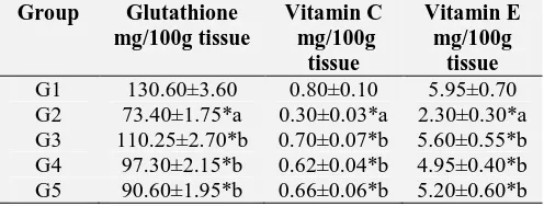
[image:3.612.54.567.170.244.2]The levels of non-enzymatic antioxidants such as reduced glutathione, Vitamin C and Vitamin E in the liver homogenate of D-Galactosamine induced hepatotoxic mice showed significant decrease (p < 0.01) when compared to normal control animals Table 3. The levels of non-enzymatic antioxidants in D-Galactosamine hepatotoxic mice significantly increased on pretreatment with Boerhaavia diffusa Linn. (HAEBD) at both doses tested when compared with untreated hepatotoxic mice.
TABLE 3: EFFECT OF HAEBD ON NON-ENZYMATIC ANTIOXIDANT LEVELS IN THE LIVER TISSUE OF D-GALACTOSAMINE INDUCED HEPATOTOXICITY
Group Glutathione
mg/100g tissue Vitamin C mg/100g tissue Vitamin E mg/100g tissue
[image:3.612.316.564.582.676.2]Histopathological Observations: Histology of liver sections of normal control animals (Group 1) showed normal liver architecture with central vein, well preserved cytoplasm and prominent nucleus and nucleolus Fig. 1A while the liver sections of galactosamine treated animals (Group 2) showed hepatic cells with serum toxicity characterized by inflammatory cell collection, scattered inflammation across liver parenchyma, focal necrosis and swelling up of vascular endothelial cells Fig. 1B.
The Vitamin-E (Group 3) exhibited protection from galactosamine induced changes in the liver Fig. 1C. While hydro alcoholic extract of Boerhaavia diffusa Linn. (HAEBD) pretreatment at a dose of 200 and 400 mg/kg (Group 4 and 5) appeared to significantly prevent the galactosamine toxicity as revealed by the hepatic cells with well-preserved cytoplasm Fig. 1D, 1E. HAEBD pre-treatment also caused marked decrease in inflammatory cells.
Liver section of GP1 Liver section of GP3
(Normal control) (Standard control)
Liver section of GP2 Liver section of GP4 Liver section of GP5
[image:4.612.86.531.200.501.2](Toxic control) (B. Diffusa L. 200 mg/kg/mice) (B. Diffusa L. 400 mg/kg/mice)
FIG. 1: HISTOPATHOLOGICAL STUDIES OF LIVER TISSUE
DISCUSSION: Liver damage induced by D-Galactosamine, reflects disturbances of liver cell metabolism, which lead to characteristic changes in the serum enzyme activities. Elevated serum enzymes are indicative of cellular leakage and loss of functional integrity of the hepatocyte 10. Their estimation in the serum is useful as a quantitative marker of the extent and type of hepatocellular damage. D-Galactosamine - induced oxidative damage is generally attributed to the formation of the highly reactive hydroxyl radical (OH·), the stimulator of lipid peroxidation and the source of destruction and damage to the cell membrane 11.
D-Galactosamine toxicity enhanced lipid
peroxidation and reduced antioxidants were reported in the kidney 12. Increased lipid peroxidation in various tissues cause functional
degradation increase oxidative stress. Treatment with HAEBD and Vitamin E showed a significant reduction in lipid peroxidation which might be due to the antioxidant ability of these compounds and free-radical scavenging effects. Oxidative stress is an imbalance between reactive oxygen species and the antioxidant defense mechanisms of a cell or tissue, which leads to lipid peroxidation, DNA damage, and the inactivation of many enzymes 13.
The enzymatic antioxidant defense system is the natural protector against lipid peroxidation that includes superoxide dismutase, catalase and glutathione peroxidase. Reduction in the activity of these enzymes may result in a number of deleterious effects due to the accumulation of superoxide radicals and H2O2. Superoxide
dismutase protects against the superoxide radical
A B
(O2∙−), which damages the membrane and its
biological structure. Catalase primarily decomposes hydrogen peroxide to H2O at a much faster rate,
sharing this function with glutathione peroxidase. Glutathione peroxidase may play an important role in the removal of lipid hydroperoxides. The balance between these enzymes is important for the efficient removal of oxygen radicals from tissues 14. Significant increases in the activities of these enzymes were observed on HAEBD administration.
The second line of defense consists of the non-enzymic scavenger’s glutathione, ascorbic acid, and α-tocopherol, which scavenge residual free radicals escaping from decomposition by the antioxidant enzymes. Glutathione a major non-protein thiol in living organisms plays a central role in coordinating the antioxidant defense process. Glutathione reacts directly with reactive oxygen species and electrophilic metabolites, protects the essential thiol group from oxidation, and serves as a substrate for several enzymes including glutathione peroxidase. Apart from glutathione, α-tocopherol and ascorbic acids are important free-radical scavengers which protect cell membrane against toxic agents. Vitamin-C functions as a free-radical scavenger of oxygen radicals and successfully prevents detectable oxidative damage under all types of oxidative stress. Ascorbic acid appears to trap the peroxyl radical in the aqueous phase with a rate large enough to lipids and dehydroascorbate is produced in this reaction. Both Vitamins C and E have a synergistic action in scavenging oxygen-derived free radicals 15.
Vitamin C functions as a free-radical scavenger of oxygen radicals and successfully prevents detectable oxidative damage under all types of oxidative stress. Ascorbic acid appears to trap the peroxyl radical in the aqueous phase with a rate large enough to lipids 16 and dehydroascorbate is produced in this reaction. A thiol cycle converts the dehydroascorbate into ascorbate. The thiol cycle consists of a GSSG / GSH couple 17. Thus glutathione in blood keeps up the cellular levels of the active form of Vitamin C. When there is a reduction in glutathione, the cellular level of ascorbic acid is also lowered. The observed decrease in the levels of α-tocopherol and ascorbic acid in the D-galactosamine mice might be due to an antioxidant defense against increased ROS or
due to a decrease in glutathione levels in D-galactosamine - hepatotoxic mice. Our study observed increase the levels of these antioxidants in HAEBD and Vitamin-E administered mice.
The ability of HAEBD to enhance the levels of antioxidants along with its anti-lipid peroxidative activity suggests that this compound might be potentially useful in counteracting free-radical-mediated tissue damage caused by hepatotoxicity. Studies on the anti-oxidative potency of various flavonoids have confirmed the importance of the distribution and quantity of the hydroxyl groups. In general, the anti-oxidative properties of polyphenols depend on hydroxylation of ring B.
The present results corroborate the protective action of HAEBD in D-Galactosamine intoxication of mice, particularly noticeable with the high dose
used by us (400 mg/kg body weight).
Supplementation with this flavonoid ameliorated the hepatoprotective and antioxidant activity in D-Galactosamine-induced hepatitis in mice.
CONCLUSION: In conclusion, our findings demonstrated that Boerhaavia diffusa linn. possesses hepatoprotective and antioxidant activity, which is evidenced by lowered serum hepatic marker enzyme activities, increased antioxidant enzymes levels which is also supported by histopathological studies. Therefore Hydro alcoholic extract of Boerhaavia diffusa Linn. showed promising hepatoprotective and antioxidant
activity against D-Galactosamine induced
Hepatotoxicity.
ACKNOWLEDGEMENT: The authors are
thankful to the authorities of K. M. College of pharmacy for providing required facilities.
CONFLICT OF INTEREST: We declare that we have no conflicts of interest.
REFERENCES:
1. Pushpavalli G, Kalaiarasi P, Veeramani C and Pugalendi KV: Effect of chrysin on hepatoprotective and antioxidant status in D-galactosamine-induced hepatitis in rats. Eur. J. Pharmcol 2010; 631: 36-41.
2. Nalini G, Chidambaranathan N, Santhanakumar M,
3. Jaishree V and Shrishailappa B: Antioxidant and hepatoprotective effect of swertiamarin from Enicostemma axillare against D-galactosamine induced acute liver damage in rats. Journal of Ethnopharmacology 2010; 130: 103-106.
4. Sandeep B, Balaji B, and Premkumar B: Hepatoprotective and antioxidant activity of Leucas aspera against
D-galactosamine induced liver damage in rats.
Pharmaceutical Biology 2012; 50(12): 1592-1595. 5. Persijn JP and VanderSlik W: A new method for the
determination of gamma-glutamyl transferase in serum. J. Clin. Chem. Clin. Biochem 1976; 14(9): 421-7.
6. Lowry OH, Rose brough, NJ, Far AL and Randall RJ: Protein measurement with the Folin phenol reagent. J. Biol. Chem 1951; 193(1): 265-75.
7. Wang Y, Li-Na G, Yuan-Lu C and Heng-Li J: Protective Effect of Danhong Injection on Acute Hepatic Failure Induced by Lipopolysaccharide and D-Galactosamine in Mice. Evidence- Based Complementary and Alternative Medicine 2014; 5: 1-8.
8. Peskin AV and Winterbourn CC: A microtiter plate assay for superoxide dismutase using a water-soluble tetrazolium salt (WST-1). Clin. Chim. Acta 2000; 293(1-2): 157-66. 9. Beers RFJ and Sizer IW: A spectrophotometric method for
measuring the breakdown of hydrogen peroxide by catalase. J Biol Chem 1952; 195: 133-140.
10. Orie Y, Yoshiaki S and Kiharu I: Hepatoprotective effect of germanium-containing Spirulina in rats with
D-galactosamine- and lipopolysaccharide-induced hepatitis. British Journal of Nutrition 2014; 111: 135-140.
11. Barry H and Gutteridge JMC. In: Barry, H. (Ed.), Oxford Clarendon Press Free radicals in biology and medicine 1989; 254-255.
12. Barrera G: Oxidative stress and lipid peroxidation products in cancer progression and therapy. ISRN Oncol. 2012; 13: 72-89.
13. Shimaa IR, Shalaby MA, Nehal A and El-Banna HA: Hepatoprotective and Antioxidant Effects of Silybum marianum Plant in Rats. IJAVMS 2011; 5(6): 541-547. 14. Sundaram R and Murugesan G: Hepatoprotective and
antioxidant activity of a mangrove plant Lumnitzera racemosa. Asian Pacific Journal of Tropical Biomedicine 2011; 348-352.
15. Wojacki JL, Samachowiec B, Gonet S, Juzwiak E and DM: Effect of buckwheat extract on free radical generation in rabbits administered high fat diet. Phytother. Res 1995; 19: 323-326.
16. Jayachandran M, Lalithapriya S and Selvam PC: Effect of ascorbic acid supplementation on tissue ascorbic acid and nucleic acid content of young and aged rats. J. Clin. Biochem. Nutr 1995; 19: 131-136.
17. Ganie SA, Aahmad B, Masood ZA and Zargar MA: Hepatoprotective and Antioxidant Activity of Rhizome of Podophyllum hexandrum against Carbon Tetra Chloride Induced Hepatotoxicity in Rats. Bio medical and Environmental sciences 2013; 26(30): 209-221.
All © 2013 are reserved by International Journal of Pharmaceutical Sciences and Research. This Journal licensed under a Creative Commons Attribution-NonCommercial-ShareAlike 3.0 Unported License.
This article can be downloaded to ANDROID OS based mobile. Scan QR Code using Code/Bar Scanner from your mobile. (Scanners are available on Google Playstore)
How to cite this article: