Krüppel-like factor 6 regulates mitochondrial
function in the kidney
Sandeep K. Mallipattu, … , Vincent W. Yang, John C. He
J Clin Invest.
2015;
125(3)
:1347-1361.
https://doi.org/10.1172/JCI77084
.
Maintenance of mitochondrial structure and function is critical for preventing podocyte
apoptosis and eventual glomerulosclerosis in the kidney; however, the transcription factors
that regulate mitochondrial function in podocyte injury remain to be identified. Here, we
identified Krüppel-like factor 6 (KLF6), a zinc finger domain transcription factor, as an
essential regulator of mitochondrial function in podocyte apoptosis. We observed that
podocyte-specific deletion of
Klf6
increased the susceptibility of a resistant mouse strain to
adriamycin-induced (ADR-induced) focal segmental glomerulosclerosis (FSGS). KLF6
expression was induced early in response to ADR in mice and cultured human podocytes,
and prevented mitochondrial dysfunction and activation of intrinsic apoptotic pathways in
these podocytes. Promoter analysis and chromatin immunoprecipitation studies revealed
that putative KLF6 transcriptional binding sites are present in the promoter of the
mitochondrial cytochrome
c
oxidase assembly gene (
SCO2
), which is critical for preventing
cytochrome
c
release and activation of the intrinsic apoptotic pathway. Additionally,
KLF6
expression was reduced in podocytes from HIV-1 transgenic mice as well as in renal
biopsies from patients with HIV-associated nephropathy (HIVAN) and FSGS. Together,
these findings indicate that KLF6-dependent regulation of the cytochrome
c
oxidase
assembly gene is critical for maintaining mitochondrial function and preventing podocyte
apoptosis.
Research Article
Nephrology
Find the latest version:
Introduction
The glomerulus is the main filtration barrier of the human body. Podocytes, which are terminally differentiated epithelial cells in the glomerulus, play a major role in the maintenance of this renal filtration barrier. Podocyte injury is implicated in many glo-merular diseases, including focal segmental gloglo-merular sclerosis (FSGS) and HIV-associated nephropathy (HIVAN) (1). In many of these conditions, the podocyte loses specific markers of differ-entiation, undergoes effacement of foot processes and eventual detachment, and loses the functional capacity to maintain the glomerular filtration barrier (2).
Mitochondrial injury has been implicated in glomerular dis-ease. Initial studies revealed altered expression of mitochondrial genes and mitochondrial dysfunction in acquired and congenital human nephrotic syndrome (3, 4). Furthermore, collapsing FSGS has been associated with mitochondrial injury with inherited mutations in genes encoding for mitochondrial function (5). Most recently, maintenance of the mitochondrial genome was shown
to be critical for preventing FSGS in a murine model of adriamy-cin-induced (ADR-induced) podocyte injury (6). However, the mechanisms mediating the regulation of the mitochondrial injury in podocytes have yet to be characterized.
Krüppel-like factors (KLFs) are a subclass of zinc finger family of DNA-binding transcriptional regulators that are involved in a broad range of cellular processes (i.e., cell differentiation, angio-genesis, and erythropoiesis) (7). Studies have revealed a novel role of KLFs in podocyte injury (8, 9). Recently, we identified the critical role of KLF15 in attenuating podocyte dedifferentiation in HIVAN (9). To explore the potential role of other KLFs in podo-cyte injury, we began by examining the expression profile of KLF transcripts in HIV-1–infected podocytes. We observed a significant reduction in KLF6 expression in HIV-1–infected human podocytes and in two independent models of podocyte injury. In addition, KLF6 expression was markedly reduced in human glomerular disease. Finally, podocyte-specific loss of Klf6 renders the kidney susceptible to mitochondrial dysfunction, resulting in activation of intrinsic apoptotic pathways and eventually glomerulosclerosis.
Results
KLF6 expression is reduced in HIV-1 transgenic mice. Since we recently identified the critical role of KLF15 in attenuating podo-cyte dedifferentiation in HIV-1–infected podopodo-cytes, we exam-Maintenance of mitochondrial structure and function is critical for preventing podocyte apoptosis and eventual
glomerulosclerosis in the kidney; however, the transcription factors that regulate mitochondrial function in podocyte injury remain to be identified. Here, we identified Krüppel-like factor 6 (KLF6), a zinc finger domain transcription factor, as an essential regulator of mitochondrial function in podocyte apoptosis. We observed that podocyte-specific deletion of Klf6 increased the susceptibility of a resistant mouse strain to adriamycin-induced (ADR-induced) focal segmental glomerulosclerosis (FSGS). KLF6 expression was induced early in response to ADR in mice and cultured human podocytes, and prevented mitochondrial dysfunction and activation of intrinsic apoptotic pathways in these podocytes. Promoter analysis and chromatin immunoprecipitation studies revealed that putative KLF6 transcriptional binding sites are present in the promoter of the mitochondrial cytochrome c oxidase assembly gene (SCO2), which is critical for preventing cytochrome c
release and activation of the intrinsic apoptotic pathway. Additionally, KLF6 expression was reduced in podocytes from HIV-1 transgenic mice as well as in renal biopsies from patients with HIV-associated nephropathy (HIVAN) and FSGS. Together, these findings indicate that KLF6-dependent regulation of the cytochrome c oxidase assembly gene is critical for maintaining mitochondrial function and preventing podocyte apoptosis.
Krüppel-like factor 6 regulates mitochondrial function
in the kidney
Sandeep K. Mallipattu,1 Sylvia J. Horne,1 Vivette D’Agati,2 Goutham Narla,3 Ruijie Liu,4 Michael A. Frohman,5 Kathleen Dickman,5 Edward Y. Chen,6 Avi Ma’ayan,6 Agnieszka B. Bialkowska,7 Amr M. Ghaleb,7 Mandayam O. Nandan,7 Mukesh K. Jain,8 Ilse Daehn,4 Peter Y. Chuang,4 Vincent W. Yang,7 and John C. He4,6,9
1Division of Nephrology, Department of Medicine, Stony Brook University, Stony Brook, New York, USA. 2Department of Pathology, Columbia University, New York, New York, USA. 3Institute for
Transformative Molecular Medicine, Case Western Reserve University, Cleveland, Ohio, USA. 4Division of Nephrology, Department of Medicine, Mount Sinai School of Medicine, New York, New York, USA. 5Department of Pharmacology, Stony Brook University, Stony Brook, New York, USA. 6Department of Pharmacology and Systems Therapeutics, Mount Sinai School of Medicine, New York, New York, USA. 7Department of Medicine, Stony Brook University, Stony Brook, New York, USA. 8Department of Medicine, Case Cardiovascular Institute Research Institute, Case Western Reserve University, Cleveland, Ohio,
USA. 9Renal Section, James J. Peters VA Medical Center, New York, New York, USA.
Related Commentary: p. 968
Conflict of interest: The authors have declared that no conflict of interest exists. Submitted: May 14, 2014; Accepted: December 9, 2014.
changes observed by light and electron microscopy is shown in Table 1. Podocyte injury was further confirmed with a loss of key podocyte differentiation markers, Nephrin and Podocin, in the ADR-treated Podocin-Cre Klf6fl/fl mice as compared with all other groups
(Supplemental Figure 2, A and B). Taken together, these findings highlight that Klf6 is required to prevent podocyte injury and even-tual glomerulosclerosis in the setting of ADR treatment.
Podocyte-specific Klf6-knockout mice exhibit mitochondrial injury with ADR treatment. Prior studies have suggested that ADR-induced mitochondrial damage plays a major role in cel-lular injury in several other tissues (11–14). Similarly, ultrastruc-tural examination of the podocyte cell bodies revealed dysmor-phic mitochondria in the ADR-treated Podocin-Cre Klf6fl/fl mice as
compared with ADR-treated Podocin-Cre Klf6+/+ mice (Figure 5,
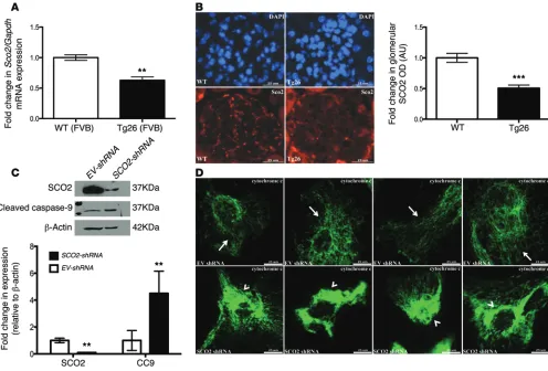
A and B). Promoter analysis was subsequently performed to assess whether transcriptional binding sites for KLF6 exist in the prox-imity of genes involved in mitochondrial function. TRANSFAC (15) promoter analysis revealed eight potential KLF6 binding sites in the cytochrome c oxidase assembly gene (SCO2) (Table 2). We initially confirmed that putative binding sites for KLF6 are present along the promoter region of SCO2 using ChIP fol-lowed by quantitative RT-PCR in cultured human podocytes (Figure 6A). Podocytes from ADR-treated Podocin-Cre Klf6fl/fl
mice exhibited a significant reduction in Sco2 mRNA expression as compared with podocytes from ADR-treated Podocin-Cre Klf6+/+
mice (Figure 6B). Similarly, SCO2 expression was significantly ined the level of other KLFs in human HIVAN by measuring the
expression profile of KLFs known to be expressed in epithelial cells (Figure 1A). In comparison to WT human podocytes, KLF6 expression was significantly reduced in HIV-1–infected podo-cytes (Figure 1A, inset). We confirmed that KLF6 expression was reduced in HIV-1 transgenic (Tg26) mice as compared with WT mice (FVB/N background) (Figure 1, B and C). These findings suggest that a loss of KLF6 may play a critical role in the podocyte injury observed in HIVAN.
Podocyte-specific loss of Klf6 increases susceptibility to ADR- induced nephropathy. To assess whether the loss of Klf6 in podo-cytes results in podocyte injury and subsequent glomeruloscle-rosis, Klf6 was specifically knocked down in podocytes using the Cre-loxP recombination system. Podocin-Cre mice (C57BL/6) were crossed with Klf6fl/fl (C57BL/6) to generate Podocin-Cre Klf6fl/fl
mice (F2). Primary culture of podocytes isolated from Podocin-Cre Klf6+/+ mice and Podocin-Cre Klf6fl/fl mice revealed a
signifi-cant reduction in Klf6 mRNA and protein expression (Figure 2, A and B). These findings were confirmed with colocalization of KLF6 with WT1, a known podocyte marker (Figure 2C). All mice were viable and fertile. Podocin-Cre Klf6fl/fl mice exhibited
a 3.5-fold increase in albuminuria as compared with Podocin-Cre Klf6+/+ mice (Supplemental Figure 1A; supplemental material
available online with this article; doi:10.1172/JCI77084DS1), but no overt histological evidence of glomerulosclerosis or tubuloin-terstitial disease was observed (Supplemental Figure 1B).
Prior studies have demonstrated that C57BL/6 mice are resis-tant to ADR-induced FSGS, unless they harbor mutations that are critical to the maintenance of the mitochondrial genome (6, 10). However, transcription factors regulating this process have yet to be identified. Therefore, we used the ADR model specifically on a resistant C57BL/6 background to determine whether the loss of Klf6 increased the susceptibility to podocyte injury, leading to FSGS. ADR-treated Podocin-Cre Klf6fl/fl mice exhibited a significant
increase in albuminuria (Figure 3A), podocyte hyperplasia, segmen-tal glomerulosclerosis, interstitial inflammation, and tubular casts with cystic dilatation as compared with ADR-treated Podocin-Cre Klf6+/+ mice (Figure 3B). In addition, electron microscopy revealed
significant podocyte foot process effacement, podocyte hypertro-phy, and microvillus transformation in the ADR-treated Podocin-Cre Klf6fl/fl mice as compared with the ADR-treated Podocin-Cre
Klf6+/+ mice (Figure 4). The quantification of these morphological
Table 2. Predicted KLF6 binding sites
Gene symbol Description Binding sites
SCO2 Cytochrome c oxidase (COX) assembly gene 8
PCSK6 Proprotein convertase subtilisin/kexin type 6 5
SHARPIN SHANK-associated RH domain interactor 4
CHODL Chondrolectin 3
LEMD2 LEM domain containing 2 2
ARFRP1 ADP-ribosylation factor-related protein 1 2
WDR46 Chromosome 6 open reading frame 11 2
KCNIP4 Kv channel interacting protein 4 2
WDR38 WD repeat-containing protein 38 2
GPRC5C G protein-coupled receptor, family C, group 5, member C 2
Table 1. Quantification of histologic and ultrastructural changes
% Segmental
global GSA hypertrophy Podocyte
(0–3+)B
Podocyte hyperplasia
(0–3+)A
Podocyte MV transformation
(0–3+)B
% Podocyte FP
effacementB inflammation% IFTA/ A % Tubular casts/cystic dilA and regen. change Tubular degen.
(0–3+)A
Cre+ Klf6+/+ 0.0 – 0.0 – – 0.0 0.0 0.0
Cre+ Klf6+/+ + ADR 0.0 0.0 0.0 0.0 5%–10% 0.0 0.0 0.0
Cre+ Klf6fl/fl 0.0 – 0.0 – – 0.0 0.0 0.0
Cre+ Klf6fl/fl+ ADR 11.0 ± 2.1 3.0 ± 0.0 1.0 ± 0.0 2.0 ± 0.0 80%–90% 1.3 ± 0.7 4.3 ± 0.7 0.7 ± 0.3 ALight microscopy (30 glomeruli per mouse; n = 6 mice per group). BElectron microscopy (n = 4 mice). All data are expressed as mean ± SEM. deg.,
tor A mitochondrial [Tfam]) and mitochondrial function (mt-Co2, COX subunit 8A [Cox8a], mitochondrial inner membrane protein [Mpv17]) were significantly reduced in ADR-treated podocytes iso-lated from Podocin-Cre Klf6fl/fl mice as compared with ADR-treated
podocytes isolated from Podocin-Cre Klf6+/+ mice (Supplemental
Figure 3, A and B). Taken together, these data suggest that Klf6 is required for the maintenance of mitochondrial function and pre-venting podocyte injury in the setting of ADR exposure.
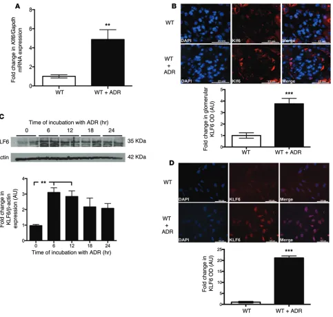
KLF6 is required to protect podocytes from ADR-induced mito-chondrial injury. In order to assess Klf6 expression pattern and activity in podocytes exposed to ADR, podocytes isolated from WT mice were treated with and without ADR in primary culture for 12 hours. We observed a significant increase in Klf6 mRNA expression in ADR-treated podocytes as compared with untreated podocytes in culture (Figure 7A). Similarly, a significant increase in KLF6 expression was observed in ADR-treated WT mice as compared with untreated mice (Figure 7B). Additionally, KLF6 protein expression was significantly increased within 6 hours of ADR exposure in cultured human podocytes (Figure 7C). reduced in ADR-treated Podocin-Cre Klf6fl/fl mice as compared
with all other groups (Figure 6C). SCO2 is a mitochondrial membrane–bound metallochaperone critical to the transport of copper atoms to cytochrome c oxidase (COX) subunits I and II; therefore, it is essential for the assembly of the catalytic core of the COX complex (16, 17). Using antibodies specific for COX subunit II (MT-CO2), we observed that MT-CO2 is expressed in the glomerular compartment (Figure 6D). Furthermore, we determined that MT-CO2 expression was significantly reduced in ADR-treated Podocin-Cre Klf6fl/fl mice as compared with
ADR-treated Podocin-Cre Klf6+/+ mice (Figure 6D).
Because our results suggest that KLF6 may play a critical role in the regulation of mitochondrial gene expression, we sought to determine the level of expression of other transcripts involved in mitochondrial function. First, podocytes were isolated from Podocin-Cre Klf6fl/fl and Podocin-Cre Klf6+/+ mice and treated with
ADR in primary culture for 12 hours. RNA transcripts involved in mitochondrial replication and transcription (nuclear respiratory tor 1 [Nrf1], polymerase mitochondrial [Polrmt], transcription
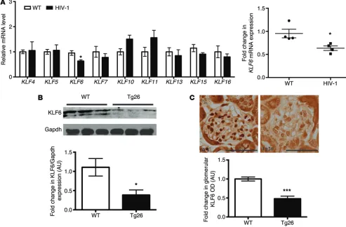
fac-Figure 1. Strong association between KLF6 and the glomerulosclerosis was observed in HIVAN. (A) mRNA levels of KLFs, previously shown to be
expressed in epithelial cells, were measured in differentiated WT and HIV-1–infected human podocytes in culture. Real-time PCR, using primers for KLF4,
KLF5, KLF6, KLF7, KLF10, KLF11, KLF13, KLF15, and KLF16, was performed using RNA isolated from WT and HIV-1–infected human podocytes. Inset:
Rela-tive mRNA expression for KLF6 (n = 4). Mann-Whitney U test, *P < 0.05. (B) Age- and sex-matched 6-week-old HIV-1 transgenic mice (Tg26) and WT mice
on the FVB/N background were used to assess KLF6 expression in the glomeruli. Western blot analysis was performed on the glomerular lysates from WT and Tg26 mice for KLF6 and GAPDH. A representative blot of six independent experiments is shown in the top panel. The bottom panel shows the quan-tification of KLF6 by densitometry (n = 6). Mann-Whitney U test, *P < 0.05. (C) The top panel shows representative images of immunohistochemistry for
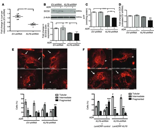
[image:4.585.46.539.59.383.2]Additionally, merely the knockdown of KLF6 shifted the morphol-ogy of the mitochondria from a tubular pattern to an intermediate pattern (Figure 8E). To assess whether the reintroduction of KLF6 can rescue the cells from mitochondrial injury, LentiORF-KLF6 was transiently transfected in EVshRNA-KLF6 human podocytes (Supplemental Figure 4C). Subsequent restoration of KLF6 in shRNA-KLF6 podocytes prevented mitochondrial fragmentation and preserved the tubular structural pattern of the mitochondria (Figure 8F). Combined, these findings suggest that KLF6 is an early inducible injury response gene, critical to the maintenance of mitochondrial function upon injury.
KLF6 mediates apoptosis in the setting of ADR treatment. ADR has been previously shown to activate intrinsic apoptotic path-ways in other models of tissue injury (18–20); we showed that a loss of KLF6 increased the susceptibility to ADR-induced mito-chondrial injury. Therefore, we hypothesized that a loss of KLF6 would result in the activation of intrinsic apoptotic pathways with ADR treatment. Initially, we observed that shRNA-KLF6 podo-cytes exhibited a significant increase in apoptotic bodies with ADR treatment (Figure 9A). This increase in apoptotic cells was quan-tified using flow cytometry with annexin V and propidium iodide labeling (Figure 9B). Furthermore, we demonstrated that release Immunostaining for KLF6 with and without ADR treatment at 12
[image:5.585.76.504.54.343.2]hours confirmed the increase in KLF6 expression (Figure 7D), sug-gesting that KLF6 is an early inducible gene. Since KLF6 activity is initially increased with ADR exposure in podocytes and the loss of Klf6 in podocytes increased the susceptibility to ADR-induced podocyte injury, we hypothesized that KLF6 is required to protect podocytes from ADR-induced mitochondrial injury. To evaluate this, we first generated human podocytes with stable knockdown for KLF6 (Supplemental Figure 4A). As compared with WT human podocytes, the loss of KLF6 resulted in a decrease in mitochon-drial membrane potential (Figure 8A). Since SCO2 was shown to be a transcriptional target of KLF6, we measured SCO2 expres-sion in this model. SCO2 expresexpres-sion was significantly reduced in shRNA-KLF6 podocytes treated with or without ADR (Figure 8B). Furthermore, shRNA-KLF6 podocytes exhibited a significant reduction in ATP activity and oxygen consumption rate with ADR treatment (Figure 8, C and D). In addition, mitochondrial biomass was not significantly different between the groups (Supplemen-tal Figure 4B), suggesting that the loss in mitochondrial func-tion was not a result of reduced mitochondrial number. We also observed that the shRNA-KLF6 podocytes exhibited an increase in mitochondrial fragmentation with ADR treatment (Figure 8E).
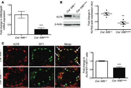
Figure 2. Podocyte-specific knockdown of Klf6 in Podocin-Cre Klf6fl/fl mice was confirmed. (A) Primary podocytes were isolated from 10-week-old
Podocin-Cre Klf6fl/fl and Podocin-Cre Klf6+/+ mice and cultured at 37°C for 1 week. RNA was extracted, and real-time PCR was performed for Klf6 mRNA
expression (n = 6). Mann-Whitney U test, ***P < 0.001. (B) Protein was also extracted, and Western blot analysis was performed for KLF6. A
representa-tive blot of four independent experiments is shown in the left panel (KLF6 and β-actin are from the same samples on the same blot, with β-actin being developed after KLF6). The right panel shows the quantification of KLF6 by densitometry (n = 4). Mann-Whitney U test, **P < 0.01. (C)
Immunofluores-cence staining for KLF6 and WT1 was performed in 10-week-old Podocin-Cre Klf6fl/fl and Podocin-Cre Klf6+/+ mice. Representative images from six mice in
each group are shown in the left panel (original magnification, ×20). Arrows show colocalization of KLF6 and WT1. Arrowheads show a lack of colocaliza-tion. Right panel: 30 glomeruli were selected in each mouse, and quantification of KLF6 staining in the podocytes was determined by the ratio of KLF6+
with both groups on the FVB/N background (Figure 10A). These findings were confirmed by immu-nostaining for SCO2 in glomeruli from WT and Tg26 mice (FVB/N background) (Figure 10B). These data suggest that KLF6 is required for the maintenance of SCO2 in the setting of injury.
Since KLF6 binding sites are present in the promoter region of SCO2 and the loss of KLF6 acti-vates the intrinsic apoptotic path-way under stress, we suspected that SCO2 is a downstream target of KLF6 and is essential to mito-chondrial stability and preventing the release of cytochrome c. To ascertain this, we first generated human podocytes with transient knockdown of SCO2 (Figure 10C). The transient knockdown of SCO2 resulted in increased cleaved caspase-9 levels (Figure 10C). As compared with WT human podocytes, shRNA-SCO2 podocytes exhibited a substantial release in cytochrome c from the mito-chondria, as shown by an increase in the diffuse pattern of cyto-chrome c staining (Figure 10D). Taken together, these data sug-gest that SCO2 is a critical downstream target of KLF6-mediated mitochondrial regulation.
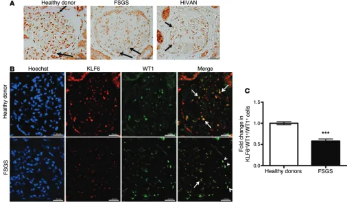
KLF6 expression is reduced in human FSGS. To ascertain the role of KLF6 in human glomerulopathy, immunohistochemistry was performed on renal biopsy specimens from healthy donor nephrectomies, HIVAN, and noncollapsing idiopathic FSGS. We observed that the staining for KLF6 had a nuclear distribution in the normal podocytes and parietal epithelial cells, with a nuclear and cytosolic distribution in tubular cells (Figure 11A, healthy donor). The staining also revealed a significant decrease in KLF6 expression in HIVAN and noncollapsing FSGS as compared with healthy donor nephrectomies (Figure 11A). Since KLF6 is a ubiquitous transcription factor expressed in majority of the com-partments in the kidney, colocalization using KLF6 and WT1 (a known nuclear-distributed podocyte marker) was performed to determine whether the reduction in KLF6 expression is specific to of cytochrome c was substantially increased in shRNA-KLF6
podocytes exposed to ADR (Figure 9C). In addition, we observed a significant increase in cleaved caspase-9 and cleaved caspase-3 levels with a decrease in procaspase-3 expression in shRNA-KLF6 podocytes treated with ADR (Figure 9D).
To assess whether the preservation of KLF6 prevents the acti-vation of caspases, we first generated human podocytes with sta-ble overexpression of KLF6 (LentiORF-KLF6) (Figure 9E). The overexpression of KLF6 in podocytes contributed to a significant increase in SCO2 and procaspase-3 expression, with a reduction in cleaved caspase-3 levels, in the setting of ADR treatment (Fig-ure 9E). We observed a reduction, albeit not statistically signifi-cant, in cleaved caspase-9 levels in LentiORF-KLF6 as compared with LentiORF-control human podocytes. Combined, these data suggest that KLF6 prevents the ADR-induced activation of the intrinsic apoptotic pathway.
SCO2, a downstream target of KLF6, is critical to the prevention of mitochondrial injury. To confirm that SCO2, a critical downstream target of KLF6, is reduced in other models of podocyte injury, we used the HIV-1 transgenic mice model. We initially observed a decrease in KLF6 expression in HIV-1–infected human podocytes and in HIV-1 transgenic (Tg26) mice. We also demonstrated that SCO2 expression was reduced in the shRNA-KLF6 podocytes and in the glomeruli of Podocin-Cre Klf6fl/fl mice (Figure 6C and
[image:6.585.42.410.55.199.2]Fig-ure 8B). Similarly, Sco2 mRNA expression was reduced in primary podocytes isolated from Tg26 mice as compared with WT mice,
Figure 3. ADR-treated Podocin-Cre Klf6fl/fl mice exhibit a significant increase in albuminuria with
glomerulo-sclerosis and tubulointerstitial injury. Podocin-Cre Klf6fl/fl and Podocin-Cre Klf6+/+ mice were treated with ADR
at 12 weeks of age. Urine was collected weekly, mice were sacrificed, and renal cortex was fixed for histology 5 weeks after ADR treatment. (A) Albuminuria (urine albumin/creatinine [cr]) was measured (n = 10). Krus-kal-Wallis test with Dunn’s post-hoc test, *P < 0.05 as compared with untreated Podocin-Cre Klf6+/+mice,
***P < 0.001 as compared with all groups. (B) PAS was used to evaluate glomerular or tubulointerstitial
changes (top panel: original magnification, ×10; bottom panel: original magnification, ×40). Representative images from six mice in each group are shown. Arrows show sclerotic glomeruli. Arrowheads show interstitial inflammation. Asterisks within images show tubular casts and dilatation.
Figure 4. Podocin-Cre Klf6fl/fl mice exhibit substantial podocyte injury
with ADR treatment. Podocin-Cre Klf6fl/fl and Podocin-Cre Klf6+/+mice
[image:6.585.320.535.602.737.2]the podocyte. Immunostaining revealed a significant decrease in KLF6 expression in podocytes of patients with FSGS as compared with healthy donor nephrectomies, as shown by the reduction in the number of cells with both KLF6 and WT1 staining (Figure 11B). Quantification of immunostaining in podocytes confirmed the decrease in KLF6 expression (Figure 11C). Furthermore, the specificity of the KLF6 staining was confirmed by utilizing a pep-tide block with the immunizing peppep-tide (Supplemental Figure 5). Collectively, with the rest of our findings, these data suggest that KLF6 expression is reduced in human FSGS, contributing to podo-cyte injury observed in FSGS.
Discussion
Several studies have characterized the critical role of mitochon-drial dysfunction in podocytopathy. However, transcriptional regulators mediating the effects of mitochondrial dysfunction in podocyte injury have yet to be characterized. A recent study by Papeta et al. (6) has identified that the maintenance of the mito-chondrial genome is essential in preventing ADR-induced FSGS. Here, we identify that KLF6 is a critical mediator of ADR-induced mitochondrial dysfunction in podocytes. This was demonstrated by the following: (a) podocyte-specific loss of Klf6 resulted in ADR-induced FSGS in a resistant mouse strain (C57BL/6) and (b) shRNA-mediated KLF6 knockdown in human podocytes resulted in mitochondrial dysfunction, as measured by reduced expression of mitochondrial transcripts, mitochondrial fragmentation, ATP levels, and oxygen consumption rate. In addition, we determined that KLF6 is an early inducible injury response gene and it is crit-ical for the prevention of the ADR-induced mitochondrial injury in podocytes. Furthermore, we showed that KLF6 is required to
prevent the activation of ADR-mediated intrinsic apoptotic pathway in podo-cytes. Finally, we confirmed that the podocyte expression of KLF6 is reduced in a murine model of HIVAN and in human HIVAN and FSGS.
Although the podocyte-specific loss of Klf6 resulted in minimal podocyte injury at baseline, these mice developed FSGS with ADR treatment on a resistant C57BL/6 background. In combination with the early increase in KLF6 expres-sion observed with ADR treatment, this suggests that KLF6 is an early inducible injury response gene that may serve to protect podocytes from injury. Recently, we published that KLF15 is a critical transcriptional regulator of podocyte differentiation (21). Furthermore, KLF4 was found to play a critical role in reg-ulating mitochondrial fusion proteins in vascular smooth muscle differentia-tion (22). In addidifferentia-tion, paralogs of KLF1 (specificity proteins) have been shown to transcriptionally regulate cytochrome c subunit genes in primary neurons (23). Future studies will focus on the potential interactions between these other KLFs in mediating mitochondrial function with KLF6 in the setting of podocyte injury.
A few transcription factors have been associated with podo-cyte apoptosis in recent years, including FOXO4 and NF-κB (24, 25). However, none of these have been implicated in mitochon-drial dysfunction leading to activation of apoptosis and eventual podocyte injury. To date, KLF6 is, to our knowledge, the first tran-scription factor that is critical to the maintenance of the mitochon-drial structure and function, and in preventing apoptosis in the setting of cell stress.
[image:7.585.38.379.54.235.2]Analysis of the promoter region of the COX assembly (SCO2) gene revealed several potential binding sites for KLF6. SCO2 is an inner mitochondrial membrane metallochaperone critical to the transport of copper ions. In fact, missense mutations in SCO2 have been found to cause fatal hypertrophic cardiomyopathy with encephalopathy (26, 27). We observed that SCO2 is a critical down-stream target of KLF6-mediated mitochondrial function, in which a loss of SCO2 resulted in the activation of the intrinsic apoptotic pathway. In normal conditions, we hypothesize that in the early phase of ADR treatment (i.e., early cell stress), the upregulation of KLF6 results in the activation of SCO2, thereby abrogating mito-chondrial injury and protecting the cell from injury. Nonetheless, we recognize that KLF6 may also regulate other genes involved in the activation of the intrinsic apoptotic pathway and mitochon-drial injury. Although ADR has typically been observed to activate the intrinsic apoptotic pathway leading to tissue damage (19, 20), the potential crosstalk between the extrinsic and intrinsic pathway in the setting of KLF6 depletion has to be further explored. Future studies will also need to determine the interaction between KLF6 and protein kinase, DNA-activated, catalytic peptide (PRKDC), a
Figure 5. Podocin-Cre Klf6fl/fl mice exhibit dysmorphic mitochondria in podocytes with ADR treatment.
Podocin-Cre Klf6fl/fl and Podocin-Cre Klf6+/+mice were treated with ADR at 12 weeks of age. All mice were
sacrificed, and renal cortex was fixed for histology 5 weeks after ADR treatment. (A) Electron
micros-copy was performed to assess ultrastructural changes in the mitochondria of the podocyte cell body (top panels: original magnification, ×50,000; bottom panels: original magnification, ×75,000). Representative images are shown from six mice in each group. Red arrows show elongated mitochondria with preserved cristae and membrane. Red arrowheads show the loss of cristae and the loss of elongated morphology in the mitochondria. (B) To quantify dysmorphic mitochondria, a total of 100 mitochondrial podocytes per
nuclear DNA repair protein recently discovered to be critical to the maintenance of the mitochondrial genome in ADR-induced FSGS (6). Alternatively, KLF6 may also participate in maintaining the mitochondrial structure and function in response to DNA damage. Therefore, we expect a wide range of future studies to decipher the precise molecular mechanism mediating the interaction between KLF6 and maintenance of mitochondrial structure and function.
We also confirmed that podocyte-specific expression of KLF6 was markedly reduced in renal biopsies from patients with HIVAN and FSGS, suggesting that a loss of KLF6 expression may contribute to the podocyte injury observed in glomerular disease. Although
podocyte depletion occurs in FSGS, the ratio of KLF6+WT1+ cells to
WT1+ cells is suggestive of reduced KLF6 expression independent
[image:8.585.49.546.57.458.2]of podocyte loss. In addition, our immunohistochemistry studies are suggestive of reduced KLF6 expression in the parietal epithe-lial cells. However, further colocalization studies with KLF6 and a known parietal epithelial cell marker need to be performed to vali-date our findings. Since there is a reduction in KLF6 expression with FSGS, further knockdown of Klf6 in HIV-1 transgenic mice may not yield a significant change in an already severely diseased pheno-type. Consequently, we demonstrated that the restoration of KLF6 attenuated mitochondrial injury in the setting of ADR treatment.
Figure 6. KLF6 binds to the promoter region of SCO2. To confirm that binding sites for KLF6 are present in the promoter region of SCO2. KLF6 was initially
overexpressed in HEK 293 cells using LentiORF transfection, and ChIP was performed. (A) Overexpression of KLF6 was confirmed with Western blot
analysis (left panel), and the presence of putative KLF6 binding sites in the promoter of SCO2 in HEK 293 cells is shown (right panel) (n = 4). Kruskal-Wallis test with Dunn’s post-hoc test, **P < 0.01 vs. the two other groups. (B) Cultured podocytes were treated with ADR for 12 hours, RNA was extracted for
real-time PCR, and Sco2 mRNA expression is shown (n = 4). Kruskal-Wallis test with Dunn’s post-hoc test, *P < 0.05, **P < 0.01. (C) SCO2 expression level
was also confirmed by immunofluorescence in Podocin-Cre Klf6fl/fl and Podocin-Cre Klf6+/+mice treated with and without ADR. Representative pictures of
six mice in each group are shown in the top panel (original magnification, ×20). A total of 30 glomeruli per mouse were selected, and SCO2 expression was quantified in the glomerular region (n = 6) shown in the bottom panel. Kruskal-Wallis test with Dunn’s post-hoc test, **P < 0.01. (D) MT-CO2 levels were
assessed in Podocin-Cre Klf6fl/fl and Podocin-Cre Klf6+/+mice treated with and without ADR. Representative immunofluorescence images of six mice in
cytes and tubular cells (31, 32), suggesting a common final pathway in podocyte injury leading to eventual podocyte loss and glomeru-losclerosis in both HIVAN and ADR nephropathy. Combined, these findings suggest that KLF6, as with ADR nephropathy, may play a critical role in protecting podocytes from injury in HIVAN.
Taken together, our results provide a potential mechanism for the transcriptional regulation of the intrinsic apoptotic pathway in podocytopathy. Our data suggest that KLF6 is a critical injury However, further studies are needed to determine whether
over-expression of Klf6 protects against podocyte injury with HIV-1 transgene expression. Although, along with other investigators, we have highlighted that podocyte dedifferentiation is a hallmark in HIVAN (28, 29), recent data suggest the potential role of parie-tal epithelial cell proliferation in podocyte injury (30). The role of KLF6 in parietal cell proliferation remains to be explored. None-theless, mitochondrial injury does occur in HIV-1–infected
podo-Figure 7. KLF6 expression is increased early with ADR treatment. Primary podocytes isolated from WT mice were treated with and without ADR for 12
hours. RNA was extracted, and real-time PCR was performed. (A) Klf6 mRNA expression was compared between primary cultured murine podocytes treated with and without ADR (n = 5). Mann-Whitney U test, **P < 0.01. (B) This was confirmed by immunofluorescence using tissue from WT mice
treated with and without ADR. Representative pictures of six mice in each group are shown in the top panel (original magnification, ×20). Bottom panel: 30 glomeruli per mouse were selected, and KLF6 expression was quantified in the glomerular region (n = 6). Unpaired t test, ***P < 0.001. (C) Cultured
human podocytes were treated with ADR for 6, 12, 18, and 24 hours. Protein was extracted, and Western blot analysis was performed for KLF6. A repre-sentative blot of three independent experiments is shown in the top panel. Densitometry analysis is shown in the bottom panel (n = 6). Kruskal-Wallis test with Dunn’s post-hoc test, **P < 0.01 vs. untreated cells. (D) Immunofluorescence staining for KLF6 with and without ADR for 12 hours is shown.
[image:9.585.55.538.54.514.2]has been described (33). Tg26 mice are in the FVB/N background. Mice generated from the same litter of Tg26 mice were used as con-trols in the studies. Genotyping by tail preparation and PCR were performed at 2 weeks of age as previously described (34).
Genotyping of Podocin-Cre Klf6fl/fl mice. Mice with Klf6-targeting
vector (C57BL/6) were generated using the targeting strategy as previously described (35). Klf6fl/fl mice (C57BL/6) were crossed
with mice expressing Cre recombinase (cre) under the control of
response gene in podocytes with a strong linkage to glomerulo-sclerosis. Further elucidation of the mechanism by which KLF6 regulates mitochondrial structure and function may uncover potential therapeutic targets in podocytopathy.
Methods
Genotyping of Tg26 mice. Derivation of a transgenic mouse line (Tg26
[image:10.585.42.546.47.466.2]mice) that bears a defective HIV-1 provirus lacking gag-pol (Tg26)
Figure 8. shRNA-mediated KLF6 knockdown increased the susceptibility to mitochondrial injury. EV-shRNA and KLF6-shRNA human podocytes were
treated with and without ADR for 24 hours. (A) Mitochondrial membrane potential was quantified (n = 6). Mann-Whitney U test, **P < 0.01. (B) Top panel:
Western blot analysis for SCO2 was performed, and representative images of six independent experiments are shown (SCO2 and β-actin are from the same samples on the same blot, with β-actin being developed after SCO2). Bottom panel: Quantification of SCO2 by densitometry (n = 6). Kruskal-Wallis test with Dunn’s post-hoc test, **P < 0.01. (C) ATP levels were quantified (n = 8). Kruskal-Wallis test with Dunn’s post-hoc test, **P < 0.01, ***P < 0.001. (D)
Extra-cellular oxygen consumption rate (OCR) was measured and expressed as fold change relative to untreated EV-shRNA podocytes (n = 6). Kruskal-Wallis test with Dunn’s post-hoc test, **P < 0.01. (E) Rosamine-based MitoTracker probe was used to assess mitochondrial structure and fragmentation. Top panel:
Representative images of six independent experiments (original magnification, ×20). Mitochondrial staining is indicated by tubular (arrows), intermediate (arrowheads), and fragmented (asterisks within images) pattern. Bottom panel: Scoring of mitochondrial morphology from 100 podocytes in each group (n = 6). Two-way ANOVA test with Tukey’s post-test, *P < 0.05 vs. tubular EV-shRNA, **P < 0.01 vs. tubular EV-shRNA, #P < 0.05 vs. all intermediate groups,
*#P < 0.05 vs. all fragmented groups. (F) To assess whether the reintroduction of KLF6 can rescue the cells from mitochondrial injury, LentiORF-KLF6 was
transiently transfected in EVshRNA-KLF6 human podocytes, and MitoTracker probe was used to assess mitochondrial fragmentation. Top panel: Represen-tative images of three independent experiments (original magnification, ×20). Bottom panel: Scoring of mitochondrial morphology from 100 podocytes in each group (n = 3). Two-way ANOVA test with Tukey’s post-hoc test, #P < 0.0001 vs. tubular KLF6-shRNA+LentiORF-control groups, **P < 0.01 vs.
Figure 9. shRNA-mediated KLF6 knockdown resulted in activation of the intrinsic apoptotic pathway. Cultured human podocytes (EV-shRNA and
KLF6-shRNA) were treated with and without ADR for 24 hours. (A) Immunofluorescence images using Hoechst staining was performed to assess for apoptotic
bodies. Representative images of six independent experiments are shown (original magnification, ×20). Arrows show apoptotic bodies. (B) To quantify
apoptosis, annexin V/propidium iodide staining in combination with FACS was performed (n = 3). Kruskal-Wallis test with Dunn’s post-hoc test, *P < 0.05.
(C) Immunofluorescence images of cytochrome c staining in EV-shRNA and KLF6-shRNA podocytes treated with and without ADR are shown.
Represen-tative images of six independent experiments are shown to demonstrate the distribution of cytochrome c staining (original magnification, ×20). Arrows show mitochondrial cytochrome c distribution. Arrowhead shows cytosolic distribution of cytochrome c. (D) Activation of the intrinsic apoptotic pathway
was assessed using Western blot analysis for cleaved caspase-9, pro–caspase-3, and cleaved caspase-3 and is shown with β-actin as a loading marker. The representative images of six independent experiments are shown in the top panel. Quantification by densitometry (n = 6) is shown in the bottom panel. Kruskal-Wallis test with Dunn’s post-hoc test, *P < 0.05 vs. treated and untreated EV-shRNA, #P < 0.05 vs. all groups, †P < 0.01 vs. all groups. (E) To
and we observed a significant increase in albuminuria at 5 weeks, we sacrificed the mice at this time point to determine the extent of glomerular injury and tubulointerstitial injury.
Measurement of urine albumin and creatinine. Urine albumin was
quantified by ELISA using a kit from Bethyl Laboratory Inc. Urine cre-atinine levels were measured in the same samples using the Quanti-Chrom Creatinine Assay Kit (DICT-500) (BioAssay Systems) accord-ing to the manufacturer’s instructions. The urine albumin excretion rate was expressed as the ratio of albumin to creatinine.
Isolation of glomeruli from mice for RNA extraction. Mouse
glom-eruli were isolated as previously described (37). Briefly, mice were perfused with HBSS containing 2.5 mg/ml iron oxide and 1% bovine serum albumin. At the end of perfusion, the kidneys were removed, decapsulated, minced into 1-mm3 pieces, and digested in HBSS
con-taining 1 mg/ml collagenase A and 100 U/ml deoxyribonuclease I. Digested tissue was then passed through a 100-micron cell strainer and collected by centrifugation. The pellet was resuspended in 2 ml HBSS, and glomeruli were collected using a magnet. The purity of the podocin promoter (B6.Cg-Tg [NPHS2-cre] 295Lbh/J; Jackson
Laboratory). After backcrossing, male offspring expressing Cre with two floxed Klf6 alleles were used as the experimental group (Podocin-Cre Klf6fl/fl). Mice with two WT alleles and Cre
expres-sion were used as controls (Podocin-Cre Klf6+/+). Genotyping by tail
preparation and PCR were performed at 2 weeks of age as previ-ously described (35).
ADR murine model. In the ADR model, Podocin-Cre Klf6fl/fl and
Podocin-Cre Klf6+/+ mice (12 weeks of age) were administered ADR
[image:12.585.46.542.55.391.2](18 mg/kg) intravenously by tail vein injection (10). Urine was col-lected weekly to assess for albuminuria, and mice were sacrificed 5 weeks after treatment. As demonstrated by previous studies (36), at 2 weeks after ADR treatment, there is typically glomerular hyper-trophy with hyaline droplets. At 4 weeks after ADR treatment, there is typically mesangial expansion, tubular vacuolization, and mild interstitial proliferation. Significant glomerulosclerosis and tubu-lointerstitial inflammation were not observed until week 6. Since our study was performed on a resistant mouse strain (C57BL/6)
Figure 10. SCO2, a downstream target of KLF6, is critical to preventing the activation of intrinsic apoptotic pathway. Primary podocytes were isolated
from WT and Tg26 mice (FVB/N). RNA was extracted, and real-time PCR was performed. (A) Sco2 mRNA expression was compared between WT and Tg26 mice (n = 5). Mann-Whitney U test, **P < 0.01. (B) SCO2 expression was confirmed using immunofluorescence. Representative images of six
indepen-dent experiments are shown in the left panel (original magnification, ×20). A total of 30 glomeruli per mouse were selected, and SCO2 expression was quantified in the glomerular region (n = 6) as shown in the right panel. Unpaired t test ***P < 0.001. (C) To assess the role of SCO2 in mitochondrial injury,
Infection of human podocytes with HIV-1 or control vector. HIV
constructs have been described previously (39). Briefly, HIV-1 gag/pol-deleted construct pNL4-3:d1443 was derived from the provi-rus pNL4-3. A fragment that contained the EGFP gene (from pEGFP-C1; Clontech Laboratories Inc.) was inserted at the SphI/MscI gag/pol deletion site. The HIV-1 gag/pol genes and the VSV-G envelope gly-coprotein were provided in trans using pCMV R8.91 and pMD.G plas-mids, respectively (gifts from Didier Trono, Salk Institute, La Jolla, California, USA). As a negative control, the virus was also produced from pHR-CMV-IRES2-GFP-ΔB, which contains the HIV-1 long-term repeat and EGFP. In all experiments, cells were grown at 37°C on type I collagen–coated dishes for 10 days to inactivate the temperature-sensitive T antigen and to allow for differentiation.
shRNA-mediated KLF6 knockdown using lentivirus. KLF6 knockdown
in human podocytes was performed using an Expression Arrest GIPZ Lentiviral shRNAmir system (Thermo Scientific). Lentiviral particles were produced by transfecting 293T cells with a combination of lentivi-ral expression plasmid DNA, pCD/NL-BH ΔΔΔ packaging plasmid, and VSV-G–encoding pLTR-G plasmid. For cell infection, viral supernatants were supplemented with 8 μg/ml polybrene and incubated with cells for a 24-hour period. Cells expressing shRNA were selected with puromy-cin for 2 to 3 weeks prior to use in all studies. GFP expression and West-ern blot analysis were performed to confirm KLF6 knockdown. glomeruli was verified by microscopy. Total RNA was isolated from
the kidney glomeruli of mice using TRIzol (Life Technologies).
Isolation of primary mouse podocytes. After glomerular isolation,
primary mouse podocytes were isolated as previously described (38). In brief, isolated glomeruli were initially cultured on collagen I–coated culture dishes in RPMI 1640 medium containing 10% fetal bovine serum (Cansera International) supplemented with 1% Insu-lin-Transferrin-Selenium-A liquid media supplement (Life Technol-ogies) and 100 U/ml penicillin. Cultures were incubated at 37°C in a humidified incubator. Subculture of primary podocytes was per-formed after 5 days of culture of isolated glomeruli. Cellular out-growths were detached with Trypsin–EDTA (Sigma-Aldrich) and passed through a 40-μm sieve to remove the remaining glomerular cores. The filtered cells were cultured on collagen I–coated dishes and processed for RNA or protein preparation.
Cell culture. Conditionally immortalized murine and human
[image:13.585.33.539.58.352.2]podocytes were gifts from Peter Mundel (Massachusetts General Hos-pital, Boston, Massachusetts, USA) and Moin Saleem (University of Bristol, Southmead Hospital, Bristol, United Kingdom). Methods for podocyte cultivation, immortalization, and differentiation were based on a previously described protocol (29). These cells proliferate under permissive conditions (gamma interferon at 33°C), but differentiate under nonpermissive conditions (37°C).
Figure 11. Reduced KLF6 expression in human HIVAN and FSGS. (A) Immunostaining for KLF6 performed on healthy donor nephrectomy specimens shows
a nuclear distribution in podocytes and parietal cells, with a nuclear and cytosolic distribution in tubular cells. By immunohistochemistry, KLF6 expression in the podocytes (arrows) is shown in the biopsy specimens from healthy donors and in patients with diagnosed HIVAN and idiopathic noncollapsing FSGS. Representative images of six subjects in each group are shown (original magnification, ×20). (B) Immunofluorescence was performed using WT1
(podo-cyte-specific marker) to colocalize for podo(podo-cyte-specific KLF6 expression in renal biopsy specimens from 10 healthy donor nephrectomies and in 10 patients with idiopathic noncollapsing FSGS. Representative images from 10 subjects in each group are shown (original magnification, ×20). Arrows show colocaliza-tion of KLF6 to WT1. Arrowheads show a lack of colocalizacolocaliza-tion of KLF6 to WT1. (C) Twenty glomeruli per biopsy specimen were selected, and quantification
ments of between 300 and 1,000 bp. Immunoprecipitation of KLF6-cross-linked chromatin was carried out using Dynabeads M-280 (Cell Signaling Technology) preincubated with rabbit anti-KLF6 (SC-7158; Santa Cruz Biotechnology Inc.) antibody. To control for nonspecific IgG binding, rabbit IgG (Cell Signaling Technology Inc.) was used. After incubation of chromatin with antibody-coupled Dynabeads, the beads were washed several times, and immunoprecipitated chroma-tin complexes were eluted from the beads. DNA-protein cross-links were reversed by incubation at 65°C for 6 hours, and then RNase A and proteinase K were added sequentially to remove RNA and pro-teins. Purified DNA was used for the analysis of the SCO2 proximal promoter region (−2 kb) by real-time PCR on an ABI PRISM 7900HT (Life Technologies) using SYBR GreenER qPCR Supermix. PCR primers for human SCO2 promoter region were derived from an available sequence (GenBank accession no. CAG30455.1), and the primers were designed using SABiosciences GPH1022775(−)02A and GPH1022775(+)01A. The relative amplification of the promoter sequence of each gene was calculated using the 2−ΔΔCT method, and
normalization was performed against the 1:100 diluted input of DNA.
Western blot analysis. Glomeruli were lysed with a buffer
con-taining 1% Triton, a protease inhibitor cocktail, and tyrosine and serine-threonine phosphorylation inhibitors. Lysates were subjected to immunoblot analysis using rabbit anti-KLF6 (SC-7158; Santa Cruz Biotechnology Inc.), rabbit anti–β-actin (A1978; Sigma-Aldrich), rabbit anti-GAPDH (MAB374; Millipore), and rabbit anti–cleaved caspase-3 (9664S; Cell Signaling Technology).
Mitochondrial fragmentation studies. To visualize mitochondrial
morphology, differentiated human podocytes were incubated with rosamine-based MitoTracker probe (Life Technologies) at 100 nM for 30 minutes. After the incubation period, cells were washed with PBS and fixed with 3.7% formaldehyde in growth medium. Mito-chondrial fragmentation was quantified using a previously described method (41). Briefly, mitochondrial morphology was categorized in each cell, by an investigator blinded to the experimental protocol, as tubular (>75% of mitochondria with tubular length >5 mm), interme-diate (25%–75% of mitochondria with tubular length >5 mm), or frag-mented (<25% of mitochondria with tubular length >5 mm).
Immunocytochemistry. Differentiated human podocytes were
ini-tially washed with PBS and subsequently fixed with 3.7% formaldehyde in growth medium. Cells were washed and permeabilized with 0.25% Triton X-100. Cells were blocked in 10% normal horse serum and incu-bated with primary antibody overnight (mouse anti-KLF6 [SC-365633; Santa Cruz Biotechnology Inc.]), rabbit anti–cleaved caspase-3 [9664S; Cell Signaling Technology], and mouse anti–cytochrome c [556432; BD Pharmingen]). On the next day, the cells were washed with PBS and incubated in secondary antibody in 10% normal horse serum (NHS). Subsequently, the cells were washed and incubated with Hoechst (Ana Spec Inc.) prior to mounting.
ATP quantification assay. ATP levels were determined in
differen-tiated human podocytes with and without ADR treatment using a luci-ferase-based ATP Determination Kit (Life Technologies) according to the manufacturer’s protocol. Briefly, differentiated human podocytes were grown in a 24-well plate. Cells were initially washed and subse-quently treated with ATP-releasing agent (Sigma-Aldrich). There were 10 μl of cells in the ATP-releasing agent used to determine ATP levels, and the remainder was used to quantify protein levels using a Pierce BCA Protein Assay Kit (Thermo Scientific) with BSA as a standard.
shRNA-mediated SCO2 knockdown. SCO2 shRNA clone was
pur-chased from Thermo Scientific, and shRNA-mediated SCO2 knock-down was achieved by transfecting human podocytes using Lipo-fectamine 3000 (Life Technologies). Cells expressing shRNA were selected with puromycin for 3 days prior to use in all studies. GFP expression and Western blot analysis were performed to confirm
SCO2 knockdown.
LentiORF-KLF6 overexpression. LentiORF-KLF6 clone was
pur-chased from Thermo Scientific, and transient KLF6 overexpres-sion was achieved by transfecting human podocytes using Lipo-fectamine 3000 (Life Technologies). Cells expressing GFP were selected with blasticidin for 3 days prior to use in all studies. GFP expression and Western blot analysis were performed to confirm
KLF6 overexpression.
ADR treatment of podocytes in culture. Human podocytes were
dif-ferentiated for 14 days at 37°C prior to all experiments. Human podo-cytes were treated with 0.4 μg/ml ADR (Sigma-Aldrich) or a control vehicle for a period of 6–24 hours, at 6 hour intervals, as previously described (40).
Real-time PCR. Total RNA was extracted by using TRIzol (GIBCO
Life Technology). First-strand cDNA was prepared from total RNA (1.5 μg) using the SuperScript III First-Strand Synthesis Kit (Life Technologies), and cDNA (1 μl) was amplified in triplicate using SYBR GreenER qPCR Supermix on an ABI PRISM 7900HT (Applied Biosys-tems). Primers for human KLF5 (SABiosciences) human KLF4, KLF6,
KLF7, KLF8, KLF9, KLF10, KLF11, KLF12, KLF13, KLF14, KLF15, and KLF16, mouse Klf6, mouse Nephrin, mouse Podocin, mouse Nrf1,
mouse Polrmt, mouse Tfam, mouse mt-Co2 (mitochondrial encoded), mouse Cox8a, mouse Mpv17, and mouse Sco2 were designed using NCBI/Primer-BLAST and were validated for efficiency prior to application (Supplemental Table 1). Light cycler analysis software was used to determine crossing points using the second derivative method. Data were normalized to housekeeping genes (GAPDH) and presented as fold increase compared with RNA isolated from the con-trol group using the 2−ΔΔCT method.
Promoter analysis. Using TRANSFAC software (BIOBASE
Biolog-ical Databases) (15), we scanned the promoters of all mouse genes in the region from −2,000 to +500 of the transcription start site with the KLF6 position weight matrix provided by the TRANSFAC system. Total counts for KLF6 binding sites for each gene were accumulated in a table and sorted by the number of identified sites.
ChIP. Prior to performing the ChIP, KLF6 was overexpressed in
HEK 293 cells using the Precision LentiORF pLOC lentiviral vectors (Open Biosystems). HEK 293 cells were acquired from ATCC (cat-alog #CRL-1573). These are lentivirus-based vectors in which the open reading frame (ORF) for KLF6 has been cloned downstream of the CMV promoter and contain GFP as a reporter gene. A scrambled LentiORF-control was used as a negative control. HEK 293 cells were transfected with Lipofectamine 3000 (Life Technologies).
The ChIP assay was performed using a kit from Cell Signaling Technology according to the manufacturer’s protocol. Briefly, 3 × 10⁷
frag-and Mount Sinai School of Medicine under a protocol approved by its Institutional Review Board. Specimens were initially baked for 20 minutes at 55–60°C and were then processed as previously described (9). Briefly formalin-fixed and paraffin-embedded sec-tions were deparaffinized, and endogenous peroxidase was inacti-vated with H2O2. Sections were then blocked in 2% goat serum in PBS for 1 hour at room temperature and were then incubated with an anti–rabbit KLF6 antibody (SC-7158; Santa Cruz Biotechnology Inc.) at 4°C overnight. The next day, sections were washed three times with PBS and then incubated with secondary antibody for 30 minutes. Positive staining was revealed by peroxidase-labeled streptavidin and diaminobenzidine substrate. The control included a section stained with only secondary antibody.
Immunofluorescence. Kidney sections from these mice were
pre-pared in identical fashion. Immunostaining was performed using rabbit anti-KLF6 (SC-7158; Santa Cruz Biotechnology Inc.) and mouse anti-WT1 antibodies (SC-7385; Santa Cruz Biotechnology Inc.). After washing, sections were incubated with a fluorophore- linked secondary antibody (Alexa Fluor 488–anti-rabbit IgG and Alexa Fluor 568–anti-mouse IgG; A10468, A10494; Life Technol-ogies). After staining, slides were mounted in Aqua Poly/Mount (Polysciences Inc.) and photographed under an AxioVision IIe microscope with a digital camera. KLF6 staining in the podocytes was quantified as the ratio of KLF6+WT1+ cells to WT1+ cells using
ImageJ 1.26t software (NIH ImageJ; http://rsb.info.nih.gov/ij/). Using a similar strategy, immunostaining for SCO2 and staining for MT-CO2 and podocalyxin were performed using mouse anti– MT-CO2 (MitoSciences, MS405) and mouse podocalyxin anti-bodies (AF1556; R&D Biosystems).
Statistics. Unpaired two-tailed t test was used to compare data
between two groups and two-way ANOVA with Tukey’s post-hoc test to compare data between more than two groups. Since we could not assume normality on some of the other data sets with smaller sam-ple sizes, nonparametric statistical tests were performed using the Mann-Whitney U test to compare data between the two groups and the Kruskal-Wallis test with Dunn’s post-hoc test to compare data between more than two groups. The exact test used for each experi-ment is noted in the figure legends. Data are expressed as mean ± SEM (X ± SEM). All experiments were repeated a minimum of three times, and representative experiments are shown. Statistical significance was considered when P < 0.05. All statistical analysis was performed using GraphPad Prism 5.0a.
Study approval. The Mount Sinai School of Medicine Animal
Insti-tute Committee approved all animal studies, and the NIH Guide for
the Care and Use of Laboratory Animals was followed strictly. The Stony
Brook University Institutional Review Board approved the use of archived, de-identified human biopsy specimens for immunostaining.
Acknowledgments
This work was supported by NIH/NIDDK 1 R01 DK078897-01 and Chinese 973 fund 2DK078897-012CB5176DK078897-01 to J.C. He; and NIH/ NIDDK 1K0801DK102519-01 and Dialysis Clinic Inc. (Paul Teschan Research Grant) to S.K. Mallipattu. We thank Ali Ghar-avi, Natalia Papeta, and Roel Sterken (Division of Nephrology, Department of Medicine, Columbia University, New York, New York, USA) for their guidance and constructive feedback on the data presented in this article.
Mitochondrial membrane potential. Mitochondrial membrane
potential was determined using MitoProbe DiIC1(5) Assay Kit accord-ing to the manufacturer’s protocol (Life Technologies). Human podo-cytes with and without KLF6 knockdown were initially labeled with DiIC1(5) for 15 minutes at 37°C. Subsequently, the cells were washed and analyzed using a flow cytometer with 633 nm excitation. Mito-chondrial membrane potential was defined as the change in DiIC1(5) fluorescence. Carbonyl cyanide 3-chlorophenylhydrazone (CCCP) served as the positive control.
Mitochondrial biomass. Mitochondrial biomass was measured as
previously described (42). Briefly, human podocytes treated with and without ADR were stained with the MitoTracker Deep Red FM dye (Life Technologies) to measure mitochondrial mass independent of membrane potential. Subsequently, the cells were washed and ana-lyzed using a flow cytometer with 644 nm excitation.
Oxygen consumption rate. An assay using the phosphorescent
oxygen-sensing probe MitoXpress (Cayman Chemical) was per-formed to measure extracellular oxygen consumption as previously described (43). Briefly, human podocytes were initially treated with serum-free medium containing either control vehicle or ADR. Min-eral oil was used to prevent the loss of extracellular oxygen. Subse-quently, fluorescence was measured with 380 nm excitation every 3 minutes over a 3-hour period. The slope of the curve represented the rate of oxygen consumption (μs/h). The relative change in oxy-gen consumption rate (OCR) as compared with untreated EV-shRNA human podocytes was presented.
Annexin V/propidium iodide assay for apoptosis. Differentiated
human podocytes treated with and without ADR were evaluated for apoptosis using the Annexin V conjugate (Alexa Fluor 647, Life Tech-nologies) and propidium iodide (PI) (BD Pharmingen) according to the manufacturer’s protocol. Stained cells were assessed for apoptotic activity using a FACSCalibur flow cytometer at Stony Brook Medi-cine with data analysis of 10,000 gated events. Apoptotic cells were defined as cells with low PI and high annexin binding.
Histopathology by bright-field light microscopy. Mice were perfused
with HBSS, and the kidneys were fixed in 10% phosphate buffered for-malin overnight and switched to 70% ethanol prior to processing for histology. Kidney tissue was embedded in paraffin by American His-tolabs, and 3-μm-thick sections were stained with periodic acid–Schiff (PAS) (Sigma-Aldrich).
Histopathology by transmission electron microscopy. Mice were
per-fused with PBS and then immediately fixed in 2.5% glutaraldehyde for electron microscopy as previously described (44). After embed-ding of kidney tissues in epoxy resin, ultrathin sections were stained with uranyl acetate and lead citrate, mounted on a copper grid, and photographed under a Hitachi H7650 microscope. Briefly, negatives were digitized, and images with a final magnification of approximately ×3,000, ×10,000, ×50,000, and ×75,000 were obtained. Quanti-fication of dysmorphic mitochondria was performed as previously described (45, 46). Briefly, a total of 100 mitochondria in the podo-cytes from all groups were initially identified. Dysmorphic mitochon-dria were defined as mitochonmitochon-dria with a focal loss of visible cristae (45), clustering of residual cristae at the peripheral mitochondrial membrane (45), and fragmented (<2 μm in length) (46).
Immunohistochemistry. Archival human biopsy specimens of
Sandeep K. Mallipattu, Department of Medicine/Nephrology, Stony Brook University, 100 Nicolls Road, HSCT17-090B, Stony Brook, New York 11790, USA. Phone: 631.638.2164; E-mail: [email protected].
Address correspondence to: John Cijiang He, Department of Medicine/Nephrology, Mount Sinai School of Medicine, One Gustave L. Levy Place, Box 1243m, New York, New York 10029, USA. Phone: 212.659.1703; E-mail: [email protected]. Or to:
1. Meyrier A. Mechanisms of disease: focal segmen-tal glomerulosclerosis. Nat Clin Pract Nephrol. 2005;1(1):44–54.
2. Wiggins RC. The spectrum of podocytopathies: a unifying view of glomerular diseases. Kidney Int. 2007;71(12):1205–1214.
3. Holthofer H, et al. Altered gene expression and functions of mitochondria in human nephrotic syndrome. FASEB J. 1999;13(3):523–532. 4. Solin ML, Pitkanen S, Taanman JW,
Holt-hofer H. Mitochondrial dysfunction in congenital nephrotic syndrome. Lab Invest. 2000;80(8):1227–1232.
5. Barisoni L, et al. Collapsing glomerulopathy associated with inherited mitochondrial injury.
Kidney Int. 2008;74(2):237–243.
6. Papeta N, et al. Prkdc participates in mito-chondrial genome maintenance and prevents Adriamycin-induced nephropathy in mice. J Clin
Invest. 2010;120(11):4055–4064.
7. McConnell BB, Yang VW. Mammalian Kruppel-like factors in health and diseases. Physiol Rev. 2010;90(4):1337–1381.
8. Cohen CD, et al. Comparative promoter analysis allows de novo identification of specialized cell junction-associated proteins. Proc Natl Acad Sci
U S A. 2006;103(15):5682–5687.
9. Mallipattu SK, et al. Kruppel-Like factor 15 (KLF15) is a key regulator of podocyte differentiation.
J Biol Chem. 2012;287(23):19122–19135.
10. Lee VW, Harris DC. Adriamycin nephropathy: a model of focal segmental glomerulosclerosis.
Nephrology. 2011;16(1):30–38.
11. Tewey KM, Rowe TC, Yang L, Halligan BD, Liu LF. Adriamycin-induced DNA damage mediated by mammalian DNA topoisomerase II. Science. 1984;226(4673):466–468.
12. Adachi K, et al. A deletion of mitochondrial DNA in murine doxorubicin-induced car-diotoxicity. Biochem Biophys Res Commun. 1993;195(2):945–951.
13. Lebrecht D, Kokkori A, Ketelsen UP, Setzer B, Walker UA. Tissue-specific mtDNA lesions and radical-associated mitochondrial dysfunction in human hearts exposed to doxorubicin. J Pathol. 2005;207(4):436–444.
14. Suliman HB, Carraway MS, Ali AS, Reynolds CM, Welty-Wolf KE, Piantadosi CA. The CO/HO sys-tem reverses inhibition of mitochondrial biogenesis and prevents murine doxorubicin cardiomyopathy.
J Clin Invest. 2007;117(12):3730–3741.
15. Matys V, et al. TRANSFAC: transcriptional regu-lation, from patterns to profiles. Nucleic Acids Res. 2003;31(1):374–378.
16. Pecina P, Houstkova H, Hansikova H, Zeman J, Houstek J. Genetic defects of cytochrome c oxidase assembly. Physiol Res. 2004; 53(suppl 1):S213–S223.
17. Banci L, et al. A structural characterization of human SCO2. Structure. 2007;15(9):1132–1140. 18. Arola OJ, Saraste A, Pulkki K, Kallajoki M,
Parvinen M, Voipio-Pulkki LM. Acute doxo-rubicin cardiotoxicity involves cardiomyocyte apoptosis. Cancer Res. 2000;60(7):1789–1792. 19. Liu TJ, et al. Ginkgo biloba extract 761 reduces
dox-orubicin-induced apoptotic damage in rat hearts and neonatal cardiomyocytes. Cardiovasc Res. 2008;80(2):227–235.
20. Yeh YC, et al. A standardized extract of Ginkgo biloba suppresses doxorubicin-induced oxidative stress and p53-mediated mitochondrial apoptosis in rat testes. Br J Pharmacol. 2009;156(1):48–61. 21. Mallipattu SK, et al. Expression of HIV transgene
aggravates kidney injury in diabetic mice. Kidney
Int. 2013;83(4):626–634.
22. Zhang R, Han M, Zheng B, Li YJ, Shu YN, Wen JK. Kruppel-like factor 4 interacts with p300 to activate mitofusin 2 gene expression induced by all-trans retinoic acid in VSMCs. Acta Pharmacol
Sin. 2010;31(10):1293–1302.
23. Johar K, Priya A, Dhar S, Liu Q, Wong-Riley MT. Neuron-specific specificity protein 4 big-enomically regulates the transcription of all mitochondria- and nucleus-encoded cytochrome c oxidase subunit genes in neurons. J Neurochem. 2013;127(4):496–508.
24. Ross MJ, Martinka S, D’Agati VD, Bruggeman LA. NF-κB regulates Fas-mediated apoptosis in HIV-associated nephropathy. J Am Soc Nephrol. 2005;16(8):2403–2411.
25. Chuang PY, Yu Q, Fang W, Uribarri J, He JC. Advanced glycation endproducts induce podo-cyte apoptosis by activation of the FOXO4 tran-scription factor. Kidney Int. 2007;72(8):965–976. 26. Papadopoulou LC, et al. Fatal infantile
cardioen-cephalomyopathy with COX deficiency and muta-tions in SCO2, a COX assembly gene. Nat Genet. 1999;23(3):333–337.
27. Jaksch M, et al. Mutations in SCO2 are associ-ated with a distinct form of hypertrophic cardio-myopathy and cytochrome c oxidase deficiency.
Hum Mol Genet. 2000;9(5):795–801.
28. He JC, et al. Nef stimulates proliferation of glo-merular podocytes through activation of Src-dependent Stat3 and MAPK1,2 pathways.
J Clin Invest. 2004;114(5):643–651.
29. He JC, et al. Retinoic acid inhibits HIV-1-induced podocyte proliferation through the cAMP path-way. J Am Soc Nephrol. 2007;18(1):93–102. 30. Smeets B, et al. Parietal epithelial cells
partici-pate in the formation of sclerotic lesions in focal segmental glomerulosclerosis. J Am Soc Nephrol. 2011;22(7):1262–1274.
31. Husain M, et al. Inhibition of p66ShcA longevity gene rescues podocytes from HIV-1-induced oxidative stress and apoptosis. J Biol Chem.
2009;284(24):16648–16658.
32. Snyder A, et al. FAT10: a novel mediator of Vpr-induced apoptosis in human immunode-ficiency virus-associated nephropathy. J Virol. 2009;83(22):11983–11988.
33. Dickie P, et al. HIV-associated nephropathy in transgenic mice expressing HIV-1 genes. Virology. 1991;185(1):109–119.
34. Feng X, et al. Reduction of Stat3 activity attenu-ates HIV-induced kidney injury. J Am Soc Nephrol. 2009;20(10):2138–2146.
35. Leow CC, et al. Prostate-specific Klf6 inactivation impairs anterior prostate branching morphogen-esis through increased activation of the Shh path-way. J Biol Chem. 2009;284(31):21057–21065. 36. Lee TI, Johnstone SE, Young RA. Chromatin
immunoprecipitation and microarray-based analysis of protein location. Nat Protoc. 2006;1(2):729–748.
37. Takemoto M, et al. A new method for large scale isolation of kidney glomeruli from mice. Am J
Pathol. 2002;161(3):799–805.
38. Katsuya K, Yaoita E, Yoshida Y, Yamamoto Y, Yamamoto T. An improved method for primary culture of rat podocytes. Kidney Int. 2006;69(11):2101–2106.
39. Husain M, et al. HIV-1 Nef induces proliferation and anchorage-independent growth in podo-cytes. J Am Soc Nephrol. 2002;13(7):1806–1815. 40. Guo J, et al. RAGE mediates podocyte injury in
adriamycin-induced glomerulosclerosis. J Am Soc
Nephrol. 2008;19(5):961–972.
41. Huang H, et al. piRNA-associated germline nuage formation and spermatogenesis require MitoPLD profusogenic mitochondrial-surface lipid signaling. Dev Cell. 2011;20(3):376–387. 42. Favre C, Zhdanov A, Leahy M, Papkovsky D, O’Connor R. Mitochondrial pyrimidine nucle-otide carrier (PNC1) regulates mitochondrial biogenesis and the invasive phenotype of cancer cells. Oncogene. 2010;29(27):3964–3976. 43. Zhdanov AV, Ward MW, Prehn JH, Papkovsky
DB. Dynamics of intracellular oxygen in PC12 cells upon stimulation of neurotransmission.
J Biol Chem. 2008;283(9):5650–5661.
44. Mallipattu SK, et al. Diabetic nephropathy in a nonobese mouse model of Type 2 dia-betes mellitus. Am J Physiol Renal Physiol. 2014;306(9):F1008–F1017.
45. Herlitz LC, Mohan S, Stokes MB, Radhakrishnan J, D’Agati VD, Markowitz GS. Tenofovir nephrotoxic-ity: acute tubular necrosis with distinctive clinical, pathological, and mitochondrial abnormalities.
Kidney Int. 2010;78(11):1171–1177.