Recruited brown adipose tissue as an
antiobesity agent in humans
Takeshi Yoneshiro, … , Toshihiko Iwanaga, Masayuki Saito
J Clin Invest.
2013;
123(8)
:3404-3408.
https://doi.org/10.1172/JCI67803
.
Brown adipose tissue (BAT) burns fat to produce heat when the body is exposed to cold
and plays a role in energy metabolism. Using fluorodeoxyglucose-positron emission
tomography and computed tomography, we previously reported that BAT decreases with
age and thereby accelerates age-related accumulation of body fat in humans. Thus, the
recruitment of BAT may be effective for body fat reduction. In this study, we examined the
effects of repeated stimulation by cold and capsinoids (nonpungent capsaicin analogs) in
healthy human subjects with low BAT activity. Acute cold exposure at 19°C for 2 hours
increased energy expenditure (EE). Cold-induced increments of EE (CIT) strongly
correlated with BAT activity independently of age and fat-free mass. Daily 2-hour cold
exposure at 17°C for 6 weeks resulted in a parallel increase in BAT activity and CIT and a
concomitant decrease in body fat mass. Changes in BAT activity and body fat mass were
negatively correlated. Similarly, daily ingestion of capsinoids for 6 weeks increased CIT.
These results demonstrate that human BAT can be recruited even in individuals with
decreased BAT activity, thereby contributing to body fat reduction.
Brief Report
Find the latest version:
Brief report
Recruited brown adipose tissue
as an antiobesity agent in humans
Takeshi Yoneshiro,1 Sayuri Aita,2 Mami Matsushita,2 Takashi Kayahara,3 Toshimitsu Kameya,4 Yuko Kawai,4 Toshihiko Iwanaga,1 and Masayuki Saito2
1Department of Anatomy, Hokkaido University Graduate School of Medicine, Sapporo, Japan. 2Department of Nutrition, School of Nursing and Nutrition, Tenshi College, Sapporo, Japan.
3Wellness Business R&D Planning Department, Ajinomoto Co. Inc., Tokyo, Japan. 4LSI Sapporo Clinic, Sapporo, Japan.
Brown adipose tissue (BAT) burns fat to produce heat when the body is exposed to cold and plays a role in
energy metabolism. Using fluorodeoxyglucose-positron emission tomography and computed tomography, we
previously reported that BAT decreases with age and thereby accelerates age-related accumulation of body fat
in humans. Thus, the recruitment of BAT may be effective for body fat reduction. In this study, we examined
the effects of repeated stimulation by cold and capsinoids (nonpungent capsaicin analogs) in healthy human
subjects with low BAT activity. Acute cold exposure at 19°C for 2 hours increased energy expenditure (EE).
Cold-induced increments of EE (CIT) strongly correlated with BAT activity independently of age and fat-free
mass. Daily 2-hour cold exposure at 17°C for 6 weeks resulted in a parallel increase in BAT activity and CIT and
a concomitant decrease in body fat mass. Changes in BAT activity and body fat mass were negatively correlated.
Similarly, daily ingestion of capsinoids for 6 weeks increased CIT. These results demonstrate that human BAT
can be recruited even in individuals with decreased BAT activity, thereby contributing to body fat reduction.
Introduction
Brown adipose tissue (BAT), a site of nonshivering thermogenesis, shows promise in combating obesity, since it contributes to the regu-lation of whole-body energy expenditure (EE) and body fat content in small rodents (1). Recent studies using fluorodeoxyglucose-PET (FDG-PET) in combination with CT revealed that adult humans have considerable amounts of BAT (2–5). The prevalence and activity of human BAT, as assessed by FDG-PET/CT, are inversely related to body fat content and decrease with age (2–4). We previously reported that the body fat content of subjects with undetectable activities of BAT increased with age, while those of subjects with detectable activi-ties of BAT remained unchanged from 20s to 40s (6), which suggests that the age-related decrease in BAT activity accelerates the accu-mulation of body fat. It is therefore expected that the reactivation and/or recruitment of BAT may protect against the onset of obesity and related metabolic disorders in humans. Vijgen et al. (7) reported increased BAT activity after weight loss in obese patients given gastric banding surgery, suggesting the effectiveness of successful recruit-ment of BAT in body fat reduction in humans.
Cold exposure is the most powerful and physiological stimulus for BAT activation, both in small rodents and in humans (2, 8–11). It is known that the stimulatory effects of cold on BAT are medi-ated through the activation of the sympathetic nervous system, initiated by peripheral stimulation of transient receptor potential (TRP) channels in sensory neurons (8, 12). This pathway is also activated by some food ingredients, such as capsaicin and caps-inoids, nonpungent capsaicin analogs (13, 14). Stimulation of TRP channels by capsinoids is effective for enhancement of BAT thermogenesis and upregulation of uncoupling protein 1 (UCP1), a key molecule of BAT thermogenesis, in mice (14).
The present study explored how to reactivate and recruit human BAT by examining the effects of chronic stimulation by cold and
capsinoids on BAT in healthy adults with low or undetectable activities of BAT. Our results showed that human BAT could be recruited even in individuals who had lost BAT, thereby contrib-uting to body fat reduction. This is the first report of successful recruitment of BAT leading to reduced body fat in humans.
Results and Discussion
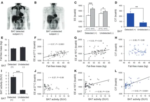
Acute effects of cold exposure on BAT thermogenesis. The metabolic activity of human BAT, as assessed by FDG-PET/CT, is undetectably low in warm conditions, but increases within hours after cold exposure (2). To examine the acute effects of cold on BAT in detail, we recruited 51 healthy young male subjects, in order to minimize any possible effects of age and sex on BAT activity (2, 4). First, the subjects under-went FDG-PET/CT after 2-hour cold exposure at 19°C to assess BAT activity. Of the 51, 27 subjects (52.9%) showed cold-activated BAT (BAT-positive subjects; Figure 1A), and 24 did not (BAT-negative sub-jects; Figure 1B). This was closely consistent with our previous results showing that cold-activated BAT could be detected in approximately 50% of young subjects (2, 6, 9). There was no significant difference between the BAT-positive and -negative subjects with respect to age, body weight, body fat mass, or fat-free mass (Figure 1E). BAT activity, assessed by FDG uptake into supraclavicular fat depots, was signifi-cantly higher in BAT-positive than BAT-negative subjects (5.5 ± 0.6 vs. 1.2 ± 0.1 standardized uptake value [SUV], P < 0.0001; Figure 1I).
To quantify the contribution of BAT to whole-body EE in humans, we measured whole-body EE under warm conditions at 27°C and after 2-hour cold exposure at 19°C using a respi-ratory gas analyzer (Supplemental Figure 1; supplemental mate-rial available online with this article; doi:10.1172/JCI67803DS1). EE at 27°C was almost the same between BAT-positive and -negative subjects (Figure 1C). After 2-hour cold exposure, EE increased significantly in both BAT-positive and -negative sub-jects. Cold-induced thermogenesis (CIT), calculated as the dif-ference between EE values at 27°C and 19°C, was significantly higher in BAT-positive than BAT-negative subjects (252.0 ± 41.1
Conflict of interest: The authors have declared that no conflict of interest exists.
brief report
vs. 78.4 ± 23.8 kcal/d, P < 0.01; Figure 1D). It is known that body composition, especially fat-free mass, is a significant determinant of EE (15). We therefore analyzed the associations of fat-free mass and BAT activity with EE using regression analyses. Fat-free mass closely related to EE at 27°C and 19°C, but not to CIT (Figure 1, F–H). BAT activity was significantly related to EE at 19°C and to CIT, but not to EE at 27°C (Figure 1, J–L). Multivariate regression models revealed independent associations of BAT activity with EE at 19°C and with CIT (P < 0.001 and P < 0.0001, respectively; Supplemental Table 1).
The metabolically active component of fat-free mass is mainly skeletal muscle (15). It is also known that shivering by skeletal muscle becomes one of the dominant components of EE in severe cold conditions, such as at 5°C–10°C (16). Orava et al. (10) report-ed that increasreport-ed FDG uptake after cold exposure at 17°C was evident in BAT, but negligible in other tissues, including skeletal muscle. This implies that muscle shivering in the present study at 19°C may be negligible. In fact, we found no significant correla-tion between fat-free mass and CIT. These results demonstrated that BAT activity becomes a dominant component of CIT at 19°C, independent of fat-free mass (Figure 1L), which indicates that CIT at 19°C is an index of BAT activity.
Chronic effects of cold exposure on BAT activity/thermogenesis and body fat. In small rodents, prolonged cold exposure induces BAT hyperplasia (1). To examine whether this is also the case in humans, 22 subjects with low or undetectable activities of BAT were randomly assigned to 2 groups: one was exposed to cold at 17°C for 2 hours every day (cold group; n = 12), while the other maintained their normal lifestyles without any exposure (control group; n = 10) (Supplemental Figure 1). Before and after a 6-week period, EE, body composition, and BAT activity were measured.
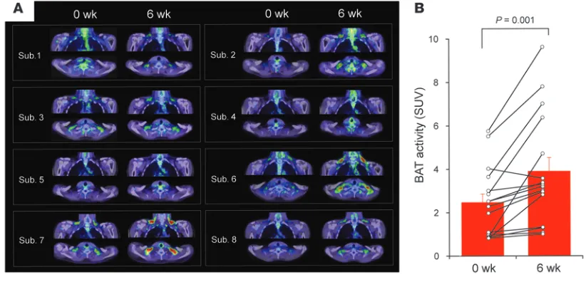
Detectable activities of BAT were found in 3 of 8 subjects at week 0 (subjects 3, 6, and 7) and in 6 subjects at week 6 (sub-jects 1, 2, 3, 6, 7, and 8; Figure 2A). BAT activity increased after the 6-week cold exposure from 2.46 ± 0.40 to 3.89 ± 0.64 SUV (P = 0.001; Figure 2B). Importantly, individuals with undetect-able activities of BAT at week 0 (subjects 1, 2, and 8) showed active BAT at week 6. In small rodents, chronic cold exposure results in induction of UCP1, hyperplasia of BAT, and a concomi-tant increase in 2-deoxy-d-glucose (2-DG) uptake into BAT (1).
[image:3.585.44.537.80.417.2]Moreover, we previously demonstrated in mice that 2-DG uptake into BAT is largely dependent on the presence of UCP1 (17). Thus, it is quite likely that the increased BAT activity (assessed by FDG uptake) is attributable to recruitment of BAT and/or
Figure 1
brief report
induction of UCP1. This is compatible with a report by Lee et al., who demonstrated higher UCP1 expression in human subjects with higher BAT activities, as assessed by FDG-PET/CT (18).
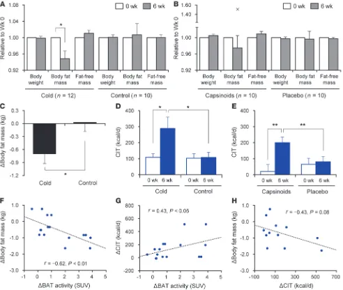
EE at 27°C did not change during the 6-week period in both the cold group and the control group. However, EE after 2-hour cold exposure of the cold group significantly increased after 6 weeks, whereas it did not change in the control group (Supplemental Fig-ure 2A). In fact, CIT of the cold group at week 6 (289.0 ± 70.0 kcal/d) was significantly higher than the cold group at week 0 (108.4 ± 22.8 kcal/d, P < 0.05) and the control group at week 6 (108.2 ± 31.1 kcal/d, P < 0.05; Figure 3D). The change in CIT dur-ing the 6-week period for the cold group was significantly higher than that for the control group (180.6 ± 69.5 vs. 5.0 ± 39.5 kcal/d,
P < 0.05), and positively correlated to the change in BAT activity (r = 0.43, P < 0.05; Figure 3G). These results, together with the high-ly positive correlation between CIT and BAT activity (Figure 1L), indicate that recruited BAT actually contributes to CIT.
Body fat mass of the cold group significantly decreased at week 6 compared with week 0 (–5.2% ± 1.9% change, P < 0.05), whereas body weight and fat-free mass did not change significantly (Figure 3A). In contrast, in the control group, there was no significant change in these parameters. During the 6-week period, body fat mass decreased more in the cold group than in the control group (–0.70 ± 0.23 vs. 0.03 ± 0.21 kg,
P < 0.05; Figure 3C). The change in body fat mass was inversely cor-related with that in BAT activity (r = –0.62, P < 0.01; Figure 3F) and insignificantly with that in CIT (r = –0.43, P = 0.08; Figure 3H), which suggests that increased BAT leads to body fat reduction.
Chronic effects of capsinoids on BAT activity/thermogenesis and body fat. Although our results are the first demonstration of the effective recruitment of human BAT, it would seem difficult to increase exposure to cold in daily life. To explore more practical methods for the recruitment of BAT, we focused on the chemi-cal stimulants of TRP channels, particularly capsinoids. Because capsinoids activate TRPV1 and TRPA1 (13) and increase BAT thermogenesis (14, 19), we reasoned that long-term capsinoid treatment could possibly recruit BAT in humans, as chronic cold exposure does.
To test this idea, we selected 10 subjects with low or undetect-able activities of BAT and examined the effects of daily ingestion of capsinoids on BAT (Supplemental Figure 1). In this experiment, we measured CIT before and 6 weeks after ingestion as a predictive index of BAT activity, instead of repeated FDG-PET/CT. Resting EE at 27°C was almost the same before and after treatment with capsinoids or a placebo. A significant increase in EE by 2-hour cold exposure was found only after capsinoid treatment (Supplemental Figure 2B). CIT after capsinoid treatment (200.0 ± 33.9 kcal/d) was higher than that before capsinoid treatment (20.6 ± 43.0 kcal/d,
P < 0.01) and after placebo treatment (81.0 ± 32.5 kcal/d, P < 0.01; Figure 3E). As CIT was proportional to BAT activity, as assessed by FDG-PET/CT, the capsinoid-induced increase in CIT appears to reflect enhanced thermogenic capacity and BAT activity.
Ono et al. reported that prolonged treatment of rats with caps-inoids resulted in increased UCP1 expression and decreased body fat (14). Body fat reduction and increased EE were also shown after capsinoid treatment for 12 weeks in mildly obese human subjects (20). In the present study in nonobese subjects, the 6-week cap-sinoid treatment increased EE, although it caused only a slight and insignificant reduction of body fat (2% reduction; Figure 3B). All these results suggest that the antiobesity effects of capsinoids are based on the thermogenic activity of recruited BAT. Thus, repeated ingestion of capsinoids can mimic the chronic effects of cold exposure on BAT and body fat in humans.
[image:4.585.86.497.84.282.2]Is BAT recruited by cold and capsinoids composed of brown adipocytes or beige cells? Current evidence suggests that rodents possess 2 types of UCP1-positive adipocytes arising from distinct develop-mental lineages: “classical brown adipocytes,” derived from Myf5-positive myoblastic cells, and “beige cells,” which reside in white adipose tissue and are induced in response to some environ-mental cues, such as cold exposure and β3-adrenergic receptor agonists (21). BAT in the supraclavicular region of human sub-jects was reported to be mainly composed of beige cells (22, 23). Lee et al. also demonstrated that preadipocytes isolated from supraclavicular fat of subjects with undetectable BAT have the ability to differentiate into UCP1-expressing adipocytes in vitro
Figure 2
brief report
(24). It therefore appears likely that the BAT recruited by cold and capsinoids in our present study is composed of beige cells.
In conclusion, we demonstrated here that human BAT can be recruited by chronic cold exposure and capsinoid inges-tion even in individuals who have lost active BAT. Our findings could contribute to developing practical, easy, and effective antiobesity regimens.
Methods
Further information can be found in Supplemental Methods.
Subjects. 51 healthy male volunteers (age, 24.4 ± 0.5 years; BMI, 22.0 ± 0.4 kg/m2;
body fat mass, 11.7 ± 0.7 kg; fat-free mass, 54.0 ± 0.7 kg) participated in the present study.
FDG-PET/CT. All subjects underwent FDG-PET/CT after 2-hour cold exposure at 19°C in winter as described previously (2, 6, 9, 19).
Indirect calorimetry. EE was measured using a respiratory gas analyzer at 27°C and after 2-hour cold exposure at 19°C as described previously (6).
Repeated cold exposure. 22 subjects showing low or undetectable activities of BAT were randomly divided into 2 groups: one was exposed to cold at 17°C for 2 hours every day for 6 weeks (cold group; n = 12); the other main-tained their usual lifestyles without cold exposure during the same period (control group; n = 10). Body fat content and EE at 27°C and after 2-hour cold exposure at 19°C were measured before and after the 6-week period (Supplemental Figure 1). 8 of the 12 subjects of the cold group underwent FDG-PET/CT examination again after 6-week cold exposure.
[image:5.585.49.541.83.499.2]Daily ingestion of capsinoids. Capsinoids were extracted from CH-19 Sweet (Capsicum annuum L.), purified, and encapsulated (19). 10 subjects with low or undetectable activities of BAT participated in this random-ized, single-blinded, placebo-controlled crossover trial. They ingested the capsules containing 9 or 0 mg (placebo) capsinoids every day for
Figure 3
brief report
Acknowledgments
This study was supported by a Grant-in-Aid for Scientific Research from the Ministry of Education, Culture, Sports, Science, and Technology of Japan (grant no. 22590227).
Received for publication November 12, 2012, and accepted in revised form May 15, 2013.
Address correspondence to: Takeshi Yoneshiro, Department of Anatomy, Hokkaido University Graduate School of Medicine, Sap-poro 060-8638, Japan. Phone: 81.11.706.5895; Fax: 81.11.706.5033; E-mail: yoneshiro@med.hokudai.ac.jp.
6 weeks. Before and after the 6-week period, body fat content and EE at 27°C and after 2-hour cold exposure at 19°C were measured (Supple-mental Figure 1).
Statistics. Data are expressed as mean ± SEM. Comparisons between groups were analyzed using t test or nonparametric test; 2-sided P values are given. Correlations were assessed using univariate and multivariate regressions. A P value less than 0.05 was considered statistically significant.
Study approval. The protocols were approved by the Institutional Research Ethics Review Board of Tenshi College (Sapporo, Japan). The trials were registered at http://www.umin.ac.jp/ctr/ (trial IDs UMIN000009005 and UMIN000006810). All participants were carefully instructed regarding the study and gave their informed consent.
1. Cannon B, Nedergaard J. Brown adipose tissue: function and physiological significance. Physiol Rev. 2004;84(1):277–359.
2. Saito M, et al. High incidence of metabolically active brown adipose tissue in healthy adult humans: effects of cold exposure and adiposity.
Diabetes. 2009;58(7):1526–1531.
3. van Lichtenbelt WDM, et al. Cold-activated brown adipose tissue in healthy men. N Engl J Med. 2009; 360(15):1500–1508.
4. Cypess AM, et al. Identification and importance of brown adipose tissue in adult humans. N Engl J Med. 2009;360(15):1509–1517.
5. Virtanen KA, et al. Functional brown adipose tissue in healthy adults. N Engl J Med. 2009; 360(15):1518–1525.
6. Yoneshiro T, et al. Age-related decrease in cold-activated brown adipose tissue and accumula-tion of body fat in healthy humans. Obesity. 2011; 19(9):1755–1760.
7. Vijgen GH, et al. Increase in brown adipose tissue activity after weight loss in morbidly obese subjects. J Clin Endocrinol Metab. 2012; 97(7):E1229–E1233.
8. Nakamura K. Central circuitries for body tempera-ture regulation and fever. Am J Physiol Regul Integr Comp Physiol. 2011;301(5):R1207–R1228. 9. Yoneshiro T, et al. Brown adipose tissue,
whole-body energy expenditure, and thermogenesis in
healthy adult men. Obesity. 2011;19(1):13–16. 10. Orava J, et al. Different metabolic responses of
human brown adipose tissue to activation by cold and insulin. Cell Metab. 2011;14(2):272–279. 11. Ouellet V, et al. Brown adipose tissue oxidative
metabolism contributes to energy expenditure during acute cold exposure in humans. J Clin Invest. 2012;122(2):545–552.
12. Tajino K, Hosokawa H, Maegawa S, Matsumura K, Dhaka A, Kobayashi S. Cooling-sensitive TRPM8 is thermostat of skin temperature against cooling.
PLoS One. 2011;6(3):e17504.
13. Shintaku K, et al. Activation of transient recep-tor potential A1 by a non-pungent capsaicin-like compound, capsiate. Br J Pharmacol. 2011; 165(5):1476–1486.
14. Ono K, et al. Intragastric administration of capsi-ate, a transient receptor potential channel agonist, triggers thermogenic sympathetic responses. J Appl Physiol. 2011;110(3):789–798.
15. Müller MJ, Bosy-Westphal A, Kutzner D, Heller M. Metabolically active components of fat-free mass and resting energy expenditure in humans: recent lessons from imaging technologies. Obes Rev. 2002; 3(2):113–122.
16. Haman F, Péronnet F, Kenny GP, Massicotte D, Lavoie C, Weber JM. Partitioning oxidative fuels during cold exposure in humans: muscle glycogen becomes dominant as shivering intensifies. J Physiol.
2005;566(pt 1):247–256.
17. Inokuma K, Ogura-Okamatsu Y, Toda C, Kimura K, Yamashita H, Saito M. Uncoupling protein 1 is necessary for norepinephrine-induced glucose utilization in brown adipose tissue. Diabetes. 2005; 54(5):1385–1391.
18. Lee P, et al. High prevalence of brown adipose tis-sue in adult humans. J Clin Endocrinol Metab. 2011; 96(8):2450–2455.
19. Yoneshiro T, Aita S, Kawai Y, Iwanaga T, Saito M. Nonpungent capsaicin analogs (capsinoids) increase energy expenditure through the activation of brown adipose tissue in humans. Am J Clin Nutr. 2012;95(4):845–850.
20. Snitker S, et al. Effects of novel capsinoid treatment on fatness and energy metabolism in humans: pos-sible pharmacogenetic implications. Am J Clin Nutr. 2009;89(1):45–50.
21. Ishibashi J, Seale P. Medicine. Beige can be slim-ming. Science. 2010;328(5982):1113–1114. 22. Wu J, et al. Beige adipocytes are a distinct type of
ther-mogenic fat cell in mouse and human. Cell. 2012; 150(2):366–376.
23. Sharp LZ, et al. Human BAT possesses molecular sig-natures that resemble beige/brite cells. PLoS One. 2012; 7(11):e49452.