stem/progenitor cell pool and induces
mammary tumors that undergo
epithelial-mesenchymal transition
Erica L. McCoy, … , Alana L. Welm, Heide L. Ford
J Clin Invest. 2009;119(9):2663-2677. https://doi.org/10.1172/JCI37691.
Six1 is a developmentally regulated homeoprotein with limited expression in most normal
adult tissues and frequent misexpression in a variety of malignancies. Here we
demonstrate, using a bitransgenic mouse model, that misexpression of human Six1 in adult
mouse mammary gland epithelium induces tumors of multiple histological subtypes in a
dose-dependent manner. The neoplastic lesions induced by Six1 had an in situ origin,
showed diverse differentiation, and exhibited progression to aggressive malignant
neoplasms, as is often observed in human carcinoma of the breast. Strikingly, the vast
majority of Six1-induced tumors underwent an epithelial-mesenchymal transition (EMT) and
expressed multiple targets of activated Wnt signaling, including cyclin D1. Interestingly,
Six1 and cyclin D1 coexpression was found to frequently occur in human breast cancers
and was strongly predictive of poor prognosis. We further show that Six1 promoted a
stem/progenitor cell phenotype in the mouse mammary gland and in Six1-driven mammary
tumors. Our data thus provide genetic evidence for a potent oncogenic role for Six1 in
mammary epithelial neoplasia, including promotion of EMT and stem cell–like features.
Research Article
Oncology
Find the latest version:
http://jci.me/37691/pdf
Six1 expands the mouse mammary epithelial
stem/progenitor cell pool and induces
mammary tumors that undergo
epithelial-mesenchymal transition
Erica L. McCoy,1 Ritsuko Iwanaga,2 Paul Jedlicka,3 Nee-Shamo Abbey,2
Lewis A. Chodosh,4 Karen A. Heichman,5 Alana L. Welm,5 and Heide L. Ford1,2,6
1Program in Molecular Biology, 2Department of Obstetrics and Gynecology, 3Department of Pathology, University of Colorado School of Medicine, Aurora, Colorado, USA. 4Department of Cancer Biology and Abramson Family Cancer Research Institute, University of Pennsylvania, Philadelphia, Pennsylvania, USA. 5Department of Oncological Sciences, Huntsman Cancer Institute and University of Utah, Salt Lake City, Utah, USA.
6Department of Biochemistry and Molecular Genetics, University of Colorado School of Medicine, Aurora, Colorado, USA.
Six1 is a developmentally regulated homeoprotein with limited expression in most normal adult tissues and
frequent misexpression in a variety of malignancies. Here we demonstrate, using a bitransgenic mouse model,
that misexpression of human Six1 in adult mouse mammary gland epithelium induces tumors of multiple
histological subtypes in a dose-dependent manner. The neoplastic lesions induced by Six1 had an in situ
ori-gin, showed diverse differentiation, and exhibited progression to aggressive malignant neoplasms, as is often
observed in human carcinoma of the breast. Strikingly, the vast majority of Six1-induced tumors underwent
an epithelial-mesenchymal transition (EMT) and expressed multiple targets of activated Wnt signaling,
includ-ing cyclin D1. Interestinclud-ingly, Six1 and cyclin D1 coexpression was found to frequently occur in human breast
cancers and was strongly predictive of poor prognosis. We further show that Six1 promoted a
stem/progeni-tor cell phenotype in the mouse mammary gland and in Six1-driven mammary tumors. Our data thus provide
genetic evidence for a potent oncogenic role for Six1 in mammary epithelial neoplasia, including promotion
of EMT and stem cell–like features.
Introduction
Normal embryogenesis and neoplasia share many of the same basic processes and molecular pathways, suggesting that tumor development is an aberrant form of morphogenesis (1). Indeed, there is now overwhelming evidence that developmental genes are often misexpressed in human cancers and that this misexpression can impact neoplastic disease through the re-initiation of develop-mental programs (2).
Recently, much attention has focused on a process typically associated with normal development, the epithelial-mesenchymal transition (EMT), as an important mechanism during tumor progression. In normal development, epithelial cells lose adhe-sion and polarity, delaminate, and acquire an invasive, so-called “mesenchymal” phenotype, allowing migration to a site appro-priate for organ formation (3). In neoplasia, a similar process is thought to occur at the tumor front, allowing for cellular inva-sion and eventual metastatic dissemination of cancer cells (4–6). Multiple signaling pathways have been implicated in both devel-opmental and oncogenic EMT, including the Notch, TGF-β, and Wnt signaling pathways (7–13). Recent evidence demonstrates that cells undergoing EMT take on stem cell characteristics (14), implicating developmental regulators of EMT as potential factors involved in stem cell maintenance. Additionally, cells that take on EMT and stem cell characteristics have increased tumorigenic and metastatic potential, underscoring the critical link between devel-opmental processes and cancer (4–6, 14).
Homeobox genes encode transcription factors that are “master regulators” of normal development and control processes such as proliferation, apoptosis, migration, and invasion. In particular, the processes of migration and invasion are associated with an EMT, and several homeoproteins have been implicated in EMT and stem cell maintenance (15–18). Our laboratory focuses on the sine oculis-related homeobox 1 homolog (Six1) homeoprotein that is expressed during early embryogenesis but lost in most adult tissues (19). It is essential for the development of numerous organs, in which it is involved in the expansion of progenitor cell populations through its ability to increase cellular proliferation and survival (19–26). In addi-tion, recent evidence demonstrates that Six1 plays a role in cellular migration and invasion during embryogenesis (20–24) through a mechanism that may involve an EMT. Interestingly, the closely relat-ed family member, Six2, regulates both a mesenchymal and stem cell population in the kidney, suggesting that Six family members may play important roles in both EMT and stem cell regulation (27), 2 processes that are now believed to be intimately related (14).
Overexpression of Six1 is observed in numerous cancers, includ- ing breast (19, 28, 29), ovarian (26), cervical (30), and hepatocel-lular carcinomas (31) as well as rhabdomyosarcomas (32–34) and Wilms tumors (35). In several of the aforementioned cancers, Six1 enhances cancer cell proliferation and survival (19, 25, 26, 28, 33), and its overexpression in immortalized mammary epithelial cells induces transformation, leading to highly aggressive and invasive tumors when transplanted into nude mice (25). Although Six1 expression is strongly correlated with neoplasia, its ability to initi-ate aggressive tumors from normal mammary epithelial cells, or any other normal cells, has not previously been examined.
Conflict of interest: The authors have declared that no conflict of interest exists.
In this paper, we test the hypothesis that Six1 overexpression in the adult mammary gland leads to activation of developmental pathways out of context, resulting in breast tumor formation. Using a mammary-specific, inducible mouse model of Six1 overexpression, we show that Six1, when misexpressed in the adult mammary epi-thelium, does indeed induce mammary hyperplasia and aggressive tumor formation. Mammary tumors formed in Six1-overexpressing mice manifest diverse histologic features, including apparent dedif-ferentiation into high-grade solid and sarcomatoid tumors. The sarcomatoid tumors display full oncogenic EMT, including com- plete loss of E-cadherin and gain of mesenchymal markers. Further- more, the majority of nonsarcomatoid tumors (80%) display a par-tial EMT, including regional loss of membranous E-cadherin, with
[image:3.585.61.525.76.467.2]concomitant gain of nuclear β-catenin and its downstream targets cyclin D1 (Ccnd1), transcription factor 7, T cell specific(Tcf7), Axin2, and myelocytomatosis oncogene (c-myc), suggesting that activation of the Wnt signaling pathway occurs with Six1 overexpression in mouse mammary tumorigenesis. As might be expected of a gene that induces EMT and is associated with Wnt signaling, mammary epithelial cells from Six1-overexpressing mice are enriched for stem cells and have increased mammosphere-forming capability. Addi-tionally, Six1-driven tumors contain cells of multiple mammary lineages and express the stem/progenitor cell marker Sca-1, sug-gesting that Six1 promotes a stem cell phenotype in the mammary gland. Thus, Six1 is able to induce both EMT and stem cell features in vivo. Consistent with our mouse model, Six1 and cyclin D1 are
Figure 1
Characterization of the inducible, mammary-specific Six1 transgenic mouse model. (A) Schematic representation of the inducible, bitransgenic mouse model system. Tet-O, tet operator. (B) Southern blot analysis shows varying copy numbers in TetSix animal lines (4910, 4922, 6239, and 6245) as compared with spiked-plasmid control. (C) qPCR, using transgene-specific primers and probe, reveals that HASix1 is not expressed in the MTB +dox control animals but is expressed at low levels in the uninduced TOSix mammary glands and at high levels in the induced TOSix mam-mary glands. Differences in expression between –dox and +dox mammam-mary glands are much greater than differences between transgenic lines (4922 and 6239). Values were transformed using log10(value+1) equation and plotted using a linear axis. Each point represents the value for 1 mammary
coexpressed in human breast cancer, and while Six1 alone correlates with poor prognosis, this correlation is significantly enhanced by coexpression of Ccnd1. Together, these data demonstrate that Six1 is a powerful inducer of mammary tumorigenesis and suggest that it may predict poor prognosis in human tumors through its abil-ity to cooperatively interact with cyclin D1 and to enable tumors to acquire both EMT and stem cell–like features.
Results
Development of an inducible, mammary-specific Six1 transgenic mouse model . Six1 is dynamically expressed in the developing mouse mam-mary gland, with the highest levels of Six1 transcript present in the late embryonic and young virgin mammary gland. In contrast, lev- els of Six1 significantly decrease as the mammary gland differenti- ates, with very low to no expression observed during pregnancy, lac-tation, and involution (28). Because Six1 overexpression has been detected in human breast cancer, aberrant expression of Six1 in the adult mammary gland may contribute to breast tumorigenesis. To directly test the role of aberrant Six1 expression in the induction of mammary tumorigenesis, and to best mimic the reexpression of Six1 that may occur in the adult human to initiate breast cancer, we developed a (doxycycline-inducible) dox-inducible, bitransgenic mouse model. To this end, we crossed the MTB transgenic line developed by Chodosh and colleagues (36) with transgenic mice containing an HA-tagged Six1 construct, in which Six1 was placed downstream of tet operator sequences (TetSix line). It should be
noted that the affect of the HA-tag on Six1 transcriptional activity was previously tested in a cell culture model to ensure that it did not interfere with the normal function of Six1 (data not shown). The MTB line expresses a mouse mammary tumor virus long termi-nal repeat region–driven (MMTV-LTR–driven) reverse tetracycline transcriptional activator (rtTA), so that treatment of bitransgenic animals (the TetSix line crossed with the MTB line, referred to here-in as the TOSix line) with dox activates rtTA, which binds the tet promoter and initiates transcription of Six1 (Figure 1A). Five TetSix founder lines were established, of which 4 transmitted through the germ line and contained variable copy numbers of the transgene (Figure 1B). In our model, Six1 was highly expressed in the mam-mary gland, with some expression in the salivary gland but not in other organs, as expected when using the MMTV promoter (Sup-plemental Figure 1; supplemental material available online with this article; doi:10.1172/JCI37691DS1).
The inducible model generates mice with no, low, or high Six1 expression. After establishing the TOSix transgenic model, we determined lev- els of expression of Six1 in the absence and presence dox. Experi-mental female and male TOSix animals from both the 4922 and 6239 lines, as well as control animals (MTB littermates), were continuously treated with 2 mg/ml dox in the drinking water at 12 weeks of age, well after the mice had reached sexual maturity. TOSix animals from both lines were continuously treated with sucrose-only water after 12 weeks of age, to establish levels of
[image:4.585.47.393.77.434.2]HA-Six1 in the absence of dox induction. A subset of female mice
Figure 2
were bred for 3 sequential pregnancies and allowed to nurse for 3 weeks before weaning. Because no phenotype was detected in male animals, the remainder of this analysis reflects phenotypes arising exclusively in female animals.
Examination of transgene expression in multiparous animals using real-time quantitative PCR (qPCR) demonstrated that no
HA-Six1 mRNA could be detected in the MTB dox-induced (+dox) cohort. Six1 transgene was, however, detectable in both the TOSix cohort that was not induced with dox (–dox) and TOSix +dox cohorts. Transgene levels were significantly higher in the induced cohort as compared with the uninduced cohort, and importantly, relative Six1 expression differences were much greater between the uninduced and induced TOSix animals than between the 4922 and 6239 transgenic lines (Figure 1C). Thus, we were able to examine dose-dependent effects of Six1 by comparing the control group (MTB +dox animals that did not express Six1, herein referred to as no Six1) with 2 different experimental groups: animals that express low levels of Six1 (herein called low Six1) (–dox animals from the
6239 and 4922 TOSix lines) and animals that express high levels of Six1 (herein called high Six1) (+dox animals from the 6239 and 4922 TOSix lines). Although Six1 mRNA expression could be detected by the sensitive method of qPCR in both young and old uninduced TOSix animals, nuclear Six1 protein expression could only be detected in older, uninduced TOSix animals (Figure 1D), suggest-ing an age-dependent increase in Six1 protein levels over time.
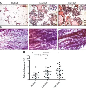
Long-term Six1 overexpression leads to precocious alveolar differentia-tion and hyperplasia . To determine the effect of Six1 overexpres-sion on the mammary gland, mammary tissue from aged no Six1–, low Six1–, and high Six1–expressing animals was taken at least 4 weeks after completion of weaning from the third sequential pregnancy. H&E-stained mammary sections from animals expressing no Six1 showed normal alveolar histology, while alveoli from both low Six1– and high Six1–expressing animals appeared to be expanded and to contain lipid droplets characteristic of secretory differentiation (Figure 2A). Analysis of
multiparous Six1-expressing and control animals showed nor-Figure 3
Mammary tumors arise in Six1-expressing animals in a dose-dependent manner and manifest histologically diverse phenotypes. (A) Kaplan-Meier analysis of the percentage of tumor-free animals reveals that both low Six1– and high Six1–expressing animals develop tumors, and tumor frequency is higher in low Six1–expressing animals. (B) Transgene-specific qPCR analysis of mammary glands and tumors taken from TOSix and control animals reveal that HASix1 expression is lower in tumors than mammary glands (including glands contralateral to tumors) but higher than that observed in control MTB +dox (no Six1) mammary glands. Values were transformed using log10(value+1) equation and plotted using
[image:5.585.65.523.78.437.2]mal postinvolution mammary gland histology after 6 months of dox treatment, suggesting that the alveolar phenotype is due to precocious differentiation, set on by long-term exposure to Six1, rather than a failure to properly involute (data not shown).
In addition to the precocious differentiation induced by Six1, presence of both low and high levels of Six1 resulted in marked mammary hyperplasia throughout the gland (Figure 2B). In the majority of cases, the hyperplastic phenotype appeared to affect the entire gland rather than discrete areas. To objectively quantify the degree of hyperplasia, H&E-stained sections were scanned and analyzed by a color-based algorithm in order to determine the epi-thelial versus fat contribution of each gland. Based on this analysis, mammary glands from both low Six1– and high Six1–expressing animals displayed a significantly higher (P < 0.04 and P < 0.006, respectively) epithelial content than age-matched control animals (>12 months of treatment) (Figure 2C).
Six-1 overexpression leads to aggressive mammary tumor formation in a dose-dependent manner. Overexpression of Six1 in the mammary gland of multiparous mice led to the development of overt mam-mary tumors in approximately 40% (9 of 25) of low Six1–expressing animals and approximately 15% (4 of 29) of high Six1–expressing animals. In contrast, only 7% (1 of 14) of control animals developed a mammary tumor, and this tumor did not occur until the mouse was over 2 years of age (Figure 3A). The average latency of tumor onset in the Six1-expressing animals was approximately 17.5 months after starting dox or vehicle treatment. Because aged, multiparous FVB animals are susceptible to mammary tumor formation due to prolac-tin-secreting pituitary abnormalities (37), we tested blood prolactin levels from a subset of low Six1– and high Six1–expressing animals that acquired mammary tumors to ensure that the tumors observed in aged animals were induced by Six1. All TOSix animals had blood prolactin levels within the normal range, providing evidence that the
tumors arising in the Six1-overex- pressing animals were not prolac-tin driven but were indeed induced by Six1 overexpression (data not shown). A small subset of nullipa-rous TOSix animals also developed mammary tumors with similar latency and histology, suggesting that mammary cancer initiation in TOSix animals is not dependent on parity. Although the sample size for the nulliparous study was insuffi-cient to reach statistical significance, tumor histology from these nul-liparous animals was similar to that
from the multiparous study groups, and this group was therefore included in all subsequent histological analysis. It should be noted that although exten-sive analysis was not done on all tissues in the animals, no overt metastases were observed in tumor-bearing animals.
Because low Six1–expressing ani-mals had a substantially higher tumor penetrance (40%) than high Six1–expressing animals (15%), we reasoned that there may be an opti-mal level of Six1 required to induce tumorigenesis. In order to further explore this finding, we used qPCR analysis to compare HA-Six1 transgene expression in all tumors to a subset of mammary glands from no Six1–, low Six1–, and high Six1–expressing animals. As expected, tumors from low Six1– and high Six1–expressing animals showed higher levels of transgene expression than the control (no Six1) mammary glands. However, the tumors expressed Six1 at lower levels than the low Six1– and high Six1–expressing mammary glands, including contralateral mammary glands taken from tumor-bearing ani-mals (Figure 3B). This finding suggests that while overexpression of Six1 leads to tumor formation, an optimal range of Six1 expres-sion may be necessary for tumor initiation.
Six1-induced tumors are histologically diverse and include high-grade adenocarcinomas and sarcomatoid (spindle cell) tumors. Histologic analysis of mammary tumors arising in both low Six1– and high Six1–expressing animals revealed neoplastic lesions of diverse his-tologies (Tables 1 and 2 and Figure 3C). The majority of tumors showed glandular (“adeno”) differentiation, as seen in human infiltrating ductal breast carcinoma. Two thirds of the tumors showed some degree of squamous differentiation. Additional histologic patterns, also with human correlates, included secre-tory and papillary differentiation. Two of fourteen tumors (14%) showed high-grade, poorly differentiated, solid areas, analogous to human poorly differentiated carcinoma. Most strikingly, 3 of 14 (21%) tumors exhibited highly aggressive spindle cell mor-phology, analogous to highly aggressive human sarcomatoid carcinoma (Table 1). While 43% of lesions exhibited features of intraepithelial neoplasia, 86% of tumors showed invasive features, characterized by loss of regular glandular architecture, stromal desmoplasia, and an infiltrative growth pattern. Interestingly, 5 of 14 (36%) individual tumors exhibited multiple histologic patterns (Table 2). Thus, overall, the neoplastic lesions appeared to have
Table 1
Summary of histological features of Six1-induced tumors
Histology No. of tumors Invasive tumors Tumors with CIS/MINA
Adenocarcinoma 2 of 14 2 of 2 (100%) 0 of 2 (0%)
Adenosquamous carcinoma 9 of 14 7 of 9 (78%) 5 of 9 (56%) Secretory carcinoma 3 of 14 3 of 3 (100%) 2 of 3 (67%) Papillary carcinoma 3 of 14 3 of 3 (100%) 1 of 3 (33%) Poorly differentiated carcinoma 2 of 14 2 of 2 (100%) 0 of 2 (0%) Sarcomatoid (spindle cell) carcinoma 3 of 14 3 of 3 (100%) 1 of 3 (33%)
[image:6.585.52.380.115.194.2]ACIS, carcinoma in situ; MIN, mammary intraepithelial neoplasia.
Table 2
Characteristics of tumors containing diverse histologies
Histology Tumor 1 Tumor 2 Tumor 3 Tumor 4 Tumor 5
Adenocarcinoma X
Adenosquamous carcinoma X X
Secretory carcinoma X X X
Papillary carcinoma X X
Poorly differentiated carcinoma X X
Sarcomatoid (spindle cell) carcinoma X X X
an in situ origin, show diverse differentiation, and exhibit pro-gression to highly aggressive malignant neoplasms, as observed in human carcinoma of the breast.
The sarcomatoid histomorphology within some Six1-overex-pressing tumors suggested that Six1 could induce an EMT in vivo, leading to a loss of polarized epithelial architecture and cell-cell contacts and a gain of mesenchymal-type cell characteristics. Thus, immunohistochemical analysis was performed on tumor sections dis- playing either sarcomatoid or epithelioid histomorphology (see H&E- stained sections in Figure 4A) for molecular markers of EMT. Strik-ingly, the sarcomatoid tumor regions displayed a complete loss of the epithelial marker E-cadherin (Figure 4B) and a concurrent gain of the mesenchymal markers SMA (Figure 4C) and nuclear Zeb1 (Supple-mental Figure 2). The cells retained the expression of the epithelial marker cytokeratin 18 (CK18) (Figure 4D), demonstrating that the mesenchymal tumors arose from an epithelial origin, which is charac-teristic of human sarcomatoid carcinoma. Thus, Six1 misexpression is able to induce oncogenic EMT in vivo in a subset of tumors.
The majority of Six1-driven tumors exhibit a partial EMT and express downstream targets of activated Wnt signaling. Although almost 80% of Six1-induced tumors did not display an overt sarcomatoid his-tomorphology, the majority did show focal loss of E-cadherin. In these tumors, E-cadherin loss correlated with the presence of nuclear β-catenin, a protein that colocalizes with E-cadherin in adherens junctions in noncancerous cells but moves to the nucleus in some tumor types (6). Nuclear β-catenin was detected in 67% (8 of 12) of tumors stained, including those arising in both nul-liparous and multiparous animals, and in 80% (8 of 10) of tumors stained arising in the multiparous subset. As expected, regions exhibiting high nuclear β-catenin expression also expressed high levels of the β-catenin transcriptional target, cyclin D1 (Figure 5A). Together, these data suggested that partial loss of E-cadherin
with concomitant activation of Wnt target genes is a predominant molecular phenotype in Six1-driven tumors. While this phenotype induced by Six1 is not an overt EMT as observed in the sarcoma-toid tumors, it possesses many molecular features of an EMT. These data suggest that Six1 drives a partial EMT, concomitant with activated Wnt signaling in most tumors.
In support of this notion, Six1-overexpressing tumors showed a statistically significant upregulation of several Wnt/β-catenin transcriptional targets, including Ccnd1, c-Myc, and Axin2 (38). The same trend is clear for another Wnt transcriptional target, Tcf7, where the data approaches statistical significance (P = 0.08) (38). Surprisingly, these β-catenin target genes were only upregulated in the tumors and not in the mammary glands overexpressing Six1 (Figure 5B), suggesting that Six1 does not directly activate Wnt signaling or Ccnd1 and c-Myc in the mammary gland, targets previ-ously reported to be activated not only through Wnt signaling but directly by Six1 in other tumor types (34). Thus, activation of Wnt signaling may act as a preferred cooperating event for Six1-driven tumorigenesis, or alternatively, Six1 may only activate these tar-gets in a tumorigenic context. In support of the hypothesis that altered Six1-mediated activation may occur in cancer, ezrin (Ezr), a reported direct transcriptional target of Six1 in rhabdomyosar- comas (34), is highly expressed in Six1-induced tumors when com-pared with control mammary glands and is only slightly, but not significantly, overexpressed in mammary glands overexpressing high levels of Six1 (Figure 5C). In contrast, a previously reported developmental target of Six1, c-Met (22), does not appear to be regulated by Six1 (Supplementary Figure 3).
Six1-overexpressing mammary glands are enriched for stem cells, and Six1-driven tumors exhibit progenitor cell characteristics. Six1 is known to stimulate proliferation in progenitor populations during normal development (19–26), and recent evidence suggests that the closely related family member Six2 may contribute to the self-renewing potential of kidney stem cells (27). The majority of Six1-overex- pressing tumors have activated the Wnt signaling pathway, a path-way that has been strongly implicated in mammary epithelial stem cell maintenance (39). Additionally, Six1-driven tumors display multiple histologies and the majority undergo an EMT, a process strongly associated with the gain of stem cell characteristics (14). Thus, we asked whether Six1 could promote a stem/progenitor cell phenotype in mammary epithelial cells and whether Six1-driven tumors would exhibit stem/progenitor cell characteristics.
To determine whether Six1 overexpression alters the percentage of mammary epithelial stem cells, primary mammary epithelial cells were isolated from both 6239 and 4922 TOSix bitransgenic lines and MTB control females aged approximately 1.5 years. Epi-thelial cells were analyzed by flow cytometry using the stem cell
markers CD24 and CD29 (39). TOSix animals from both trans-Figure 4
[image:7.585.49.284.80.361.2]genic lines displayed an increased CD29hiCD24+
mammary popu-lation compared with MTB controls (32.0% and 28.4% vs. 16.1%, respectively) (Figure 6A).
To determine whether Six1 overexpression led to an increased population of functional mammary epithelial stem cells, mammo-sphere assays were performed using epithelial cells isolated from TOSix (4922 and 6239 lines) and MTB animals. The secondary mammosphere assay, which measures capacity for self-renewal (40–42), revealed that Six1 bitransgenic mammary glands con-tain an increased number of functional mammary epithelial stem cells when compared with control mammary glands. Similar to the results from flow cytometry, mammary cells isolated from TOSix animals formed substantially more mammospheres than cells isolated from MTB controls (Figure 6B). Similar results were obtained for both flow cytometry and mammosphere assays,
regardless of whether the animals were sucrose treated or induced with dox, such that bitransgenic Six1-expressing animals always displayed a higher percentage of stem cells than the MTB control counterparts (data not shown). Together, our results demonstrate that mammary glands overexpressing Six1 are enriched for stem cells, suggesting that Six1 may play a role in the establishment or maintenance of mammary epithelial stem cell populations.
[image:8.585.41.533.78.480.2]To determine whether Six1-driven tumors also manifest a stem/ progenitor cell phenotype, we examined the TOSix tumors for expression of the luminal epithelial cell marker CK18, the myoepi-thelial cell marker CK5, and the mammary progenitor cell marker CK6 (43). Strikingly, 6 of 12 (50%) Six1-driven tumors exhibited areas of mixed cytokeratin expression, including CK18, CK5, and CK6 (Figure 7A). Additionally, 9 of 12 (75%) TOSix tumors expressed the stem cell marker Sca-1 (Figure 7B). These data, in
Figure 5
conjunction with the observation that individual tumors driven by Six1 frequently show multiple histologic patterns and activation of Wnt signaling, suggest that Six1-induced tumors arise from a progenitor/stem cell–like cell population.
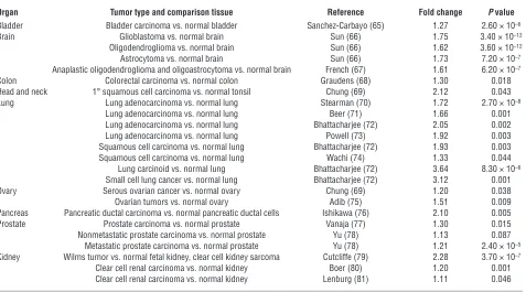
Six1 overexpression is observed in many human malignancies . Inappro- priate expression of Six1 has been reported in breast (19, 29), ovar-ian (26), cervical (30), and hepatocellular carcinomas (31) and in rhabdomyosarcoma (32–34) and Wilms tumor (35). The fact that Six1 is overexpressed in several, unrelated tumor types led us to examine its expression in an even more diverse set of tumor types. Using the Oncomine database (44), we evaluated Six1 expression in 23 microarray data sets that profile one or more tumor types against normal tissue. Strikingly, in 15 out of 20 tumor types that we were able to evaluate, Six1 was overexpressed compared with the normal tissue. In many cases this was confirmed in multiple datasets (Table 3). Interestingly, the levels of Six1 overexpression
were relatively low, ranging from a 1.1- to 4-fold increase, but in each case the data were highly statistically significant (Table 3). This observation suggests that, similar to the results from our transgenic model, low levels of Six1 overexpression are more likely to promote tumorigenesis than high levels of Six1.
The malignancies in which Six1 was overexpressed included brain tumors (glioblastoma, oligodendroglioma, and astrocytoma), lung tumors (adenocarcinoma, squamous cell carcinoma, carcinoid, and small cell carcinoma), kidney tumors (clear cell carcinoma and Wilms tumor), and bladder, colon, tonsillar squamous cell, ovarian, pancre-atic, and prostate carcinomas (Table 3). These data strongly support the notion that inappropriate expression of Six1 is an important tumor-initiating or -promoting factor in many human tumors.
[image:9.585.46.404.77.379.2]Overexpression of Six1 and Ccnd1 correlates in human breast tumors and strongly predicts poor prognosis. Because we observed an increase in cyclin D1 expression in regions undergoing EMT in Six1-induced
Figure 6
Six1-overexpressing mammary glands exhibit stem/progenitor cell characteristics. (A) Flow cytometric analysis of mammary epithelial cells harvested from sucrose-treated, nul-liparous TOSix animals (4922 and 6239 lines) and MTB animals aged approximately 1.5 years. Three ani-mals were pooled for each group. Antibodies used to perform flow cytometry include CD24 and CD29, markers found on mammary epi-thelial stem cells. TOSix animals have substantially more stem cells than MTB controls. Percentages denote the CD29hiCD24+ population.
APC, allophycocyanin. (B) Second-ary mammosphere assays were performed using mammary epithe-lial cells isolated from the above groups. Mammosphere numbers are increased in TOSix animals com-pared with MTB controls. Experi-ments in A and B were repeated with mammary epithelial cells from either sucrose- or dox-treated animals, yielding similar results.
Figure 7
[image:9.585.79.508.594.680.2]mammary tumors, we sought to determine whether cyclin D1 and Six1 proteins are coexpressed in human breast cancer using tissue microarrays. Immunohistochemical staining using antibodies gen- erated against Six1 and cyclin D1 was performed on arrays contain-ing primary tumors (mixed invasive ductal carcinoma and invasive lobular carcinoma) from 160 patients, and staining was scored for intensity on a 0–4 scale. Representative examples of anti-Six1 and anti–cyclin D1 staining in human breast cancers are shown in Figure
8A. Importantly, intensity of cyclin D1 antibody staining increased with the intensity of Six1 antibody staining in a statistically signifi-cant manner (Figure 8B). These results demonstrate that Six1 and cyclin D1 immunoreactivity correlate in human breast cancer.
[image:10.585.57.534.111.376.2]Our data show that Six1 is capable of initiating tumors that are highly invasive and aggressive, and clinical studies show that Six1 overexpression occurs in 90% of breast cancer metastatic lesions compared with 50% of primary breast tumors (19, 29). In addition,
Table 3
Many human tumor types overexpress Six1
Organ Tumor type and comparison tissue Reference Fold change P value Bladder Bladder carcinoma vs. normal bladder Sanchez-Carbayo (65) 1.27 2.60 × 10⁻⁶
Brain Glioblastoma vs. normal brain Sun (66) 1.75 3.40 × 10⁻¹²
Oligodendroglioma vs. normal brain Sun (66) 1.62 3.60 × 10⁻¹²
Astrocytoma vs. normal brain Sun (66) 1.73 7.20 × 10⁻⁷
Anaplastic oligodendroglioma and oligoastrocytoma vs. normal brain French (67) 1.61 6.20 × 10⁻⁷
Colon Colorectal carcinoma vs. normal colon Graudens (68) 1.30 0.018
Head and neck 1° squamous cell carcinoma vs. normal tonsil Chung (69) 2.12 0.043
Lung Lung adenocarcinoma vs. normal lung Stearman (70) 1.72 2.70 × 10⁻⁸
Lung adenocarcinoma vs. normal lung Beer (71) 1.66 0.001
Lung adenocarcinoma vs. normal lung Bhattacharjee (72) 2.05 0.002
Lung adenocarcinoma vs. normal lung Powell (73) 1.92 0.003
Squamous cell carcinoma vs. normal lung Bhattacharjee (72) 1.93 0.003
Squamous cell carcinoma vs. normal lung Wachi (74) 1.33 0.044
Lung carcinoid vs. normal lung Bhattacharjee (72) 3.64 8.30 × 10⁻⁶ Small cell lung cancer vs. normal lung Bhattacharjee (72) 3.12 0.001
Ovary Serous ovarian cancer vs. normal ovary Chung (69) 1.20 0.038
Ovarian tumors vs. normal ovary Adib (75) 1.51 0.009
Pancreas Pancreatic ductal carcinoma vs. normal pancreatic ductal cells Ishikawa (76) 2.10 0.005
Prostate Prostate carcinoma vs. normal prostate Vanaja (77) 1.30 0.015
Nonmetastatic prostate carcinoma vs. normal prostate Yu (78) 1.13 0.087 Metastatic prostate carcinoma vs. normal prostate Yu (78) 1.21 2.40 × 10⁻⁵ Kidney Wilms tumor vs. normal fetal kidney, clear cell kidney sarcoma Cutcliffe (79) 2.28 3.70 × 10⁻⁷
Clear cell renal carcinoma vs. normal kidney Boer (80) 1.20 0.001
Clear cell renal carcinoma vs. normal kidney Lenburg (81) 1.11 0.046
Figure 8
[image:10.585.47.541.511.668.2]a recent examination of the van de Vijver dataset, profiling 295 early stage (I and II) breast carcinomas (45), and the Pawitan and Ivshina dataset, profiling 240 invasive breast carcinomas unselect-ed for disease stage (46, 47), showed that Six1 is a predictor of poor prognosis in breast cancer (48). Together, these observations sup-port a causal role for Six1 in human breast cancer progression.
[image:11.585.62.525.82.548.2]Because results from our mouse model and from human breast cancer samples suggest that Six1 and cyclin D1 are often coex- pressed in mammary cancer, we used the aforementioned datas-ets to test the hypothesis that cyclin D1 may cooperate with Six1 in human breast cancer. In the van de Vijver dataset, patients whose tumors expressed above mean levels of Ccnd1 exhibited
Figure 9
Six1 overexpression significantly correlates with poor clinical outcome in breast cancers, and this significance is increased by coexpression of
no significant trends in any of the prognostic indicators, includ-ing time to metastasis, time to relapse, and overall survival due to breast cancer–related death (Figure 9B). However, in patients whose tumors had above mean expression of both Ccnd1 and Six1, adverse outcomes were exacerbated and concomitant overexpres-sion of both Six1 and Ccnd1 increased the prognostic value when compared with Six1 overexpression alone, with respect to the fol-lowing parameters: shortened time to metastasis (P = 0.003 for Six1 and Ccnd1 vs. P = 0.03 for Six1 alone), shortened time to relapse (P = 0.021 for Six1 and Ccnd1 vs. P = 0.03 for Six1 alone), and short-ened survival due to breast cancer–related death (P = 0.011 for Six1 and Ccnd1 vs. P = 0.03 for Six1 alone) (Figure 9, A vs. C). In the Pawitan and Ivshina dataset, while Ccnd1 overexpression alone did not predict shortened time to relapse, above mean levels of Six1 and Ccnd1 together cooperatively worsened prognosis and were more strongly associated with shortened time to relapse than Six1 alone (P < 0.0004 for Six1 and Ccnd1 vs. P = 0.009 for Six1 alone) (Figure 9D). These results, obtained with 2 independent datasets comprising a total of 535 patients, show that the combination of
Six1 and Ccnd1 are powerful predictors of poor prognosis.
Discussion
Six1 is aberrantly expressed in a number of cancers, including breast (19, 28, 29), ovarian (26), cervical (30), and hepatocellular carcinomas (31) as well as in rhabdomyosarcomas (32–34) and Wilms tumors (35). In these cancers, reexpression of Six1 is believed to reinstate a developmental program out of context, increasing proliferation, migration, invasion, and survival to stimulate cancer initiation as well as progression. To determine whether Six1 is in fact able to initiate breast cancer, we established a mammary-spe-cific transgenic mouse model that allowed us to study the effects of both low and high levels of Six1 overexpression. In both low Six1– and high Six1–expressing animals, the transgene induced precocious alveolar differentiation and mammary epithelial hyper-plasia. In addition, chronic Six1 overexpression, particularly in the low Six1–expressing animals, resulted in the formation of frank tumors, most of which had activated Wnt signaling and exhibited EMT and stem cell features.
qPCR results revealed that Six1 transgene expression, while increased compared with control MTB +dox (no Six1) mammary glands, is consistently overexpressed at lower levels in the TOSix tumors as compared with TOSix mammary glands. Transgene silencing has been reported to occur in MMTV-LTR–driven mouse models in cases in which the transgene facilitates an EMT (49). In these cases, as cells undergo an EMT, epithelial-specific promoters, such as MMTV-LTR, become silenced. Because Six1 is capable of initiating an EMT or partial EMT in the majority of the mammary tumors, this silencing mechanism may result in reduced levels of the Six1 transgene expression within the tumors as compared with that in the contralateral mammary glands. Alternatively, selective pressure may contribute to the outgrowth of tumor cells express-ing the optimal level of Six1 required to promote tumorigenesis. Regardless of mechanism, the consistent and striking correlation between low levels, as opposed to high levels, of Six1 overexpres-sion and tumor frequency suggests that there is an optimal level of Six1 for inducing tumorigenesis.
Interestingly, recent evidence suggests that Six1 can confer dose-dependent effects in cranial placodes during development (50). Small-to-moderate increases in the levels of homeobox gene expression are known to have substantial effects on target gene
transcription with significant phenotypic consequences. For example, a 1.5-fold increase of Oct3/4 expression is sufficient to activate and repress multiple target genes (51), and both Oct3/4 and another homeoprotein, Nanog, display dose-dependent effects during development (51–53). Additionally, Oct3/4 influences the genesis of germ cell tumors in a dose-dependent manner (54). We hypothesize that the effects of Six1 are dose dependent, at least in part, due to its required interactions with available cofactors with-in the cell. Six1 has no intrinsic activation or repression domains and thus is dependent on the Eya and Dach transcriptional cofac-tors to activate and/or repress transcription (21). When Six1 exists in a complex with Dach, it represses transcription. However, if Eya is added to the complex, it can switch Six1 from a repressor to an activator (21). Like Six1, Eya1 also acts in a dose-dependent man- ner during development (55). Thus, it is likely that the stoichiom-etry of Six1 with its cofactors, as well as the availability of cofactors in the cell, modulates the ability of Six1 to activate and/or repress transcription and to induce tumorigenesis. Future experiments are required to determine how the ratio of Six1 to its cofactors may differentially affect tumorigenesis.
Tumors arising in TOSix animals have diverse histologies, rang- ing from relatively well-differentiated tumors (squamous, secre-tory, and papillary) to poorly differentiated solid and sarcomatoid tumors. Interestingly, the sarcomatoid tumors undergo a complete EMT, as determined by loss of E-cadherin, gain of mesenchymal markers Zeb1 and SMA, and retention of the epithelial marker CK18. Although this is the first evidence to our knowledge that Six1 is directly capable of driving an EMT, a recent Six1/Six4 –dou-ble-knockout study is suggestive of a role for Six1 in EMT during normal development. While Six4- knockout mice display no pheno-typic abnormalities, myogenic precursor cells in Six1/Six4 –double-knockout mice fail to delaminate from the dermomyotome (an epithelial structure) and migrate to the limb bud (22). This defect is suggestive of a failure to undergo a developmental EMT. Fur-thermore, the Six2 protein, which is 96% conserved to Six1 across the Six domain and homeodomain at the amino acid level, is criti-cal for maintaining the mesenchymal progenitor cell population in the developing kidney, and loss of Six2 results in premature and ectopic differentiation of mesenchymal cells into kidney epithe-lial cells (56). While these developmental studies suggest a role for Six1 in regulating EMT, our model provides what we believe to be unique evidence that overexpression of Six1 can induce an EMT in vivo, particularly in a tumorigenic context.
(34). Likewise, because the majority of Six1-driven tumors dis-play characteristics of Wnt signaling, activation of this signaling pathway is most likely either a preferred cooperating oncogenic pathway for Six1 or the result of Six1 activation that only occurs in the tumorigenic state.
Wnt and its downstream effector β-catenin are known to induce tumorigenesis when overexpressed in the mouse mammary gland. Although some characteristics of Wnt or activated β -catenin trans- genic mice phenocopy TOSix mammary phenotypes, such as pre-cocious lobuloalveolar development, Six1-driven tumors display greater histologic diversity than the tumors arising from animal models of activated Wnt signaling (57–60). Additionally, while Wnt signaling is known to induce EMT in development and in cell culture models (7–11), oncogenic EMT is not induced by activated Wnt signaling in the mammary gland, as animals overexpressing Wnt in this context maintain E-cadherin on the surface of the mammary tumor cells (57). These data suggest that it is Six1, or Six1 in concert with Wnt, that is the primary driving force for the complete and partial EMTs observed.
Because recent studies demonstrate that cells undergoing EMT take on stem cell characteristics (14), and because Six1 drives the formation of tumors with multiple histomorpholo-gies, we sought to determine whether Six1 is involved in the regulation of mammary epithelial stem cells. Recent evidence demonstrates that Six2 regulates a stem cell population in the kidney, suggesting that the Six family may play important roles in stem cell regulation (27). Interestingly, our findings indicate that Six1 may play a role in mammary epithelial stem cells, both in the normal gland as well as in tumorigenesis. Mammary epithelial cells from Six1-overexpressing mice are enriched for stem cells and have increased mammosphere-forming capability. Additionally, Six1-driven tumors contain cells of multiple mam-mary lineages and express Sca-1. Together, these data strongly suggest that Six1 promotes a stem cell phenotype in the normal mammary gland and that Six1-driven tumors arise from a stem/ progenitor cell population.
Finally, since Six1 is not normally expressed in most adult tissues, and since its misexpression can initiate mammary tumorigenesis, we investigated whether inappropriate expression of Six1 may be involved in malignancies in addition to breast cancer. We found that a large number of cancers overexpress Six1 compared with the expression of the normal tissue from which they arise. Although the increase in Six1 transcript levels in these tumors is moderate (1.1- to 4-fold increase), at least as measured on microarray plat-forms, it is highly significant and is observed in multiple tumor types. These data strongly support a critical role for Six1 in human tumorigenesis and further support the model whereby low levels of Six1 overexpression drive tumorigenesis.
Given the increased expression of cyclin D1 in tumors arising from animals overexpressing Six1, we hypothesized that Six1 and cyclin D1 may be coexpressed in human breast cancers. Indeed, immunohistochemical analysis of breast cancer tissue microarrays revealed that tumors that stained intensely with an anti-Six1 anti-body also stained intensely with an anti–cyclin D1 antibody. It should be noted, however, that the Six1 antibody (Atlas Antibod-ies) used for this analysis is made against a region of Six1 that may allow cross-reactivity with other Six family members. Repeated attempts to generate antibodies uniquely specific to Six1 have resulted in antibodies that do not work for immunohistochemical analysis. Thus, we can only confidently say that Six family expres-sion correlates with cyclin D1 in human tumors. However, since
Six1 and Ccnd1 mRNA together are highly predictive of poor prog-nosis (see Figure 9), we conclude that Six1 and cyclin D1 are likely coexpressed in human breast tumors.
In work described elsewhere (48), we demonstrate that Six1 is an indicator of poor prognosis in human breast cancers. When
Six1 overexpression is combined with overexpression of Ccnd1, poor prognosis in breast cancer patients, as assessed by shortened time to metastasis, shortened time to relapse, and decreased sur-vival, is exacerbated, even though Ccnd1 alone is not predictive of poor prognosis. These results strongly suggest that the coopera-tion occurring between Six1 and cyclin D1 in our mouse model is recapitulated in human breast cancers. In our analysis, we also attempted to identify a correlation with other Wnt signaling tar-gets, including Axin, Tcf7, and c-Myc. However, overexpression of these molecules did not seem to cooperate with Six1 as prognostic indicators. These data suggest that, unlike what is observed in the mouse model, Six1 may not cooperate with canonical Wnt signal-ing in human breast cancer, but rather, only the association with cyclin D1 is conserved. However, more association studies must be performed to fully rule out a cooperative interaction between Six1 and Wnt signaling in human breast cancer.
In closing, our data are the first to our knowledge to demon- strate that Six1 overexpression is sufficient to induce tumorigen-esis when expressed out of context in a normal adult cell, leading to highly aggressive and invasive mammary tumors with EMT and stem cell features. We conclude that Six1 is promoting tumorigen-esis by reemploying its developmental program to drive not only proliferation and survival but also migration and invasion through EMT. Additionally, our data are the first to our knowledge to sug-gest that Six1 acts to promote a stem cell phenotype. Because Six1 is overexpressed in a large number of different cancers, we propose that this homeoprotein may act to initiate numerous types of can-cer in addition to breast cancer, acting as a more global regulator of tumorigenesis than originally anticipated.
Methods
Generation of mice. The full-length human HA-Six1 cDNA was inserted into pTetSplice vector using EcoRI adapters to generate the transgenic plasmid. Microinjections were performed by the Cancer Center Transgenic Mouse Core Facility at University of Colorado Denver (UCD) Anschutz Medical Campus. Transgenic progeny were identified by Southern blot analysis using a full-length Six1 cDNA probe and standard protocols (29). Four lines trans-mitted the transgene through their germline (4910, 4922, 6239, and 6245). Although all lines expressed Six1 after dox induction, the 2 lines with highest expression levels were chosen for further analysis (4922 and 6239). PCR geno-typing was performed at 3 weeks of age using genomic DNA isolated from a tail biopsy and the REDExtract-N-Amp Tissue PCR Kit (Sigma-Aldrich). Primers used to detect the TetSix transgene were 5′ -AGCTCCAAGACTCTCT-GCTC-3′ (forward) and 5′-TCCGGCGAATTTCTGCCATT-3′ (reverse).
treated with water containing 2% sucrose as a vehicle control (5% sucrose caused increased intake of control water). Water was provided to animals in a light-protected bottle and changed weekly. A cohort of female animals from the MTB and TOSix genotypes were bred for 3 sequential pregnan-cies and were allowed to nurse for 3 weeks before weaning. Animals on long-term tumor studies (treated for 11–22 months) were palpated weekly. TOSix (4922 and 6239 lines) and MTB nulliparous animals that began 2% sucrose treatment at 12 weeks of age were used to perform flow cytometry and mammosphere assays.
Whole-mount staining analysis of mammary gland. Inguinal mammary fad pads were excised from euthanized mice and stretched on a histologic glass slide. The glands were fixed for at least 4 hours in Carnoy’s fixative and then transferred to a cassette and placed in 70% ethanol for 15 minutes. Glands were rinsed with water and carmine-stained overnight. Tissue was dehydrated in graded alcohols and xylene and then coverslipped using Per-mount mounting media (Fisher).
Histology and immunohistochemistry. Tumors and mammary glands were fixed in 4% paraformaldehyde, processed on a standard histology processor, embedded in paraffin, and cut into 5 micron sections. For histologic analy- sis, sections were H&E stained. For immunohistochemistry, paraffin sec-tions were dewaxed in xylene and rehydrated in decreasing concentrations of alcohol. Zeb-1 immunostaining was performed as previously described (61). For Sca-1 immunostaining, TSA biotin system (Perkin Elmer) was used according to the recommended protocol and an anti-rat biotinyl-ated secondary antibody was obtained from Vector Laboratories (used at 1:250). For remaining targets, sections were exposed to citrate buffer and heat antigen retrieval and then blocked and incubated with appropriate primary antibody, using the recommended protocol for ImmPRESS kit or rabbit primary antibodies and M.O.M. kit for mouse primary antibodies (Vector Laboratories). Primary antibodies used included those against Six1 (1:100; Atlas Antibodies), β-catenin (1:100; BD Biosciences — Transduction Laboratories), E-cadherin (1:500; BD Biosciences — Transduction Labora-tories), cyclin D1 (1:25; Thermo Scientific), CK18 (1:50; Epitomics), CK5 (1:500; Abcam), CK6 (1:500; Covance), Zeb-1 (1:1,500; gift from Doug Darling, University of Louisville, Louisville, Kentucky, USA), Sca-1 (Ly-6A/E, clone D7) (1:100; BD Biosciences — Pharmingen), and SMA (1:500; Sigma-Aldrich). DAB substrate (Dako) was used to develop slides. Slides were counterstained with hematoxylin (Dako) and coverslipped using Per-mount mounting media (Fisher).
Mammary epithelial versus fat content algorithm analysis . H&E-stained mam- mary gland sections were scanned using an Aperio T3 Scanscope at an effec-tive resolution of 0.46 microns/pixel square. Resulting images were sampled down to a resolution of 2.43 microns/pixel square to facilitate subsequent analysis. Image analysis was performed using ImageJ version 1.37 (http:// rsbweb.nih.gov/ij/), running on Java version 1.5. All image analysis steps were executed using custom written ImageJ plugins. Preliminary analysis steps isolated mammary gland biopsy tissue from white field background. Addi-tional steps identified and isolated breast epithelial tissue based on specified signal color values. The composition of mammary gland epithelial tissue in the biopsy tissue was expressed as a percentage of the whole-tissue sample.
RNA isolation. Tissues were excised and placed in 4 ml of TRIzol Reagent (Invitrogen) and homogenized using a power homogenizer, and total RNA isolation was performed per manufacturer’s recommendation. All RNA samples were treated with DNase I in order to eliminate genomic DNA contamination using RNeasy Micro Kit (Qiagen). Two micrograms of total RNA per sample were used to generate cDNA using random hexamer prim-ers and SuperScript III Reverse Transcriptase (Invitrogen).
qPCR and Northern blot analysis. qPCR was performed using a model 7500 Real-Time PCR instrument (Applied Biosystems). Amplicons were detected using TaqMan fluorescence probes per manufacturer’s protocol (Applied
Biosystems). The primers and probes used for this study were as follows: HASix1 5′-GCTTGCCACCATGGGATATC-3′ (sense), 5′ -ACGCACGC-CACTTGCTC-3′ (antisense), and 5′ -CCTGATTATGCTATGTCGATGCT-GCCGTCGT-3′ (probe); Axin2 5′-ACGCACTGACCGACGATTC-3′ (sense), 5′-CCATGCGGTAAGGAGGGAC-3′ (antisense), and 5′ -TGTCCATGAC-GGACAGTAGCGTAGATGG-3′ (probe); and Tcf7 5′ -AAGGTCATTGCT-GAGTGCACAC-3′ (sense), 5′-TGCATGCCACCTGCGAC-3′ (antisense), and 5′-AGAGCGCTGCCATCAACCAGATCCT-3′ (probe). TaqMan Gene Expression Assays were purchased for Ccnd1 (Mm00432359_m1), c-Myc (Mm00487804_m1), Ezr (Mm01307018_m1), and c-Met (Mm01156980_m1) (Applied Biosystems). Target genes were analyzed using standard curves to determine relative levels of gene expression, and individual cDNA samples were normalized according to the levels of cyclophilin B using following primers and probe: 5′-GCAAAGTTCTAGAGGGCATGGA-3′, 5′ -CCCG-GCTGTCTGTCTTGGT-3′, and 5′-TGGTACGGAAGGTGGAG-3′ (probe). Analysis was performed using Sequence Detection Software (SDS) version 1.3.1 (Applied Biosystems). Northern blot analysis was performed as previ-ously described (19).
Preparation of primary mouse mammary epithelial cells . Primary mouse mam-mary epithelial cells were isolated as previously described (62), except that cells were digested in collagenase overnight, rather than for 1 hour, to enrich for stem cells, as has been performed by other groups when exam-ining stem cell populations (63, 64). Mammary cells were isolated from TOSix (4922 and 6239) and MTB females, aged 1.5 years (sucrose treated) (3 animals per group). Cells were used to perform flow cytometry analysis and mammosphere assays (described below).
Flow cytometry. Isolated cells were cultured overnight in DMEM/F12 medium with l
-glutamine (Hyclone), containing 5% FBS (Hyclone), peni-cillin-streptomycin (Hyclone), 10 mM HEPES (Cellgro), and 50 μg/ml gentamicin (Cellgro). Mammary epithelial cells were washed in PBS containing 0.5% FBS. For 30 minutes 106 cells were stained in 20 μl of
antibody solution (1:100 dilution) on ice. Cells were washed in 1 ml of 0.5% FBS-PBS and resuspended in 400 μl of 1 μg/ml DAPI/0.5% FBS-PBS. The following antigens were used: FITC-linked anti-Ter119 (catalog no. 557915; BD Biosciences — Pharmingen), anti-CD45 (catalog no. 553079; BD Biosciences — Pharmingen), anti-CD31 (catalog no. 558738; BD Bio-sciences — Pharmingen), biotin-linked anti-CD24 (catalog no. 553260; BD Biosciences — Pharmingen), PE-linked streptavidin (catalog no. 554061; BD Biosciences — Pharmingen), and allophycocyanin-linked anti-CD29 (catalog no. 102215; BioLegend). Fluorescence was detected with CyAn (Beckman Coulter).
Mammosphere assays . Primary mammary epithelial cells were plated imme-diately after isolation in 6-well, ultra-low attachment plates (2 ml per well) at a concentration of 10,000–30,000 cells/ml in serum-free DMEM/F12 media (Hyclone), supplemented with 20 ng/ml bFGF (BD Biosciences), 20 ng/ml EGF (BD Biosciences), 4 μ g/ml heparin (Sigma), penicillin-strep-tomycin (Hyclone), and B27 (Gibco) as described previously (40–42). The cells were fed every 3–4 days. After 7 days, following trypsin digestion using 0.05% trypsin and 0.53 mM EDTA-4Na (Invitrogen), single cells from the primary mammospheres were plated at 1,000 cells per well to perform sec-ondary mammosphere assays. The mammospheres (colonies >50 nm) were counted 7–10 days after passage 2.