ScholarWorks @ Georgia State University
ScholarWorks @ Georgia State University
Chemistry Dissertations Department of Chemistry
Spring 5-9-2014
Using Molecular Dynamics to Elucidate the Mechanism of
Using Molecular Dynamics to Elucidate the Mechanism of
Cyclophilin
Cyclophilin
Lauren McGowan
Follow this and additional works at: https://scholarworks.gsu.edu/chemistry_diss
Recommended Citation Recommended Citation
McGowan, Lauren, "Using Molecular Dynamics to Elucidate the Mechanism of Cyclophilin." Dissertation, Georgia State University, 2014.
https://scholarworks.gsu.edu/chemistry_diss/88
This Dissertation is brought to you for free and open access by the Department of Chemistry at ScholarWorks @ Georgia State University. It has been accepted for inclusion in Chemistry Dissertations by an authorized
administrator of ScholarWorks @ Georgia State University. For more information, please contact
by
LAUREN MCGOWAN
Under the Direction of Dr.Donald Hamelberg
ABSTRACT
Cyclophilins are ubiquitous enzymes that are involved in protein folding, signal transduction,
vi-ral proliferation, oncogenesis, and regulation of the immune system. Cyclophilin A is the prototype of
the cyclophilin family. We use molecular dynamics to describe the catalytic mechanism of cyclophilin A
in full atomistic detail by sampling critical points along the reaction coordinate, and use accelerated
mo-lecular dynamics to sample cis-trans interconversions. At these critical points, we analyze the
conforma-tional space sampled by the active site, flexibility of the enzyme backbone, and modulation of binding
interactions.We use Kramer’s rate theory to determine how diffusion and free energy contribute to
lowering the activation energy of prolyl isomerization. We also find preferential binding modes of
sever-al cyclophiln A inhibitors, and compare the conformationsever-al space sampled by inhibited cyclophilin A to
the conformational space sampled during wild-type interactions. We also analyze the mechanism of the
next family member cyclophilin B in order to probe differences in enzyme dynamics and intermolecular
sterics, electrostatic interactions, and multiple reaction timescales in order to speed up reaction rate.
Conformational space sampled by cyclophilin when inhibited and when undergoing wild-type
interac-tions share significant similarity. Cyclophilins A and B do have notable differences in enzyme dynamics,
due to variation in intramolecular interactions that arise from variation in primary structures. This work
demonstrates how computational methods can be used to clarify catalytic mechanisms.
by
LAUREN MCGOWAN
A Dissertation Submitted in Partial Fulfillment of the Requirements for the Degree of
Doctor of Philosophy
in the College of Arts and Sciences
Georgia State University
Copyright by Lauren Christina McGowan
by
LAUREN MCGOWAN
Committee Chair: Donald Hamelberg
Committee: Stuart Allison
David Wilson
Giovanni Gadda
Electronic Version Approved:
Office of Graduate Studies
College of Arts and Sciences
Georgia State University
DEDICATION
To my mother, Lorraine Naughton, who taught me to be victorious (not a victim); to my father,
James McGowan, for being my first science teacher; and my grandparents Carroll and Allyne Evans, who
ACKNOWLEDGEMENTS
I will begin by acknowledging all of the mentors, faculty, and staff that have assisted and
devel-oped me along the journey of doctorship: Dr. Donald Hamelberg, Ph.D., Dr. David Wilson, Ph.D., Dr.
Stu-art Allison, Ph.D., Dr. George D. Kennedy, Ph.D., Dr. Dabney Dixon, Ph.D., Dr. Giovanni Gadda, Ph.D., Dr.
Susanna Greer, Ph.D., Dr. Pedro Vasquez, Ph.D., Stephanie Lewis, Robert Daniel, Stephanie Young, Will
Lovett, and Rita Bennett. I would also like to acknowledge several of my colleagues that have uplifted
my spirits, treated me with continual kindness, and shared in the aspirations of achieving great work and
making better lives for themselves and others: Adam Velazquez, Jennifer Kelley, Safieh Tork Ladani,
Kathleen Carter, Nikita Burrows, Sarah Burroughs, Gala Chapman, Dominique Williams, Yao Xin, Xiaojun
(Max) Xu, and Alice Zhang. Finally, I give thanks to the Molecular Basis of Disease Area of Focus for
TABLE OF CONTENTS
ACKNOWLEDGEMENTS ... v
LIST OF TABLES ... ix
LIST OF FIGURES ... x
1 INTRODUCTION ...1
2 INTRICATE COUPLING BETWEEN CYCLOPHILIN A DYNAMICS AND SUBSTRATE TURNOVER .3 2.1 Methods ...5
2.2 Conformational selection in CypA recognition during catalysis ...7
2.3 Substrate binding alters the conformational dynamics of CypA beyond the active site ... 13
2.4 Intermolecular interactions are tightly coupled to the chemical step ... 15
2.5 CypA preferentially binds the substrate in the transition state ... 22
2.6 On the role of Trp 121 ... 23
2.7 Copyright Notice ... 25
3 THE COMPLEX ROLE OF ENZYME CONFORMATIONAL DYNAMICS IN CATALYTIC FUNCTION ... 27
3.1 Probing the influence of CypA dynamics on the chemical step ... 28
3.2 Accelerating CypA Dynamics and its Effects on the Chemical Step ... 31
3.3 Using Kramer’s Rate Theory to Explain the Effects of CypA Dynamics... 34
3.5 Estimating Binding Free Energies of the CypA-substrate Complexes ... 40
3.6 Methods ... 41
3.6.1 Preparation of the model structures for MD simulations ... 42
3.6.2 Normal molecular dynamics (nMD) simulations ... 44
3.6.3 Accelerated molecular dynamics (aMD) simulations ... 45
3.6.4 Kinetic studies of prolyl isomerization in free and enzyme-bound substrate ... 46
3.6.5 Umbrella sampling for obtaining potentials of mean force (PMF)... 46
3.6.6 Decomposition of the hyper-dimensional conformational space of CypA active site to a few relevant and vital dimensions ... 47
3.6.7 Estimation of the binding free energies of the enzyme with substrate in trans, transition state, and cis configurations ... 47
3.6.8 Calculation of the order parameters (S2) ... 48
3.7 Characterization of CypA dynamics ... 48
3.8 Copyright Notice ... 50
4 CYCLOPHILIN A INHIBITION: TARGETING TRANSITION STATE BOUND ENZYME CONFORMATIONS FOR STRUCTURE BASED DRUG DESIGN ... 51
4.1 Conformational Changes in the Active Site of Cyclophilin A ... 55
4.2 Potent inhibitors are recognized by functionally relevant active site conformations of CypA ... 57
4.4 Methods ... 65
4.5 Copyright Notice ... 67
5 JUXTAPOSING THE MECHANISMS OF HUMAN CYCLOPHILINS A AND B ... 68
5.1 Methods ... 71
5.2 Enzyme dynamics of CypA and CypB during substrate recognition and catalysis ... 74
5.3 Variations in active site conformational dynamics in CypA and CypB ... 88
5.4 Characterization of intermolecular interactions and the effect of substrate dynamics on enzyme function ... 96
5.5 Implications of CypA and CypB function in isoform-specific drug design ... 101
6 CONCLUSIONS ... 103
7 REFERENCES ... 108
8 APPENDICES ... 124
8.1 Appendix A: Supporting Figures for Chapter 2 ... 124
8.2 Appendix B: Supporting Figures for Chapter 3 ... 128
8.3 Appendix C: Supporting Figures for Chapter 4 ... 136
LIST OF TABLES
Table 1. Free energy barriers, diffusion and time constants for the uncatalyzed and catalyzed
LIST OF FIGURES
Figure 1 ...9
Figure 2 ... 12
Figure 3 ... 14
Figure 4 ... 17
Figure 5 ... 19
Figure 6 ... 21
Figure 7 ... 24
Figure 8 ... 26
Figure 9 ... 30
Figure 10... 32
Figure 11... 36
Figure 12... 39
Figure 13... 54
Figure 14... 56
Figure 15... 58
Figure 16... 59
Figure 17... 61
Figure 18... 63
Figure 19... 64
Figure 20... 76
Figure 21... 79
Figure 22... 80
Figure 24... 83
Figure 25... 87
Figure 26... 89
Figure 27... 93
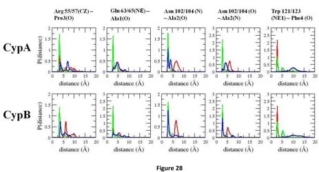
Figure 28... 98
1 INTRODUCTION
Our research group studies the dynamics and reaction mechanisms of several types of
mole-cules and biomolemole-cules using theoretical biophysical chemistry, including DNA, ribozymes, enzymes, and
smaller medicinal compounds. The focus of my graduate work is understanding enzyme function,
name-ly, the enzyme cyclophilin A (CypA). In the same way that biology is the study of life, chemistry is the
study of matter, and physics is the study of the motion of matter through space and time. At the
cross-roads of chemistry and biology was birthed the field of biochemistry in the 20th century, which began to
give insight into the chemical reactions that occur within living organisms – most importantly the human
body and the pathogens that can invade it. The crossroads of chemistry and physics provided us with the
knowledge of how heat, work, organization, molecular interactions, and molecular motions affect the
energy required for chemical reactions to occur. Scientists began to learn how to quantitatively and
qualitatively express chemical reactions of interest; however, these quantitative descriptions are often
tedious and required multiple calculations for manual proof or demonstration of concept.
The invention of computers in the mid 20th century was the first transition from manual to
au-tomated calculations. The computer became the fundamental tool for molecular dynamics (MD), and
relieved much of the labor required for these mathematical proofs. The pioneers of molecular dynamics
began to publish results in the late 20th century, modeling ideal and real molecular interactions.(1) The
first molecular dynamics simulation of a protein was successfully implemented into the literature in
1977,(2) revealing the plasticity and dynamical nature of biomolecules, the molecules that sustain our
life. As computer memory and speed began to increase over time, theoretical scientists began to study
more complex systems of interest such as DNA and proteins. While experimental in vitro techniques
provided a way to study biomolecules on a macroscopic scale, theoretical in silico techniques such as
The development of theoretical science also provided a way for scientists to validate their
pre-dictions in alignment with experimental results. The theoretical approaches yield an idealized, modeled
environment while the environment of experimental approaches is provided by nature. Success in
ex-perimental endeavors requires extensive knowledge of scientific techniques as well as their potential
errors and safety hazards. Although errors can be minimized, they cannot be completely avoided; even
our equipment has inherent error that reveal how precise our measurements are. The amount of
con-trol that we introduce into theoretical methods by algorithm design is based on our prior knowledge of
chemical behavior and is often times compared to the results we obtain from experimental procedures.
Moreover, scientists began to study dynamics of chemical and biochemical systems at timescales
previ-ously inaccessible by experimental techniques, down to the femtosecond. Being able to study dynamics
on significantly faster timescales unfolds aspects of reaction mechanisms that could not be effectively
2 INTRICATE COUPLING BETWEEN CYCLOPHILIN A DYNAMICS AND SUBSTRATE TURNOVER
The importance of enzymes in biology cannot be overstated. Enzymes are critical to a broad
range of functions, including metabolism(3), gene regulation(4), cell survival(5), intracellular
communi-cation(6), and hormone regulation(7). They catalyze specific biochemical reactions, increasing reaction
rates by many orders of magnitude to more biologically relevant timescales. Enzymes can act to form or
break covalent bonds, perform acid-base chemistry, transfer functional groups, and switch
configura-tions around bonds to yield isomers(8-10). Certain enzymes can perform these functions alone, while
others need cofactors or prosthetic groups to assist in the catalytic function. Fully understanding the
mechanism of action of enzymes could provide valuable insights into engineering proteins and designing
new drugs.
In vitro experiments have provided valuable insights into the mechanisms of enzymes(11).
How-ever, detailed atomistic understanding of the mechanism along the catalytic pathway is not always
pos-sible with current experimental techniques. Therefore, computational simulations are routinely used to
complement experiments(12), usually starting from well-characterized atomic x-ray crystal structures.
Nonetheless, classical MD presents several challenges in studying enzyme mechanisms with catalytic
turnover times in the millisecond timescale. In addition to the sub-microsecond timescale limitation, MD
cannot be used to study chemical reactions involving bond formation or breakage without the use of
more demanding hybrid quantum mechanical methods. As of this writing, it is not believed possible to
directly simulate most enzymatic reactions without using some form of course-graining or advanced
sampling techniques.
Peptidyl-prolyl cis-trans isomerases (PPIases) are a class of enzymes that take part in many
cellu-lar processes and catalyze their reactions without any bond formation or breakage. This characteristic
makes them tractable and ideal to study using classical MD. PPIases catalyze cis-trans isomerization of
sub-strate along the reaction coordinate are well defined at ω-bond angle values of 0˚, +180˚, and 90˚,
re-spectively. Also, the cis-trans interconversion can be simulated directly using accelerated MD, without
any conformation bias, as was previously shown(13, 14). PPIases consist of cyclophilins, FK-506-binding
proteins, and parvulins(15). The tertiary structure of the catalytic domain of cyclophilins is structurally
conserved among all of the familial isoforms(16). Human cyclophilin A (CypA), this smallest prototypic
cyclophilin of ~18 human isoforms, is the most studied and characterized isoform(17). Uncatalyzed
prolyl cis-trans isomerization has an activation free-energy barrier of ~20 kcal/mol (~84 kJ/mol)(18) and
a half-life on the second timescale(19, 20). Human CypA speeds up the reaction rate from seconds to
milliseconds(21). CypA catalyzes the peptide bond of a -X-Pro- motif (where X is any amino acid), and
differences in catalytic turnover rates are mainly due to the identity of the amino acid in the X
position(21, 22).
Human CypA has a range of specific functions in vivo. The immunosuppressive drug
cyclospor-ine A (CsA) binds to CypA, and the CypA-CsA complex inhibits calccyclospor-ineurin, suppressing the transcription
of cytokine genes by inhibiting calcineurin’s native phosphatase function(15). CypA is the first human
protein that has been found to be both enclosed within the HIV-1 virion and crucial for viral
replication(23, 24). An interaction between CypA and the HIV-1 capsid core protein, CAN, facilitates viral
replication by accelerating destruction of the capsid(24, 25). Hepatitis C virus also uses CypA to replicate
by forming a critical contact with the HCV NS5B RNA polymerase(26). A role of CypA in signal
transduc-tion involves regulating the functransduc-tion of the prolactin receptor in mammary cells, impacting the
interac-tion of the prolactin receptor with Janus-activated tyrosine kinase(27). Also, CypA can form a complex
with interleukin-2-tyrosine kinase inside of Jurkat T-cells, which is disrupted upon addition of CsA(28).
Cyclophilins are also involved in protein folding (15)and oncogenesis(27, 29).
The exact catalytic mechanism of CypA is not fully understood. Several hypotheses have been
enzyme(27, 30-35). It has been suggested that conformational heterogeneity that occurs during enzyme
catalysis provides the means by which an enzyme complements its substrate(36). Therefore, in order to
fully understand the mechanism of CypA, there is a need to fully understand how enzymes make use of
a large ensemble of conformations in recognition and catalysis at different points along the chemical
step. We have therefore simulated the substrate-free enzyme and enzyme-substrate complexes of the
cis, trans, and transition state configurations of the substrate – three important segments along the
cat-alytic pathway. We have also carried out accelerated MD simulations on the enzyme-substrate complex
in order to freely sample cis-trans isomerization during catalysis and investigate the coupling between
the conformational dynamics of CypA and the chemical step. Altogether, we carried out >2 μs of MD
simulations in full atomistic detail, sampling conformational changes beyond the nanosecond timescale.
Moreover, these computational approaches provide a way to study the short-lived ensemble of
confor-mations of the enzyme-substrate transition state complex. These studies provide further insight into the
importance of enzyme flexibility in catalysis, as well as the coupling between the chemical step and the
stabilizing polar and nonpolar intermolecular interactions.
2.1 Methods
All simulations were carried out using the AMBER 10 suite of programs(37) in explicit TIP3P
wa-ter(38) using the PARM99SB(39) modified version of the force-field parameters from Cornell et al. (40).
Additional modifications to the dihedral parameters for the peptide ω-bond angle were also employed
(41). A 1.58 Å resolution x-ray crystal structure with PDB:1AWR was used for the simulations (25). An
experimentally well-studied substrate analog, Ace-Ala-Ala-Pro-Phe-Nme (AAPF), was used in these
stud-ies (21, 22, 33, 34, 42-45). The Ace-AAPF-Nme substrate analog was introduced by keeping common
backbone and side-chain atoms of the substrate analog (HAGPIA) in the PDB file and adding the missing
atoms using the xleap module in AMBER. The complex was then solvated with ~5500 TIP3P water
mini-mized for 1000 steps with a harmonic constraint of 100 kcal/mol/Å2 applied to the atoms of the protein,
followed by two short (400-ps) MD simulations with harmonic constraints of 50 kcal/mol/Å2and 25
kcal/mol/Å2, respectively, applied to all of the atoms of the protein. The system was then equilibrated
for an additional 200 ps without any constraints using the isothermal-isobaric ensemble at 300 K and 1
bar. The pmemd module in AMBER 10 was used to carry out all of the conventional MD simulations. An
integration time step of 0.002 ps was used to integrate Newton’s equation of motion.
The SHAKE algorithm (46) was used to restrain all bonds involving a hydrogen atom during the
simulations. Langevin dynamics was used to maintain the temperature at 300 K with a collision
frequen-cy of 1 ps-1. This temperature and a constant pressure of 1 bar were used throughout all simulations.
The long-range electrostatic interactions were treated using particle-mesh Ewald summation (47-49).
The ω-bond angle of the substrate in the transition state complex was maintained at ~90˚ using a flat
bottom-well torsional restraint with a force constant of 1000 kcal/mol/Å2 between 89˚ and 91˚.
Re-straints were not required to maintain the substrate in the trans and cis configurations in their
enzyme-substrate complexes, because a high barrier separates the two low-energy states. The enzyme-substrate in the
crystal structure was in the trans configuration. Thus, the cis configuration of the substrate was
equili-brated with the same torsional restraint in order to shift the substrate from the trans configuration to
the cis, and subsequently simulated with no restraint.
The aggregate simulation time for all of the conventional MD simulations of substrate-free CypA
and the enzyme-substrate complexes exceeded 1.5 μs. Four independent simulations were carried out
for each enzyme-substrate complex. Each simulation was carried out for at least 110 ns. The first 10
na-noseconds were discarded as part of the equilibration phase. One long 350-ns simulation was carried
out on substrate-free CypA, using a similar setup and equilibration procedure as was done for the
All accelerated MD simulations (50) were carried out using a modified version of the pmemd
module in AMBER 10, in order to accelerate the rate of cis-trans isomerization of the substrate while in
complex with the enzyme. Eight independent accelerated MD simulations were carried out for a total of
~1 μs of simulation time. The total torsional potential of the substrate was selectively boosted(13), using
a boost energy, E, of 60 kcal/mol above the average total dihedral energy calculated after equilibration
and a tuning parameter, α, of 10 kcal/mol. Each configuration was reweighted using the strength of the
Boltzmann factor of the bias potential energy, eβΔV(r), calculated on-the-fly during the simulation to
cal-culate the probability distributions(51).
Principal component analysis (PCA) was carried out using the ptraj module in AMBER (37). The
implementation of this method has been extensively discussed (52-56), and ptraj was used to calculate
and diagonalize the covariance matrix. The ptraj module was also used to calculate the torsional angles,
root-mean-square fluctuations, and hydrogen-bonding distances. For residues containing equivalent
δ-carbons (such as Leu or Phe), the Cδ1 atom was selected when measuring the torsional angle. The
bind-ing free energies were estimated usbind-ing the Molecular Mechanics/Poisson-Boltzmann Surface Area
ap-proach (57, 58). The relative changes in translational, rotational, and conformational entropies were
assumed to be negligible in estimating the relative binding free energies.
2.2 Conformational selection in CypA recognition during catalysis
In general, conformational selection (59-62) and induced fit(63) can be used to describe the
mechanism of enzyme-substrate recognition. Conformational selection implies that equilibria between
weak- and tight-binding conformations of the substrate-free enzyme exist before substrate binding,
whereas substrate binding is a prerequisite for the formation of a tightly bound enzyme-substrate
com-plex in the induced-fit mechanism(64). However, these two mechanisms are limiting extremes for
dy-namical systems, and it is difficult to ascribe either one as the sole contributor to biomolecular
regard given to conformational changes in the substrate. It has been previously noted that the
free-energy landscape of the enzyme and substrate are transformed upon complex formation(61). Thus,
var-iation in the conformation of the substrate should also be considered when describing the
conforma-tional heterogeneity of the complex because the conformations of the enzyme can affect those of the
substrate, and vice versa.
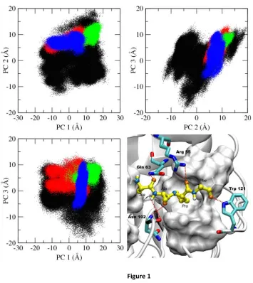
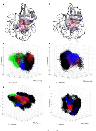
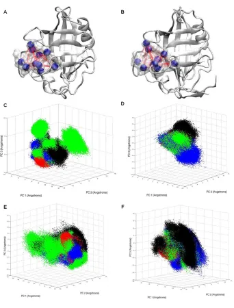
We have used principal component analysis (PCA) to characterize the mechanism of recognition
of CypA upon binding the cis, trans, and transition state configurations of the substrate. PCA allows us to
project the conformational phase space sampled by the active site residues of the substrate-free and
substrate-bound enzyme using a reduced set of degrees of freedom. It helps us to determine if the
con-formations of the active site residues of the substrate-free enzyme can effectively bind the substrate by
revealing similarities between substrate-free and substrate-bound ensembles of enzyme conformations
(Fig. 1). Active site residues of CypA consist of Arg 55, Phe 60, Met 61, Gln 63, Ala 101, Asn 102, Ala 103,
Phe 113, Leu 122, and His 126. These residues were selected because they are no more than 4 Å away
from the peptide ω-bond angle and form the binding cavity for the substrate. Also, most of these
resi-dues are fully conserved across species and have been identified as participating in substrate turnover
(66).
Fig. 1 shows that the active site residues of substrate-free CypA sample a large conformational
space involving several rotameric states (Fig. 2 and Fig. A1). Upon binding the substrate, the active site
residues of CypA lose a tremendous amount of conformational freedom that was present in the
sub-strate-free enzyme. The phase space sampled by the active site residues of the enzyme-substrate
com-plexes in the cis and trans configurations (ground states) of the substrate is slightly broader than that of
Figure 1
Figure 1. Principal component analysis (PCA) of free CypA and CypA enzyme-substrate complexes.
The top three dominant motions of the ten active site residues are shown, with each data point representing a different conformation of the active site. The free enzyme is depicted in black, and trans complex is depicted in red, the transition state complex is depicted in green, and the cis complex is depicted in blue. The
with few differences in the enzyme-substrate intermolecular interactions from one conformation to the
other. These enzyme-substrate intermolecular interactions involve several key hydrogen bonds, as
shown in Fig. 1D. These well-optimized hydrogen bonds localize the transition state ensemble of the
enzyme conformations to a single region of phase space, as we show later. It can be seen that the active
site of the substrate-free enzyme also samples the majority of the transition state conformations (Fig.
1).
The active site of the enzyme does not necessarily have to be induced to some exclusive
con-formations for catalysis to occur. The active site concon-formations of the ground-state complexes overlap
quite well with each other, while the transition state shares a smaller subset of conformations with the
ground states. The substrate-bound active site conformations of the enzyme are subsets of that of the
substrate-free enzyme conformations based on the top three principal components (Fig. 1 and Fig.
A2)that represent ~70% of the motions of the top 10 principal components of the active site residues
(see Fig. A3). The connection between the substrate-free and substrate-bound active site conformations
is indicative of an existing equilibrium between weak- and tight-binding conformations of the enzyme.
Localization of the active site residues of the bound ensembles is characteristic of a population shift
to-ward a subset of the substrate-free enzyme conformations. The results suggest that CypA has evolved to
complementarily shift its active site conformation alongside the configuration of the substrate with only
slight changes to the rotameric state of the active site residues as the reaction progresses. Whether
CypA binds the substrate in the trans, transition state, or cis configuration, the needed enzyme
confor-mations already exist in the ensemble of the substrate-free enzyme. Thus, the binding mechanism of
CypA is predominantly conformational selection, as was previously suggested by NMR studies (42, 67).
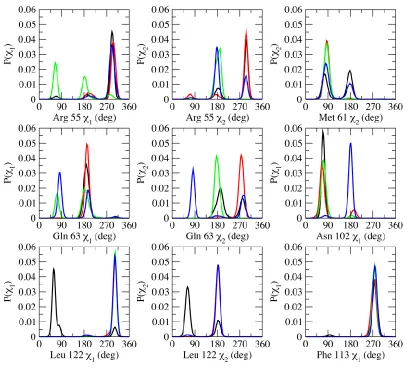
Examining the rotameric states of the active site residues in the substrate-free and the different
substrate configurations of the substrate-bound CypA suggests similar conclusions (Fig. 2). In general,
residues. Various intramolecular and intermolecular interactions in the enzyme affect the
conforma-tional preference of the active site residues, which are also dependent on the state of the substrate. The
rotamers sampled by the active site residues of the substrate-free enzyme overlap with the rotamers
sampled by the substrate-bound enzyme complexes, also demonstrating that the bound conformations
of the enzyme are a subset of the free enzyme conformations. Active site residues that form
hydrogen-bonding interactions with the substrate tend to sample more rotameric states than hydrophobic
resi-dues. Unlike the hydrophilic residues, the hydrophobic residues in the bound states of the enzyme
pre-dominantly sample a single rotameric state and are less sensitive to the configuration of the substrate.
The most flexible active site residues are Arg 55, Gln 63, and Asn 102 – three residues that form
key hydrogen-bonding interactions with the substrate (Fig. 1D). Arg 55 has been shown to be important
for substrate recognition and catalysis by forming a bifurcated hydrogen bond with the carbonyl oxygen
of proline in the substrate (13, 33, 35). Gln 63 and Asn 102 also participate in hydrogen-bonding
interac-tions with the substrate. However, only the hydrogen-bonding interacinterac-tions between residues Arg 55 and
Asn 102 and the substrate have been identified as being important in stabilizing the transition state(33).
The backbone amine group of Asn 102 forms a hydrogen bond with the carbonyl oxygen of Ala in the
-Ala-Pro- motif only in the transition state and cis enzyme-substrate complexes. The side chain amide
group of Gln 63 forms a hydrogen bond with the carbonyl oxygen of the Ala residue preceding the
-Ala-Pro- motif of the Ace-Ala-Ala--Ala-Pro-Phe-Nme substrate analog used in this study.
Phe 113 sits at the base of the proline-binding pocket and has been suggested to play a key role
in catalyzing cis-trans isomerization (42). Phe 113 can rotate in the free enzyme, and rotation to the
mi-nor rotameric state was suggested to be coupled to the catalytic step (42). Our studies suggest that,
once the substrate binds to the enzyme, the side chain of Pro in the substrate pushes directly against
Figure 2
Figure 2. Probability distributions of backbone torsional angles of some active site residues in free and bound CypA.
conformation of the substrate (Fig. 2. and see Fig. A1). The minor rotameric state of Phe 113 seems to
obstruct the proline-binding pocket. Interestingly, the less active Ser99Thr mutation of CypA was shown
to increase the population of the minor rotameric state of Phe 113, which sits on top of Ser 99 (42).
An-other hydrophobic residue, Leu 122, in the active site forms intermolecular contact with the substrate in
the proline-binding pocket and predominantly samples only one rotameric state in all of the
substrate-bound enzyme complexes (Fig. 2). In the substrate-free enzyme, Leu 122 participates in loose
hydphobic contacts with other active site residues in the proline-binding pocket, visiting more than one
ro-tameric state.
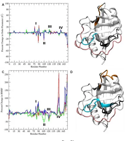
2.3 Substrate binding alters the conformational dynamics of CypA beyond the active site
In addition to the conformational changes observed in the active site of CypA upon substrate
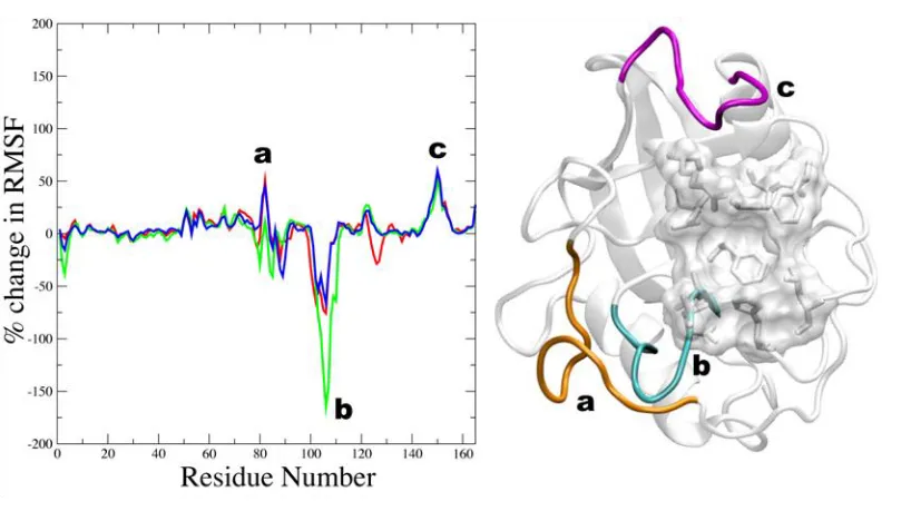
binding, the dynamics and fluctuations of the enzyme beyond the active site are also altered, as shown
in Fig. 3. Here, Fig. 3 shows the average percent-change in the root-mean-square fluctuations of the
en-zyme backbone atoms upon binding the substrate. Enen-zyme residues that become more localized upon
binding have positive change; conversely, residues that become more flexible upon binding have
nega-tive change. The loop region containing residues 75-85 becomes overall less flexible upon binding the
substrate in the cis and trans ground states (orange; Fig. 3). This region is part of a larger loop (residues
66-96) that has been reported to undergo fast conformational exchange in the substrate-free enzyme
(42). It appears that Lys 82 acts as a hinge for this loop, becoming more flexible upon binding the
sub-strate in the transition state configuration. This residue has also been found to undergo significant
devi-ation upon binding in other studies (31, 68). These results suggest that the motions of this loop have
significant impact on complex formation. Several residues of CypA within the loop region 75-85 are well
conserved across species, with the exception of residues Lys 76, Glu 81, and Glu 84 (66). Another loop
Figure 3
Figure 3. Average percent change in root mean square fluctuations upon substrate binding of CypA backbone atoms.
gion, in combination with the α-helix from residues 136-146, acts as a hinge region to the two β-sheets
contributing to the closed β-barrel fold. The stability of this loop region upon substrate binding suggests
that the two β-sheets become more compact in the enzyme-substrate complex and less likely to
sepa-rate as much as would be expected in the breathing motions of the substsepa-rate-free enzyme.
Of particular interest is the loop region consisting of residues 101-110 (cyan; Fig. 3). This region
becomes more flexible upon substrate binding and most flexible in the transition state
enzyme-substrate complex. Moreover, these residues are well conserved across different species (66) and have
been shown to contribute to the dominant motions of CypA (69). The motion of this loop may be critical
to catalytic turnover, and this motion may be required to introduce enough deviation to allow Asn 102
to form hydrogen bonds with the substrate. This hypothesis may be tested experimentally by modifying
the dynamics and flexibility of that loop to determine how the catalytic turnover rate is impacted. Very
few deviations are apparent in the backbone of the active site residues upon binding the substrate in
the different configurations, as opposed to the relatively large side-chain rearrangements that are
ob-served. The results therefore suggest that the backbone of the active site in the substrate-free enzyme is
pre-organized to bind the substrate in its different configurations that requires rearrangements of
sev-eral key side chains during catalysis. Also, many of these loop residues have been shown to contribute to
the top three vibrational modes of CypA (66, 68).
2.4 Intermolecular interactions are tightly coupled to the chemical step
It is well established that enzymes lower the free energy barrier of a reaction by stabilizing the
transition state during catalysis (10). However, it is not always clear as to how exactly this barrier
reduc-tion is achieved. In the active site of CypA, the conformareduc-tions that complement the transireduc-tion state are
very ordered, resulting in a low entropy, yet high affinity complex. This order is achieved due to key
en-zyme-substrate in intermolecular interactions. The key intermolecular interactions of CypA include the
spe-cific enzyme-substrate hydrogen bonds (Fig. 1D). The smaller conformational space sampled by the
ac-tive site residues of the substrate-bound complexes (as compared to the substrate-free enzyme) is a
result of the formation of these intermolecular interactions.
Initially, two main hydrogen bonds involving Arg 55 and Asn 102 of CypA were identified as
be-ing responsible for stabilizbe-ing the transition state relative to the ground state (13). These two hydrogen
bonds flank the proline residue of the -Ala-Pro- motif of the substrate analog. In this work, we have
identified at least four intermolecular hydrogen bonds between the enzyme and substrate that are
deemed to be important in stabilizing the transition state (Figs. 1D and 4). The bifurcated hydrogen
bond between the guanidinium group of Arg 55 and the carbonyl oxygen of proline in the -Ala-Pro- motif
of the substratein the -Ala-Pro- continually forms and breaks in the ground (cis and trans) states.
How-ever, this hydrogen bond is always well formed in the transition state complex and is almost never
bro-ken (Fig. 4 and see Fig. A4). While Arg 55 can undergo fast conformational changes in the free enzyme
(31), it becomes less mobile in the enzyme-substrate complexes, especially upon binding the transition
state.
Similarly, Gln 63 and Asn 102 are involved in several hydrogen-bonding interactions with the
substrate that are loosely formed in the cis and trans states, but well formed in the transition state. The
side-chain amide proton of Gln 63 forms a hydrogen bond with the carbonyl oxygen of the Ala residue
preceding the -Ala-Pro- motif of the substrate (Fig. 1D). This hydrogen-bonding interaction is difficult to
form in the trans and cis states. However, in the transition state, it is difficult to break (Fig. 4 and see Fig.
A4). The backbone amine group of Asn 102 forms a tight hydrogen-bonding interaction with the
carbon-yl oxygen of alanine in the -Ala-Pro- motif of the substrate in the cis and transition states, but not in the
trans state. We believe this tightly formed hydrogen-bond interaction between Asn 102 and the cis
Figure 4
Figure 4. Enzyme-substrate intermolecular hydrogen bonding interactions contributing to the transition state stabilization of CypA.
configuration to the enzyme, as shown below and previously observed computationally (13) and
exper-imentally (31, 67).
In the substrate-free enzyme, Arg 55 can move freely. In the enzyme-substrate complexes of the
cis and trans states, Arg 55 can either orient downward, interacting with the substrate; or upward (away
from the active site), interacting mainly with residue Asn 149 in a loop (violet; Fig. 3). In one out of the
four independent normal MD simulations carried out on the enzyme-substrate complex when the
sub-strate is in the trans state, Arg 55 is consistently in the downward position, interacting with the
sub-strate. In the other three simulations, Arg 55 spends most of the time upward, away from the active site
and substrate. In all of the independent normal MD simulations of the enzyme-substrate complex when
the substrate is in the cis state, Arg 55 sampled both downward and upward conformations. The
interac-tion between Arg 55 and the substrate in the transiinterac-tion state complex was hardly ever broken in all four
independent normal MD simulations. The results suggest that the behavior of Arg 55 is sensitive to the
state of the substrate and changes along the catalytic pathway. To fully understand the conformational
preference of Arg 55, and other key residues, and determine how the intermolecular interactions are
coupled to the state of substrate during catalysis, we carried out accelerated MD simulations on the
en-zyme-substrate complex. Accelerated MD allowed us to observe the back-and-forth cis-trans
isomeriza-tion of the catalytic process while sampling the conformaisomeriza-tional space of the enzyme.
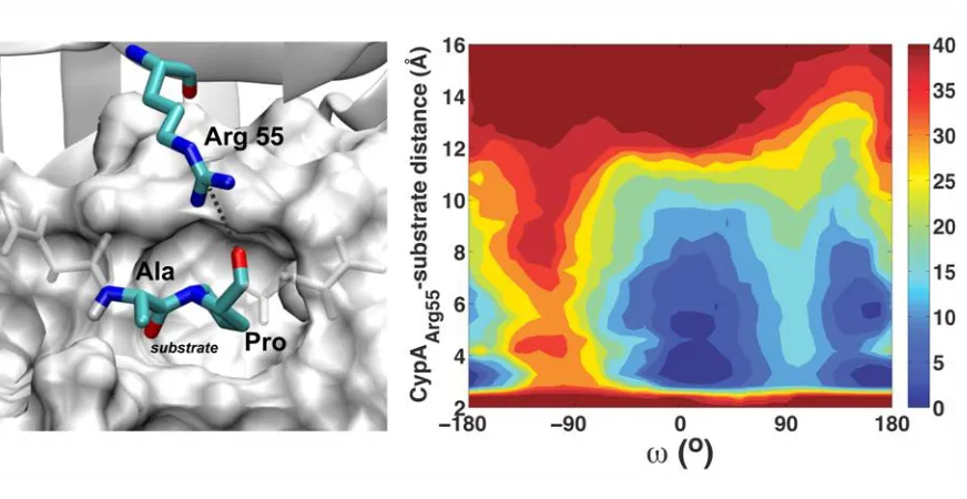
The hydrogen-bonding interaction between Arg 55 and the substrate is coupled to the chemical
step along the reaction coordinate, ω, as can be seen in Fig. 5. Also, it is clear from Fig. 5 that, in the
trans state, Arg 55 has to overcome an energetic barrier of ~5 kcal/mol in order to switch between the
upward and downward conformers, unlike the cis configuration. This barrier is small in the cis
enzyme-substrate complex, and Arg 55 in the transition state complex consistently stays in the downward
posi-tion due to optimized contact with the substrate. These results are consistent in all eight, independent,
Figure 5
Figure 5. Coupling between the intermolecular hydrogen bonding interaction of Arg 55 and the reaction coordi-nate (ω).
The barrier observed in the trans state also explains why the hydrogen-bonding interaction between Arg
55 and the substrate in one of the four independent normal MD simulations of the trans complex stayed
formed during the entire simulation. Our results suggest that the barrier separating the formed and
un-formed state of Arg 55 with the substrate in the trans enzyme-substrate complex exists because Arg 55
can equally form long-lasting interactions downward (with the substrate) and upward (with Asn 149),
corresponding to the two observed wells in Fig. 5. Because there is a barrier separating the formed and
the unformed states of the intermolecular interaction between Arg 55 and the trans substrate, these
simulations can get trapped in the formed or unformed well for a long period of time. It is interesting to
see that the configuration of the substrate can directly affect the enzyme dynamics. Fig. 5 also confirms
that the hydrogen-bonding interaction between the guanidinium group of Arg 55 and the carbonyl of
proline in the -Ala-Pro- motif of the substrate can form and break in the trans and cis states and is well
formed as the substrate goes through the transition state.
The hydrogen-bonding interaction between Gln 63 and the substrate is also coupled to the
chemical step, as shown in Fig. 6. The hydrogen bond between Gln 63 and the substrate readily forms
and breaks when the substrate is in the trans or cis states, and stays consistently formed in the
enzyme-substrate complex of the transition state (Fig. 6). Gln 63 is completely conserved across the human
cy-clophilin isoforms (17) and across species (33, 70), and our results suggest that it is primarily important
for stabilizing the transition state of the substrate, along with Arg 55 and Asn 102, two other
well-conserved amino acids of CypA (17, 71, 72). The suggested role of Gln 63 in stabilizing the transition
state can be tested by mutagenesis experiments or chemical modification of the side chain in order to
abolish the hydrogen-bonding interaction with the substrate and measurements of the effect on the
Figure 6
Figure 6. Coupling between the intermolecular hydrogen bonding interaction of Gln 63 and the reaction coordi-nate (ω).
2.5 CypA preferentially binds the substrate in the transition state
We have estimated the binding free energies of the enzyme-substrate complexes with different
configurations of the substrate using the Molecular Mechanics/Poisson-Boltzmann Surface Area
ap-proach(57, 58), in order to further understand how CypA speeds up the rate of cis-trans isomerization
(Fig. 7). The relative changes in conformational, translational, and rotational entropies are not included
in these estimates and are assumed to be negligible. The average free energies of binding the substrate
in the trans configuration are ~–13.1 kcal/mol (~–54.8 kJ/mol) in the simulation where Arg 55 does not
interact with the substrate and ~–22.1 kcal/mol (~–92.5 kJ/mol) in the simulation where Arg 55
continu-ously interacts with the substrate, as shown in Fig. 7A. The average free energies of binding the
sub-strate in the cis and transition state configurations are estimated to be ~–24.1 kcal/mol (~–100.8 kJ/mol)
and ~–31.4 kcal/mol(~–131.4 kJ/mol), respectively. The enzyme binds the transition state better than
the cis and trans states, as was previously shown (13, 73). These results suggest that CypA is designed to
preferentially bind and stabilize the transition state of the substrate. CypA lowers the free-energy
barri-er by ~10 kcal/mol (~42 kJ/mol), similar to estimates from Figs. 5 and 6 and in line with previous
simula-tions and experiments (13, 34, 67), which is achieved by binding the transition state configuration of the
substrate better than the cis and trans states by thisamount. Also, our results provide a quantitative
es-timate of the critical role of the interaction of Arg 55 with the substrate in forming a stable
enzyme-substrate complex.
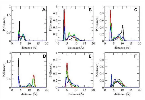
A breakdown of the energetic components (Fig. 7, B-E) reveals that the enzyme stabilizes the
transition state configuration using optimized van der Waals and electrostatic interactions. The trans
substrate forms slightly better van der Waals contact with the enzyme than the cis substrate, but the
trans substrate forms the worst electrostatic contact with the enzyme out of the three configurations.
Interestingly, when Arg 55 forms its hydrogen bond with the substrate, the electrostatic contacts
than the trans complex, yet the cis complex forms the weakest van der Waals interactions. This implies
that the cis complex formation is characterized by better hydrogen-bond formation, while the trans
complex formation is characterized by a better grip on the proline ring. The difference in electrostatic
contacts between the ground states is much greater than the difference in nonpolar contacts. This
indi-cates that electrostatic contributions have the biggest impact on complex formation. The importance of
electrostatic contacts in complex formation and catalytic turnover has been previously highlighted(74).
Interestingly, and somewhat expected, the tighter the electrostatic interaction between the enzyme and
the substrate (Fig. 7C), the less favorable the change in the polar free energy of solvation (Fig. 7E).
How-ever, the electrostatic and van der Waals interactions overcompensate for the unfavorable change in
the polar solvation free energy. The change in the nonpolar solvation free energy is relatively small (Fig.
7D).
2.6 On the role of Trp 121
Tryptophan 121 forms a hydrogen bond with the substrate, but does not help to carve out the
hydrophobic proline-binding pocket. Therefore, it had not always been considered an active site residue,
but mutating Trp 121 has been shown to adversely affect the catalytic activity of CypA (44, 75, 76). The
amine group of the indole ring can form a hydrogen bond with the backbone carbonyl oxygen of the
res-idue (Phe) after proline of the -Ala-Pro- motif of the substrate (Fig. 8C). The side chain of Trp 121
pre-dominantly populates a single rotameric state, mainly forming hydrophobic contact with the outside of
the proline-binding pocket, and the hydrogen of the indole ring points toward the substrate (Fig. 8, A-C).
Unlike the other residues that form hydrogen-bonds with the substrate, the hydrogen-bonding
interac-tion between Trp 121 and the substrate is well formed in the trans enzyme-substrate complex and
mostly formed in that of the transition state, but completely unformed in the complex of the cis state
Figure 7
Figure 7. Probability distributions of free energy of CypA binding the substrate in different configurations.
the configuration of the substrate and not on the dynamics of the residue. A fluorescence study of
sub-strate-free CypA and CypA-CsA complex resulted in a twofold increase in Trp 121 fluorescence upon
binding CsA, suggesting that Trp becomes more localized upon substrate binding (77). Therefore, our
results suggest that Trp 121 may be critical for recognizing the substrate in the trans configuration and
help stabilize the transition state.
Trp 121 is moderately conserved across human cyclophilin isoforms; however, it is well
con-served across orthologous CypA species(17, 70, 71). Interestingly, human cyclophilin isoforms without
tryptophan at this position are normally substituted with a histidine or tyrosine, residues that can also
form hydrogen bonds with their side chains(17). A Trp121Phe mutation, which abolishes this
hydrogen-bonding interaction, causes CypA to bind cyclosporine with much lower affinity, resulting in
CsA-resistant strains of Saccharomyces cerevisiae(75). The Trp121Ala(44) and Trp121Tyr(76) mutations
re-tain 9% and 19% of wild-type catalytic activity, respectively; to the best of our knowledge, there is no
experimental data for the catalytic activity of the human Trp121His mutant.
2.7 Copyright Notice
This work is published in Biophysical Journal, 2013, 104(1): 216-226.
(http://dx.doi.org/10.1016/j.bpj.2012.11.3815) This work is supported in part by the National Science
Foundation CAREER grant (No. MCB-0953061), the Georgia Cancer Coalition of the Georgia Research
Alliance, and the Molecular Basis of Disease program at Georgia State University. L.C.M. is a Molecular
Basis of Disease fellow. This work was also supported by Georgia State’s IBM System p7 supercomputer,
acquired through a partnership of the Southeastern Universities Research Association and IBM
Figure 8
Figure 8. Backbone torsional angle distributions of Trp 121 and intermolecular hydrogen-bonding interaction of Trp 121 with the substrate.
3 THE COMPLEX ROLE OF ENZYME CONFORMATIONAL DYNAMICS IN CATALYTIC FUNCTION
Enzymes accelerate reaction rates by several orders of magnitude, allowing them to occur at
timescales relevant for cellular functions(10). One of the long-standing issues in biochemistry is how
en-zymes achieve this remarkable speedup. It is commonly accepted that the most dominating effect arises
from significant reduction in the free energy barrier compared to the corresponding noncatalyzed
reac-tion in solureac-tion. It is also well established that this predominant effect is mainly electrostatic in nature
(74, 78), which is more favorable for the transition state than the reactant or the product (10). However,
to what degree and how other factors such as desolvation, steric strain, and enzyme dynamics
contrib-ute to catalysis remains disputable. Of particular interest is the role of enzyme dynamics in catalysis that
has stirred considerable debate(32, 70, 79-84) partly because it has not been clearly defined, leading to
a semantic issue. Also, the link between enzyme dynamics and catalysis is difficult to address both
ex-perimentally and theoretically. Currently, the implications of enzyme dynamics are from ensemble- and
time-averaged experiments, as the temporal behavior of every atom cannot be observed directly.
Alt-hough standard MD simulations can provide an atomistic picture of enzyme dynamics, they are still not
amenable to study catalytic reactions that usually occur in milliseconds. Computational approaches that
have investigated the effects of millisecond-timescale enzyme dynamics on the chemical reaction have
been possible only with the use of coarse-grained models (79, 83). NMR relaxation dispersion
experi-ments that can probe microsecond-millisecond timescale motions have detected backbone and
side-chain motions in and around the active site that occur on the same millisecond timescale as the
chemi-cal step (85). It has been further shown that such slow motions are already present in the free enzyme
(31). Furthermore, loss of conformational fluctuations occurring in milliseconds in the active site of
mu-tant enzyme has been observed with concomimu-tant reduction in activity (84). Single molecule studies on
enzymes have also revealed that catalytic rates can fluctuate over five orders of magnitude – from
(86).These observations are not surprising given that protein dynamics comprise motions that span
mul-tiple timescales and occur in either a more localized or collective manner (87, 88). Nevertheless, protein
dynamics has been suggested to directly contribute to catalytic function and rate enhancement. The
exact nature of this dynamical contribution cannot be understood, unless specific questions regarding
whether dynamical motions of enzymes help in lowering the activation barrier (i.e., barrier effects) or
aid the substrate to surmount the barrier (i.e., prefactor effects) are addressed.
The energy landscape of proteins is characterized by several energy minima that represent
con-formational substates separated by barriers of varying heights (89, 90). Simultaneous motions of many
degrees of freedom constitute protein dynamics and bring about equilibrium interconversions (88, 91).
We sought to understand the role of enzyme conformational dynamics (ECD) in catalytic functions by
employing a combination of normal MD (nMD) and accelerated MD (aMD) (50) approaches that provide
atomistic detail with extended timescale. We chose to study CypA (Fig. 1A), an extensively studied
pep-tidyl-prolyl cis-trans isomerase, that catalyzes isomerization of the peptide ω-bond preceding proline
residues in proteins. Such system is ideal to study using classical molecular mechanics because no bond
breaking or formation is involved in the catalytic process.
3.1 Probing the influence of CypA dynamics on the chemical step
The uncatalyzed isomerization reaction (RO) is an extremely slow process with an activation
bar-rier of approximately 20 kcal/mol and occurs readily in hundreds of seconds in solution(20, 92).
Cyclo-philins are known to accelerate prolyl isomerization by 105 – 106 times, reducing the timescale to around
milliseconds(67, 93).It is not feasible to simulate even the catalyzed reaction (RC) with nMD, since it is
currently limited to only hundreds of nanoseconds. Therefore, to probe the effects of ECD in catalysis,
we used several lowered torsional energy barriers around the -Ala-Pro- ω-bond of a well-studied
sub-strate analogue, Ace-Ala-Ala-Pro-Phe-Nme (Fig. 9A). We then took advantage of the linearity of the
optimum temperature range and extrapolating to a temperature outside that range. The lower barriers
allowed us to track the dwell times in the trans well before going over the barrier with sufficiently good
statistics. Using nMD we investigated the kinetics of prolyl isomerization in the free and the
enzyme-bound substrate with the same value of the AMBER force field parameter V2 (see section 3.6), which is
the main determinant of torsional barrier. We found that the decay of the survival probability function,
S(t), of dwell times for the reference RO in the free substrate unambiguously exhibited single exponential
behavior (Fig. 9B). Progressively slower kinetics resulted as V2was systematically increased from 0 to 11
kcal/mol. Interestingly, for RC, the resulting kinetics exhibited multi-exponential decays (Fig. 9C).
In view of the notion that protein dynamics is not independent of its environment (91), we
ob-served that peptide isomerization dynamics was influenced by environment, whether it be the
fluctua-tions in the solvent or the active site of the environment. From Fig. 9B and D, it became evident that in
terms of timescale, the dynamics of the aqueous environment was relatively uniform with the solvent
motions occurring on a single or a very narrow timescale. In contrast, enzymatic motions were dispersed
over an extensive timescale and were coupled to substrate dynamics. Consequently, the different
en-zyme dynamic modes became apparent in the isomerization kinetics, yielding multi-exponential
behav-ior (Fig. 1C and E). As the torsional barrier became progressively greater (with the increase in V2), the
distributions of timescales showed the following trend: the relative amplitudes of the faster phases
di-minished and those of the slower phases showed a gradual increase. These results suggested that,
de-pending on the barrier, and hence characteristic timescale, of the chemical step, the reaction dynamics
got coupled to the slightly faster and slower enzyme motions, resulting in multi-exponential decays.
Figure 9
Figure 9. Structure of CypA and the influence of its dynamics on the kinetics of prolyl isomerization in its sub-strate.
step in the enzyme-bound substrate, RC (Table I). It was evident from our results that the chemical step
was coupled to, and would be affected by, the enzyme motions.
3.2 Accelerating CypA Dynamics and its Effects on the Chemical Step
Another question of interest is whether the contribution from ECD can significantly enhance the
rate of the chemical step as compared to that in solution. In answering this question, we subjected only
CypA to increasing levels of aMD with the substrate still simulated with nMD (see section 3.6.3). In order
to confirm that aMD indeed resulted in faster ECD in CypA, we characterized the fluctuations in free
CypA (CO) from independent nMD and aMD (see section 3.6.2 and 3.6.3). Accelerated MD brought about
an increase in not only conformational plasticity; i.e., greater amplitudes of fluctuations as depicted
from the shift of backbone (Fig. 10A and B) and side chain (Fig. B1) order parameters (S2) to lower
val-ues, but also conformational heterogeneity (Fig. B2) at the active site of CO. Similar to our results, aMD
has been shown to successfully increase the rate of conformational sampling, thereby characterizing
millisecond-timescale protein/peptide dynamical motions and achieving notable agreement with
exper-imental data (94-98). Our simulations further confirmed recent experimental observations (31, 86) that
ECD in CypA takes place over a broad range of timescales even in the substrate free state (Fig. B1).
Ac-celerating CypA dynamics clearly affected the kinetics of prolyl isomerization in the bound substrate,
resulting in faster decay of the survival probability (Fig. 10C). The decays fitted to multi-exponential
functions with only three phases as opposed to five phases in the nMD of CypA (Fig. 2D). Since the
en-zyme modes sped up, the relative contribution of the faster phases increased as slower phases became
faster (Fig. 2D). The net result was a gradual speed up in the average lifetimes as the extent of
Figure 10
Figure 10. Effects of accelerating CypA dynamics on prolyl isomerization in the substrate.
Table 1. Free energy barriers, diffusion and time constants for the uncatalyzed and catalyzed prolyl isomerization
RO RC
V2 (kcal/mol) ΔG# (kcal/mol) <τ> (ns) Deff
(deg2 /s)
ΔG# (kcal/mol)
<τ> (ns)
Deff
(deg2 /s)
nMD 0.0 0.81 0.05 18.1 x 10¹⁴ - 0.01 1.37 x 10¹⁴
nMD 4.0 2.3 0.28 18.1 x 10¹⁴ - 0.07 1.37 x 10¹⁴
nMD 5.0 3.16 0.54 18.1 x 10¹⁴ - 0.16 1.37 x 10¹⁴
nMD 7.0 3.93 2.48 18.1 x 10¹⁴ 0.99 0.50 1.37 x 10¹⁴
nMD 9.0 5.57 16.11 18.1 x 10¹⁴ 2.31 1.35 1.37 x 10¹⁴
nMD 11.0 7.42 61.65 18.1 x 10¹⁴ 3.49 6.24 1.37 x 10¹⁴
aMD 28.0 20.0a 1.22 x 10¹¹b 10.74a 2.75 x 10⁵b
aMD I 7.0 2.09 5.443 x 10¹⁴
aMD II 7.0 2.37 13.362 x 10¹⁴
aMD III 7.0 2.7 31.229 x 10¹⁴
ΔG#s were calculated from potentials of mean force obtained from umbrella sampling except for (a) where they were obtained after reweighting free energy profiles resulting from aMD. (b) Time constants extrapolated from linear fits (red and green lines) in figure 11c. AMD levels I, II, and III are the lowest, intermediate, and the highest extents of acceleration (same as in Fig. 10D) subjected only on CypA. Deff
3.3 Using Kramer’s Rate Theory to Explain the Effects of CypA Dynamics
Although the usage of traditional rate theories to explain enzyme kinetics has been a
conten-tious matter (82, 99, 100), our results could be rationalized within the framework of Kramers’
theory(101, 102) in the high friction regime:
where k is the rate of escape from the trans well with curvature ωo over the free energy barrier ΔG#
with curvature ωb, kB is the Boltzmann constant and T is the temperature. Deff is the effective diffusion
coefficient on a one-dimensional free energy profile and assumed to be independent of the reaction
coordinate. Deff incorporates the effects of the environment, as well as those inherent in proteins, for
example, frictional, dynamical effects and energetic roughness. Frictional effects arise from solvent
vis-cosity and internal friction that impede protein motions. Dynamical effects originate from enzyme with
inhomogenous diffusivity, arising from ECD occurring on a wide continuum of timescales, or an aqueous
medium that offers a more homogeneous environment with essentially single (or very narrowly
distrib-uted) diffusion coefficient. The substrate undergoes desolvation while moving into the active site of the
enzyme from aqueous solvent, as a result the energetic roughness may reduce, leading to a less
hin-dered substrate (103). The above Kramers’ relation can be rearranged in the log form, i.e.,
From the plots of vs. ΔG#,V2 (where rate constants, curvatures, and free energy barrier
heights correspond to various values of V2) with a well-defined slope of 1/kBT, the relative contributions
from the pre-exponential factor and the barrier effects were estimated for RO and RC (Fig. 11). Clearly,
to the corresponding RO (Fig. 11A). The speedup was the consequence of two opposing effects: Increase
in rate as a result of the reduction of the free energy barrier, for example, 237 times from a barrier
re-duction of 3.26 kcal/mol in case of V2= 9.0 kcal/mol, which was offset by approximately 2.6 times due to
the modification in the curvatures of the free energy profiles and by approximately 13 times due to the
differences in the Deff in solution and in enzyme-bound substrate, bringing the net rate enhancement to
only about seven times. It can be seen from Fig. 11A that the y-intercept, from which the effective
diffu-sion coefficient can be estimated, is smaller for RC than RO (Table I). Therefore, ECD does not enhance
but rather hinders the rate enhancement. When the dynamics of CypA was accelerated, we clearly
no-ticed an increase in the isomerization rates (Table I). Enzymatic CD does not directly modify the
proper-ties of the free energy landscape, as that scenario would violate Boltzmann statistics. However, for each
level of acceleration subjected on CypA, the solvated CypA-substrate complex should be considered a
distinct system associated with its Hamiltonian and characteristic free energy profile. And indeed, the
curvatures and barrier heights were modified (Table I) when ECD was accelerated. As the levels of
accel-eration were raised, both ΔG# and Deff showed an increase (Fig. 11B) relative to the case in which ECD
was not sped up (i.e., RC). Noticeably, even with the highest level of acceleration, Deff was not
significant-ly faster (i.e., by onsignificant-ly approximatesignificant-ly 1.7 times) than the one in aqueous solution, given the errors
associ-ated with the calculation of quantities from logarithmic scale and MD simulations. Further acceleration
of CypA dynamics could reach a limiting case where the integrity of the active site might be lost as a
re-sult of the unfolding of the enzyme brought about by very fast dynamical motions. Our analysis
there-fore suggested that altering motions associated with ECD made the enzyme’s active site environment
more aqueous-like and directly affected the pre-exponential factor. At the same time, the favorable
in-teractions between CypA and its substrate were possibly perturbed such that the free energy height of
Figure 11
Figure 11. Comparison of prolyl isomerization kinetics in the free and the enzyme-bound substrate.Kramers’ plots are shown in the form of ln (k/ωoωb) vs ΔG#.
In an independent aMD study, setting V2to the reoptimized value of 28 kcal/mol (41), we
calcu-lated the free energy profiles with the expected actual barriers for RO and RC (see section 3.6.3 and Fig.
B4). We estimated the rate constants (Table I) corresponding to the actual RO and RC from their
respec-tive linear fits in Kramers’ plots (Fig. 11C). RC showed a speed up of approximately 4.5 x 10⁵ times over
RO, which was strikingly similar to experimental estimates (67). The above result therefore validated
Kramers’ theory in the analysis of prolyl isomerization kinetics and its catalysis. We would like to note
from Fig. 11C, that in the case of CypA, as one goes to the higher barrier regime, the relative and
domi-nant contribution from the reduction in barrier heights to the rate enhancement will continue to
in-crease while the difference in Deffbetween RO and RCwill remain at approximately 13 times.
Recently, ambient temperature X-ray and relaxation NMR studies on CypA have shown that
im-pediment of motions that help in the interconversion of conformational substates in a mutant enzyme is
accompanied with the reduction in catalytic rate (42). It was therefore concluded that protein dynamic
motions contribute directly to the catalytic power of the enzyme. Such results can now be explained
with Kramers’ theory that allows us to understand the nature of the dynamical contribution. The CypA
mutant with slower dynamics implies that the isomerization reaction would take place on a distinct
free-energy profile with barrier heights and curvatures that are different from the wild-type CypA and with
an effective diffusion coefficient that is perhaps slower than the wild-type enzyme (Fig. 11C). As we
show above, it is equally important to investigate the dynamical effects as well as the free-energy
barri-er effects of the mutant, which, in most cases, is missing from expbarri-erimental analyses. The recent studies
linking enzyme dynamics to catalysis have focused on mutants with either slower or total absence of
fluctuations in the active site as compared to the wild-type enzyme (42, 84). It would be interesting to
investigate mutants with faster dynamics (Fig. 11C) and observe whether the catalytic rates are