C A S E R E P O R T
Open Access
The eye in dengue fever, a rarely
appreciated aspect of dengue expanded
syndrome: a case report
Jevon Yudhishdran
1*, Isurujith Kongala Liyanage
1,2, Mitrakrishnan Rayno Navinan
1, Sandamalee Herath
1,
Danushka Withanage
1, Sivakumar Jeyalakshmy
3and Aruna Kulatunga
1Abstract
Background:Dengue fever is a mosquito-borne illness prevalent mainly in the tropics. It is feared for causing the dengue hemorrhagic spectrum of the disease leading to significant morbidity and mortality. Its rarer manifestations are categorized as the expanded dengue syndrome, and though being recognized, they are not fully appreciated and understood. The involvement of the eye in dengue fever is one such phenomenon.
Case presentation:A 27-year-old South-Asian woman presented on day 2 of dengue fever, without capillary leakage, for further management. Despite developing hepatitis, she had an otherwise uncomplicated
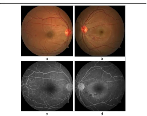
progression of the illness because she did not develop capillary leakage. On day 8 of the illness, she had the lowest platelet count and developed bilateral blurred vision. Examination revealed that only gross movements were detected in the left eye, and the right eye had a visual acuity of 6/9. She was diagnosed with foveolitis in the right eye and central serous chorioretinopathy in the left eye, along with hemorrhages in both eyes. These were confirmed by funduscopy, fluorescein angiography, optical coherence tomography, and macular scans. She received systemic and intravitreal steroids and was assessed regularly. After 6 months of
observation, her visual acuity was 6/6 in the right eye and 6/9 in the left eye, which remained the same thereafter.
Discussion: The exact mechanism of eye involvement in dengue viral infection is poorly understood. Multiple causes have been suspected and include viral factors, immune mediation, capillary leakage, stress, and hemorrhage. Eye involvement is classically seen at the lowest platelet count and when the count begins to rise. Though symptoms are nonpathognomonic, blurring of vision is the commonest complaint, but the range of presentation is extensive and variable. Ophthalmological assessment and funduscopy are very useful in addition to advanced assessments. There is no clear consensus on management; suggestions range from conservative care to aggressive steroid therapy with immune modulation and even ophthalmological intervention. Recovery can be full or partial with a variable time scale. Conclusion:The extensive spectrum of possible visual symptoms should prompt the clinician to suspect any visual complaint as potential dengue eye involvement. Guided studies and screening are needed to better understand the true incidence of eye involvement in dengue fever.
Keywords:Dengue, Dengue fever, Dengue retinopathy, Dengue maculopathy, Expanded dengue syndrome, Foveolitis, Central serous chorioretinopathy
© The Author(s). 2019Open AccessThis article is distributed under the terms of the Creative Commons Attribution 4.0 International License (http://creativecommons.org/licenses/by/4.0/), which permits unrestricted use, distribution, and reproduction in any medium, provided you give appropriate credit to the original author(s) and the source, provide a link to the Creative Commons license, and indicate if changes were made. The Creative Commons Public Domain Dedication waiver (http://creativecommons.org/publicdomain/zero/1.0/) applies to the data made available in this article, unless otherwise stated. * Correspondence:yudhishdran@yahoo.com
1National Hospital of Sri Lanka, Colombo, Sri Lanka
Background
Dengue fever (DF) is a mosquito-borne disease with ex-tensive worldwide reach [1]. Because urbanization pro-motes its spread, it is not surprising that it is found endemically in more than 100 countries globally [2]. Fever remains at the core of its numerous and varied presenta-tions. Its progression to dengue hemorrhagic fever (DHF) should be managed promptly; otherwise, it leads to deadly complications. With growing vigilance, previously known but less appreciated atypical presentations such as neuro-logical, gastrointestinal, respiratory, cardiac, renal, and eye involvement are gaining greater recognition as an extended spectrum of its manifestations, classified as ex-panded dengue syndrome [3]. Within this spectrum, the severe organ involvement can cause significant morbidity, which may become permanent or by itself result even in death. Though international and regional guidelines have begun to incorporate these varied presentations, there is still a lack of understanding of these guidelines as path-ways of management, especially in management of complications.
The eye is not commonly involved in dengue viral infec-tion, and patients with this presentation experience signifi-cant distress and pose an additional challenge to the physician. Furthermore, complications involving the eye can outlast the short-lived dengue viral infection [4]. In this case report, we describe a patient with dengue viral infection who developed ocular complications (for ex-ample, retinopathy) with residual deficit attributable to the disease, and we stress the importance of appreciating the potential rarer and less recognized complications that can occur due to dengue viral infection which can have a per-manent impact.
Case presentation
Our patient was a 27-year-old South Asian woman work-ing as an intern medical officer. She is a teetotaller and nonsmoker, who was otherwise previously healthy, and has no significant family or social history of medical rele-vance. She presented with fever of 2 days’duration associ-ated with arthralgia and myalgia, for which she had taken only acetaminophen 1 g on an as-needed basis. On initial evaluation, she was febrile to touch, with a temperature of 100.6 °F. Her blood pressure on admission was 110 mmHg systole and 70 mmHg diastole, with a pulse rate of 96 beats per minute. A thorough general and systemic exam-ination failed to elicit any other significant findings. DF was suspected and confirmed with a positive NS1 (non-structural protein 1) antigen test on the second day. She was managed in accordance with national guidelines with precise fluid replacement, both orally and intravenously, with 0.9% normal saline. In addition to fluids, the only other medication administered was acetaminophen 1 g as needed based on her febrile state, which was stopped upon
Table 1Blood cell counts and liver enzyme levels during hospital stay
Day 2 Day 5 Day 6 Day 7 Day 8 Day 9 Day 12
Whole-blood analysis
White cell count
(reference range 4–11 × 109) 4.59 3.35 3.77 18
Platelet count
(reference range 150–450 × 109)
186 27 34 336
Liver function
Aspartate aminotransferase (reference range 10–35 U/L)
51 1215 1872 1020 531
Alanine aminotransferase (reference range 10–40 U/L)
34 630 1145 805 617
Activated partial thromboplastin time (reference range 28–34 seconds)
45.4 57.6
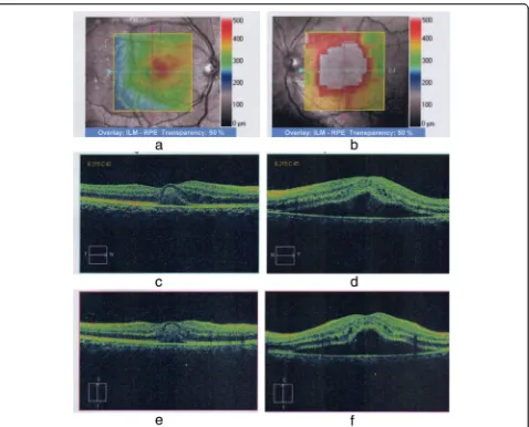
[image:3.595.59.538.287.665.2](OCT) and macular scans (Fig.2a–f) were used to assess the patient’s baseline status, which showed increased ret-inal thickness centering on the foveal region (more severe in the left eye) and elevation of the retinal pigment epithelial layer with collection of fluid with changes com-patible with central serous choroid retinopathy in the left eye and foveolitis in the right eye (Fig.2a–f). Repeat liver function tests on day 8 showed declining liver enzyme values (Table 1). Incidentally, her whole-blood analysis showed rising counts with WBC of 3.77 × 10⁹/L (normal range, 4–10 × 109/L) and platelet value of 34 × 109/L (150–450 × 109/L) (Table 1), heralding recovery. A focused ultrasonographic study failed to reveal evidence of
fluid leakage in the thoracic or abdominal cavities. How-ever, her visual deficit remained. As treatment for the eye manifestations, she received a single 2-mg intravitreal tri-amcinolone injection into the right eye and 3 days of intravenous methylprednisolone 1 g following an ophthal-mology consult.
[image:4.595.61.539.85.471.2]protein was 0.4 mg/dl (< 8), and her erythrocyte sedi-mentation rate was 22 mm for the first hour. Oph-thalmological reassessment prior to discharge revealed reduced but persisting macular edema and subretinal fluid collection in the eye. In follow-up, her VA was assessed frequently along with funduscopic examina-tions. Gradual recovery was observed. Despite normalization of her VA in her right eye, VA in her left eye remained at 6/9 at the end of 6 months. Similarly, assessment with OCT at 6 months to the day of initial assessment also demonstrated improve-ment from the previously seen changes (Fig. 3a, b). When she was reviewed 1 year later, her VA remained unchanged with persistence of 6/9 in the left eye.
Discussion
This case report highlights the less frequently seen but nevertheless important complications that can occur with dengue viral infection. Ocular complica-tions of dengue viral fever are quite uncommon, and when a combination of complications is seen, such as foveolitis and central serous chorioretinitis (CSCR)
with profound visual impact, the role of active treat-ment vs. passive approach becomes questionable, es-pecially when strict guidance is unavailable. In this case report, we attempt to give some insight into the etiopathogenesis, clinical picture, progression, investi-gations, and treatment options available following ocular involvement in dengue viral fever, and we de-scribe the decisions we made for the ocular complica-tions we encountered based on available evidence, in addition to the patient’s outcome.
[image:5.595.60.540.86.428.2]mounted immune response might vary depending whether it is a primary or secondary infection, which may contribute to the observed extensive spectrum of the clinical picture [5, 6]. The usual timeline of eye involvement is seen in close association with the low-est platelet count values in both DF and DHF, a phenomenon observed by Chan et al. [7] and others, just as it occurred in our patient, and this reinforces the suspected platelet hypothesis, which suggests that complications in dengue virus infection occur at the lowest platelet count. However, Tan et al. [8] stated that this time frame also favors the immune-mediated hypothesis because it coincides with the production of immunoglobulin G, when the clinical picture her-alds recovery signaling close association with the body’s immune response. None of the many suggested hypotheses, when considered in isolation, fully ex-plains the spectrum of manifestations visualized in ocular involvement in dengue. This subject is still under study and is beyond the scope of this article.
Involvement of the eye, when it occurs, is usually bilat-eral, though it can also present unilaterally. The common-est symptom is blurring of vision; other symptoms include ocular pain, redness, metamorphopsia, impaired color vi-sion, diplopia, eye flashes and floaters, haloes, and photo-phobia [4,5,9,10]. The involvement of the macula results in the patient being symptomatic, but peripheral retinal involvement, such as retinal hemorrhage, may not be obvious and may be missed by the unaware clinician and the nonsymptomatic patient, implying that the true inci-dence of eye involvement in dengue may be underre-ported [11].
Observed signs on examination are vast and include hemorrhage, which can be sub-conjunctival or retinal; reduced VA; scotomas; inflammatory maculopathy with chorioretinitis; possible macular edema; and foveolitis. Vasculitis is also seen either focally or even in a panret-inal distribution. Other signs include exudative retpanret-inal detachment, perifoveal telangiectasia, anterior uveitis, cotton wool spots, optic disc swelling, hyperemia, and neuritis [5,7,8,12–14]. Uncommon occurrences include CSCR, which has been observed in dengue [15]. These manifestations can at times occur for the first time, months after the recovery of an otherwise uncompli-cated dengue viral infection, as noted by Guptaet al. in their case series in which uveitis was seen as a delayed phenomenon, stressing the need for vigilance [6].
Funduscopic evaluation of the eye will help identify obvious retinal changes, such as retinal hemorrhage, cotton wool spots, and optic disc swelling. However, advanced investigations such as OCT have been found to be very useful, especially to define macular involvement and assess retinal thickness and morph-ology. Sometimes, infrared fundus photography (IFG)
can shed more light and better delineate suspicious lesions which were appreciated on funduscopy that were not elucidated by OCT, because they can appear as dark patches in the retinal territory in the IFG re-port. The Amsler grid chart, a basic tool used to as-sess the visual field, can also help further delineate the scotomas, and this can be better appreciated by using an automated Humphrey field analyzer, which provides a comprehensive visual field assessment. Angiography with fluorescein or indocyanine green can further aid in identifying vascular lesions such as occlusion, leakage, and vasculitis [9, 16].
The definitive management is still disputed. Conserva-tive management with close observation and follow-up is usually acceptable and has shown that recovery can occur unaided with complete resolution [7,8]. In severe eye in-volvement such as that in our patient, however, active treatment has been chosen. Glucocorticoid therapy has been given as intravenous pulses, tapered oral regimens, or topical instillations [7], or even as intravitreal or sub-Tenon triamcinolone injections [6]. Immunosuppressive therapy with steroids has been shown to confer favorable overall outcomes with marked improvement, albeit with some residual deficit [9]. Intravenous immunoglobulin when intensive steroid therapy has failed or with worsen-ing clinical picture has also been trialed and has been shown to be beneficial with improved VA and outcome [17, 18]. Thus, the positive response seen following immunosuppressive treatment possibly supports the im-mune-mediated hypothesis in ophthalmic manifestations of DF. However, the outcomes seen following immuno-suppressive therapy, though encouraging, were not always uniform in their response. Furthermore, the strength of evidence is anecdotal at best, because most are based on case series and case reports, and none of the modalities of management are fully endorsed in either national or inter-national guidelines. Intervention beyond conservative medical management has also been required with pars plana vitrectomy and panretinal photocoagulation for worsening hemorrhage and iridotomy for glaucoma [9]. The duration of recovery ranges from a few days to a few months. Full recovery has been observed. Similarly, re-sidual deficit has also been noted, ranging from persistent mild central scotoma to impaired VA.
Although our patient did not develop DHF, she did de-velop hepatitis along with minor derangement in her coagulation profile. Willset al. observed that though coagu-lation derangement is a known phenomenon complicating dengue, even the presence of thrombocytopenia by itself does not contribute to hemorrhage unless complicated by shock and hypoxia with acidosis predisposing to dissemi-nated intravascular coagulation precipitating bleeding [19]. Thus, the signs observed are not directly attributable to im-paired coagulation and instead depend on the complex mechanisms discussed above. Our patient’s manifestations were uncommon, with foveolitis in the right eye and central serous chorioretinopathy in the left eye along with hemor-rhages in both.
Central serous chorioretinopathy is a poorly under-stood phenomenon and more so when it occurs in dengue viral infection. The potential etiologies for CSCR include steroids, either exogenous or endogen-ous in origin; stress; psychological makeup; pregnancy; and genetic risk [20, 21]. CSCR has also been ob-served in cases of autoimmune disease and infections [22, 23]. In pregnancy, one of the postulated causes is the increased permeability [21]; though the mechan-ism is different, a similar increase in permeability is appreciated in the spectrum of dengue viral infection. Similarly, dengue viral fever can cause significant stress and a subsequent rise in catecholamine levels [15]. One could postulate that these are potential mechanisms for CSCR to occur in patients with den-gue viral infection in addition to the patient factors such as psychological makeup. Taking this into con-sideration, the challenge we faced was to determine the treatment strategy for our patient: whether ag-gressive treatment was to be chosen or to let the dis-ease run its course and await spontaneous resolution. Our patient developed foveolitis and CSCR due to dengue viral infection. The clinical problem we faced was that foveolitis is known to benefit from steroids, whereas CSCR may potentially worsen with steroid therapy [24, 25]. Though evidence favoring treatment is anecdotal, owing to the severity of the presentation, a combined clinical decision was made to opt for systemic glucocorticoid therapy. The clinical response was favorable; the patient was able to engage in daily activities with very little in the way of limitation due to a visual deficit, which can be elucidated only with Amsler charting. However, whether the observed im-provements were either fully or partially attributable to the treatment is speculative. The clinical problem of choosing not to treat or choosing a less intensive treatment is a difficult decision, considering the risk of permanent visual deficit. Thus, with lack of clearly defined guidelines, the decision should be at the dis-cretion of the clinical team and the patient.
Conclusion
Involvement of the eye in dengue viral infection, though uncommon, should garner a greater awareness and ap-preciation. Any visual complaint should be regarded as potential dengue eye involvement, especially around the time platelet counts drop to their lowest value and begin to rise. To understand the true incidence of dengue eye involvement and the patterns of presentation, as well as to formulate a clear protocol for management, further focused research is necessary.
Abbreviations
ALT:Alanine aminotransferase; AST: Aspartate aminotransferase;
CBC: Complete blood count; CSCR: Central serous chorioretinitis; DF: Dengue fever; DHF: Dengue hemorrhagic fever; Hb: Hemoglobin; HCT: Hematocrit; IFG: Infrared fundus photography; OCT: Optical coherence tomography; VA: Visual acuity; WBC: White blood cell count
Acknowledgements
We acknowledge Dr. Charith Fonseka, consultant eye surgeon, for contribution to the care of the patient. We also acknowledge Dr. D. M. S. V. Dissanayake’s contribution to the manuscript. We acknowledge the contribution of the nursing staff of ward 56B of the National Hospital of Sri Lanka and also the patient for consenting to publication of this case report.
Authors’contributions
JY, SH, MRN, IK, and DW diagnosed the clinical scenario. MRN, JY, SH, SJ, JS, and IK conducted research and drafted the manuscript. All authors provided care for the patient. AK, RN, and JY revised the manuscript. All authors read and approved the final manuscript.
Authors’information
IK is a registrar of medicine at the National Hospital of Sri Lanka, Colombo, and lecturer in the Department of Pharmacology, Faculty of Medical Sciences, University of Sri Jayewardenepura. SH is a registrar of medicine at the National Hospital of Sri Lanka, Colombo. MRN is a registrar of medicine at the National Hospital of Sri Lanka, Colombo. JY is a senior registrar in medicine at the National Hospital of Sri Lanka, Colombo. JS is a senior registrar in medicine at the Colombo South Teaching Hospital. DW is an intern house officer at the National Hospital of Sri Lanka, Colombo. AK is a consultant physician in acute medicine at the National Hospital of Sri Lanka, Colombo.
Funding Not applicable
Availability of data and materials
All the data used and/or analyzed during case report development are included in this case report.
Ethics approval and consent to participate Not applicable.
Consent for publication
Written informed consent was obtained from the patient for publication of this case report and any accompanying images. A copy of the written consent is available for review by the Editor-in-Chief of this journal.
Competing interests
The authors declare that they have no competing interests.
Author details
1
National Hospital of Sri Lanka, Colombo, Sri Lanka.2Department of Clinical Pharmacology, Faculty of Medical Sciences, University of Sri
Jayewardenepura, Nugegoda, Sri Lanka.3Colombo South Teaching Hospital,
Received: 2 October 2018 Accepted: 4 July 2019
References
1. Gubler DJ. The global pandemic of dengue/dengue haemorrhagic fever: current status and prospects for the future. Ann Acad Med Singapore. 1998; 27(2):227–34.
2. World Health Organization (WHO). Dengue and severe dengue. Fact sheet 117. Geneva: WHO Media Center; 2015 [updated May 2015].http://www. who.int/mediacentre/factsheets/fs117/en/. Accessed 7 Sept 2015. 3. World Health Organization (WHO) Regional Office for South-East Asia.
Comprehensive guidelines for prevention and control of dengue and dengue haemorrhagic fever. Revised and expanded edition. Geneva: WHO; 2011.
4. Lim WK, Mathur R, Koh A, Yeoh R, Chee SP. Ocular manifestations of dengue fever. Ophthalmology. 2004;111(11):2057–64.
5. Teoh CB, Chan PL, Laude A, Chee KLC, Lim HT, Goh YK. Dengue
chorioretinitis and dengue-related ophthalmic complications.https://uveitis. org/wp-content/uploads/2017/05/dengue_chorioretinitis_and_dengue_ related_ophthalmic_complications.pdf. Accessed 7 Sept 2015.
6. Gupta A, Srinivasan R, Setia S, Soundravally R, Pandian DG. Uveitis following dengue fever. Eye. 2009;23(4):873–6.
7. Chan DP, Teoh SC, Tan CS, Nah GK, Rajagopalan R, Prabhakaragupta
MK,et al.Ophthalmic complications of dengue. Emerg Infect Dis. 2006;
12(2):285–9.
8. Tan CS, Teoh SC, Chan DP, Wong IB, Lim TH. Dengue retinopathy manifesting with bilateral vasculitis and macular oedema. Eye. 2007; 21(6):875–7.
9. Yip VC, Sanjay S, Koh YT. Ophthalmic complications of dengue fever: a systematic review. Ophthalmol Therapy. 2012;1(1):2.
10. Siqueira RC, Vitral NP, Campos WR, Orefice F, de Moraes Figueiredo LT. Ocular manifestations in dengue fever. Ocul Immunol Inflamm. 2004; 12(4):323–7.
11. Chlebicki MP, Ang B, Barkham T, Laude A. Retinal hemorrhages in 4 patients with dengue fever. Emerg Infect Dis. 2005;11(5):770–2.
12. Loh BK, Bacsal K, Chee SP, Cheng BC, Wong D. Foveolitis associated with dengue fever: a case series. Ophthalmologica. 2008;222(5):317–20. 13. Ng AW, Teoh SC. Dengue eye disease. Surv Ophthalmol. 2015;60(2):106–14. 14. Deutman AF, Bos PJ. Macular bleeding in dengue fever [in German]. Klin
Monatsbl Augenheilkd. 1979;175(3):429.
15. Goel N, Bhambhwani V, Jain P, Ghosh B. Massive retinal pigment epithelial detachment following acute hypokalemic quadriparesis in dengue fever. J Ophthalmic Vis Res. 2016;11(2):231–3.
16. Young SM, Santiago P, Chee CK. A patient presenting with bilateral central scotomas after dengue fever. World J Retina Vitreous. 2012;2(1):18–21. 17. Chang PE, Cheng CL, Asok K, Fong KY, Chee SP, Tan CK. Visual disturbances
in dengue fever: an answer at last? Singapore Med J. 2007;48(3):e71–3. 18. Bacsal KE, Chee SP, Cheng CL, Flores JV. Dengue-associated maculopathy.
Arch Ophthalmol. 2007;125(4):501–10.
19. Wills BA, Oragui EE, Stephens AC, Daramola OA, Dung NM, Loan HT,et al. Coagulation abnormalities in dengue hemorrhagic fever: serial investigations in 167 Vietnamese children with dengue shock syndrome. Clin Infect Dis. 2002;35(3):277–85.
20. Khan Y, Mapani A, Islam N, Tufail A. Central serous chorioretinopathy (CSCR). In: Trust MEHNF, editor. 1 ed. London: Moorfields Eye Hospital; 2018. p. 2. 21. Choudhary T, Chawla S, Ray R, Pereira S. Hypercortisolism induced atypical
central serous chorioretinopathy in pregnancy. Med J Armed Forces India. 2015;71(Suppl 1):S55–9.
22. Eckstein MB, Spalton DJ, Holder G. Visual loss from central serous retinopathy in systemic lupus erythematosus. Br J Ophthalmol. 1993;77(9):607–9. 23. Cassel GH, Brown GC, Annesley WH. Central serous chorioretinopathy: a
seasonal variation? Br J Ophthalmol. 1984;68(10):724–6.
24. Ling TJ, Krishnan DR, Isa H, Din NM. Early treatment of dengue foveolitis resulting in good visual outcome. Asia Pac J Trop Dis. 2017;7(11):696–8. 25. Carvalho-Recchia CA, Yannuzzi LA, Negrao S, Spaide RF, Freund KB,
Rodriguez-Coleman H,et al.Corticosteroids and central serous chorioretinopathy. Ophthalmology. 2002;109(10):1834–7.
Publisher’s Note