ScholarWorks @ Georgia State University
ScholarWorks @ Georgia State University
Chemistry Theses Department of Chemistry
Summer 8-7-2012
Studying the DNA Binding and Conformation of Metal-Binding Site
Studying the DNA Binding and Conformation of Metal-Binding Site
Mutations in Pirin
Mutations in Pirin
Imran J. Rehmani
Georgia State University, [email protected]
Follow this and additional works at: https://scholarworks.gsu.edu/chemistry_theses
Recommended Citation Recommended Citation
Rehmani, Imran J., "Studying the DNA Binding and Conformation of Metal-Binding Site Mutations in Pirin." Thesis, Georgia State University, 2012.
https://scholarworks.gsu.edu/chemistry_theses/53
This Thesis is brought to you for free and open access by the Department of Chemistry at ScholarWorks @ Georgia State University. It has been accepted for inclusion in Chemistry Theses by an authorized administrator of
MUTATIONS IN PIRIN
IMRAN REHMANI
Under the Direction of Dr. Aimin Liu
ABSTRACT
The transcription factor NF-κB interacts with many other co-regulator proteins that
modulate its binding and transcriptional activity. One of these co-regulators, Pirin, is an
iron-dependent metalloprotein that has been shown to enhance the DNA binding of NF-κB
homodimers. Here, we characterize the interactions between Pirin and its known NF-κB binding
partners and examined the role of Bcl-3, a protein that is required for Pirin’s interaction with
p50. In addition, we use site-directed mutagenesis to alter conserved residues within Pirin’s
metal binding environment and observed how it affected the DNA binding and conformation of
the Pirin-NF-κB complex. These studies show that, while a similar enhancing effect on DNA
binding is observed, the interactions of Pirin with different NF-κB members are distinct from
each other and could possibly have different physiological purposes.
MUTATIONS IN PIRIN
by
IMRAN REHMANI
A Thesis Submitted in Partial Fulfillment of the Requirements for the Degree of
Master of Science
in the College of Arts and Sciences
Georgia State University
Copyright by Imran Jabbar Rehmani
MUTATIONS IN PIRIN
by
IMRAN REHMANI
Committee Chair: Dr. Aimin Liu
Committee: Dr. David Wilson
Dr. Donald Hamelberg
Electronic Version Approved:
Office of Graduate Studies
College of Arts and Sciences
Georgia State University
ACKNOWLEDGEMENTS
Where I am today is the result of the support and influence of a countless number of
people. But, since I doubt any of them will read this (other than the ones I might force), I’ll stick
to the most immediate people.
I would like to first thank my advisor, Dr. Aimin Liu, for giving me the opportunity to
work on this project and for the support (and patience) he has provided throughout my time here.
Working in his lab has been a valuable experience that has changed the way I understand and
view science.
I would also like to thank my colleagues who also worked on the Pirin project, Fange
Liu and Xiaoxi Wei, for their constant help and insightful conversations that have played a large
part in my understanding of this project.
Finally, I have to thank my girlfriend Amanda, who for nearly four years has allowed
stacks of journal articles scattered everywhere, listened to my complaints about failed
experiments, and has driven me to lab at an unreasonable hour to “just check on something”
TABLE OF CONTENTS
ACKNOWLEDGEMENTS ... iv
LIST OF TABLES ... vii
LIST OF FIGURES ... viii
1 INTRODUCTION ... 1
2 NF-κB: STRUCTURE, FUNCTION, AND REGULATION ... 9
2.1 Overview... 9
2.2 Structural Features ... 10
2.3 Primary components of the NF-κB activation pathway ... 12
2.4 Interaction with DNA ... 14
2.5 Interaction with other Bcl-3 and other proteins... 19
2.6 Summary ... 23
3 RESULTS ... 25
3.1 Introduction ... 25
3.2 Alteration of NF-κB-DNA conformation by Pirin ... 27
3.3 Bcl-3 interferes with the DNA interaction of the Pirin-p65 complex ... 33
3.4 Altering the metal-binding environment ... 37
4 DISCUSSION ... 44
4.2 Why might Pirin interact directly with p65 but not p50? ... 46
4.3 Bcl-3: more than just a bridging molecule... 49
4.4 Structural role of Q115A and W117A ... 50
4.5 Concluding Remarks ... 53
REFERENCES ... 55
APPENDIX A: MATERIALS AND METHODS ... 60
LIST OF TABLES
Table 2.1: Examples of Class I and II κB sequences ... 14
Table 3.1: Viscoelastic properties of Pirin-NF-κB-DNA complexes ... 29
Table 3.2: Shear and viscosity of p65 + Bcl-3 measurements ... 35
Table 3.3: Viscoelastic properties of mutant-p50/p65 complexes ... 39
LIST OF FIGURES
Figure 1.1: Structure of Pirin and other cupin family proteins ... 2
Figure 1.2: Octahedral coordination of iron ion bound by Pirin ... 3
Figure 1.3: Sequence alignment of Pirin ... 4
Figure 2.1: Structure of the RHD of p50 ... 10
Figure 2.2: Comparisons of the dimerization domains of NF-κB. ... 10
Figure 2.3: Structural domains of NF-κB family members ... 12
Figure 2.4: Scheme of canonical NF-κB activation ... 12
Figure 2.5: NF-κB uses flexible loops to recognize and interact with DNA ... 16
Figure 2.6: Small changes in a κB sequence affect the conformation of DNA-bound NF-κB ... 18
Figure 2.7: Structures of IκBα and Bcl-3 ... 20
Figure 3.1: Preparation of QCM-D sensor surface... 27
Figure 3.2: Pirin enhances the DNA binding of p50 and p65 homodimers ... 28
Figure 3.3: Additional CD spectra features seen with p65-Pirin interaction ... 30
Figure 3.4: Effects of DNA binding on conformation of p65 and p65-Pirin. ... 31
Figure 3.5: Bcl-3 interferes with Pirin-p65 binding to DNA. ... 33
Figure 3.6: DNA binding models for Bcl-3 interference with Pirin/p65 interaction 36 Figure 3.7: Location of Q115 and W117A residues relative to iron center ... 37
Figure 3.8: Effect of metal-center mutations on p50 and p65 binding ... 38
Figure 3.9: Dissipation vs. frequency plots for p50 and p65 complexes ... 40
Figure 3.10: CD spectra differences of mutant Pirin/p65 complexes ... 42
Figure 4.2: Electrostatic map of Pirin. ... 48
Figure 4.3: Model of wild-type and W117A Pirin interactions with immobilized
1 INTRODUCTION
The term “NF-κB” was coined in 1986 to describe an interactor of the κ light chain gene
enhancer element in B cells1, known as a κB site. Since then, it has been the subject of tens of
thousands of publications and directly associated with over a hundred different stimuli and target
genes in nearly every type of cell2. NF-κB is a family of transcription factors (p50, p52,
p65/RelA, RelB, and c-Rel) that form both homo- and heterodimers that are sequestered in the
cytoplasm by inhibitory proteins in the IκB (inhibitor of κB) family and are released in response
to cytokines or growth factors, among other stimuli2. Upon activation and translocation to the
nucleus, NF-κB is capable of activating or inhibiting hundreds of genes involved in cell survival
and proliferation. With so many different sources of activation and genes that it can potentially
affect, there is an obvious need for tight regulation of NF-κB activity so that only certain subsets
of genes are activated in response to a specific cellular signal. To make this possible, NF-κB has
many other proteins that it interacts with to modulate its activity, allowing the formation of a
complex signaling network that integrates multiple pathways. One of these regulatory proteins,
Pirin, will be the focus of this study.
Pirin is a metalloprotein with two domains containing anti-parallel beta-sheets
characteristic of the cupin superfamily of proteins (Figure 1.1)3. The cupin superfamily, one of
the most functionally diverse families, includes both enzyme and non-enzyme members and
contains proteins that have been seen to bind a variety of metals as well as no metal at all4. Pirin
octahedrally coordinates an iron ion within its N-terminal domain (but not its C-terminal
domain) with three histidine residues (H56, H58, and H101) along with a glutamate residue
3-His-1-Glu motif is a deviation from the 2-His-3-His-1-Glu/Asp coordination seen in most other non-heme
iron-dependent proteins and is somewhat rare5, but it can be found in other cupin family
metalloprotein enzymes that share structural features with Pirin. In particular, oxalate oxidase
and quercetin 2,3-dioxygenase share the same 3-His-1-Glu coordination of their metal ion and
have a similar fold and binding pocket located at their metal binding sites that is present in Pirin,
although these two enzymes have their coordinating Glu in a different position relative to their
[image:13.612.92.500.277.553.2]metal and utilize transition metals other than iron3.
Figure 1.1: Structure of Pirin and other cupin family proteins (A) Pirin (PDB: 1J1L), (B) Cysteine dioxygenase (2IC1), (C) Quercetin 2,3-dioxygenase (1JUH), (D) Oxalate oxidase (1FI2). Note the anti-parallel β-barrel structures characteristic of this family.
Even though Pirin was initially discovered as an interactor of the NFI/CTF box
transcription factor6, the later finding that it is also an interactor of p50 homodimers in
conjunction with Bcl-3, another NF-κB regulator, has attracted much more attention to this
homodimers and other coregulator proteins such as Tip60, Jab1, and Bard1. However, among
these proteins, the p50/Bcl-3/Pirin complex was unique in displaying a significantly enhanced
level of binding to the κB DNA sequence tested compared to p50 and p50/Bcl-3 alone7
[image:14.612.244.371.171.352.2].
Figure 1.2: Octahedral coordination of iron ion bound by Pirin
In spite of being discovered over a decade ago, knowing other proteins it interacts with,
and the 3-D structure and protein family being known, the relationship between the structure and
function of Pirin remains relatively uncharacterized. Pirin and its orthologs found in other
mammals, plants, fungi, and prokaryotes (but, interestingly, not yeast) contain no known
functional motifs or significant sequence homology with other proteins. Even when aligned with
its orthologs, Pirin displays only a low to moderate degree of sequence conservation outside of
its iron-binding N-terminal domain (Figure 1.3)6. Most of the residues that show nearly complete
conservation across all species are located in proximity to the metal center or within the binding
Figure 1.3: Sequence alignment of Pirin Alignment includes eukaryotic (XePirin, CapPirin, AraPirin, ZeaPirin) and prokaryotic (CCPirin, P1205Pir) homologs (NCBI identifiers listed in Materials and Methods). Metal ligands are highlighted in yellow, residues lining binding pocket surrounding metal highlighted in green. Obtained with CLUSTALW:
The Pirin orthologs found in plants and bacteria, which do not contain the same
transcription factors as mammals and thus lack NF-κB and other proteins mammalian Pirin
interacts with, have still been observed to have functions relating to cell survival that are similar
to the functions associated with the NF-κB pathway in humans. An ortholog of Pirin found in the
cyanobacterium Synechocytis was found to be upregulated in conditions of high salinity8 and,
similarly, the expression of a homolog of Pirin found in tomatoes is increased in response to
specific apoptosis-inducing stimuli9.
Because of its central position in multiple signaling pathways, dysreguation of NF-κB has
been implicated in many pathological conditions including cancer, inflammation, and septic
shock10,11, making κB an attractive target for drug development. Thus, as a modulator of
NF-κB binding, Pirin could also potentially be a target protein for the development of new therapies
for disease. In particular, multiple studies have established links between Pirin and cancer12-14,
although the exact signaling pathways involved or how Pirin affects their activity remains vague.
Pirin’s role in oncogenesis, and perhaps the cell itself, is highly dependent upon the cell line
being examined. The expression of Pirin varies greatly across different cancer cell lines14,15,
generally showing higher expression in breast, cervical, lung, and skin cancers and reduced
expression in colon cancer and leukemia cell lines. The significance of the expression levels of
Pirin to a given type of cancer and its contribution to a cancerous phenotype is not clear-cut. The
development of specific types of cancer has been linked to both overexpression and the absence
of Pirin. While the high expression of Pirin in subsets of melanoma cell lines contributes to the
disease by inhibiting the senescence of melanocytes15, its reduced expression in leukemia cell
development of acute myelogenous leukemia (AML)12. Knockdown of Pirin in melanoma cell
lines that have high Pirin expression reverses the lack of senescence associated with these cells15.
In addition, Pirin knockdown reduces the metastatic phenotype of these cells by causing a
corresponding decrease in the expression of the SLUG protein14, further revealing its potential as
a therapeutic target. A small-molecule inhibitor which binds to Pirin’s binding pocket in
proximity to its metal center and disrupts its association with Bcl-3 has been shown to inhibit
melanoma cell migration in a way similar to its siRNA knockdown, showing promise for future
drug-development14.
Since the discovery that Pirin interacted with p50 homodimers through Bcl-3 and
enhanced DNA binding, almost no other information regarding the specific nature or mechanism
of Pirin’s effect upon NF-κB has been published. Other coregulator proteins that interact with
p50 through Bcl-3 have been linked to enzymatic functions. One of these proteins, Tip60, has
been shown to have histone deacetylase activity16 and Bard1 is needed to stabilize BRCA1, a
DNA-repair enzyme17. Although Pirin’s metal binding site and putative binding pocket
resembles that of other enzymes in the cupin superfamily, it has not been found to have a
prominent enzymatic role in vivo. One study revealed that Pirin and one of its prokaryotic
orthologs found in E. Coli, yhhW, is capable of quercetinase activity in vitro in a way similar to
quercetin 2,3-dioxygenase18, and overexpressing Pirin in NKE cells increased the quercetin
resistance of poliovirus replication in those cells19. However, the extent of how physiologically
relevant Pirin’s quercetinase activity is, or whether or not it is an important factor in the
regulation of NF-κB, has yet to be determined. Based on the evidence present at this time, it is
Since its discovery, a majority of the investigation of Pirin has been primarily approached
from a biological perspective. While this has identified its role as a transcriptional regulator and
a possible oncogene, it has left the questions surrounding the specific mechanisms of Pirin’s
interactions with NF- κB and DNA relatively untouched. An aspect of Pirin’s function that has
received little attention is its potential interactions with other NF-κB members, which have a
similar overall structure to p50. It was initially believed that Pirin only interacted with p50 in a
Bcl-3 dependent manner. However, our group has found that Pirin interacts with p65
homodimers as well as p50 homodimers and confers a similar enhancing effect on the ability of
p65 to bind DNA (Liu, unpublished data). Interestingly, unlike p50 homodimers, the interaction
with p65 does not require Bcl-3, which only interacts with NF-κB dimers consisting of p50 or
p5220. This would imply that the interactions with p50 and p65 are distinct from each other and
may have different characteristics such as stoichiometry, the structural features of the
protein/protein or protein/DNA complexes, or mechanism of action. These two members of the
Rel family have distinct differences in the consensus sequence they favor, the proteins they can
potentially interact with, and overall physiological function. With at least two NF-κB dimers that
it can interact with, Pirin’s potential role in cell signaling has been expanded greatly. With the
aforementioned differences between p50 and p65, along with the different compositions of their
known complexes with Pirin, understanding the differences in the nature of Pirin’s interactions
with these two proteins will help facilitate future studies that form more precise links between
Pirin and its physiological role.
The focus of this study will be to obtain an initial characterization of the complexes Pirin
forms with p50 and p65 to find distinguishing characteristics between the two interactions. In
gain more insight on how this highly conserved region, and perhaps the metal itself, contributes
to Pirin’s function. These topics will be addressed from a perspective that focuses on the
conformation of the Pirin-NF-κB-DNA complex in addition to how much it enhances the DNA
binding of NF-κB. As we will learn in the next chapter outlining the structure and function of
NF-κB, the conformation of an NF-κB-DNA complex is crucial in determining its activity and
varies from one promoter site to another. Thus, by studying changes conformation in conjunction
with DNA binding resulting from mutagenesis, we are able discern between protein-DNA
complexes that resemble the wild-type and are likely to be transcriptionally active in vivo and
2 NF-κB: STRUCTURE, FUNCTION, AND REGULATION
2.1 Overview
Even after over two decades of intensive study and over 36,000 publications on the
subject, NF-κB remains a highly active area of research with many unanswered questions. The
five NF-κB proteins (p50, p52, p65/RelA, RelB, c-rel) display a remarkable versatility that
allows them to participate in numerous signaling pathways and physiological responses, some
which appear to contradict each other such as how NF-κB activity has been shown to promote
both cell survival and apoptosis. This versatility is due to the myriad layers of regulation that
fine-tune NF-κB activity across different cell types and physiological conditions.
The most basic level of regulation over the NF-κB response is that the five proteins can
form many different dimer combinations that are similar in overall structure but have different
preferences for the DNA sequences they bind and non-redundant functions. Thus, a cell
producing a greater amount of one type of subunit would have a direct influence on the genes
that are likely to be activated or repressed.
Interactions with other proteins provide a much more complex layer of regulation and
influence how NF-κB dimers are activated by various stimuli, how they are deactivated, and how
they influence other signaling pathways through direct interaction with other transcription
factors.
In addition to the above methods of NF-κB regulation, the DNA sequences themselves
have been shown to also play a role in determining the exact action an NF-κB-DNA interaction
will yield. The κB consensus sequence that NF-κB binds to is remarkably flexible, with a single
base pair substitution being capable of affecting the conformation and activity of a bound NF-κB
This chapter will attempt to review the fundamentals of NF-κB structure and function as
well as go into more detail on the different layers of regulation mentioned above.
2.2 Structural Features
As mentioned previously, the term NF-κB is given to any hetero- or homodimer
combination of five proteins: p50, p52, p65/RelA, RelB, and c-Rel. The defining feature of the
NF-κB family is a 300 amino acid sequence at their N-terminal domain referred to as the Rel
homology domain (RHD) which consists of a pair of immunoglobin-like domains which are
[image:21.612.163.451.311.658.2]required for dimerization, nuclear localization, and DNA binding. (Figure 2.1)
Figure 2.2: Comparisons of the dimerization domains of NF-κB. (A) Dimerization domain of NF-κB Rel homology domains highlighted with red box (p65 homodimer shown, PDB code 1RAM); (B) Asp 254 and Asn 200 interact more favorably with each other in p50/p65 heterodimers (pink) than they do when they are paired with copies of themselves in p50 (blue) and p65 (green) homodimers; (C) Additional hydrogen bonding contributed by Y267 in p50 (blue), p65 shown in green
NF-κB proteins form dimers through hydrophobic interactions along a β-sheet interface
between the C-terminal domains of each subunit’s RHD (Figure 2.2A). These hydrophobic
dimer interfaces are highly conserved across the NF-κB family members, allowing most dimer
combinations to exist in physiological conditions22. However, there are minor differences
between the dimerization domains of each NF-κB protein that make some members more
complimentary for each other than others, thus making them more stable and increasing their
population relative to other dimer combinations. The p50/p65 heterodimer, the most common
NF-κB dimer combination, is more stable than the homodimers of either subunit due to a
favorable interaction between Asp 254 of p50 and Asn 200 of p65 that does not exist in the
many of the same residues, but a few minor differences make p50 homodimers more stable22.
F213 in p65 is replaced with a tyrosyl (Y267) in p50, which further stabilizes the p50
homodimer with the additional hydrogen bonding contributed by the hydroxyl group (Figure
2.2C). Also, F307 in p50 is replaced with V248 in p65, which reduces the hydrophobic
interaction contribution to the stability of the p65 homodimer.
The dimer combinations that do not exist in vivo are those that contain RelB, which has
structural features that set it apart from the other NF-κB subunits which includes an additional
N-terminal leucine zipper domain and a patch of nonpolar residues at its surface. RelB forms
intertwined homodimers that are unstable in physiological conditions and are hypothesized to
exist only transiently as a way of avoiding degradation until it can form a stable heterodimer24.
RelB has not been found to form heterodimers with p65 or c-rel, but can form stable
heterodimers with either p50 or p5225 and are important in specific NF-κB signaling pathways,
which will be discussed later.
While the RHDs and the DNA binding motifs of each NF-κB protein are similar to each
other, the C-terminal domains have crucial differences that further specify the roles of each
family member. NF-κB proteins can be divided into two groups: the proteins containing a
transactivation domain (TAD) at their C-terminal (p65, RelB, and c-Rel) and the proteins that
lack a TAD (p50 and p52) (Figure 2.2). Thus, homodimers of p50 and p52 are typically
repressors of transcription while dimers containing p65, RelB, and c-Rel are transcription
activators in most cases26.
Another difference between the two groups of NF-κB proteins is that p50 and p52 are
processed from larger proteins named p105 and p100, respectively27,28. p105 and p100 contain
inhibitor of κB (IκB) family, and are capable of interacting with other NF-κB proteins and
[image:24.612.88.509.155.363.2]sequestering them in the cytoplasm.
Figure 2.3: Structural domains of NF-κB family members. RHD: Rel homology domain, TAD: transactivation domain, LZ: leucine zipper motif. Taken from Wan, 200929.
2.3 Primary components of the NF-κB activation pathway
While the different dimer combinations are selectively activated by specific stimuli, they
all reside in the cytoplasm in an inactive form and are shuttled to and from the nucleus in a
regulated manner30. With the exception of p50/RelB and p52/RelB heterodimers, NF-κB
activation is accomplished through what is referred to as the “canonical” pathway. To briefly
summarize, this pathway is initiated by a membrane receptor protein being stimulated by its
ligand which activates IκB kinase (IKK), resulting in the degradation of the IκB protein that
sequesters the NF-κB dimer in the cytoplasm, allowing it to translocate to the nucleus where it
Figure 2.4: Scheme of canonical NF-κB activation
IKK has three different subunits: IKKα and IKKβ, which serve a catalytic role, and
IKKγ/NEMO, which has no catalytic activity but plays a regulatory role31. IKK targets IκB
proteins, resulting in their degradation by the 26S proteasome and release of the NF-κB dimer it
was bound to30. IKK is activated in response to stimulation of receptor proteins by various
cytokines, such as TNF-α and IL-1, and IKKγ recruits kinases that phosphorylate the IKKβ
conserved serines on IκB proteins that sequester NF-κB dimers in the cytoplasm, causing their
degradation. Phosphorylation of IκB by IKK is the only regulated step in its degradation32, thus
dysregulation of IKK has deleterious consequences such as tumor development33.
The IκB family has five members (IκBα, IκBβ, IκBε, p105, and p100) that are considered
to be “typical” IκB proteins since they operate in the same general manner by binding to the
RHDs of NF-κB dimers with their ARDs and rendering them inactive. Two more “atypical” IκB
family members exist: IκBζ and Bcl-3. These atypical members, while sharing structural features
with other members of the IκB family, do not repress NF-κB activity in the same way and
instead act primarily as coregulator proteins that can potentially activate transcription as well as
repress it. Bcl-3 will be discussed further in another section. Another structural feature present
in many IκB members that is absent in Bcl-3 is the C-terminal PEST domain, a disordered region
rich in proline, glutamate, serine, and threonine34. This region is constitutively phosphorylated
and is thought to be important for both protein function and stability35.
It was originally believed that IκB proteins were only important in the cytoplasm,
inhibiting NF-κB dimers by concealing their nuclear translocation domains. While that is still an
important aspect of NF- κB inhibition, it was later found that IκB proteins also act in the nucleus
by removing DNA-bound NF-κB and that the IκB-NF-κB complex was capable of being shuttled
back and forth between the cytoplasm and the nucleus36. The gene for IκBα is activated by
NF-κB itself, creating a feedback loop that ensures that NF-NF-κB activation is transient37
. After NF-κB
activation, IκBα is produced to replace that which was degraded and binds to the NF-κB dimers
attached to their κB sequences, removing them and shuttling them back to the cytoplasm where
they await activation once again. Each IκB family member has a preference for which NF-κB
some overlap38. There are also differences in the kinetics of their activation. IκBα is rapidly
produced in response to NF-κB activation while IκBβ and IκBε take more time to accumulate,
allowing for the length of an NF-κB response to be regulated38.
Upon stimulation by cytokines or other inducers of NF-κB activity, IκB proteins are
phosphorylated at specific residues (Ser32 and Ser36 in the case of IκBα39). This
phosphorylation event targets them for ubiquitination, which makes them substrates for the 26S
proteasome and results in their degradation40. Once released from IκB repression, the NLS of the
NF-κB dimer is exposed and can be translocated to the nucleus.
2.4 Interaction with DNA
The target gene sequences of NF-κB dimers, referred to as κB sites, generally conform to
the consensus sequence 5’-GGGRNWYYCC-3’ where R, N, W, and Y represent purines, any
base, A or T, and pyrimidines, respectively. Many κB sites only loosely conform to the
consensus sequence, resulting in a very broad assortment of identified κB sites for different
[image:27.612.175.441.472.724.2]genes (Table 2.1).
Table 2.1: Examples of Class I and II κB sequences
Class I Sequences
IgκB 5’-GGGAC T TTCC-3’
IFN-β 5’-GGGAA A TTCC-3’
IL-6 5’-GGGAT T TTCC-3’
Class II Sequences
κB-33 5’-GGAA AT TTCC-3’
IL-2 5’-A AGAA T TTCC-3’
As one would expect, the sequence of a κB site determines its preference for which
NF-κB dimer combination it interacts with. Similar to how NF-NF-κB subunits can be divided into two
groups based on their structural domains, κB sites can be divided based on whether they prefer to
interact with p50 or p65. The RHDs of NF-κB subunits are mostly similar from one family
member to another, but small differences between their DNA binding domains result in p50 and
p52 preferring 5’-GGGRN-3’ sequences and p65 and c-rel having a preference for 5’-YYCC-3’
sequences. Combined, these two half-sites form the κB consensus sequence. Each monomer unit
of an active NF-κB dimer is capable of binding half of a κB site. Since p50/p65 is the most
common NF-κB dimer combination, the ideal consensus sequence is a 10 bp sequence contains
the 5 bp sequence targeted by p50 or p52 and the 4 bp sequence preferred by p65 and c-rel
connected with an A-T bp in the center. κB sequences that have a preference for homodimers are
composed of either two of the 5 bp half sites, resulting in an 11 bp sequence that attracts p50 or
p52 homodimers, or contain two of the 4 bp sites in a 9 bp sequence that attracts p65 or c-rel
homodimers. The 10 or 11 bp sequences are referred to as Type I κB sites while the 9 bp
sequences that prefer p65 or c-rel homodimers are referred to as Type II sites.
NF-κB interacts with DNA in a somewhat unusual way that is not classified by any of the
typical motifs seen on DNA-binding proteins (helix-turn-helix, helix-loop-helix, etc.). Instead of
binding DNA through α-helices that are inserted into DNA grooves, NF-κB uses an
immunoglobin-like fold that consists of β-sheets and large connecting loops that interact with the
major groove41 (Figure 2.5). The key residues responsible for the specific protein-DNA contacts
are mostly similar between NF-κB subunits. In p50 they are R54, R56 Y57, E60, H64, and K241,
alanine23. This minor alteration is the primary reason for the different half site preferences of p50
and p65 subunits23. H64 hydrogen bonds with the additional G-C base pair found in Type I sites,
creating a more favorable interaction that is absent in p65.
Figure 2.5: NF-κB uses flexible loops to recognize and interact with DNA. p65 bound to DNA shown (PDB: 1RAM). DNA-binding loops highlighted in red.
The loose adherence of κB sequences to the consensus sequence is made possible by the
flexible nature of the RHD. NF-κB homodimers, which should ideally bind κB sequences that
are symmetrical to reflect that each half of the dimer has the same DNA binding preferences, are
still able to have high binding affinity for asymmetrical sites because of the flexibility allowed
by the linker region connecting the two domains of the RHD that enables the subunit that
[image:29.612.84.529.149.492.2]and form contacts with the DNA backbone42. These non-base-specific contacts allow for an
NF-κB homodimer that only forms base-specific contacts with half the NF-κB site to still bind the DNA
with a high affinity. This binding mechanism is what allows p50 and p65 homodimers to bind to
the IgκB sequence, which includes half-sites for both p50 and p65 and normally prefers p50/p65
heterodimers.
As our understanding of NF-κB has developed over the past two decades, the importance
of the sequence of a κB site has become more apparent. Because of NF-κB’s flexibility, the κB
sequence itself is very pliable and is still recognizable by NF-κB if a few base-pairs deviate from
the consensus sequence. This would suggest that there would not be many consequences
resulting from a single base-pair mutation. But, in spite of the flexibility of the κB consensus
sequence and the diversity of κB sequences found in the genome, a κB site in the promoter of
any specific gene displays almost complete conservation from one species to the next. If the
sequence of a κB site were truly flexible, one would expect a lower level of sequence
conservation. Consistent with this observation, multiple studies have revealed that even a single
base pair mutation in a κB site has significant effects upon both the structure42
and function43 of
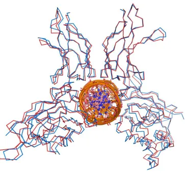
the NF-κB/DNA complex. Crystal structures of NF-κB bound to different κB sequences has
revealed that changing the sequence of a κB site by even a single base pair will also change its
structure, resulting in a different conformation of the protein-DNA complex when NF-κB binds.
An example is the difference in the conformation of a p50/p65 heterodimer bound to the IgκB
sequence23 and one bound to the IFN-β sequence44. These are both Class I κB sequences, each
containing a consensus half-site for p50 (5’-GGGRN-3’) and p65 (5’-TTCC-3’). The sequence
for IFN-β (5’-GGGAAATTCC-3’) only differs from IgκB (5’-GGGACTTTCC-3’) by only the
DNA sequence translates to significant differences in the conformation of the NF-κB
[image:31.612.109.472.135.475.2]heterodimer bound to it.
Figure 2.6: Small changes in a κB sequence affect the conformation of DNA-bound NF-κB. p50/p65homodimer bound to IgκB (red, PDB: 1VKX) and IFN-β (blue, PDB:1LE5).
This illustrates how the protein-DNA complex can vary greatly between individual genes
regulated by the same NF-κB dimer21. The structural conformation of a protein-DNA complex is
important for the recruitment of transcription factors, such as Pirin, creating the diverse
co-factor requirements observed between genes that are regulated by the same NF-κB dimer
2.5 Interaction with other Bcl-3 and other proteins
In addition to the sequestration and activation of NF-κB in response to various stimuli,
there are numerous coactivator and corepressor proteins that further modulate NF-κB activity
once it is active and within the nucleus. NF-κB alone cannot activate transcription; it requires the
recruitment of multiple coactivator proteins to form a transcriptionally active enhancer
complex45. NF- κB coregulator proteins include both enzymes and non-enzymes and the proteins
required for transcription at a given promoter are determined by both the NF-κB dimer
combination and the sequence of the promoter, which affects the conformation of the NF-κB
dimer21.
There are dozens of known protein-protein interactions that NF-κB is involved in; even
some that include other transcription factors. This section will primarily focus on Bcl-3, the
protein that is required for Pirin to interact with p50 homodimers, as well as a few of the more
prominent coregulators of p65.
Bcl-3 is considered as a member of the IκB family due to its sequence similarity and the
presence of ARDs that interact with the RHD. However, unlike other IκB proteins, the result of
Bcl-3 interacting with NF-κB does not necessarily result in inhibition. The PEST domain present
in IκBα that is required for its interaction with NF-κB is absent in Bcl-3, being replaced by an
additional ARD (Figure 2.7). Another important difference is that Bcl-3 is primarily located
within the nucleus while other IκB proteins are mostly cytoplasmic and are shuttled between the
Figure 2.7: Structures of IκBα and Bcl-3
Bcl-3, which exclusively interacts with p50 and p52, contains a TAD that we now know
can potentially give transcriptional activity to these NF-κB subunits that normally lack it46.
However, whether or not Bcl-3 was an activator or inhibitor of transcription and the specific
mechanism behind its activity was initially a subject of controversy. Early studies suggested that
Bcl-3 influenced transcription of NF-κB-regulated genes by inhibiting the binding of p50
homodimers to DNA47 and that Bcl-3 could bind to and remove inhibitory p50 homodimers that
lacked a TAD from DNA, allowing the binding of dimers containing a TAD48. Other studies
presented opposite findings, revealing that Bcl-3 activated transcription in a p50-mediated
process49. The discrepancy between these findings was found to be due to variations in the
sources of their recombinant Bcl-3 as well as differences in truncations. Along with its ARDs,
Constructs of Bcl-3 that contain these regions show a streaked gel migration pattern, indicating
the variable degrees of phosphorylation, while truncated versions that only contain the ARDs
migrate as a single band due to the lack of phosphorylation49. The transactivation function of
Bcl-3 is dependent upon its phosphorylation, so truncated Bcl-3 that only contained the ARDs or
recombinant Bcl-3 expressed in bacteria or insect cells where they would undergo the necessary
post-translational modifications displayed an inhibitory effect while Bcl-3 expressed in
mammalian cells was capable of activating transcription via p50 homodimers49.
Bcl-3 has been studied for almost as long as NF-κB has, but its biological function still
remains somewhat unclear. Bcl-3 knockout mice were still viable and were only noticed to be
more susceptible to infection, similar to the phenotype observed in p50 knockout mice50 which
indicated that Bcl-3 acts through p50 in its role in immunity. However, the phenotypes of Bcl-3
and p50 deficient mice do not overlap completely, with Bcl-3 deficient mice having
abnormalities in their spleens while mice lacking p50 have normal spleens, so Bcl-3 also has
p50-independent functions. Constitutive expression of Bcl-3 in LyH7 and BAF-B03 cells
increased nuclear translocation and DNA binding of p50 homodimers, but this effect was found
to be dependent on cell type and is proposed to require a significant pool of p50-p105
heterodimers in the cytoplasm for Bcl-3 to act upon51. Although previously mentioned studies
showed a transactivating role for Bcl-3, later studies revealed that it could potentially act as a
repressor of IL-2 expression in tolerant CD4+ T cells52.
Bcl-3 also has been found to act as a bridging molecule from p50 homodimers to other
proteins, such as Pirin7. Initially, four proteins (Pirin, Tip60, Jab1, Bard1) were found to interact
with p50 in a Bcl-3 dependent manner. Pirin greatly increased the amount of DNA binding by
the other hand, the histone acetyltransferase (HAT) Tip60 did not increase DNA binding but did
increase transcriptional activity. Recently, IRS-3 was also found to use Bcl-3 as a bridging
molecule to interact with p50 homodimers and increased the DNA binding of p50 in a manner
similar to Pirin53. The same study found that IRS-3 increased the activity of p50 as well, but only
in a TNF-α dependent manner. IRS-3, while it increases the DNA binding and activity of p50
homodimers, was not detected in the protein-DNA complex during immunoprecipitation,
suggesting that its interaction with Bcl-3 and p50 is transient. This is an important difference
from Pirin, which was demonstrated to remain in the DNA-bound complex with p50 and Bcl-3.
p65 also has many proteins that interact with it, some that are required for its activity on
certain promoters. Chromatin remodeling by HATs have been known to promote the binding and
activity of transcription factors by loosening the chromatin structure. The HAT Tip60, which
was previously mentioned as one of the proteins that interacts with p50 homodimers via Bcl-3,
also interacts with p65 homodimers and promotes its binding and activation of specific promoter
sites54 and is another example of a protein that requires Bcl-3 when bound to p50 but can
associate with p65 on its own. Curiously, Tip60 interacts with the N-terminal domain of p65’s
RHD instead of the C-terminal domain, which is where it would associate with Bcl-3 and p50.
Another HAT that also functions as a bridge between p65 and the basal transcription
machinery is CBP and its homolog p300. CBP/p300, which has many known interactions with
other transcription factors55,56, was found to also interact directly with the C-terminal
transactivation domain of p65 and enhanced p65-dependent transcription57. Along with its HAT
activity that modifies chromatin structure58, CBP/p300 can connect p65 to neighboring
transcription factors bound to other promoters on a gene, allowing the transcription factors to
Phosphorylation of S276 on the RHD of p65 by protein kinase A (PKA) has been shown to be
needed for efficient binding of CBP/p300 to p6560, illustrating how post-translational
modifications by other proteins also contribute to the regulation of NF-κB signaling.
Just as HATs act as coactivators of NF-κB activity, histone deacetylases (HDACs) act as
repressors of gene expression. HDAC1 and HDAC2 were found to interfere with p65 activity,
although only HDAC1 was found to interact directly with p65 while HDAC2 only bound to
HDAC1 to form a complex61.
Sometimes, the coactivators of p65 are transcription factors that have their own promoter
sites and are independent from NF-κB under a different context. One example of this is p65’s
interaction with Fos and Jun members of the AP-1 transcription factor family, which act
synergistically to enhance DNA binding and activation at certain promoters62.
2.6 Summary
While the mechanisms that sequester and induce NF-κB signaling exist in the cytoplasm,
activation and translocation of NF-κB to the nucleus to bind a DNA sequence in response to a
stimulating signal is only the beginning of what ultimately determines the physiological result of
the protein-DNA interaction. The κB sequence itself, which has dozens of variants, acts as an
allosteric regulator of NF-κB by affecting the conformation of the bound protein. The flexible
linker region connecting the two domains of the RHD makes this possible by allowing an
amazing level of conformational versatility when recognizing κB sequences and modifying the
orientation of the RHD domains relative to DNA to form an optimal interaction with suboptimal
sequences. Furthermore, by each NF-κB-DNA complex having a different conformation, it
allows for selectivity among the many protein coregulators of NF-κB which modulates the
are combined, it creates a highly complex system of regulation that allows only five NF-κB
3 RESULTS
3.1 Introduction
As we have described in the previous chapter, not only is the binding of an NF-κB dimer to
a κB sequence important but the resulting conformation of the NF-κB/DNA complex is also
important in determining the outcome of the interaction. p65 homodimers, which contain a
TAD, are normally considered potent activators of transcription. However, in the case of IL-2,
p65 homodimers compete with the transcription factor NF-AT for binding to its DNA sequence,
which is similar enough to the κB consensus sequence to function as a low-affinity site for p65.
Increasing expression of p65 results in reduced expression of IL-263, illustrating how DNA
binding by a NF-κB dimer containing a TAD is not always sufficient for transcription.
Furthermore, when inhibiting Pirin with a small molecule inhibitor and analyzing changes in
gene expression, Pirin inhibition resulted in an 0.8 fold increase of IL-8 expression14, which is a
gene containing a Class II κB promoter sequence that is regulated by p65 and implies that Pirin
could potentially have a repressor function as well as an activating one. Therefore, when
studying Pirin and its interaction with NF-κB, especially when in vitro techniques that study the
interaction outside the context of gene expression, it is important to consider how the
Pirin/NF-κB complex is interacting with DNA in addition to how much of that interaction is taking place.
Learning how Pirin affects the interaction and conformation of different NF-κB dimers can
contribute to a greater understanding of Pirin’s structural features as well as its physiological
role. Keeping this in mind, this study will address the following questions:
How does Pirin alter the conformation of NF-κB and the NF-κB/DNA complex?
In what significant ways are the interactions of p50 and p65 with Pirin different
How does Pirin’s metal-binding environment contribute to the conformation of
these higher order complexes?
These questions are primarily studied using quartz crystal microbalance with dissipation
(QCM-D). QCM-D, similar to surface plasmon resonance (SPR), is capable of monitoring very
small changes in mass added to a sensor surface modified to capture a specific ligand/molecule,
making it ideal for studying binding activities of biomolecules. For this study, κB DNA was
immobilized onto the surface of the QCM-D sensor and the binding of NF-κB and/or Pirin to the
DNA was measured by the change in mass added to the sensor. Unlike SPR, which strictly
measures changes in mass, QCM-D can also measure how rigid or floppy the mass attaching
itself to the sensor is. Layers on the sensor surface formed by a well-organized protein-DNA
complex that show less freedom of conformation are distinguishable from less-organized layers
that are in a less rigid conformation, thus giving information on the conformational nature of a
protein-DNA interaction as well as its quantity. (For a more detailed explanation of the QCM-D
technique and how to interpret data obtained from it, please refer to the appendix.)
To supplement the structural information obtained with QCM-D, which only measures
the overall conformation of the DNA layer and cannot differentiate between
protein-protein and protein-protein-DNA interactions, circular dichroism (CD) spectra in both the near and far
UV regions are also obtained. While CD also has the limitation of being unable to determine the
specific locations of the conformational changes taking place, contributions from protein and
DNA to a change in conformation can be looked at separately by taking advantage of the
dominance of protein secondary structure in the features seen in the 200-260 nm region and the
3.2 Alteration of NF-κB-DNA conformation by Pirin
Pirin was first observed to enhance the binding of p50 homodimers to the MHC enhancer
sequence (5’-GGGGATTCCCC-3’)7
. We have previously found that Pirin is also capable of
interacting with p65 homodimers and has a similar enhancing effect upon its ability to bind the
IgκB sequence (Liu, unpublished). Interestingly, Pirin’s interaction with p50 homodimers
requires the presence of Bcl-37 while its interaction with p65 does not appear to require any other
protein, suggesting that these two NF-κB proteins interact with Pirin differently.
To explore this possibility, QCM-D was utilized to examine the DNA binding affinity
and conformation of p50 and p65 homodimers and how these characteristics were altered by
Pirin. The IgκB sequence was immobilized to the sensor surface through a biotin-streptavidin
linkage (Figure 3.1). When flowing the Pirin/NF-κB complex over the immobilized DNA,
binding of the DNA results in an increase in mass deposited on the sensor and causes a
[image:40.612.117.502.428.576.2]corresponding shift in frequency (Δf).
Figure 3.1: Preparation of QCM-D sensor surface. Abiotin-functionalized QCM-D sensor (purchased from Q-Sense) was sequentially treated with streptavidin and biotinylated DNA
Consistent with both published results and our own previous results, the increased shift in
frequency indicates that adding Pirin to p50-Bcl-3 or p65 increases the amount of protein bound
the complex binding DNA which results in a larger shift in frequency per unit bound, assuming
that one monomer of Pirin binds each NF-κB subunit the increase in frequency is beyond what
[image:41.612.91.520.164.338.2]one would expect if the effect was only due to the increase in mass.
Figure 3.2: Pirin enhances the DNA binding of p50 and p65 homodimers Pirin was incubated with p50 and Bcl-3 or p65 at room temperature before flowing over QCM-D sensor at 0.1 mL/min. (n=3)
To obtain more information on the characteristics of the protein-DNA layer of the
Pirin/p50/Bcl-3 and Pirin/p65 complexes, the shear and viscosity of each measurement was
modeled using Q-Tools software. The shear and viscosity of a protein-DNA layer describes how
easily the shape and structure of the layer is deformed when shear stress is applied. Higher shear
and viscosity values indicate that the layer is more resistant to shear stress, making it likely that
the layer is more dense, stiff, and/or organized and has a more constrained conformation. Lower
values, meaning the layer is less able to resist shear stress, would indicate that the layer is less
dense and organized and likely has higher water content. Low shear values could also result from
incomplete coverage of the layer, giving the bound proteins more conformational freedom in
response to shear stress without a neighboring protein-DNA complex to restrict its movement.
Comparing the effect of wild-type Pirin on the shear and viscosity of p50/Bcl-3 and p65,
higher), suggesting that it stabilizes the interaction of the protein complex to DNA and forms a
rigid, organized layer (Table 3.1). Conversely, the Pirin/p65 complex has a lower shear and
viscosity than p65 alone. This difference between Pirin’s apparent effect on p50 and p65 can
partially be explained by the p65-DNA complex having higher shear and viscosity values than
the p50-Bcl-3-DNA complex. This can possibly be attributed to this experiment using
recombinant Bcl-3 that was obtained from bacteria, which is truncated to only include the ARD
segment of the protein and does not include the flanking regions that are normally
phosphorylated in mammalian cells. Unlike Bcl-3 produced in mammalian cells,
unphosphorylated recombinant Bcl-3 has been known to inhibit or remove p50 homodimers from
DNA and could be making the interaction between the p50-Bcl-3 complex and DNA unstable
[image:42.612.65.547.405.506.2]until Pirin is present and forms the complete, stable complex.
Table 3.1: Viscoelastic properties of Pirin-NF-κB-DNA complexes
Shear (fold of NF-κB) Viscosity (fold of NF-κB)
p50 + Bcl-3 + Pirin 7.729 ± 0.7137 1.564 ± 0.3510
p65 + Pirin 0.3256 ± 0.0535 0.5051 ± 0.0051
Adding Pirin to p65 reduces the shear and viscosity, but this can be attributed to Pirin
doubling the size of the protein complex bound to DNA, creating a thicker layer containing more
trapped water that is more likely to give lower modeled values for shear and viscosity than the
smaller complex. However, it is notable that adding Pirin to the p50/Bcl-3 complex would also
make it significantly bulkier but the shear/viscosity values drop significantly in spite of this,
To verify the structural change induced by Pirin interacting with p65, circular dichroism
was used to detect new secondary structure features resulting from interaction with Pirin. To do
this, the CD spectrum of a mixture of Pirin and p65 were taken and had the individual spectra of
each protein subtracted. Anything remaining on the spectra after subtraction is a result of a
[image:43.612.85.526.214.425.2]change in the secondary structure of one or both of the proteins.
Figure 3.3: Additional CD spectra features seen with p65-Pirin interaction. (A) Spectra of Pirin and p65. Calculated sum of spectra plotted in brown, actual spectra of Pirin + p65 obtained experimentally plotted in green; (B) Difference spectra of p65 and 2.5 and 5 μM Pirin
The individual CD spectra of Pirin and p65 have a broad peak extending from 212 nm to
222 nm, characteristic of proteins that have a mixture of α-helices and β-sheets. As seen in
Figure 3.3A, there is a small, but reproducible, change in the CD spectra of Pirin and p65
measured together from the sum of the two individual spectra. The difference spectra (Figure
3.3A and 3.3B) shows a broad negative peak ranging from 210 to 220 nm, indicating that the
changes in conformation involve both α-helices and β-sheets. Adding a lower concentration of
Figure 3.4: Effects of DNA binding on conformation of p65 and p65-Pirin. (A) Far UV CD spectra of p65 and p65 + DNA; (B) Far UV CD spectra of p65 + Pirin and p65 + Pirin + DNA; (C) Comparison of difference spectra of p65 + DNA and p65 + Pirin + DNA; (D) Near UV spectra of DNA, p65+ DNA, and p65 + Pirin + DNA. The spectrum for DNA was subtracted from all far UV measurements involving both DNA and protein. Protein spectra were subtracted from all near UV measurements.
DNA binding also has an effect on the conformation of p65 (Figure 3.4A). NF-κB
proteins are known for their conformational flexibility that allows them to recognize a wide
range of κB sequences42. Interestingly, binding DNA has a smaller effect on the Pirin-p65
complex than it does with p65 alone (Figure 3.4B and 3.4C). Comparing the difference spectra of
two negative peaks are different; the shapes of the peaks show differences as well. The
difference spectrum from adding DNA to p65 has a broad signal between 210 and 222 nm,
similar to the difference spectrum seen in Figure 3.3B. However, the peak minimum of the
difference spectrum of the p65-Pirin-DNA complex is shifted forward to about 225 nm and the
peak itself is not as broad. The similarity in shape and magnitude of the difference spectrum of
p65+DNA compared to p65+Pirin could suggest that p65 has an optimal conformation for
binding this particular κB sequence that takes place upon DNA binding. However, Pirin could
induce that conformation on its own, making the Pirin-p65 complex bind the κB sequence more
favorably with less conformational change required.
Another interesting observation is that Pirin, when complexed with p65, appears to alter
the secondary structure of DNA (Figure 3.4D). Features seen at wavelengths longer than 260 nm
on CD spectra are dominated by DNA when it is present. Protein structural features such as
aromatic residues and disulphide bonds also contribute to the signal in this region, but the
concentration of protein used in these experiments (5 μM or less) makes their contribution
negligible. Therefore, the effects on DNA secondary structure can be isolated from the changes
in protein secondary structure. The signal of IgκB was compared to the signal of p65 + IgκB and
p65 + Pirin + IgκB. The spectra of each protein were subtracted before comparison. By itself,
p65 induces a small decrease in the magnitude of the peak at ~275 nm. However, when
complexed with Pirin, a significantly larger change is seen. When measuring DNA, the
magnitude of a CD band at 275 nm has been found to be a function of both winding angle and
twist64. The magnitude of the band is directly proportional to the twist angle and inversely
proportional to the winding angle. Therefore, the IgκB sequence has higher winding and lower
which increases the winding of the specific DNA sequence it binds to and enhances the stability
of the protein-DNA complex65.
3.3 Bcl-3 interferes with the DNA interaction of the Pirin-p65 complex
Since Bcl-3 is required for Pirin to interact with p50 but not p65, it is possible that Bcl-3
could interfere with the formation of a Pirin-p65 complex through competing for access to the
same binding region on Pirin or by Bcl-3 interaction altering the conformation of Pirin in such a
way that it is no longer able to properly interact with p65. To examine this, Pirin was incubated
with an equimolar concentration of Bcl-3 at room temperature for 10 minutes before adding p65
to the mixture and flowing it over a QCM-D sensor containing immobilized IgκB DNA. The
results of this can be seen in Figure 3.5A. Preincubating Pirin with Bcl-3 before adding p65
resulted in the mass bound to the sensor to dissociate almost immediately upon rinsing with
buffer, a large contrast to the normally slow dissociation seen with NF-κB-DNA complexes and
suggestive of a weakly-bound or non-specific interaction. Adding Bcl-3 to p65 without the
presence of Pirin has no effect on the dissociation of p65 from DNA (Figure 3.5B), consistent
[image:46.612.79.538.498.658.2]with the observation that p65 does not interact with Bcl-3.
Interestingly, the almost-complete removal of protein from the sensor surface upon
rinsing suggests that the Pirin-Bcl-3 complex continues to interact with p65. If no interaction
between the Pirin-Bcl-3 complex and p65 took place, as would be the case if Bcl-3 and p65
competed over the same binding region on Pirin, one would expect the Figure 3.5A to look
similar to Figure 3.5B since neither Pirin or Bcl-3 bind DNA and the p65 would bind to the
sensor surface as if no other protein was present and would dissociate normally (Figure 3.6A).
Instead, it appears that the protein complex binding to the DNA in Figure 3.5A is a
Pirin-Bcl-3-p65 complex that has a much lower affinity for DNA as indicated by its lower frequency shift
(2.01 vs. 3.2) and almost immediate dissociation (Figure 3.6B). An indicator that the
protein-DNA complex formed in Figure 3.5A is different from 3.5B is the spacing of the frequency
traces of each harmonic. Each harmonic penetrates a certain depth into the biomolecules film
formed on the QCM sensor surface. Thus, relatively uniform shifts in frequency from each
harmonic, as seen in Figure 3.5B, indicates a more homogenous layer. Conversely, deviations in
Δf from one harmonic to another indicate that the mass is not distributed evenly perpendicular to
the sensor, a characteristic of non-specifically bound protein that is only loosely attaching itself
to the end of the DNA. Viscoelastic modeling of shear and viscosity further supports that the
complex binding DNA in Figure 3.5A is different from a p65 homodimer (Table 3.2). Mixing
p65 with Bcl-3 alone gives shear and viscosity values that are only about 10-15% less than p65
alone, suggesting that most of the protein bound to the surface is p65. The small reduction in
shear and viscosity of the measurement of p65 with Bcl-3 could be attributed to nonspecific
interaction between Bcl-3 and p65 or the sensor surface. Adding Pirin to the mixture produces
shear and viscosity values that are very similar to the ones obtained from mixing p65 and Pirin
and Bcl-3 bind on different regions of Pirin and their interactions are not mutually exclusive.
Pirin appears to retain its ability to interact with p65 homodimers when bound to Bcl-3, but the
[image:48.612.78.545.154.284.2]p65-Pirin-Bcl-3 complex is unable to effectively bind DNA.
Table 3.2: Shear and viscosity of p65 + Bcl-3 measurements
Shear (fold of p65) Viscosity (fold of p65)
p65 + Bcl-3 0.8298 ± 0.1389 0.8469 ± 0.0414
p65 + Bcl-3 + Pirin 0.2856 ± 0.0016 0.5326 ± 0.0212
3.4 Altering the metal-binding environment
We have previously shown through both SPR and crystallography studies that altering the
metal-binding environment of Pirin by replacing its iron with other transition metals have
structural consequences throughout the entire protein and affect the DNA binding affinity of the
Pirin/NF-κB complex (Liu, unpublished). The structural changes induced by transition metal
replacement are most likely mediated by alterations in the side-chain positions of the
highly-conserved residues immediately surrounding the metal and its ligands.
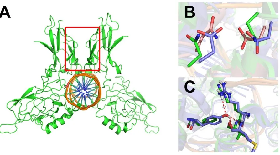
To examine the effects of perturbing the metal-binding environment, two Pirin mutants
were generated using site-directed mutagenesis: Q115A and W117A. These two residues are not
metal ligands, but are among the closest non-ligand residues to the iron (Figure 3.1) and show
100% conservation across all species (Figure 1.3). W117 is located near the protein surface while
Q115 is further inside the binding pocket and interacts with one of the iron’s water ligands
[image:50.612.162.477.427.666.2]through hydrogen bonding.
QCM-D was used to examine the differences in DNA binding and conformation resulting
from each mutation. Surprisingly, the mutations had different effects when they were combined
with p50 or p65 (Figure 3.3). When combined with p50 and Bcl-3, the W117A mutant showed a
reduced level of binding. However, when W117A was in a complex with p65, an opposite trend
was observed where the binding of the protein complex to the immobilized DNA was nearly
doubled. Similarly, the Q115A mutant also resulted in an increase of protein binding when
complexed with p65 but resulted in nearly no effect when combined with p50 and Bcl-3. Thus,
perturbing the metal-binding environment of Pirin has a different effect upon Pirin’s ability to
enhance the binding of p50 and p65 and further suggests that Pirin interacts with these two
[image:51.612.88.519.368.563.2]homodimers differently.
Figure 3.8: Effect of metal-center mutations on p50 and p65 binding
To further examine the differences between the interactions of p50 and p65 with the Pirin
mutants, the shear and viscosity of each protein-DNA complex was modeled using Q-Tools
Table 3.3: Viscoelastic properties of mutant-p50/p65 complexes
Viscosity (fold of WT) Shear (fold of WT)
p50 + Bcl-3 + W117A 0.7008 ± 0.0101 0.5553 ± 0.05532
p50 + Bcl-3 + Q115A 0.9746 ± 0.0547 0.7880 ± 0.1011
p65 + W117A 0.7320 ± 0.1253 0.5862 ± 0.2597
p65 + Q115A 0.8526 ± 0.1706 1.033 ± 0.0158
The Q115A mutant, which did not cause a significant change in the amount of protein
bound to the immobilized DNA when in a complex with p50 and Bcl-3, has a very similar shear
and viscosity to the wild-type complex. This suggests that the protein-DNA complex containing
the Q115A mutant adapts a conformation that is similar to the wild-type complex. The
similarities between their dissipation vs. frequency plots further support this (Figure 3.4). The
W117A-p50 complex, which has significantly reduced shear and viscosity compared to the
wild-type complex, has a noticeably different dissipation vs. frequency plot. Unlike Q115A, this
mutation appears to affect the conformation of the protein-DNA complex, likely resulting in the
reduced binding DNA binding affinity seen in Figure 3.3.
Similar to the Q115A-p50 complex, the Q115A-p65 complex also appears to have a
similar conformation to the complex containing wild-type Pirin, as evidenced by the nearly
identical shear, similar viscosity, and the similar overall shape of their dissipation vs. frequency
Figure 3.9: Dissipation vs. frequency plots for p50 and p65 complexes
The bend in the dissipation vs. frequency plots of the wild-type and Q115A indicates a
change in the conformation of the layer after a certain amount of binding, going from a relatively
rigid, organized conformation to a floppier, more disordered conformation with higher
dissipation. Since both plots bend at roughly the same point (around -10 Hz), it is likely that the
sensor surface becomes saturated at that point and any additional binding is either unable to take
the correct conformation due to crowding or is nonspecific binding to the existing layer of
protein adsorbed to the sensor, causing the sudden upshot in dissipation when the remaining
proteins bind in a disordered manner on the crowded surface. Although the assumed ratio of
protein complexes to DNA molecules is 1:1, the protein complexes are much larger and bulkier
than the immobilized DNA molecules so surfaces with high densities of immobilized DNA will
saturate at a protein:DNA ratio that is lower than 1:1. Interestingly, the dissipation vs. frequency
plot of the W117A-p65 complex does not have this bend, implying that the sensor surface isn’t
To further characterize the differences in DNA binding and conformation, the relative
thickness of the protein-DNA layer was obtained. Interestingly, the thickness of the layer formed
by the W117A-p65 complex is nearly twice as thick as the layer formed by the wild-type
complex. This observation could help explain why W117A-p65 does not experience surface
saturation. If the protein complex is binding to DNA in such a way that it forms a thicker layer of
protein, it could mean that the complexes are binding in a disordered manner that does not
restrict them to a specific sequence on the immobilized DNA strand which would allow for more
possible binding locations. In correlation with other measurements, the thickness of the layer
formed by the Q115A-p65 complex resembles the wild-type complex more than it does W117A.
Table 3.4: Thickness of Pirin mutant complexes with p65 and DNA Thickness (fold of WT+p65)
p65 + Q115A 1.16 ± 0.22
p65 + W117A 1.79 ± 0.45
The CD spectra of the Pirin-p65 complexes were compared to further explore the changes
in conformation resulting from the Q115A and W117A mutations (Figure 3.10). Measurements
of Pirin (or its mutant) were taken alongside p65 followed by measurements of each Pirin-p65
complex. The spectra of Pirin and p65 were subtracted from each Pirin-p65 spectra, leaving
behind the difference in spectra resulting from the change in secondary structure upon the two
Figure 3.10: CD spectra differences of mutant Pirin/p65 complexes
The additional features in the CD spectra of Pirin and p65 upon complex formation are
very similar to those seen in the formation of a Q115A-p65 complex (Figure 3.10A), supporting
the QCM data that showed that the Q115A mutant had similar viscoelastic properties (and thus a
similar conformation) to the wild-type protein. Also consistent with the QCM data was the
observation that the differences spectrum of the W117A-p65 complex did not follow the same
trend as the complexes formed from wild-type or Q115A Pirin, its difference spectrum having a
larger magnitude than those two complexes.
The effect of IgκB DNA binding upon the conformation of a Pirin-p65 complex and how
each mutation could affect this was also examined. In this case, DNA binding produced different
results with each complex (Figure 3.10B). As expected with W117A, the difference spectrum
showing a decrease in the negative ellipticity of the W117A/p65 complex rather than the increase
seen with the wild-type and Q115A complexes reveals that this mutation has significant
consequences on the structure of the protein-DNA complex. While the magnitude of the
the Pirin/p65/DNA complex shows a distinct minimum near 224 nm while the difference
spectrum of the Q115A/p65/DNA complex has a broader minimum ranging from 210-220 nm.
This finding reveals that, in spite of their similar conformations when bound to p65 alone, DNA
binding induces a conformational change with the wild-type complex that does not take place in