Journal of Chemical and Pharmaceutical Research, 2015, 7(8):61-66
Research Article
ISSN : 0975-7384
CODEN(USA) : JCPRC5
Antibiogram of methicillin resistant staphylococcus aureus (MRSA) in
healthcare settings
*Sarika Gupta
1, Aman Dongre
2, Anshuja Charvi Pandey
2, Rajesh Biswas
2and
Saksham Gupta
21
Banasthali University, Banasthali
2
Dr. B. Lal Institute of Biotechnology, Jaipur
_____________________________________________________________________________________________
ABSTRACT
MRSA is, by definition, any strain of Staphylococcus aureus that has developed resistance to beta-lactam antibiotics which include the penicillins (methicillin, dicloxacillin, nafcillin, oxacillin, etc.) and the cephalosporins. MRSA is capable of resisting Beta-Lactamase resistant Antibiotics via the mecA gene. This is a gene that encodes Penicillin-binding-protein 2a (PBP2a). β-lactam antibiotics have a low affinity for PBP2a, therefore cell wall synthesis is able to proceed in their presence. This study has been designed to look for the presence of MRSA and their correlation with drug resistance. In addition, MRSA producing organisms exhibit coresistance to many other classes of antibiotics resulting in limitation of therapeutic option. We bacteriologically analyzed 30 samples susceptible for MRSA. The samples were cultured using selective media and identification, susceptibility tests MRSA producing strains were made with microbiological methods. Susceptibility testing of MRSA isolates was done for various beta lactam, cephalosporins and methicillin antibiotics. MRSA was detected in 40% (12 out of 30 isolates). Among the samples under study conduct 17% were HA-MRSA and 83% were CA-MRSA. Among the MRSA isolates 67% were hemolytic and 33% were non-hemolytic. MRSA producing strains not only showed high-level resistance to beta lactam antimicrobial agents like Ampicillin (82.35%) followed by Ciprofloxacin (70.58%), Co-trimoxazole (58.82), Cefixime (52.94), Doxycycline/ Pefloxacin (41.17%), Ofloxacin (35.29%) Norfloxacin (29.41%), Cefuroxime/ Cephadroxil/ Amoxiclave/ Cefazolin/ Cephalexin/ Amikacin/ Netilmicin (17.64%). MRSA are often multidrug-resistant. Currently, the majority of S. aureus strains in communities are beta-lactamase producers, hence resistant to penicillin and ampicillin. Incidence of these organisms is being continuously increasing through out the world with limited treatment alternatives. Therefore, regular surveillance of hospital-associated infections including antimicrobial susceptibility pattern of MRSA and formulation of a definite antibiotic policy may be helpful in reducing the burden of MRSA infections in the hospital.
Keywords: Antibiogram, MRSA, Multiple Drug Resistance, healthcare settings
_____________________________________________________________________________________________
INTRODUCTION
emergence of drug-resistant organisms is now a major concern. Methicillin-resistant Staphylococcus aureus (MRSA) was endemic in hospitals by the late 1960s, but it appeared rapidly and unexpectedly in communities in the 1990s and is now prevalent worldwide. [9,17] S. aureus is notorious for its ability to become resistant to antibiotics. Infections that are caused by antibiotic-resistant strains often occur in epidemic waves that are initiated by one or a few successful clones. MRSA features prominently in these epidemics. Historically associated with hospitals and other health care settings, MRSA has now emerged as a widespread cause of community infections. Community or community-associated MRSA (CA-MRSA) can spread rapidly among healthy individuals. Outbreaks of CA-MRSA infections have been reported worldwide.[6]
The frequency of MRSA infections continues to grow in hospital-associated settings, and more recently, in community settings globally. The increase in the incidence of infections due to S. aureus is partially a consequence of advances in patient care and also of the pathogen's ability to adapt to a changing environment. Infection due to S. aureus imposes a high and increasing burden on health care resources. A growing concern is the emergence of MRSA infections in patients with no apparent risk factors.[7]The growing problem in the Indian scenario is that MRSA prevalence has increased from 12% in 1992 to 80.83% in 1999.[13] MRSA in tonsils may serve as a potential source for the spread of these organisms to other body sites as well to other individuals.[9] MRSA is prevalent in many hospitals and often reflects the difficulties in hospitals and the health service generally, in terms of the control and prevention of healthcare-associated infection.[14]Multidrug-resistant bacteria, such as MRSA, are endemic in healthcare settings in the United States and many other countries of the world. The prevalence rate of MRSA was found to be 29.1% in our study, which is in accordance with investigators from India (32.8%) [19] and Nepal (26.14%)[16]. On the contrary, some studies have reported alarmingly high incidence of MRSA infection. The epidemiology of MRSA over different parts of India is not uniform. Reports from a Delhi hospital showed a prevalence rate of 51.6% in 2001, whereas it was reported as 38.44% in the same hospital in 2008. [27]
Early detection of MRSA and formulation of effective antibiotic policy in tertiary care hospitals is of paramount importance from the epidemiological point. The present study has been carried out with an aim to know the prevalence and antibiotic sensitivity pattern of Staphylococcus aureus and MRSA (both Community acquired- MRSA and Hospital acquired MRSA) from the clinical samples isolates, in order to utilize the information obtained and formulate antibiotic policy and appropriate control measures.
EXPERIMENTAL SECTION
Collection of sample
Thirty samples were collected from Dr. B. Lal Clinical Laboratory, Jaipur susceptible for MRSA infection.
Isolation and characterization of the organisms
All the samples were subjected to microbiological analysis and were cultured on Mannitol Salt Agar & Blood Agar and then incubate at 37°C overnight. Further microbiological characterization was established.
Detection of multiple drug resistance (MDR pattern)
Antimicrobial susceptibility testing of isolates was done by the reference agar diffusion method, on Muller Hinton agar (Hi Media Laboratory-Mumbai, India.)as described by the National Committee for Clinical Laboratory Standards (NCCLS) andCLSI guidelines.[2,3,22]MRSA were identified using seventeen different antibiotic disks as Amoxiclave (30 mcg), Ampicillin (10 mcg), Carbenicillin (100 mcg), Cefixime (5 mcg), Ceftriaxome (30 mcg), Cefuroxime (30 mcg), Cephalexin (10 mcg), Cephotaxine (30 mcg), Amikacin (30 mcg), Ciprofloxacin (5 mcg), Cotrimoxazole (25 mcg), Doxycycline (30 mcg), Lomefloxacin (10 mcg), Netilmicin (30 mcg), Norfloxacin (10 mcg) and Ofloxacin (5 mcg). The susceptibility testing results were interpreted according to the recommendation of CLSI.[3,22]
RESULTS AND DISCUSSION
protocols. [21]Life-threatening sepsis, endocarditis, and osteomyelitis caused by MRSA have also been reported.[7] Surveillance for MRSA and eradication of the carrier state reduces the rate of MRSA SSI.[24] The outbreak remained uncontrolled despite rigid infection control measures. Subsequent emphasis on hand washing, in-service education and provision of weekly review of the MRSA colonization rates have failed to eliminate the organism from the unit.[29]
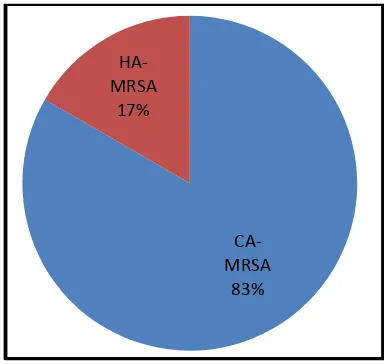
[image:3.595.119.479.241.423.2]In the present study 30 samples of Staphylococcus aureus were collected from Dr. B. Lal Clinical Laboratory, Jaipur. Out of 30 samples 24 were CA-MRSA (community acquires) and 6 were HA-MRSA (Hospital acquired). Isolation, identification & characterization of the clinical isolates were performed. All the samples were preserved in mannitol salt agar slants.
Fig 1: Total number of MRSA and Staphylococcus aureus isolates isolated from clinical samples
Fig 2: Total number of CA-MRSA, HA-MRSA and Staphylococcus aureus isolates from various samples
During the study period, 30 samples that were analyzed, out of them samples were urine (14), inanimate swab (6), semen (4), ear swabs (3), corneal swab (2) and cervical swabs (1) among them 12 were found to be infected with MRSA isolates respectively. (Fig 1) There were 40.0% (12) incidences of MRSA in all samples collected. From which 80% were CA-MRSA and 20% were HA-MRSA. (Figure 2).Similar distribution of MRSA was encountered in a study involving clinical isolates of SA maximum infections find in urine sample 39% or as follow Pus 17%, Inanimate environment 17%, Semen 11%, Ear 6%, Eye cornea 6% and Cervical infection 4%. MRSA isolated in patients in tertiary care institutions in these three Canadian provinces usually is acquired prior to admission.[6] A study conduct reported common genetic markers existed among 117 CA-MRSA isolates from the United States,
3
14
4
1
2
6
0
4
3
1
2 2
3
10
1
0 0
4
ear swab urine semen cervical
swab
corneal
swab
inanimate
swab
TOTAL
MRSA
SA
CA-MRSA
83%
HA-MRSA
[image:3.595.201.393.447.629.2]France, Switzerland, Australia, New Zealand, and Western Samoa by performing polymerase chain reaction for 24 virulence factors and the methicilline resistance determinant. Within each continent, the genetic background of CA-MRSA strains did not correspond to that of the hospital-acquired CA-MRSA.[8] The prevalence rate of CA-MRSA was found to be 40 % in our study, which is in accordance with investigators from India (32.8%)[19] and Nepal (26.14%)[16]. On the contrary, some studies have reported alarmingly high incidence of MRSA infection. The epidemiology of MRSA over different parts of India is not uniform. Reports from a Delhi hospital showed a prevalence rate of 51.6% in 2001, whereas it was reported as 38.44% in the same hospital in 2008.[27] Another study from north India has reported an incidence comparable to our study.[20] Community-acquired methicillin-resistant Staphylococcus aureus (MRSA) infections are not commonly recognized in healthy patients without predisposing risk. In our population, the majority of community-acquired MRSA infections occurred in previously healthy individuals without characteristics suggestive of MRSA transmission.[28] On the other hand Methicillin resistant Staphylococcus aureus (MRSA) is now a major cause of adult bacteraemia. All reports of Staphylococcus aureus bacteraemia to the Health Protection Agency were analysed from 1990 to 2001. There were 376 cases of MRSA bacteraemia in children <15 years over this time. The proportion of Staphylococcus aureus bacteraemia due to MRSA increased steadily from 0.9% in 1990 to 13% in 2000. The proportion was higher in infants. MRSA bacteraemia is now a serious problem in children in England and Wales. More data on the risk factors for acquisition and spread of MRSA in children are required.[15]
Fig 3: Total number of Hemolytic and Non-hemolytic MRSA isolated from various samples
All the samples were cultured in Blood agar media, D-Mannitol salt agar media. Multiple Drug Resistant (MDR) pattern of the isolates were identified on various groups of antibiotics as Methicillin, Cephalosporin & others. Now in all the MRSA incidences 67.0% were Hemolytic and 33% were non hemolytic.(Figure 3) whereas a study indicates that Coagulase-negative staphylococci (CoNS) are recognized as the a etiological agents of an important range of infections in humans. Most developed countries have reported an increase in CoNS infections in hospitalized patients that are resistant to meticillin and other antibiotics.[18]
During MDR analysis isolates were found to be MRSA on the basis of resistance to third generation antibiotics as Cefixime, Ceftriaxon, Cefuroxime, Cephadroxil, Cephotaxime and Ceftazidime. MDR pattern of the isolates were evaluated using seventeen antibiotics of Cephalosporin, Mithicillin group to confirm the presence of MRSA. The clinical isolates revealed maximum resistance to Ampicillin (82.35%) followed by Ciprofloxacin (70.58%), Co-trimoxazole (58.82), Cefixime (52.94), Doxycycline/ Pefloxacin (41.17%), Ofloxacin (35.29%) Norfloxacin (29.41%), Cefuroxime/ Cephadroxil/ Amoxiclave/ Cefazolin/ Cephalexin/ Amikacin/ Netilmicin (17.64%). (Fig 4) Whereas Clinical strain of methicillin-resistant Staphylococcus aureus (MRSA) with reduced susceptibility to vancomycin (MIC = 8 mg/L). The strain was isolated from a surgical wound infection which was refractory to vancomycin therapy. The MRSA strain (Mu50), which was isolated from the purulent discharge at the sternal incision site and from the debridement sample, had a vancomycin MIC of 8 mg/L by the broth microdilution method. The exact mechanism of the organism's reduced susceptibility to vancomycin remains to be determined but it may be due to an intrinsic mechanism of augmented cell-wall synthesis.[10] In a study at Aligarh, India, it was shown that 35.1% of S. aureus and 22.5% of coagulates-negative staphylococcal isolates were resistant to
HEMOLYT IC
67%
NON-HEMOLYT IC
methicillin. Highest percentage of MRSA (35.5%) was found in pus specimens (n=151). In case of both methicillin-resistant as well as methicillin-sensitive Staphylococcal isolates, zero resistance was found to vancomycin.[8] But, methicillin resistance is uncommon in community-acquired primary pyodermas in Mumbai.[23] In major southern districts of Tamilnadu, out of 906 strains of S. aureus isolated from clinical samples, 250 (31.1%) were found to be methicillin resistant. However, all strains of clinical and carrier subjects were sensitive to vancomycin.[25] Researchers in other part of the globe also observed that many of these MRSA isolates were becoming multidrug resistant and were susceptible only to glycopeptide antibiotics such as vancomycin. Low level resistance even to vancomycin was emerging.[1] A study from Maharashtra has reported that more than 90% isolates from South Maharashtra have been found resistant to penicillin, ampicillin, erythromycin, gentamicin, and tobramycin, whereas only 39.1% were resistant to methicillin.[12] Similar findings have been reported from other studies as well.[11] Multidrug resistance among MRSA strains was higher than those that were sensitive to methicillin. Ciprofloxacin was proposed to be an alternate therapy for MRSA infection.[26]
Fig 4: Histogram indicating the Resistance index of the clinically isolated MRSAunder study conduct
CONCLUSION
There is a progressive increase in MRSA positivity and multi-drug resistance in strains of Staphylococci. Theoretically, vancomycin is still the drug of choice for MRSA infections. The major reservoir of methicillin resistant staphylococci in hospitals is colonized/infected inpatients and colonized hospital workers. The field practitioners should be judicious enough in terms of use of antimicrobials so that the growing problem of antibiotic resistance of organism isolated does not reach a level of public health concern in this part of India. We recommend that frequent monitoring of susceptibility patterns of MRSA and the formulation of a definite antibiotic policy may be helpful in decreasing the incidence of MRSA infection. The dissemination of this information will help the treating clinicians for the primary care level physicians. Since resistance to multiple antibiotics among MRSA isolates is very common, there is a possibility of extensive outbreaks, which may be difficult to control. Early detection of MRSA and formulation of effective antibiotic policy in tertiary care hospitals is of paramount importance from the epidemiological point. We hope to undertake the study in community perspective to be acquainted with the actual prevalence and pattern of MRSA colonization and ascertain the extent of the menace at a larger level so that the information can be disseminated down the path, even to primary care health workers right at the grass root level before it becomes a public health problem.
Acknowledgement
We are grateful to the Department of Science and Technology, Government of Rajasthan for providing financial support for the study. The authors wish to acknowledge the kind assistance and contribution of Dr. B. Lal Gupta,
11.76 82.35
0 5.88
0 11.76
52.94
0
17.6417.64 11.76
0 11.76
70.58
58.82
41.17
0
11.7611.76 29.41
35.29 41.17
Managing Director, Dr. B. Lal Clinical Laboratory, Jaipur for providing samples for the case study and the guidance during the study conduct.
REFERENCES
[1]Assadullah S, Kakru D K, Thoker M A, Bhat F A, Hussain N and Shah A. Indian J Med Microbiol, 2003, 21,196-8.
[2]Behrooozi A, Rahbar M, Yousefi JL. African Journal of Microbiology Research, 2010, 4 (9), 881-884. [3]Bhattacharya S. Indian Journal of Medical Microbiology, 2006, 24 (1),20-24.
[4]Boucher H W, Corey G R. Clin Infect Dis., 2008, 46(5),S344-S349.
[5]Brook I and Foote P A. Int J Pediatr Otorhinolaryngol, 2006, 70(12),2099-2102. [6]Chambers H F and Deleo F R. Nat Rev Microbiol, 2009, 7(9),629-641.
[7]Cox R A, Conquest C, Mallaghan C and Marples R R. J Hosp Infect, 1995, 29(2),87-106.
[8]Dar J A, Thoker M A, Khan J A, Ali A, Khan M A, Rizwan M, Bhat K H, Dar M J, Ahmed N and Ahmad S. Ann Clin Microbiol Antimicrob., 2006, 14,5-22.
[9]DeLeo F R and Chambers H F. J Clin Invest., 2009, 119(9),2464-74.
[10]Edward Gorak J, Yamada S M and Brown J D. Clinical Infectious Diseases, 1999, 29(4), 797-800.
[11]Embil J, Ramotar K, Romance L, Alfa M, Conly J, Cronk S, Taylor G, Sutherland B, Louie T, Henderson E and Nicolle L E. Infection Control and Hospital Epidemiology, 1994, 15(10), 646-651.
[12]Henderson D K. Am J Med., 2006, 119(6),S45-52.
[13]Hiramatsu K Hanaki H, Ino T, Yabuta K, Oguri T and Tenover F C. Journal of Antimicrobial Chemotherapy, 1997, 40(1), 135-136.
[14]Humphreys H. Eur J Clin Microbiol Infect Dis., 2008, 27(6),409-13.
[15]Khairulddin N, Bishop L, Lamagni T L, Sharland M G and Duckworth G. Arch Dis Child, 2004, 89(4),378-379.
[16]Kumari N, Mohapatra T M and Singh Y I. JNMA J Nepal Med Assoc., 2008, 47(170),53-6. [17]Liebowitz L D. Int J Antimicrob Agents, 2009, 34(3),S11-3.
[18]Machado A, Reiter K C, Rodrigo M P R M and Barth A L. J Med Microbiol, 2007, 56(10), 1328-1333. [19]Mehta A P, Rodrigue C, Sheth K, Jani S, Hakimiyan A, Fazalbhoy. J Med Microbiol., (1998), 16,31-4. [20]Mohanty S, Kapil A and Dhawan B. Indian J Med Microbiol., 2004, 58(1):10-5.
[21]Nadig S, Namburi P, Raghunath D and Arakere G. Current Science, 2006, 91(10); 1364-1369.
[22]National Committee for Clinical laboratory Standards. Performance Standards for antimicrobial disk susceptibility tests; approved standard. NCCLS, 6th ed. 1999.
[23]Patil R, Baveja S, Nataraj G and Khopkar U. Indian J Dermatol Venereol Leprol., 2006, 72(2),126-8.
[24]Pofahl W E, Goettler C E, Ramsey K M, Cochran M K, Nobles D L and Rotondo M F. J Am Coll Surg. 2009, 208(5),981-6.
[25]Rajaduraipandi K, Mani K R, Panneerselvam K, Mani M, Bhaskar M and Manikandan P. Indian J Med Microbiol; 2006, 24(1),34-8.
[26]Sharon M S, Robert H K and Flor T T. Antimicrob Agents Chemother., 1989, 33(2),181-4. [27] Tiwari H K, Sapkota D and Sen M R. Infection and Drug Resistance, 2008, 1,57-61.
[28]Vandenesch F, Naimi T, Enright M C, Lina G, Nimmo G R, Heffernan H, Liassine N, Bes M, Greenland T, Reverdy M E and Etienne J. Emerging Infectious Diseases, 2003, 9(8), 978-84.