Journal of Chemical and Pharmaceutical Research, 2016, 8(2):773-779
Research Article
CODEN(USA) : JCPRC5
ISSN : 0975-7384
Renoprotective effect of curcumin on acetaminophen-induced
nephrotoxicity in rats
Amel Fouad Mohamed Ismail
1and Asmaa A. M. Salem
21
Drug Radiation Research Department, National Center for Radiation Research and Technology(NCRRT), Atomic Energy Authority, Atomic Energy Authority, Nasr City, Cairo, Egypt
2Regional Center for Food and Feed (RCFF), Agriculture Research Center, Giza, Egypt
_____________________________________________________________________________________________
ABSTRACT
This study investigated the nephroprotective effect of curcumin against acetaminophen (APAP)induced nephrotoxicity in rats. APAP-induced nephrotoxicity was evident by a significant increase (P<0.01) of urea and creatinine in serum. Furthermore, a significant increase (P<0.01) of lipid peroxidation as malondialdehyde (MDA), nitric oxide (NO), protein carbonyl (PC) contents, interleukin-1β(IL-1β) and tumor necrosis alpha (TNF-α)accompanied by a significant decrease in superoxide dismutase (SOD), catalase (CAT), glutathione peroxidase (GPx) activities and glutathione (GSH) content were observed in the kidney tissues. In addition, the cytochrome-P2E1 (CYcytochrome-P2E1) and inducible nitric oxide synthase (iNOS)gene expression ratios showed a significant elevation (P<0.01) in the kidney tissues. Oral administration of curcumin (100 mg/kg,b.wt.) ameliorated these adverse effects and showed nephroprotective activity against APAP toxicity. The nephroprotective activity of curcumin could be attributed to the regulation of the renal antioxidant balance, the down-regulation of CYP2E1 and iNOSgene expressions and the improvement of IL-1β and TNF-α levels. In conclusion, oral administration of curcumin showed protective effect against APAP-induces nephrotoxicity in rats.
Key words: Curcumin, acetaminophen, nephrotoxicity, CYP2E1, iNOS, IL-1β and TNF-α.
_____________________________________________________________________________________________
INTRODUCTION
The kidney is a vital organ that plays a fundamental role in the health and disease conditions, particularly in development and growth. The main function of kidney is to maintain the total body fluid volume, its composition and acid base balance. A number of environmental variables, including certain drugs could influence these functions[1].Acetaminophen (paracetamol, N-acetyl-para-aminophenol, APAP) is one of the most widely used pharmaceutical analgesics and antipyretic agents that is fairly inexpensive, safe and effective for treatment of common aches and colds[2]. The APAP overdose is a chief clinical problem that resulting in severe liver injury and potentially acute liver failure [2,3,4]. However, renal insufficiency occurs in approximately 1-2% of patients with APAP overdose [5].Renal APAP toxicity could evokemultisystem organ damage [6]. Moreover, in sensitive individuals, as those persons with renal insufficiency, the therapeutic doses of APAP have been implicated in kidney damage [7].
EXPERIMENTAL SECTION
Chemicals
Acetaminophen, curcumin, 1,1,3,3‐tetraethoxypropane and all other chemicals and reagents used in this study were of analytical grade and purchased from Sigma-Aldrich Chemical Co. (St. Louis, MO, USA).
Animals
Male Wistar rats weighing 180-200 g each were obtained from the Nile pharmaceutical Co., Cairo, Egypt. Animals were housed at the animal facility, at the National Centre for Radiation Research and Technology (NCRRT), Atomic Energy Authority, Cairo, Egypt. They were kept under standard laboratory conditions of light/dark cycle (12/12 h), a temperature of 25±2°C and humidity of 60±5% with free access to food (standard laboratory-pellet-diet) and drinking water ad libitum. The animals were allowed to acclimatize for 1 week before starting the experiment.The study was conducted in accordance with international guidelines for animal experiments and approved by the Ethical Committee at the NCRRT.
Experimental design
Forty Male Wistar rats were randomized and divided into four groups of ten animals each. The first group of rats served as control and received only vehicle (Arabic gum 1% suspension) along the experimental period. The second group received curcumin (100 mg/kg b. wt.),suspended in Arabic gum 1%, by gavage for 21 consecutive days. The third group received a vehicle(Arabic gum 1% suspension) by the same route of administration for 21 days and 1 h after the last dose of curcumin, a single dose of APAP (3 g/kg) suspended in Arabicgum 1% was orally administered [13]. The fourth group received curcumin (100 mg/kg, b. wt.) for 21 days and 1 h after curcumin, on the last day, they received the single dose of APAP (3 g/kg).After 6 h fasting period, with free access to water, all animals were then euthanized by cervical dislocation. The blood was collected in EDTA-free tubes then the blood was allowed to clot for 30 min at 25°C, centrifuged at 1200 g at 4°C using universal centrifuge (16R, Germany) and sera were separated for the biochemical assessment. The kidneys from each rat were quickly removed, rinsed in ice-cold physiological saline, blotted dry on a filter paper and stored at -80°C.
Determination of urea and creatinine levels:
The biochemical blood analyzer (ALFA Wassermann Diagnostic Technologies, LLC, ACE, Alera, USA at RCFF) was used for the determination of urea and creatinine in the serum.
Assessment of antioxidant status in the Kidney tissues:
The kidney from each rat was weighed and homogenized in ice-cold 0.15 M KCl solution using a Teflon homogenizer (Universal laboratory aid, Type MPW-309, Poland) to obtain 10% (w/v) homogenates. The homogenates were centrifuged at 1200 g for 15 min at 4°C. Aliquots of the homogenates kidney were used in the following determinations:
Superoxide dismutase (SOD)activity was determined according to Nishikimi et al. [14].Catalase(CAT) activity was assessed according to Aebi[15]. Glutathione peroxidase(GPx)activity was determined according to Paglia and Valentine’s method[16].Glutathione (GSH) concentration was measured according to Beutler et al[17]. Lipid peroxidation in terms of malondialdehyde (MDA) were measured according to the method of Satoh [18],using 1,1,3,3‐tetraethoxypropane as a standard. Nitric oxide (NO) was measured based on Griess colorimetric assay according the method of Montgomery and Dymock[19]. Protein carbonyl content (PC) was assessed by the procedure of Floor and Wetzel [20].
Determination of the proinflammatory cytokines, IL-1β and TNF-α in the kidney tissues:
The proinflammatory cytokines IL-1β and TNF-α were determined in the kidney homogenates using an ELISA kits for rat (Glory Science Co., Ltd, USA). The measurements were done according to the catalogue-instruction guidelines.
Detection of CYP2E1 and iNOS relative gene expression in the kidney tissues: -RNA isolation and reverse transcription
spectro-photometrically by measuring the absorbance at 260 nm. Total RNA was incubated at 70°C for 10 min to prevent secondary structures until the analysis of cDNA synthesis reaction, which was performed using the Reverse Transcription System (Promega, Leiden; The Netherlands). The RNA was supplemented with MgCl2 (25 mM),
RTease buffer (10X), dNTP mixture (10mM), oligo d(t) primers, RNase inhibitor (20 U) and AMV reverse transcriptase (20 U/µl). This mixture was incubated at 42°C for 1 h.All samples were stored at -80°C
-Quantitative real time PCR
qRT-PCR was performed using an optical 96-well plate with an ABI PRISM 7500 fast sequence detection system (Applied Biosystems, Carlsbad, California) and universal cycling conditions min 95°C, 40 cycles of 15 s at 95°C and 60 s at 60°C). Each 10 µl reaction contained 5 µlSYBR Green Master Mix (Applied Biosystems), 0.3 µl gene-specific forward and reverse primers (10 µM), 2.5 µl cDNA and 1.9 µl nuclease-free water. The sequences of PCR primer pairs used for each gene as follows, CYP2E1was carried out in kidney' tissues with the oligonucleotide 5’ ACT TCT ACC TGC TGA GCA C 3’ (forward) and 5’ TTC AGG TCT CAT GAA CGG G 3’ (reverse) [21],
iNOS: Forward 5’-GGG CCA CCT TTA TGT TTG TG-3’, reverse: 5'-CCG GTG GGT TTC TTCTTC TTG AA-3'
[22].GAPDH: Forward 5’-ATG GGA GTT GCT GTT GAA GTC A-3’, reverse: 5'-CCG AGG GCC CAC TAA AGG-3’ [23].
-Gel electrophoresis,
Ten µl of PCR product was analyzed on 2% agarose gel with ethidium bromide staining and the product was visualized on ultraviolet transilluminator, then gel documentation was performed. PCR products were semi-quantified by using a gel documentation system (Bio Doc Analyze) supplied by Biometra, Germany. Then, the data were analyzed with the ABI Prism sequence detection system software and quantified using the v1·7 Sequence Detection Software from PE Biosystems (Foster City, CA). The relative expression of the studied genes was calculated using the comparative threshold cycle method. All values were normalized to the GAPDH genes [24].
Statistical Analysis
All statistical analyses were conducted by using the statistical package for Windows Version 15.0 (SPSS Software, Chicago, IL). The results for continuous variables were expressed as mean±SEM. Values were compared by one-way analysis of variance (ANOVA). Post hoc testing was performed for inter-group comparisons using the least significant difference (LSD) test, significance of P values < 0.05 were considered statistically significant.
RESULTS AND DISCUSSION
The current experiment showed that a single dose of APAP(3 g/kg) induced severe kidney toxicity after6 h of administration. This toxicity was reflected by high levels of urea and creatinine in the serum. Moreover, administration of the nephron-toxic dose of APAP to rats induced oxidative stress in the kidney tissues, where MDA, NO and PC levels showed high elevation levels in parallel with reduced GSH level, SOD and GPx activities. Furthermore, the acute APAP overdose induced high degree of inflammation manifested as elevation of the pro-inflammatory cytokines TNF-α and IL-1β in the kidney tissues of the intoxicated rats. Furthermore, to explore the oxidative stress mechanism, iNOS and CYP2E1 gene expressions were studied. The data showed a high elevation in the gene expression ratios of these two enzymes. These results of APAP-induced nephrotoxicity model is comparable to those reported by other researchers[6,8,25,26,27,28,29].
.
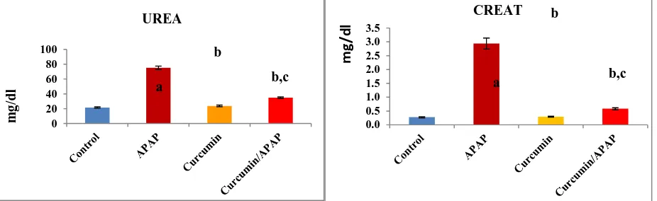
Fig. (1): Effect of acetaminophen and curcumin on serum urea and creatinine levels. The results were expressed as mean ± S.E, n=6 rats/group. a non-significant to control (p > 0.05), b significant to control (p < 0.01) and c significant to APPA (p < 0.01)
0 20 40 60 80 100
UREA
m
g
/d
l b,c
b
a
0.0 0.5 1.0 1.5 2.0 2.5 3.0 3.5
CREAT
b,c b
a
m
g
/d
[image:3.595.76.539.567.709.2]Current investigation revealed that the rats developed renal toxicity after 6 h of APAP administration as judged from a high significant elevation (P<0.01) of urea and creatinine in serum of the intoxicated rats approximately with 4 and 11 folds, respectively as compared to the control animals (Fig. 1). APAP overdose triggers acute renal failure, and chronic exposure to APAP has been associated with chronic renal failure [30].
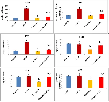
Our results exerted the oxidative stress and antioxidant status in the kidney tissues of the different experimental groups. The oxidative stress markers: MDA, NO and PC contents were significantly elevated by 5, 3 and 3.3 folds, respectively, as compared to the untreated animals. GSH level was diminished to 44 % of the control value. Moreover, the activities of SOD and GPx were inhibited to 50 % and 66 %, respectively, as compared to the control values (Fig. 2).
..
.
.
Fig. (2): Effect of acetaminophen and curcumin on antioxidant status in the kidney tissues. Lipid peroxides in term of malondialdehyde (MDA), nitric oxide (NO), protein carbonyl content (PC), reduced glutathione (GSH), superoxide dismutase (SOD) and glutathione peroxidase (GPx). The results were expressed as mean ± S.E, n=6 rats/group. a non-significant to control (p > 0.05), b significant to
control (p < 0.01) and c significant to APPA (p < 0.01)
The medicinal doses of APAP are metabolized via glucuronidation and sulfuration reactions occurring primarily in the liver, and results in water-soluble metabolites, which are excreted through the kidney. Because of the metabolic conversion of APAP by the microsomal P-450 enzyme system, a highly reactive intermediate, N-acetyl-p-benzoquinone imine (NAPQI) is produced. NAPQI directly reacts with glutathione (GSH) and at overdoses of APAP that evoke the depletion of the cellular GSH. This allows NAPQI to bind to cellular proteins and initiate lipid peroxidation, leading to renal injury [27,31]. Previous studies demonstrated that acute APAP overdose increased lipid peroxidation and suppressed the antioxidant defense system in the renal tissues of experimental animals
[image:4.595.74.536.197.628.2][8,9,32]. Nitric oxide, a free radical of oxygen, significantly increased in the kidney due to APAP intoxicated rats in the present study.
The data indicated that the nephrotoxic doses of APAP in rats increased the production of NO. The overproduction of NO could play an important role in the pathogenesis of APAP-induced renal damage [27].In addition, reactive NO may combine with O2•- to form peroxynitrite, which produces3-nitrotyrosine in protein. This scavenging effect
on O2•-by NO may be a mechanism by which tissues of host are protected from the deleterious effects of O2•- andO2
•--derived reactive oxygen species [33]. Peroxynitrite is also known to initiate LPO, which cause direct or indirect oxidative damage in nucleic acids or promote apoptosis[34,35,36].Other studies showed that treatment with toxic doses of APAP increased NO production in the liver and kidney tissues and this was correlated with the expression of the inducible nitric oxide synthase (iNOS) protein [37].
Free radicals can lead to the formation of protein/protein cross-linkages, oxidation of protein backbone resulting in protein fragmentation and modification of amino acid side chains, which includes oxidation of sulfhydryl moieties and formation of protein carbonyls (PC) [38].
The antioxidant defense system copes with the oxidative stress and operates for scavenging ROS to avoid this oxidative stress. Among the different antioxidant molecules, SOD and GPx mutually perform as the principal enzymes in the removal of the ROS. Reduction in SOD activity in APAP exposed animals may be due to the overproduction of superoxide radical anions (O2•-) [39]. In order to eliminate the excess free radicals, GPx utilize
GSH during their course of reactions. Decrease in GSH content due to the APAP simultaneously decreased the activity of GPx and the other glutathione-dependent enzymes: glutathione-s-transferase and glutathione reductase [27].
.
Fig. (3): Effect of acetaminophen and curcumin on serum Tumor Necrosis Factor alpha (TNF-α) and interleukin 1β (IL-1β) levels in kidney tissues. The results were expressed as mean ± S.E, n=6 rats/group. a non-significant to control (p > 0.05), b significant to control
(p < 0.01) and c significant to APPA (p < 0.01)
.
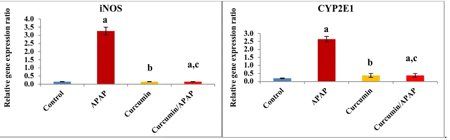
Fig. (4): Effect of acetaminophen and curcumin on the relative gene expression ratio of inducible nitric oxide synthase (iNOS) and the cytochrome P2E1 (CYP2E1) in the kidney tissues. The results were expressed as mean ± S.E, n=6 rats/group. a non-significant to
control (p > 0.05), b significant to control (p < 0.01) and c significant to APPA (p < 0.01)
The present study showed that the rats developed a high significant degree of inflammation after 6 h of APAP administration as judged from a high significant elevation (P<0.01) of TNF-α and IL-1β levels in the kidney tissues of the intoxicated rats, approximately with 380 % and 406 %, respectively as compared to the control animals (Fig.
[image:5.595.79.537.348.491.2] [image:5.595.78.539.538.682.2]3). In addition, the data showed that iNOS gene expression relative ratio was significantly increased (P<0.01) with 21 folds as compared to the control value. However, CYP2E1 gene expression relative ratio was significantly increased (P<0.01) with 13 folds as compared to the control value (Fig. 4).
Ghosh et al[27]showed that APAP overdose could induce renal inflammation with increased generation of TNF-α. This fact can be explained by the ability of TNF-α to up-regulate the iNOS and that in turn increases NO production [40].In the current study, the elevated level of CYP2E1 gene expression due to APAP-intoxication is in accordance with the results of Kim et al.[41]where expression of CYP2E1 proteins in rat liver was elevated significantly following APAP administration. CYP2E1, is one of the members of cytochromes P450 (CYPs) family, which could metabolize numerous small molecules of toxicological interest, including APAP generating highly reactive oxygen radicals[42].
Curcumin administration in a dose of 100 mg/kg of body weight for 21 days showed significant reduction of the nephrotoxicity induced by APAP. The present study showed that curcumin pretreatment markedly ameliorated the renal damage induced by APAP overdose as evidenced by regulating of the biochemical parameters in renal tissue (improved oxidant/antioxidant status and prevention of the detrimental increase in TNF-α and NO levels). Curcumin pretreatment, on the other hand, ameliorated these events and helps the organ remaining in its normal physiological state. The renal protective effect of curcumin against the serious renal complication due to APAP overdose could be attributed to the antioxidant and anti-inflammatory activity of curcumin which scavenging ROS. Furthermore, the results of this study indicated that pre-treatment of rats with curcumin suppressed NO over-production induced by the acute toxic dose of APAP via down-regulated iNOS expression. The mechanism of iNOS down-regulation could be attributed to the direct radical scavenging property of curcumin or indirect mechanism, as a consequence of decreased the TNF-αand IL-1β formation (as TNF-αand IL-1β have the ability to up-regulate the iNOS that increases NO production) [40].The antioxidant properties of curcumin are mainly based on the suppression of inducible nitric oxide synthase [43]. Curcumin cytoprotective, anti-inflammatory, antitumor, and antioxidant property is based on several mechanisms. The reported ones are mainly based on the suppression of pro-inflammatory mediators, including inducible nitric oxide synthase (iNOS), nuclear factor kappa B (NF-κB), tumor necrosis factor alpha (TNF-α) [44].In addition, curcumin treatments to PAPA-intoxicated rats normalized these values and prevent their elevation, which may be attributed to the anti-inflammatory activity of curcumin. Curcumin has inhibitory activity on nuclear factor kappa B (NF-κB) that induces these proinflammatory cytokines. It was demonstrated that curcumin down-regulates the expression of various inflammatory cytokines, including IL-1, IL-6, IL-8 and chemokines, and inhibits the action of TNF-α, one of the most pro-inflammatory cytokines [45]. Curcumin suppresses the activation of the transcription factor Nuclear Factor Kappa Beta (NF-κB), which regulates the expression of pro-inflammatory gene products [46]. Also, the data demonstrated that curcumin down-regulated CYTPE1 gene expression, which elevated in PAPA-intoxicated rats. Therefore, curcumin could prevent the production of ROS and the oxidative stress inside the kidney tissues that mediated by CYP2E1 and maintained the cellular antioxidant defense mechanisms of kidney tissues.
CONCLUSION
The results of this study demonstrate that curcumin has the ability to protect against the nephrotoxicity induced by APAP. The ability of curcumin to provide this protective effect is positively correlated with its ability to suppress APAP-induced NO overproduction, depletion of intracellular GSH, inhibition of SOD and GPx activities and increase of LPO level. Thus, the protective effect of curcumin against APAP-induced renal damage may due to inhibition of NO and ROS overproduction and maintenance of cellular antioxidant defense mechanisms via down-regulation of iNOS and CYP2E1 gene expressions that could be due to the antioxidant, anti-inflammatory activities of curcumin
REFERENCES
[1]S Priyamvada; Priyadarshini M; Arivarasu NA; NFarooq; S Khan; SA Khan; MdWasim Khan; ANK Yusufi.
Prostaglandins, Leukotrienes and EssentialFatty Acids,2008, 78, 369-381.
[2]AM Larson; J Polson; RJ Fontana; TJ Davern; E Lalani; LS Hynan; JS Reisch;; FV Schiodt; G Ostapowicz; AO Shakil; WM Lee. Hepatology,2005, 42, 1364-1372.
[3]WM Lee. Semin Liver Disease,2003, 23, 217-226.
[4]T Uehara; O Kosyk; E Jeannot; BU Bradford; K Tech; JM Macdonald; GA Boorman; S Chatterjee; RP Mason; SB Melnyk; VP Tryndyak; IP Pogribny; I Rusyn. Toxicology and Applied Pharmacology,2013, 266, 224-232. [5]M Mazer; J Perrone. J Med Toxicol,2008, 4, 2-6.
[6]IO Aycan; O Tokgöz; A Tüfek; U Alabalık; O Evliyaoğlu; H Turgut; F Çelik; A Güzel. International Journal of
[7]HL Bonkovsky; RE Kane; DP Jones; RE Galinsky; B Banner. Hepatology, 1994, 19, 1141-1148. [8]OA Abdel-Zaher; RH Abdel-Hady; MM Mahmoud; MMY Farrag. Toxicology,2008, 243, 261-270. [9]A Ghosh; PC Sil. J Biochem Mol Biol., 2007; 40: 1039-1049.
[10]AK Tuba; I Gulcin. ChemBiol Interact,2008, 174, 27-37.
[11]Y Xia; L Jina; B Zhanga; H Xuea; Q Lic; Y Xu. Life Science,2007, 80, 2161-2169.
[12]SS Sandur; KM Pandey; B Sung; SK Ahn; IA Murakami; G Sethi. Carcinogenesis, 2007, 28, 1765-1773. [13]MM El-Shafey; GM Abd-Allah; AM Mohamadin; GI Harisa; AD Mariee. Pathophysiology,2015, 22, 49–55. [14]M Nishikimi; NA Roa; K Yogi. BiochemBiophysRes Commun.,1972, 46, 849-854.
[15]H Aebi; Catalase. In: Methods in Enzymatic Analysis. Bergmeyer HU (ed); Chemic Academic Press; Inc. Verlag. 1984, 2, 673-678.
[16]ED Paglia; WN Valentine. J Lab Clin Med.,1967, 70, 158-169. [17]E Beutler; O Duron; BM Kelly. J Lab Clin Med., 1963, 61, 882-890. [18]K Satoh. ClinicaChimicaActa.,1978, 90, 37-43.
[19]HA Montgomery; JF Dymock. Analyst., 1961, 86, 414-416. [20]E Floor;MG Wetzel. JNeurochem.,1998, 70, 268-275.
[21]XB Man; L Tang; XH Qiu; LQ Yang; HF Cao; MC Wu; HY Wang. World J Gastroenterol.,2004, 10, 1565-1568.
[22]HS Koo; KC Kim; YM Hong. Korean Circ J.,2011, 41, 83–90.
[23]H Hayase; A Ishizu; H Ikeda; Y Miyatake; T Baba; M Higuchi; A Abe; U Tomaru; T Yoshiki.
IntImmunol.,2005, 17,677-684.
[24]KJ Livak; TD Schmittgen. Methods,2001, 25, 402-408.
[25]MBhadauria; SKNirala. Environmental Toxicology and Pharmacology,2009, 27, 17-25.
[26]AA Fouad; MT Yacoubi; MH El-Bidawy. Environmental Toxicology and Pharmacology,2009, 27, 277-282. [27]J Ghosh; J Das; P Manna; PC Sil. Toxicology,2010, 268, 8-18.
[28]S Pradhan; S Mandal; S Roy; A Mandal; K Das; DKNandi.Saudi Pharmaceutical Journal,2013, 21, 187-192. [29]YL Zhao; GD Zhou; HB Yang; JB Wang; LM Shan; RS Li; XH Xiao. Food and Chemical Toxicology,2011, 49, 1705-1710.
[30]CM Fored; E Ejerblad; P Lindblad; JP Fryzek; PW Dickman; et al. N Engl J Med.,2001, 345, 1801-1808. [31]SG Hart; WP Beierschmitt; DS Wyand; EA Khairallah; SD Cohen.ToxicolApplPharmacol., 1994, 126, 267-275.
[32]C Lorz; P Justo; A Sanz; D Subira. J Am SocNephrol.,2004, 15, 380-389.
[33]JS Beckman; TW Beckman; J Chen; PA Marshall; BA Freeman. ProcNatlAcad Sci.,1990, 87, 1620-1624. [34]SY Lee; SJ Lee; C Han; AA Patkar et al. ProgNeuro-PsychopharmacolBiolPsychiat.,2012, 46, 224-235. [35]P Pacheret; JS Beckman; L Liaudet. Physiol Rev.,2007, 87, 315-424.
[36]D Huang; A Shenoy; JK Cui; W Huang et al. J Fed Am SocExp Biol.,2009, 14, 407-417.
[37]CR Gardner; JD Laskin; DM Dambach; M Sacco; SK Durham; MK Bruno; SD Cohen; MK Gordon; DR Gerecke; P Zhou; DI Laskin. ToxicolApplPharmacol.,2002, 184, 27-36.
[38]BS Berlett; ER Stadtman. J Biol Chem.,1997, 272, 20313-20316.
[39]K Yamanaka; A Hasegawa; R Sawamura; S Okada. ToxicolApplPharmacol.,1999, 1108, 205-213. [40]SM Morris; TR Billiar. Am J Physiol., 1994, 266, 829-839.
[41]SN Kim; JY Seo; DW Jung; MY Lee; YS Jung; YC Kim. Drug Metabolism and Disposition,2007, 35, 1754-1758.
[42]FJ Gonzalez. Mutation Research,2005, 569, 101-110.
[43]D Černý; N Lekić; K Váňová; L Muchová; A Hořínek; E Kmoníčková; Z Zídek; L Kameníková; H Farghali.
Fitoterapia,2011, 82, 786-791.
[44]VP Menon; AR Sudheer. AdvExp Med Biol.,2007, 595, 105-25.