2969
A REVIEW STUDY OF METHODS UTILIZED FOR
IDENTIFYING AND SEGMENTING THE BRAIN TUMOR
FROM MR IMAGERIES
AHMED SAIFULLAH SAMI1 , MOHD SHAFRY MOHD RAHIM2 , FALAH Y H AHMED3 AND
GHAZALI BIN SULONG4
1Faculty of Computing, Universiti Teknologi Malaysi Universiti Teknologi Malaysi,UTM Skudai,81310,
Johor Bahru, Malaysia
2IRDA Digital Media Center, Universiti Teknologi Malaysi Universiti Teknologi Malaysi,UTM Skudai,
81310, Johor Bahru,Malaysia
3,4Department of Information Science and Computing, Faculty of Information Sciences and Engineering
(FISE), Management and Science University, 40100 Shah Alam, Malaysia
E-mail: [email protected], [email protected], [email protected], [email protected]
ABSTRACT
This paper provides a detailed analysis of the existent methods and approaches utilized in medical image segmentation. Also integrate a comparative study of the automated brain tumor coupled through the utilization of tuomr detection techniques. Additionally, the paper will provide an analysis of the process integrated pertaining to the retrieval of brain images through the identification of the specific data sets selected in the process to identify the stipulated features., [1]maintains that the utilization of a computer-aided diagnosis in medical imaging influences the decision-making capacity of specialists in the provision of accurate images pertaining to the existent volume and distance. Also maintains that the detection of brain ailments remains difficult among radiologists and neurologists due to the existence of similar brain abnormalities. The existence variances influence the level of difficulties experienced pertaining to the tasks due to the existence of differential diagnosis coupled with the complexity attached to manual segmentation. The processes delimit the accuracy levels leading to the identification of the need for the provision of additional time considered necessary in meeting the stipulated needs of the process. The process necessitates the integration of extensive processes necessary in the identification of reliable and accurate algorithms necessary in the provision of solutions pertaining to the existent questions. This part of the study seeks to integrate an illustrative analysis, which will provide illustration that will identify the existent needs relating to automatic detection. The approaches will influence the evaluation processes integrated pertaining to the existent human brain abnormalities including injuries, tumor and edema. The process will also remain instrumental in the identification of additional abnormalities that can be extracted from computerized images of human brain.
Keywords: Five Keywords are Required Separated By Commas (Capitalize Each Work Italic)
1. INTRODUCTION
Nowadays, various type of Magnetic Resonance Imaging (MRI) images are usually used for segmenting brain tumor, because MRI image gives extra details on tumor and its areas. Tthis paper focus on the challenges in current medical imaging investigates which faces with a tough effort in the detection of tumor through using MRI , the accurate segmentat0ion of any brain tumor is the most significant task. For this purpose, the brain tumor image is divided into a number of regions.
2970 which will provide illustration that will identify the existent needs relating to automatic detection. The approaches will influence the evaluation processes integrated pertaining to the existent human brain abnormalities including injuries, tumor and edema. The process will also remain instrumental in the identification of additional abnormalities that can be extracted to, this paper only focuses on two dimensional images the volumetric visualization was not included in this review in addition the Anomalous progression of tissue in an intensified manner within a living organism accounts for tumor. The cells existing within the tissue of a malignant tumor is responsible for self-multiplying itself and replicate all over feasible regions of a human physique. Tumor existing at varied stage causes a diversified effect in the human body. Tumor cells usually demolish the well-being nature of a normal tissue by means of creating diverse symptoms like inflammation in the affected portion or to generate an added pressure in other organs of physique consequently causing a surge of pressure within the infected portion of the tumor. In particular, a brain tumor prevailing in the stage of a metastatic level is designated as cancer that is traversed from other organs to the brain. During its preliminary stages, the transferring nature of a tumor that resembles a simple communicable disease is actually terminal (if not recognized). Hence, a better brain tumor processing methodology is a prerequisite of the present scenario. In general, images acquired through Magnetic Resonance (MR) are utilized for identification of brain tumor. In order to construct a perfect and robust brain tumor segmenting procedure, the faults surviving in the pre-existing techniques are assessed in this paper for suggesting further improvisations in the methodology that is to be proposed in this research.
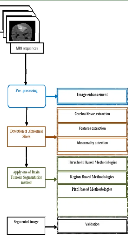
[image:2.612.98.283.550.684.2]Figure 1: Brain tumor in the T1 with against T2 by
Figure 2:mapping literature of Brain tumor segmentation
2. RELATED WORK
2.1 Techniques utilized for preprocessing
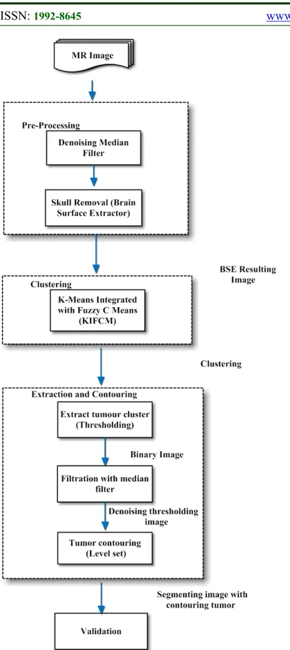
2971 Figure 3: Overall Flow of proposed technique
De-noising- A non-linear fashion of filtering methodology was utilized to perform the process of alleviating noise from those MR images by means of implementing median filtering. This non-linear fashion of filtering technique was best suited for the image being processed with edges. On completion of this preprocessing stage, the outcome obtained was devoid of any disorders available with the image.
Skull Removal- the process of eliminating the background part of the brain MR image like scalp portion, skull was done with this stage of preprocessing. The quantity of memory space
utilized was significantly diminished and also the occurrence of delay in processing the acquired MR image was completely eradicated. A specific Brain Surface Extractor (BSE) algorithm was utilized for accomplishing this sort of skull stripping methodology. The procedure of brain segregation and morphological erosions were performed in skull removal methodology. The noiseless outcome of this procedure was obtained by means of taking away the abnormalities in the imagery and the edges prevailing in that was identified.
Moreover, the further process of clustering was executed through an implementation of K-means Integrated with Fuzzy C Means (KIFCM) algorithm. The associated pixel points existing within the image was gathered and clustered through a proficient clustering methodology like hybrid clustering. A technique termed as thresholding segmentation relying upon the approach of intensity-based segmentation was imposed for fragmenting the image accompanied with a darkened background area and brighter arena highlight the tumor that was to be investigated. The process of contouring was performed by means of applying level set methodology. The outcome of the proposed KIFCM methodology was significantly better than other prevailing techniques like Expectation Maximization (EM), K-means, Fuzzy C-means (FCM) in terms of acquiring a fragmented and isolated brain tumor imagery. Though, the drawback observed with the methodology was its inability to carry out the process of identifying brain tumor with an enhanced intensity from the MR imageries.
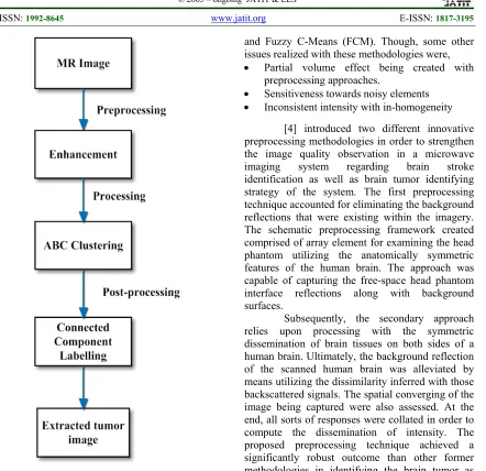
2972 Figure 4: Proposed ABC Segmentation methodology
Afterward, it was accomplished by means of implementing ABC procedure. Afterward, the mechanism of post-processing was implied to abstract the complete segment of brain tumor from those MR imageries being acquired. A grey level thresholding was employed at this stage in order to eradicate the segmented brain tumor imagery that comprised of a vast amount noisy elements within it. Finally, an associated element based labeling methodology relying upon the pixel connectivity was recognized to assist in segregating the tumor infected portion from the MR imagery. The performance metrics provided by the proposed ABC algorithm based brain tumor segmenting methodology was comparatively better in terms of accuracy than segmented imageries obtained former approaches like Genetic Algorithm (GA), k-means,
and Fuzzy C-Means (FCM). Though, some other issues realized with these methodologies were,
Partial volume effect being created with preprocessing approaches.
Sensitiveness towards noisy elements
Inconsistent intensity with in-homogeneity
[4] introduced two different innovative preprocessing methodologies in order to strengthen the image quality observation in a microwave imaging system regarding brain stroke identification as well as brain tumor identifying strategy of the system. The first preprocessing technique accounted for eliminating the background reflections that were existing within the imagery. The schematic preprocessing framework created comprised of array element for examining the head phantom utilizing the anatomically symmetric features of the human brain. The approach was capable of capturing the free-space head phantom interface reflections along with background surfaces.
Subsequently, the secondary approach relies upon processing with the symmetric dissemination of brain tissues on both sides of a human brain. Ultimately, the background reflection of the scanned human brain was alleviated by means utilizing the dissimilarity inferred with those backscattered signals. The spatial converging of the image being captured were also assessed. At the end, all sorts of responses were collated in order to compute the dissemination of intensity. The proposed preprocessing technique achieved a significantly robust outcome than other former methodologies in identifying the brain tumor as well as identifying the symptoms regarding a brain strokes. The drawbacks prevailing with the proposed methodology were
Incapability in realizing the scattered features of some other complicated human brain
Unable to sense the heterogeneity in between surfaces of the human brain
2973 labelling for brain MR imageries by means of utilizing those perceptual concepts. These labels being generated were further utilized to create the scientific values of the imageries exposed for processing.
The proposed perceptual procedure was capable of synchronizing expertized knowledge with those image processing methodologies in an automated fashion. This automated prioritizing methodology was proficient in terms of tumor identification accuracy than former methodologies like prioritization using N-cut segmentation and saliency weighted segmentation. Beyond all these accomplishments, the proposed perceptual methodology lacked in identifying some sort of peculiar lesions found as a symptom of abnormality which was found under the human supervision.
[6] designed a clinical system that would highly assist in generating decision regarding meningioma brain histopathological image categorization of brain tumors by means of utilizing fractal dimensions of subband possessed by a human brain. Subbands of brain tissues were opted for categorization procedure owing to its unaltered nature of tissue structure on behalf of rapid modifications occurring with a brain. The acquired brain imageries were subjected for pre-processing by means of utilizing a wavelet transform approach. Thus, the attributes found unrelated were eliminated at this stage. The attributes assisted for categorizing those infected brain tissues were more highlighted using this preprocessing level. The difficulties involved in segregating varied textures of tissues were mitigated and hence, an ideal joint spatial frequency localization was acquired. An appropriate classifier model was adapted to handle the variation occurring with brain tissues. The projected wavelet tree-structured decomposition methodology was capable of segregating histopathological meningioma imageries with an enhanced accuracy. The drawback prevailing with this approach was archiving of unrelated assessments on experiencing an inconsequential variation between subbands.
[7]surveyed on various brain tumor segmentation methodologies. Methodologies that were utilized for accomplishing a perfect preprocessed brain MR imagery were exhibited.
Anisotropic Diffusion Filtering (ADF): ADF methodology was highly proficient in eliminating the noisy constituents from the acquired brain MR imageries. Furthermore, it imposed many sorts of destructive impact on segregating brain tumors.
Skull Stripping: Vital approach for eradicating the presence of non-cerebral tissues like meninges obtained from softest portions of brain, scalp, and skull and delineating the overall structure of the brain. A crucial trade-off was experienced with an accuracy of preprocessing and accuracy of the tumor identification on implementing this approach.
Intensity Normalization: Assisted for categorizing and clustering of tumor infected portions of the brain in an effectual manner. The drawback found with this approach was, disintegrating the overall form of the preprocessed brain image ultimately causing a degradation for identifying the presence of a tumor.
Bias field correction: Formulated for mitigating the influence of inhomogeneities owing to the presence of the magnetic field that existed at the time of attaining a brain MR imagery.
Pathology robust normalization: Devised to enhance both sorts of indigenous and global constrictions of the MR imageries.
Likewise, varied methodologies employed in fragmentation and segmentation was also surveyed in this research. A robust Machine Learning (ML) technique that was deployed for segmenting the abnormal tumor tissue from the normal brain tissue was significantly proficient.
[8]surveyed on varied methodologies utilized for segmenting and classifying the acquired brain MR imageries. Moreover, the usefulness of Computer Aided Diagnosis or Detection methodology was examined for assisting the medical practitioners in an effectual identification of brain tumors. In addition, an automated brain tumor identification methodology was devised for CAD. The endorsed automated Machine Learning (ML) brain tumor identification methodology was carried out in a sequential procedure given as,
2974 filtering was its tendency leaving the edges of the imagery undisturbed without any sort of disruptions with the appearance of the image concerned. Actually, this procedure accounted for a typical image that would permit further sequential process for obtaining the boundary level.
Fragmenting the MR Imagery: Feedback Pulse Coupled Neural Network (FPCNN) methodology was utilized to fragment the portions of an image that tended to comprise brain tumor out of complete imagery. The vital benefit obtained with the procedure applied was its capability in altering the input fed into the system in accordance with the outcome achieved in the previous trial that was fed in as feedback.
Abstracting the necessitated attributes from brain image: Discrete Wavelet Transform (DWT) was employed on fragmented imagery in order to realize the attributes of the imagery two sorts of frequency as well as time domain from the spatial plot. Consequently, this criterion would decompose the image concerned into appropriate sub-bands comprising of interlinked DWT coefficients.
Curtailing the overall dimension of dimension of an image: Principal Component Analysis (PCA) methodology was exploited to mitigate the attributes that were available in excess, in order to alleviate the barrier indicated by “the curse of dimensionality”. Ultimately, the complexity that would arise at the time of trailing categorization procedure was totally eradicated.
Categorization of the input image being fed: Back Propagation Neural Network (BPNN) approach was deployed on those imageries in order to categorize them into tumor affected or a normal brain tissue. Categorization was accomplished by means of supervised learning methodology accompanied by a non-linear generalization technique.
The proposed hybrid methodology that was the assimilated outcome of highly proficient methodologies accounted for a better categorized product with an improvised robustness. The drawbacks prevailing with this automated approach developed on the basis of CAD were,
Lack in highly necessitated accuracy in a specifically enhanced way exceeding manual diagnosis
Processing with diversified image qualities
[9]presented an effectual methodology for identifying the presence of brain tumor by means of realizing statistical features of the image built on the basis of curvelet transform incorporated along with another group of textured features known as Grey Level Cooccurrence Matrix (GLCM). Tumor cells found in the brain was totally abnormal and would get altered with no time. Hence, the vital attributes that were left unaltered easily based on dual set domain specifically termed as frequency and spatial were abstracted. Moreover, those abstracted features were given into the system for training purpose. The feature acquired from the spatial domain that was belonging to GLCM was collated with the statistical features that were obtained from frequency domain based on curvelet transform. The complete part of the brain was segregated from other irrelevant constituents like the scalp, skull was eradicated through the procedure of preprocessing. It was accomplished by means of deploying skull stripping methodology on the acquired brain ME imagery. The efficacy of the segmenting procedure was empowered by means of pre-processing MR imagery. The hardest portion probably skull was detached off from the brain imagery. Subsequently, another methodology called Gaussian filter was exploited on the same brain MR imagery in order to eliminate the noisy factors from the brain image. Afterward, the imagery was treated with an effectual segmentation procedure that was built on the basis of a watershed transform. Support Vector Machine (SVM) approach was utilized to categorize the preprocessed imagery into the normal tissue or a tumor infected one. The promptness of the categorizing procedure was enhanced by means of proposed image mosaic methodology framed on the basis of abstracted features. Though the accuracy of the classifying process was increased, the space occupancy of those abstracted features was very high.
[10]projected an inventive methodology for identifying and segregating brain tumor from the acquired imageries. A regular dataset that comprised of 17 patients’ medical record was accounted for processing and segregating tumor portion out of it. The complete imageries available alone were considered for brain tumor abstraction and moreover, modified into its grey scale imagery in order to get processed with subsequent procedures.
The process of preprocessing was accomplished by means of deploying a methodology based on linear normalization.
2975 be employed in order to obtain only the brain tissues and to get rid of the skull bone.
In addition, a morphological operational filter was utilized to clean the noisy constituents existing within the imagery and also to acquire the Glioblastoma (GBM) arena in a 2Dimaensional form of axial imagery
Furthermore, the process of segmentation was accomplished by means of multithresholding segmentation procedure. Principal Component Analysis (PCA) methodology was collated along with wavelet features was exploited to categorize the imageries of the brain tumor. The drawback sustaining with this proposed filtering approach was, realizing a difficulty in acquiring a filtered imagery at the instance whenever the overall covering area of GBM was in a comparatively diminished state than the thickness of skull.
[11]surveyed on various approaches available for performing an efficient skull stripping process in order to enrich the quality of overall preprocessing strategy. Thus, the entire portion of the brain was absolutely segregated out of the portions held by skull and other cranial tissues within head portion. The methodologies assessed were,
Morphology based approach
Intensity based approach
Deformable-surface based approach
Atlas/ Template based approach
Hybrid approaches
On enriching the proficiency of skull stripping methodology, the tissues whichever do not belong to the brain was eliminated and consequently some other noisy factors were convoluted within the brain imagery was also removed off from the MR Imagery. The skull stripping methodologies assessed so far with this research was capable of performing the preprocessing procedure in a robust manner with some sort of inbuilt constrictions.
[12] suggested a stochastic framework for distinguishing varied attributes of the MR imagery of a brain tumor. The surface of the brain tumor was framed by means of implementing a multifractal attributes incorporated into a single methodology termed as multifractal Brownian motion (mBm). The process of preprocessing was accomplished by means of merging an inventive Piecewise Triangular Prism Surface Area (PTPSA) along with features possessing multi- Fractal Dimension (FD). An enhanced and modified AdaBoost methodology was utilized to fragment discrete set of brain tumor imageries possessed by
varied patients. The overall performance of the devised merging approach was enhanced through a methodology of slice resolution. Any sort of definite atlas designated by deformable imagery registration was not necessitated for a robust tumor segmentation procedure. The drawback prevailing with this approach was its inability to process and segregate some sort of complicated tumors on the brain surfaces.
2.2 Feature Extraction
[13] implemented the brain module segmentation and feature extraction with the help of digital image processing for classification of the disease stages to minimize the brain tumour percentage distribution. They used the segmented modules for feature extraction at once when the segmentation is performed. A Feature was dealt as the important section of information extracted from the image with the help of detailed image. The extracted features were used for the disease classification. The condition of brain cancer at early stages were determined by the nodule features. This method remained easy for implementation, less costly and less time consumption.
[14]illustrated the classification of brain tumour in Artificial Neural Network by Feature extraction. He explained the techniques and methods involved in data reduction in order to identify the subset of variables based on the image. Seven textural features based on grey level co-occurrence matrix were extracted from the set of images. Co-occurrence matrix was computed in four directions namely 0o, 45o, 90o, 135o degrees. Angular Second Moment(ASM)/Energy
Contrast
Inverse Difference Moment (IDM)/ Homogeneity
Dissimilarity
Entropy
Maximum Probability
Inverse
[15]proposed a method to overcome the challenges to utilize the coarse to fine analysis of localized characteristics in pathology images. They analysed the diversity of coarse regions and included the extraction of spatially localized features of color, outline and texture from tiled regions covering the slide. This scheme presented great steadiness and robust nature to parameter discrepancy with exactness and widespread range of parameters.
2976 proposed in order to remain quite useful in context of detection and classification of brain tumours. In classification, features of each image was extracted from MR image by Fast Fourier Transform (FFT) algorithm. FFT is used to implement image conversion from spatial domain to the frequency domain. Image decomposition into real and imaginary components were considered as the representation of image in the frequency domain. The advancement in increasing the size of dataset by adding ages, gender and symptoms.
[16]introduced an efficient MRI brain image analysis methodology where, MRI image is classified into normal, non-cancerous brain tumour and cancerous brain tumour. This system proposed that the tumour diagnosis process remains accurate and fats. This classifier based MRI brain image processing approach produced at the best MRI brain image classification by the usage of feature extraction and segmentation results. The feature extraction methods produced the best result of brain tumour diagnosis in advance in the medical fields. The proposed MRI image based tumour analysis dealt with the segmentation and classification process. The technique used for feature extraction used texture feature extraction based on sobel and canny methods.
[8]examined Computer-aided detection / diagnosis (CAD) systems to improve the diagnostic competences of physicians and decrease the time essential for exact diagnosis. The review revealed that the CAD systems of human brain MRI images are still an open problem. So, they suggested a hybrid intelligent machine learning technique for computer-aided detection system for automatic detection of brain tumour by magnetic resonance images with the help of feature extraction.
The proposed technique was implemented based the following methods.
The feedback pulse-coupled neural network
The discrete wavelet transform
The feed forward back-propagation neural network
The principal component analysis
The proposed technique applied feedback pulse-coupled neural network as a front-end processor for image segmentation and discovering the region of interest, and the discrete wavelet transform was applied on images to remove features. Additionally the principal component analysis was performed to reduce the dimensionality of the wavelet coefficients which resulted in a more efficient and accurate classifier. The reduced features were forwarded to
back-propagation neural network to classify inputs into normal or abnormal based on feature selection parameter.
The classification accuracy on training and test images was significantly high. Furthermore, the proposed technique established its effectiveness compared with the other machine learning techniques. The results revealed that the proposed hybrid approach was exact and fast and robust.
[17]signified a model, image inhomogeneity rectification and intensity standardization before feature extraction of images. Image intensities in MRI images lacked in fixed meaning and extensively varied within or between subjects. A patch-based technique was used in mining the image feature. The intensity values represented in a patch around a voxelwere attained and reorganised as a feature vector. Four modalities of MRI images were used, so the feature used a cubic patch with a size of w w w was (w3 × 4)-dimensional. Locality was considered in defining whether local anchor embedding was more applicable in solving linear projection weights compared with other coding methods.
2977 The function of feature extraction was to reduce the features of original dataset by estimating some specific properties or features that differentiate one input pattern from another. The extracted features delivered the input type to the classifier by considering the depiction of the significant properties of the image. The analysing methods used the pixel value intensities, pixels coordinates and nearly supplementary statistic features like mean, variance or median, which possessed more number of errors in determination process and low precision efficiency in classification.
[19]explained the performance of texture feature extraction for brain tumor image retrieval in MRI. The chief objective of this study was to examine and calculate an effective and strong approach for texture representation. The techniques used for texture feature extraction were
The curvelet transform,
Contourlet transform and
Local ternary pattern.
They applied DNN and ELM to categorize the brain tumor image for supervised learning systems. It was concluded from the results that contourlet transform by DNN and ELM classifier outperformed other techniques like Curvelet transform and Local ternary pattern. In terms of time, ELM classifier outperformed DNN. Contourlet transform achieved superior performance and ELM classifier accomplished better performance. It reduced the retrieval time and upgraded the repossession accurateness considerably.
[20]investigated the segmented regions of medical image and the usage of different techniques for detection and classification of image regions. After the completion of analysis on some segmentation methods categorized by researchers they suggested Region classification in the visualization of brain tissues of MRI. Brain image was basically classified into three regions;
WM,
GM and
CSF.
The forth region was named as tumor region, if the image was not normal. The segmentation and categorisation of Brain MR image regions with the help of SOM and neuro fuzzy techniques. They combined Self Organizing Map (SOM) and Neuro Fuzzy scheme to spontaneously extract WM, GM, CSF and tumor region of brain MRI . This system was also examined and verified on axial view images to classify the regions of brain MRI.
2978 The proposed method used (SWT) to remove features from MR brain images and principal component analysis (PCA) was coupled to reduce the SWT coefficients. Finally, two classifiers named generalized eigenvalue proximal support vector machine (GEPSVM), and GEPSVM with RBF kernel were also used. These methods were detected and verified on three benchmark datasets. The results exhibited that SWT-based classifier was more accurate and performed better than state-of-the-art methods.
[22]proposed algorithms for Brain Tumor Detection based on MRI Images with greater accuracy and low error proportions. The statistical analysis of the experimental results indicated that the developed algorithm divided brain MR images with good precision. Detecting the occurrence of tumours at faster rate to improve survival rates remained as challenging task ever. Though, variety of different neural network-based algorithms available for texture based segmentation and classification methodology. Still most of these neural network-based algorithms always required the widespread observation, training and their performance relied on the training method and data used in training. Finally, it was observed that the medical image segmentation and classification algorithms should comprise the following features:
Accuracy,
Consistency,
Repeatability,
Healthiness and
Least dependence on the operator.
[23]focused to improve the segmentation algorithms and enhance tumor boundaries prior to segmentation. The proposed method explained two paradigms to progress the presentation of segmentation. At first, the proposed GLCM-CA extracted the features from an original MRI and then transformed this feature space to the target image. Then an improved Tumor-Cut (ITC) was suggested to handle the sturdiness of seed growing encountered in Tumor-Cut. ITC directly implemented on the target edge. For performance evaluation, training and testing datasets were empirically executed in evaluation using dice evaluation metrics. In this regard, the proposed method performed better than the state-of-the-art compared methods.
Based on the state-of-the-art compared methods and performance assessment reports, the proposed method afforded the out- standing results. There are issues to take into account for future research works. Firstly, GLCM recognized a texture space of intensities. This produced the
proposed GLCM-CA offered the non- promising results in the white matter regions with similar intensities with tumor region. In this regard, the proposed GLCM-CA was changed to the transition function and neighborhood to extract the tumor feature. Moreover, the proposed ITC exhibited more complexity to compute the patch weight distance depending on the tumor voxels. The other patch distances, like Chi- square, histogram, for example, reduced the computational time complexity.
[24]have presented a novel semi-automatic approach for multi-modality medical image segmentation using CRF framework. In the energy function. They introduced purely probabilistic regional and boundary terms estimated from logistic regression models. The case-specific models were trained from expert user inputs with brush strokes and accepted segmentations on the training slices, allowing for the method to learn from human guidance adaptively.
They showed that tumour segmentations from method on multi-modality images were more exact than a related automatic method in terms of Dice coefficients when compared to ground truths. Since the regional term in CRF was communal among methods using Bayesian approach, the results suggested the major advantages also. They sprightly modelled target specific boundary probability distribution forming the boundary term in CRF rather than a boundary term constructed from dissimilarity between two tissue classes which do not represented the true boundary of the target. Further, semi-automatic approach with a tool familiar to users much welcomed in the clinical practice. Using the same purposed framework, future works included addressing multi-class segmentation and investigating substitute statistical representations for the regional and boundary.
2979 was demanded to quickly complete tasks. Brain MRI image was taken and pre-processing using filters. Finally, after segmentation it was resulted that, the performance of the canny edge detector is superior in general to the edge detectors.
[26] incorporated an automated framework to detect MR images containing tumour and then segment the tumour implemented on T1-w and FLAIR sequences (separately). The prominent exactness of the algorithm in tumour segmentation in concert with its low computational complexity validated the efficiency of the proposed method. Another advantage of the proposed method was the use of single-spectral MRI. For classification, four supervised robust classification techniques like the SVM, NSC, SRC, KNN and one unsupervised clustering method, k-means were used and the results were compared After training the classifier, the recognition rate of the classifier on independent data was used as the indicator of algorithm’s performance in tumor segmentation, and also each feature set’s suitability for tumor segmentation This made the proposed algorithm much more robust and general than other methods.
[27]proposed a new approach based on Saritha’s scheme for MR brain image classification and a novel FS method called as BPSO-MT, to discover optimal feature amalgamation from the entropy of calculation and sub-bands of 8-level DWT decomposition. The results presented that the BPSO-MT produced better fallouts than existing methods. They extracted wavelet entropy (WE) features from the detailed sub-bands of eight-level decomposition. Feature selection discovered the grouping spaces to invent the optimal feature combination. Subsequently, the proposed BPSO-M, BPSO-T, and BPSO-MT helped to select features. Lastly, the selected features were served into a probabilistic neural network (PNN).
2.3 Techniques Utilized For Segmentation
[28]exhibited a comprehensive analysis regarding three varied segmenting approaches meant specifically for identifying the presence of tumor in brain MR imageries. The techniques dealt here were,
K-means clustering accompany with watershed segmentation procedure
Optimized K-means clustering merged along with Genetic Algorithm (GA)
Optimized C-means clustering compounded with Genetic Algorithm (GA)
The sensitivity towards preliminary clustered centers was possessed by conventional k-means clustering methodology. Both K-means and
C-means procedures assimilated with GA was doing well in identifying a brain tumor out of a human brain MR imagery.
The drawback specified owing to excess segmentation was alleviated by means if implementing a Genetic C-means algorithm.
The defined assimilated procedure was proficient in delivering accurate results within a minimized time span.
Convergence of the brain MR imagery was enriched significantly
Another issue existing with the proposed automated ML approach endorsed was its inability to process a brain tumor MR imagery in a three dimensional fashion.
[29]presented a comparative analysis based on the productive quality of two different methodologies typically known as K-means and Fuzzy C-means procedures that were utilized for segmenting the acquired brain MR imageries. The MR images of the brain that comprised of tumor in it were subjected to further processing on the basis of initialization procedure directed by histograms constructed on complicated edema MR imageries. The promptness of any sorts of segmenting algorithms was defined means of checking for its ability in segregating the tumor cells from those normal ones without any reservations. Hence, both the investigated approaches were subjected to segregate several portions of human brain such as,
White matter
Cerebro-Spinal fluid
Grey matter
Necrotic Focus of Glioblastoma Mutliforme (GBM)
Perifocal Vasogenic edema
It was evidently identified that K-mans was proficient than FCM approach. The FCM methodology was capable of detecting white matter of brain along with vasogenic edema collated into a unique tissue category. Likewise, necrotic focus and grey matter were incorporated into one.
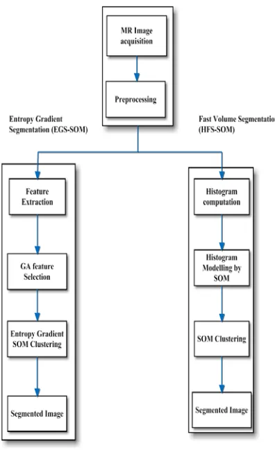
2980 sequential procedure. It was also proficient in categorizing the tissues existing within brain into diversified classes. The complete procedure of the proposed techniques was illustrated in Figure 4.
Figure 6: Overall flow of both EGS-SOM and HFS-SOM procedure
The segregated classes into which the brain images would get categorized was,
Acquiring brain MR imageries
Feature extraction by means of utilizing overlapping window methodology
Feature selection accomplished on the basis of evolution assessment
Clustering on the basis of conventional SOM procedure.
EGS-SOM approach was capable of segregating all sorts of brain tissues into normal or abnormal even there existed a significant disruption with noisy elements. The proposed methodology exhibited a significantly enhanced accuracy rating regarding the irregularities affecting the brain. Issues prevailing with the proposed segmentation procedure was its high sensitivity towards tissue
homogeneity which also managed to incline toward the wrong classification.
[31]endorsed an effective methodology for segmenting the tumors from those MR imageries of a brain. The procedure was framed by means of gaining knowledge from those existing population that comprised of attributes which were precise enough to elaborate the details of a patient picked out from the multimodal MR imageries. Ultimately, the procedure of segmentation was successfully accomplished through a graph cut methodology. Moreover, those feature sets were utilized to gain knowledge so as to instruct both global and conventional classifiers. The proposed segmentation procedure was comparatively robust than other traditional results provided by medical practitioners. The drawbacks inferred with this devised methodology were,
The graph cut methodology would not function in a completely automated fashion
Requiring vast time span utilization for instructing the global classifier that lack in providing instant outcomes
Delivering instructions to other testing classifiers was done through global classifiers and it was updated only once by an offline fashion. Hence, the outcomes were not optimized.
[32]introduced a semi-automated segmenting approach for processing tumors of brain imageries acquired through multi-channel MR imageries. The slices were opted in a completely automated fashion and labelled. Every single labelled slice was utilized at every instance of time whenever the imageries were permitted for processing in an asymmetric manner. Consequently, a requisite for the previously framed structure was neglected. Moreover, a multi-label Markov Random Field (MRF) Optimization methodology accompanied with hard constrictions was utilized for cataloguing other leftover labels. Specific global attributes from the brain tumor imageries were abstracted through a programmed optical stream. The process of tumor segmentation was robustly accomplished by means of acquiring those pre-acquired global attributes. Hence, a significantly enhanced accuracy over segmentation was achieved. The drawback prevailing with the approach was adapting with a dynamic user interpretations were inferred as hugely complicated tasks.
2981 key feature of the devised technique mitigating the overall energy consumption in processing. The aforementioned feature was achieved by means of collating both supervised classification approach and ordered conditional random field. The proposed methodology was proficient in isolating those highly available outliers within the imagery. It would also provide the temporal linkages that were capable of affording the specific information bound curves. The devised completely automated approach was effectual in processing the brain tumor imageries obtained from the intra-patient report with and enhanced promptness. The disadvantages prevailing with this technique was its limited capability in acquiring only the information regarding indigenous attributes leaving out the global framework in order to accomplish a better knowledge.
[34]designed an inventive approach toward segmenting brain in a proficient manner. The process of segmenting the brain was carried out in a “within brain” mode in a generalized manner. The imageries were acquired from BRATS datasets [35]. Hence, both sorts of training the system and testing it for a brain imagery along with a mitigated form of user interruptions were performed only in the interior area of a brain and not in a completely covered skull area. The proposed semi-automated mechanism would possible convert all sorts of acquired attributes into N-Dimensional voxels. Those achieved voxels were furthermore, categorized into a diverse group of categories with the utilization of two dissimilar models relying upon the pre-defined labels. The models utilized in this research were,
Markov Random Fields (MRF) accompanied with K-Nearest Neighbour (K-NN) Classifier (MRF-kNN)
Condition Random Fields (CRF) accompanied with K-Nearest Neighbour (K-NN) Classifier (CRF-kNN)
The proposed within brain generalization methodology would possibly get trained with each and every brain imagery that was opted for processing within it. It was also designating tumor portion with a mitigated quantity of two slices. Thereby, achieving the robustly segmented tumor with a minimized computational overhead. The problem being identified with this methodology was attainment of such accurate outcomes only because of the manual interruptions and it was not a completely automated approach that was devoid of any sort of human assistance.
[36]recommended an inventive approach that was built on the basis of fuzzy logic was
designed in order to segment the tumor in brain MR imagery in an automated fashion. The former Fuzzy C-Means algorithm was considered and enhanced by means of integrating a priori data within the procedure termed as Prior Information Guided Fuzzy C-Means (PIGFCM) procedure. The procedure trailed in this approach were,
Assessing the intensified tumor by means of incorporating MR imagery that possessed a T1 weight
Moreover, a defuzzification procedure was deployed on those processed imageries in order to accomplish an enhanced membership within the imageries
Furthermore, a post-processing strategy was trailed on the segregated group of tumors so as to eliminate the isolated voxels
An indigenous kind of interconnections was provisioned within those segregated tumors and hence, the completely interconnected portion ultimately obtained was designated as the tumor portions
The PIGFCM algorithm was capable of segregating the tumors from the MR brain imagery with an supplemented accuracy prevailing in it within a minimized time constraint. The drawback inferred with the devised PIGFCM approach was the typical detachment between the grouped voxels possibly would lead to an issue regarding local optima.
[37]surveyed and presented benchmarked framework of Multimodal Brain tumor Image fragmentation with a dataset comprising both elevated and least complexity levelled glioma patients who were accounted for a count of 65 in a dataset. Those images existing within the dataset concerned was visualized by means of a virtualization software capable of generating images with a reality. It was evident that no single algorithm was capable of producing a robust result when deployed in an in an individual manner. An inventive pre-processing mechanism accomplished by performing an intensity multiplication nearby the points of Region of Interest (ROI). An automated mechanism deployed for the purpose of accomplishing a successful segmentation consumed a magnified amount of time span. The classification was performed by means of utilizing Leave-one-out cross validation methodology in training the data exhibited within a single class. The attributes lying out of the significantly clustered sets were classified with a poor promptness.
2982 The devised methodology was instructed to work under a single kind of anatomical MR imageries. The utilization of multi-spectral information regarding brain MR imageries was assuring the registration with those acquired imageries. The performance of the devised techniques was significantly better than the former Gabor wavelets and multi-scale classification algorithms in terms of tumor segmenting accuracy.
[2]presented a proficient image segmentation approach by means of utilizing a K-means clustering approach that was also assimilated with fuzzy C-means algorithms. The promptness of the segmentation procedure was further enhanced by means of subjecting the imagery to a trailing sequence of methodologies such as level set segmentation and thresholding approaches. The proposed methodology was capable of acquiring the mitigated computational time and an enhanced accuracy.
[38]suggested an enhanced edge identification methodology for improvising the quality of brain tumor segmenting procedure. The devised methodology was completely relying upon the Sobel edge identification approach. Closed contour algorithm and Soble approach relying upon the thresholding technique were collated into a single approach in order to explore varied portions of imagery. Those closed contours were utilized to abstract tumors from those brain imageries. The tumor regions that were abstracted by means of implementing this approach was of enhanced accuracy. The drawback still remaining with this approach was incapable of assessing the brain tumor that possessed a complicated shape and dimensionally mitigated boundary lines.
[39]presented a finer and completely automated segmenting methodology that was devised particularly for MR images termed as Multi-Atlas Label Propagation with Expectation Maximization based refinement (MALP-EM). This approach devised was actually trailing the former registration approach named as (MAPER). An inventive pattern such as probabilistic segmentation accompanied with that spatial amalgamation of weights was realized in this approach. The quality of predefined labels was significantly enhanced with this MALP-EM mechanism.
[40]introduced a procedure for skull stripping methodology posed by means of collating both the thresholding and other morphological attributes of a brain MR imagery. The upcoming sequential process of segmentation was performed by implementing Self Organizing Maps (SOM) procedure. A practice of unsupervised learning
procedure termed as Learning Vector Quantization (LVQ) was trailed to train and upgrade the SOM procedure. The projected unsupervised approach was proficient in categorizing all sorts of complete, enhancing and core graded imageries in a robust manner that was typically finer that other state-of-art methodologies Brain Surface Extractor (BSE) on the BRATS dataset. The limitation observed with this procedure was incapable of separating brain tumor when it was in some sort of complex stage.
[41]suggested an inventive methodology that was capable of permitting the user interruptions while segregating brain tumors from an MR imagery. The issue of intensified bias modification was resolved in this research by means of implementing an approach termed as “within brain generalization”. It was accomplished through semi-automated brain tumor identification methodology. The proposed generalization methodology was capable of enhancing the performance of certain classifying methodologies like random forests, Support Vector Machine (SVM) and K-Nearest Neighbour. Devised generalization approach would beneficially deliver a significantly robust specificity value.
[42]recommended a productive probabilistic segmentation framework that was capable of segregating the lesions of the brain out of the multi-dimensional imageries. The introduced generative brain lesion segmentation model was capable of segregating the lesions of a brain in an effectual manner by implementing an Expectation Maximization (EM) segementer. A collective methodology that collated both Gaussian mixtures and an atlas created out of realizing EM approach was designated as EM segmenter. At the end, an estimation algorithm was designed by means of incorporating closed arrangement of EM updates. The proposed generative algorithm was capable of segregating the high-dimensional data with an enhanced efficacy. The acquired result was better than other former methodologies.
[15]introduced a resolving methodology in order to overcome the hurdles involved in the imagery such as,
Dimensionally huge imageries possibly leading to an unproductive mechanism in segregating the tumor
Presence of diversified tissues in the imagery was unable to be associated with each other based on any sort of interconnections
2983 deploying coarse-to-fine investigating analysis that accomplished of stages such as,
Preprocessing: The process of preprocessing was performed by means of resizing the methodology named as bicubic interpolation
Procedure of altering imageries into tiles: The acquired brain tumor MR imageries were subdivided into equidimensional tiled regions that were capable of providing a minimized resolution of an entire imagery devoid of flaws
Unmixing of stains: Very basic RGB stains named as hematoxylin and eosin were obtained by means of implementing a color deconvolution approach called pseudo inverse matrices.
The devised coarse-to-fine profiling methodology realized a significantly mitigated constraint in processing the imageries typically stated as the minimized computational time. The drawbacks observed with the devised approach were its auxiliary values added in addition by means of tiling that would lead to the enhanced complication in processing a brain tumor imagery.
[43]analysed varied brain tumor segmenting approaches that were accomplished by means of implementing Fuzzy C-Means clustering algorithm. The investigation was also proceeded towards,
Resolving the issue of rectifying the intensity inhomogeneity
Eradicating the stronger noisy attributes: The former issue was mitigated through series of methodologies relying upon FCM were given by,
FCM clustering approaches collated with spatial constrictions: The spatial constrictions were amalgamated along with FCM mechanisms. Hence, the issue regarding intensified inhomogeneity was modified in order to accomplish the objective of the devised technique.
Altered techniques constructed on the basis of FCM approach: Enriched FCM clustering procedures were collated along with a process of brain tumor segmentation. Hence, the procedure designated was fastened to reach the end by means of incorporating deformable structures based on the fuzzy spatial associativity.
Generalized methodologies relying on FCM: The procedure of segregating tumor from the acquired brain images was made proficient by means of implementing intuitionistic mechanism relying on a fuzzy set clustering approach.
The noisy attributes existing within the imagery was avoided by means of implementing sequence of procedures given as,
Non-local Filters: These filters were capable of utilizing the obviously redundant data prevailing inside the imagery through the methodology implemented termed as Optimized Block-wise nonlocal means denoising filter. This sort of methodology was specifically designated to get along with the 3 Dimensional MR imageries of a human brain comprising the tumor. The Rician noise feature dwelling within the M imagery was consumed in order to mitigate the fluctuating noise levels that were disbursed in a spatial manner.
Anisotropic Diffusion Filters: The MR imageries were possibly enriched by means of implementing these designated anisotropic filters. The edges were identified with an added promptness with a utilization of a scale-space methodology. A probabilistic model of an anisotropic diffusion filter opted for a completely automated stream of segregating the tumors from brain imageries.
Methodologies constructed on the basis of Wavelets: The methodologies that were completely depending upon the wavelets were specifically devised to mitigate the influence of noisy elements in an examining imagery. A strategy of an unsupervised global-to-local segmenting procedure was adapted in processing a 3-Dimensional MR brain imagery. In this procedure, a fuzzy clustering methodology accomplished on the basis of the spatial feature was employed in order to achieve an entirely processed and noiseless imagery. Though the analysed strategies were capable of delivering an accurate outcome within a diminished time constraint the issue of intensified inhomogeneity was still existing with 3D imageries.
2984
The approach formulated that comprised of completely automated architecture and was capable of processing the imageries acquired from BRATS dataset
The proposed methodology was capable of accomplishing the tumor being segregated with a significantly quicker speed.
A dual-phased training procedure was realized on two-pathway architecture in order to
disseminate the labels on all sorts of categorized portions in a sensible manner.
A cascaded framework was utilized in order to segment the tumor in a proficient way
The proposed framework constructed on the basis of DNN was proficient enough to segregate the entire brain portion with a highly minimized time constraint. The drawback found with the proposed procedure was its complexity in processing that occupied the very huge space for archiving the attributes.
3.1 Future work
After reviewing the basic methods utilized in tumor segmentation the most significant was the
Technique Advantages Disadvantages
Threshold Based Methodologies
Local thresholding Technique
Global thresholding Technique
Minimized computation span Unable to boost the visibility of
brain tumor area to an enlarged form
Region Based Methodologies
Region growing technique Capable of fragmenting the brain
tumor images in a proficient manner
Able to associate the portions of imagery that possessed analogous features and correlating the alike one into single cluster with an efficacy
Incapable of realizing the outcome of segmentation technique in efficacious way owing to presence of noisy elements
dissimilarity with the intensity of imagery would lead to generation of holes or excess segmentation
Watershed methodology Proficient in segregating several
sections at single instance of time
Able to generate complete contour of brain MR imageries
Eliminated the necessity of
implementing contour aggregation approach
Realized excess of segmentation in imageries
Pixel based Methodologies
Fuzzy C Means Boundaries of the tumor portions
were converged with efficiency by means of utilizing an unsupervised approach
Elongated computational span
Complicated on realizing the
presence of noisy constituents
Artificial Neural networks Facilitate to form non-linear
dependencies
Capable of framing non-trivial
distributions
Collection of sample images for make system trained was little bit complicated
Time span required to learn the image content was significantly high
Markov Random Fields Capable of signifying data instances
using complicated dependencies Opting for attributes that were capable of managing the robustness of spatial collaborations
Only adaptable with the algorithms
that were computationally
demanding Model-based methodologies
Parametric Deformable Models Cooperative with the biological
unevenness or inconsistency
inferred in accordance with time
Patch up with irrelevant boundaries on realizing inhomogeneity
Insensitive towards confining the boundaries of infected portions
Level Sets Able to accommodate with all sorts
of topological alterations
Incurred very high computational cost
2985 accuracy of segmentation, also the shape and location of tumor before and after treatment so the volume is major factor in the treatment process so 3D visualization should be considered in any future study about tumor in MRI ,in addition there are promising development in GPU power which will help the use of unsupervised learning algorithm such as deep learning and machine learning will play a great role in increasing the accuracy of segmentation specially in 3D MRI techniques.
3.2 limitation
This work investigate the method utilized in segmentation of tumor in MR images only such TI,T2,falair T2,and t1c there are more type of MRI that can produce more details about tumors and edema but the gold standard of such datasets are not available as needed for segmentation validation
4. CONCLUSION
This paper reviewed on various methodologies existing in present scenario for identifying and segmenting the brain tumor from brain MR imageries. The tumor segmentation is performed by sequence procedures stated as pre-processing, feature extraction and segmenting the brain tumor from MR imageries. These processing methodologies actually pave the way for a better classification of brain tissues under two different categories as normal or abnormal one. The pre-processing of brain MR imageries is accomplished by means of a two-fold procedure is effectual in mitigating the noisy elements in brain imageries but the incapability found with those approaches are its insensitivity towards recognizing the lesions of brain imagery with a varied intensity. The pre-processing methodology constructed upon the microwave imaging framework is capable of alleviating the background reflection but issues found with this approach is its inability in identifying the scattered features of some other complicated tumor. It is also incapable of exploring the heterogenic attributes that are existing along with the human brain. Afterward, the process of feature extraction is performed with utilization of Gaussian Mixture Model (GMM) features by means of implementing Glioblastoma (GBM) feature extraction methodology. The drawback inferred with this approach is its lack of ability in recognizing the perfect features regarding tumor size variations. The process of segmentation is accomplished by implementing an unsupervised learning methodology called Entropy Gradient Segmentation Self Organizing Maps (EGS-SOM). Any sort of minimal disruptions are also detectable
by this EGSSOM along with the drawback of experiencing high sensitivity towards brain tissue homogeneity which leads to a misclassification. A segmentation process based on Mutli-label Markov Random field (MRF) optimization methodology is capable of achieving the accurate brain tumor segmentation. But, the drawback inferred is inability to adapt with user interpretations which in turn makes the process more complex. Hence, to eradicate all these above mentioned issues, a perfect segmentation methodology is devised in this paper with the advantages of proficient segmentation, minimized circuit complications and mitigated power utilization level.
REFRENCES:
[1] N. Elaiza, A. Khalid, S. Ibrahim, and M. Manaf, “Brain Abnormalities Segmentation Performances Contrasting : Adaptive Network-Based Fuzzy Inference System ( ANFIS ) vs K-Nearest Neighbors ( k-NN ) vs Fuzzy c-Means ( FCM ),” 15th WSEAS Int. Conf. Comput., pp. 15–17, 2011.
[2] E. Abdel-Maksoud, M. Elmogy, and R. Al-Awadi, “Brain tumor segmentation based on a hybrid clustering technique,” Egypt. Informatics J., vol. 16, no. 1, pp. 71–81, 2015. [3] E. Hancer, C. Ozturk, and D. Karaboga,
“Extraction of brain tumors from MRI images with artificial bee colony based segmentation methodology,” in 2013 8th International Conference on Electrical and Electronics Engineering (ELECO), 2013, pp. 516–520. [4] S. Mustafa, B. Mohammed, and A. Abbosh,
“Novel preprocessing techniques for accurate microwave imaging of human brain,” IEEE Antennas Wirel. Propag. Lett., vol. 12, pp. 460–463, 2013.
[5] I. Mehmood, N. Ejaz, M. Sajjad, and S. W. Baik, “Prioritization of brain MRI volumes using medical image perception model and tumor region segmentation,” Comput. Biol. Med., vol. 43, no. 10, pp. 1471–1483, 2013. [6] O. S. Al-Kadi, “Assessment of texture
measures susceptibility to noise in conventional and contrast enhanced computed tomography lung tumour images,” Comput. Med. Imaging Graph., vol. 34, no. 6, pp. 494– 503, 2010.
2986 [8] E.-S. A. El-Dahshan, H. M. Mohsen, K.
Revett, and A.-B. M. Salem, “Computer-aided diagnosis of human brain tumor through MRI: A survey and a new algorithm,” Expert Syst. Appl., vol. 41, no. 11, pp. 5526–5545, 2014. [9] R. Karthik, R. Menaka, and C. Chellamuthu,
“A comprehensive framework for classification of brain tumour images using SVM and curvelet transform,” Int. J. Biomed. Eng. Technol., vol. 17, no. 2, pp. 168–177, 2015.
[10] A. Chaddad, “Automated feature extraction in brain tumor by magnetic resonance imaging using gaussian mixture models,” J. Biomed. Imaging, vol. 2015, p. 8, 2015.
[11] P. Kalavathi and V. B. S. Prasath, “Methods on skull stripping of MRI head scan images— a review,” J. Digit. Imaging, vol. 29, no. 3, pp. 365–379, 2016.
[12] A. Islam, S. M. S. Reza, and K. M. Iftekharuddin, “Multifractal texture estimation for detection and segmentation of brain tumors,” IEEE Trans. Biomed. Eng., vol. 60, no. 11, pp. 3204–3215, 2013.
[13] V. Prema, M. Sivasubramanian, and S. Meenakshi, “Brain cancer feature extraction using Otsu’s thresholding segmentation,” BRAIN, vol. 6, no. 3, 2016.
[14] S. Jain, “Brain Cancer Classification Using GLCM Based Feature Extraction in Artificial Neural Network,” vol. 4, no. 07, pp. 966–970, 2013.
[15] J. Barker, A. Hoogi, A. Depeursinge, and D. L. Rubin, “Automated classification of brain tumor type in whole-slide digital pathology images using local representative tiles,” Med. Image Anal., vol. 30, pp. 60–71, 2016.
[16] A. Shenbagarajan, V. Ramalingam, C. Balasubramanian, and S. Palanivel, “Tumor diagnosis in MRI brain image using ACM segmentation and ANN-LM classification techniques,” Indian J. Sci. Technol., vol. 9, no. 1, 2016.
[17] M. Huang, W. Yang, Y. Wu, J. Jiang, W. Chen, and Q. Feng, “Brain tumor segmentation based on local independent projection-based classification,” IEEE Trans. Biomed. Eng., vol. 61, no. 10, pp. 2633–2645, 2014.
[18] S. Damodharan and D. Raghavan, “Combining Tissue Segmentation and Neural Network for Brain Tumor Detection.,” Int. Arab J. Inf. Technol., vol. 12, no. 1, 2015. [19] A. A. Pandian and R. Balasubramanian,
“Performance Analysis of Texture Image
Retrieval in Curvelet, Contourlet, and Local Ternary Pattern Using DNN and ELM Classifiers for MRI Brain Tumor Images,” in Proceedings of International Conference on Computer Vision and Image Processing, 2017, pp. 239–248.
[20] A. Ahirwar, “Study of techniques used for medical image segmentation and computation of statistical test for region classification of brain MRI,” IJ Inf. Technol. Comput. Sci., vol. 5, no. 5, pp. 44–53, 2013.
[21] M. S. Hamoud Al-Tamimi, G. Sulong, and I. L. Shuaib, “Alpha shape theory for 3D visualization and volumetric measurement of brain tumor progression using magnetic resonance images,” Magn. Reson. Imaging, vol. 33, no. 6, pp. 787–803, 2015.
[22] P. B. Kanade and P. P. P. Gumaste, “Brain Tumor Detection Using MRI Images,” vol. 3, no. 2, pp. 146–150, 2015.
[23] “samiahmedpaper21.” .
[24] Y.-C. Hu, M. Grossberg, and G. Mageras, “Semiautomatic tumor segmentation with multimodal images in a conditional random field framework,” J. Med. Imaging, vol. 3, no. 2, pp. 1–10, Jun. 2016.
[25] N. Manasa, G. Mounica, and B. D. Tejaswi, “Brain Tumor Detection Based on Canny Edge Detection Algorithm and it ’ s area calculation,” vol. 5, no. 3, pp. 3–6, 2016. [26] N. Nabizadeh and M. Kubat, “Brain tumors
detection and segmentation in MR images: Gabor wavelet vs. statistical features,” Comput. Electr. Eng., vol. 45, pp. 286–301, 2015.
[27] S. Wang, P. Phillips, J. Yang, P. Sun, and Y. Zhang, “Magnetic resonance brain classification by a novel binary particle swarm optimization with mutation and time-varying acceleration coefficients,” pp. 1–11, 2016. [28] K. Sinha and G. R. Sinha, “Efficient
segmentation methods for tumor detection in MRI images,” 2014 IEEE Students’ Conf. Electr. Electron. Comput. Sci. SCEECS 2014, pp. 1–6, 2014.
[29] S. Madhukumar and N. Santhiyakumari, “Evaluation of k-Means and fuzzy C-means segmentation on MR images of brain,” Egypt. J. Radiol. Nucl. Med., vol. 46, no. 2, pp. 475– 479, 2015.