Development 139, 1744-1753 (2012) doi:10.1242/dev.075804 © 2012. Published by The Company of Biologists Ltd
INTRODUCTION
The pancreas consists of two functionally distinct exocrine and endocrine compartments. Despite this functional distinction, both pancreatic compartments are derived from a common pool of progenitor cells during embryogenesis (Pan and Wright, 2011). The embryonic pancreas becomes evident as a dorsal and ventral outgrowth of the foregut epithelium at ~E9.5. These early multipotent progenitor cells are marked by the expression of Pdx1 and Ptf1a, both of which are required for pancreatic fate specification (Afelik et al., 2006; Burlison et al., 2008; Kawaguchi et al., 2002). Following an initial growth phase, by E11.5 the pancreatic epithelium goes through a process of organ domain patterning that results in a branched epithelial network that can be segregated into spatially defined ‘tip’ (TipPC) and ‘trunk’ (TrPC) domains (Villasenor et al., 2010; Zhou et al., 2007) that express specific cell-intrinsic markers. Early tip multipotent pancreatic progenitor cells (MPCs) contribute to endocrine, acinar and duct fates, but become restricted to acinar fate by E14.5 (Zhou et al., 2007). At the molecular level this is evidenced by the global expression of Ptf1a and Nkx6.1 in MPCs, in contrast to the tip-restricted Ptf1a and trunk-specific Nkx6.1 expression patterns that are observed as branching morphogenesis and organ domain patterning ensue (Hald et al., 2008). A recent study by Schaffer et al. (Schaffer et al., 2010) provides compelling evidence that the Nkx6 factors (Nkx6.1 and Nkx6.2) expressed in the trunk domain
and Ptf1a at the tip mutually antagonize each other to specify trunk and tip fates, respectively, thus ensuring an effective split of the MPC population.
Although there is mounting evidence for patterning of the early pancreatic epithelium on the molecular level, the signaling event(s) that operate upstream of these key transcriptional regulatory factors is not fully understood. This study focuses on the role of Notch signaling during domain patterning of the early pancreatic epithelium, with an emphasis on how this signaling pathway controls the expression of transcription factors that are pivotal during the specification of the initial subdomains of progenitor cells in the embryonic pancreas, namely those of the tip and trunk. The conventional view of the role of Notch signaling has been that it is inhibitory to the differentiation of endocrine cells. This view is based on loss-of-function studies of Notch signaling components, which result in the premature differentiation of MPCs into endocrine cells (Apelqvist et al., 1999; Jensen et al., 2000), as well as on constitutive activation of Notch signaling, which blocks the differentiation of pancreatic progenitor cells. Yet, this cannot fully account for the role of Notch signaling during pancreas development.
A number of recent studies have provided cues to suggest a possible role for Notch signaling in domain patterning of the embryonic pancreas, although detailed analyses of the molecular and developmental mechanisms involved have not been undertaken. Genetic lineage-tracing analysis in zebrafish indicates that Notch-responsive cells in the developing pancreas differentiate almost exclusively to endocrine cells (Wang et al., 2011). This is in agreement with an earlier study performed in mouse in which targeted deletion of presenilin 1 (Psen1) and Psen2, which encode the catalytic core of -secretase, in neurogenin 3 (Ngn3)-positive progenitor cells was shown to route these cells from an endocrine to acinar fate (Cras-Meneur et al., 2009). However, the causal role 1Lerner Research Institute, Cleveland Clinic, 9500 Euclid Ave, Cleveland, OH 44195,
USA. 2Department of Molecular Biology, University of Texas Southwestern Medical Center, Dallas, TX 76390, USA.
*Author for correspondence (jensenj2@ccf.org)
Accepted 23 February 2012 SUMMARY
Early pancreatic morphogenesis is characterized by the transformation of an uncommitted pool of pancreatic progenitor cells into a branched pancreatic epithelium that consists of ‘tip’ and ‘trunk’ domains. These domains have distinct molecular signatures and differentiate into distinct pancreatic cell lineages. Cells at the branched tips of the epithelium develop into acinar cells, whereas cells in the trunk subcompartment differentiate into endocrine and duct cells. Recent genetic analyses have highlighted the role of key transcriptional regulators in the specification of these subcompartments. Here, we analyzed in mice the role of Notch signaling in the patterning of multipotent pancreatic progenitor cells through mosaic overexpression of a Notch signaling antagonist, dominant-negative mastermind-like 1, resulting in a mixture of wild-type and Notch-suppressed pancreatic progenitor cells. We find that attenuation of Notch signaling has pronounced patterning effects on multipotent pancreatic progenitor cells prior to terminal differentiation. Relative to the wild-type cells, the Notch-suppressed cells lose trunk marker genes and gain expression of tip marker genes. The Notch-suppressed cells subsequently differentiate into acinar cells, whereas duct and
endocrine populations are formed predominantly from the wild-type cells. Mechanistically, these observations could be explained by a requirement of Notch for the expression of the trunk determination gene Nkx6.1. This was supported by the finding of direct binding of RBP-jto the Nkx6.1proximal promoter.
KEY WORDS: Patterning, Pancreas, Notch, Nkx6.1, Ptf1a, Cell fate, Mouse
Notch-mediated patterning and cell fate allocation of
pancreatic progenitor cells
Solomon Afelik1, Xiaoling Qu1, Edy Hasrouni1, Michael A. Bukys1, Tye Deering2, Stephan Nieuwoudt1, William Rogers1, Raymond J. MacDonald2and Jan Jensen1,*
D
E
V
E
LO
P
M
E
N
of Notch signaling in the patterning of MPCs into pro-acinar and pro-endocrine/duct progenitors, which forms the basis for subsequent terminal cell fate allocation, was not evaluated in these studies.
We have created a novel model that provides a more specific means of inhibiting Notch than the -secretase inhibition achieved by genetic or pharmaceutical means. By overexpressing the Notch-interacting helix mastermind-like 1 (Maml1), specific inhibition of Notch transcriptional targets could be achieved. Utilizing a built-in viable marker for cells expressing the transgene we were able to perform a mosaic analysis of Notch-deficient pancreatic progenitor cells relative to wild-type cells as these populations co-exist in the developing pancreas. Our findings reveal that Notch signaling is required to establish a trunk field identity through activation of Nkx6.1 and is consequently needed to establish a pool of endocrine/ductal bipotential progenitors. We conclude that during pancreas development, Notch signaling is the molecular basis for the patterning of MPCs into pro-acinar and pro-endocrine/duct progenitor subcompartments.
MATERIALS AND METHODS
Animals
The dominant-negative Maml1 fragment (dnMAML1) consisting of amino acids 13 to 74 of mouse Maml1 was generated by PCR amplification using cDNA from mouse E14.5 pancreas. The forward primer was initiated with ATG and a FLAG tag sequence was incorporated at the end of the reverse primer to generate FLAG-tagged dnMAML1. The resulting fragment was ligated into a modified version of the pTRE2 (Clontech Laboratories) vector that contains an IRES-nGFP downstream of the multiple cloning site (Fig. 1B). To validate doxycycline inducibility, this construct was cotransfected with pCMV-rtTA (a gift from J. A. McDonald, Mayo Clinic, AZ, USA) into HEK293 cells. Nuclear EGFP (nEGFP) was detected in the presence of doxycycline and the functional ability of the construct to inhibit Notch-driven activation was assessed (supplementary material Fig. S1). The pTRE-dnMAML1-IRES-nEGFP fragment was linearized and injected into fertilized one-cell mouse embryos at the Case Western Reserve University transgenic and gene targeting facility. Four founders (F0) out of a total of 22 were identified as carrying the transgene by PCR-based genotyping using DNA from ear notches and primers specific to the transgene. Transmission and pancreas-specific expression of the transgene were tested by mating founders to Pdx1-tTA mice. Following genotyping of embryos from these matings, two of the founders were found to transmit the transgene. Double transgenic (DTG) embryos (positive for dnMAML1 and Pdx1-tTA) showed pancreas-specific expression of the transgene through the expression of EGFP in the pancreas and duodenum, in accordance with the expression domain of Pdx1.
Histology, immunohistochemistry and microscopy
Histological analysis was performed as described (Kobberup et al., 2010), based on n≥3 samples at all time points. Dissected tissue was fixed in 4% paraformaldehyde at 4°C for 4 hours or overnight. All histological analysis was performed on 6 m frozen sections. Antigen retrieval was achieved by microwave treatment (two 5-minute heatings in 0.01 M citrate buffer pH 6). Microwave treatment was avoided for stainings that involved visualization of nEGFP because this leads to quenching of nEGFP. Primary antibodies were applied overnight. Secondary antibodies (pre-absorbed antibodies coupled to Cy2, Texas Red or AMCA; Jackson ImmunoResearch, West Grove, PA, USA) were applied at 1:100 dilution for 1 hour at room temperature. Following three 5-minute washes with PBS, slides were mounted in glycerol mount (20% glycerol in PBS). Controls without added primary antibody were included in all setups. Imaging was performed with an Olympus BX51 equipped with digital image acquisition using ImagePro 4.1-7.0 (Media Cybernetics, Bethesda, MD, USA). Antibodies and dilutions are provided in supplementary material Table S1.
Dissociation of embryonic pancreas and flow cytometry
Embryonic pancreas was isolated in cold Hank’s Balanced Salt Solution (HBSS), DTG and wild-type tissues were sorted based on EGFP and pooled into batches of 6-10 pancreata. Pancreatic tissue was chopped into pieces in 500 l collagenase P (1 mg/ml in HBSS) and incubated for 5 minutes at 37°C with shaking. Collagenase activity was stopped by adding 500 l 5% FBS in HBSS and centrifuged at 1800 rpm (250 g) for 4 minutes at 4°C. Tissue samples were then treated with 0.05% trypsin/EDTA for 5 minutes at 37°C followed by a rinse in 5% FBS. Dissociated cells were pelleted at 1800 rpm for 4 minutes at 4°C and stained for EpCam by incubating with APC-EpCam antibodies (Biolegend, San Diego, CA, USA) for 40 minutes on ice. Stained cells were spun down and resuspended in HBSS with 5% FBS and filtered through a 30 m pore size cell strainer prior to FACS sorting.
Morphometry and cell counting
For quantitative morphometry, pancreata of age-matched wild-type and various genotyped embryos were sectioned and every fifth section was used for immunostaining using the antibodies described in supplementary material Table S1. The total areas of stained cells and pancreas from five equally spaced sections were quantified using ImagePro software. To assess the effects of Notch inhibition in transgene-expressing cells, the protein of interest was immunostained in the presence of the transgene-derived EGFP and the total numbers of cells positive for nEGFP versus those positive for nEGFP and the protein of interest were manually counted.
Quantitative real-time RT-PCR
DTG pancreatic epithelial cells were sorted into transgene-expressing (EGFP+) and non-transgene-expressing (EGFP–) cells. Total RNA was isolated, treated with DNase and reverse-transcribed using Superscript II (Invitrogen). Quantitative real-time PCR was performed using an ABI 7500 Real-Time PCR System (Applied Biosystems, Foster City, CA, USA). The primers used are listed in supplementary material Table S4. PCR reactions (25 l) were performed using RT² Real-Time PCR SYBR Green/ROX Master Mix (SABiosciences, Frederick, MD, USA). The relative concentration of each RNA to GapdhmRNA was determined using 2⌬CT, where ⌬CTCTmRNA–CT
GapdhmRNA.
ChIP-Seq analysis
Chromatin immunoprecipitation sequencing (ChIP-Seq) was performed as described (Masui et al., 2010) using an affinity-purified rabbit polyclonal antibody for Ptf1a (Rose et al., 2001), RBP-jantiserum and a CTD-specific mouse monoclonal for RNA polymerase II (05-623, Millipore, Billerica, MA, USA). The RBP-jantiserum was raised against the peptide sequence NSSQVPSNESNTNSE and does not cross-detect the related Rbpjl protein (M. Borromeo, T.D., R.J.M. and J. Johnson, unpublished). Chromatin was purified from pooled pancreatic rudiments dissected from C57BL/6 embryos at E12.5, E15.5 and E17.5. Amplified libraries were prepared from the immunoprecipitated DNAs and sequenced with an Illumina/Solexa Genome Analyzer II. The RBP-j ChIP-Seq was performed once for each stage and twice for Ptf1a. The number (millions) of aligned 32 bp sequence tags for the tracks shown in Fig. 5 were 14.3, 16.8 and 37.2 for RBP-jE12.5, E15.5 and E17.5, respectively; 24.8 for Ptf1a E15.5; 19.9 for RNA polymerase II E15.5; and 26.3 for the E17.5 input sample.
RESULTS
Notch signaling is required for pancreatic endocrine cell differentiation
The role of Notch signaling in pancreas development is currently ambiguous. Although previous studies of mouse mutants of Notch signaling component genes such as RBP-j (Rbpj – Mouse Genome Informatics) have suggested that Notch signaling is inhibitory to endocrine cell differentiation (Apelqvist et al., 1999; Jensen et al., 2000), other observations indicate that Notch signaling may positively influence the endocrine fate (Cras-Meneur et al., 2009; Stetsyuk et al., 2007; Wang et al., 2011). Given that
D
E
V
E
LO
P
M
E
N
this difference could be explained by sequential, but temporally distinct, roles of Notch in organogenesis, we generated a conditional transgenic model that would allow temporal control of Notch signaling. In order to achieve specificity for the Notch pathway, this strategy was aimed at targeting the Notch intracellular domain (NICD) DNA-binding complex. NICD functions as a transcriptional co-activator and becomes tethered to target promoter regions through the formation of a trimeric complex comprising RBP-jand a bridging factor of the mastermind-like (MAML) family (Fig. 1A). MAML is required for complex formation, and a truncated version of the transcriptional co-activator mastermind-like 1 (Maml1), which consists of the N-terminal domain (amino acids 13 to 74) that interacts with NICD and RBP-j, acts effectively as an antagonist of Notch signaling by sequestering NICD and RBP-j into an inactive transcriptional complex (Maillard et al., 2003; Weng et al., 2003) (Fig. 1A).
We generated transgenic mice with a truncated Maml1 fragment, referred to as dominant-negative Maml1 (dnMAML1), cloned downstream of the tetracycline-responsive promoter pTRE and followed by an IRES-nEGFP reporter cassette (Fig. 1B). We assessed conditional expression of transgenic founders by crossing pTRE-dnMAML1-IRES-nEGFP mice to Pdx1-tTA mice, which express the pTRE transactivating protein tTA under the Pdx1 promoter (Holland et al., 2005). Two independent transgenic founder lines were characterized in more detail, and the phenotype was consistent between both lines. In the absence of doxycycline, we observed pancreas/distal foregut-specific activation of the transgene in double-transgenic (DTG) embryos through the
expression of nEGFP (Fig. 1D). The lines did not reveal expression leakage of the transgene cassette, as embryos carrying only the transgene for pTRE-dnMAML1 did not show EGFP expression or any pancreatic phenotype at any point in our study. Furthermore, the expression of the transgene cassette was extinguished in DTG embryos by administration of doxycycline to pregnant dams throughout gestation.
[image:3.612.56.499.383.643.2]Measurement of pancreatic mass relative to body weight at E18.5 revealed that, on average, the pancreas of DTG embryos was 79% that of wild-type littermates (supplementary material Fig. S3). Focusing here on the pancreas, we performed a quantitative analysis of the five endocrine cell types (, , , PP, ), acinar and duct cells of pancreatic tissue of E18.5 dnMAML1 DTG embryos in comparison to wild-type littermates (Fig. 1E-J). The relative area of pancreatic acinar cells (~70% of total) was unchanged (Fig. 1I). Although pancreatic duct cell density was slightly reduced in DTG embryos, this was not statistically significant (Fig. 1J). However, immunofluorescence staining revealed a relative paucity of four out of the five endocrine cell types (, , , ) in DTG embryos as observed through histological assessment for insulin, glucagon, somatostatin and ghrelin (Fig. 1E-H). Morphometric quantification based on these stainings demonstrated a reduction in pancreatic endocrine cells in dnMAML1 DTG embryos, down to one-third of that in wild-type littermates (Fig. 1J). Intraperitoneal glucose tolerance testing performed on 1-month-old animals revealed that DTG mice expressing dnMAML1 protein exhibited impaired blood glucose clearance compared with wild-type controls (supplementary material Fig. S2).
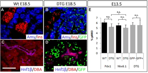
Fig. 1. Notch signaling is required for endocrine differentiation.(A)The Notch transcriptional complex and truncated Maml1 (dnMAML1) lead to a dominant-negative effect on the expression of Notch target genes. (B)Transgenic overexpression of dnMAML1. Pdx1promoter-driven expression of tTA results in transcriptional activation of the dnMAML1-IRES-nEGFP mRNA. Presence of doxycycline prevents tTA-mediated expression of the transgene. (C,D)Mid-gut dissection of wild-type (Wt) and dnMAML1;tTA double transgenic (DTG) embryos at E18.5 (C). Under fluorescent light (D), green fluorescence of the DTG pancreas in C is visible. (E-H)Immunofluorescence staining for pancreatic differentiation markers in wild-type (E,G) and DTG (F,H) pancreas at E18.5. (I,J)Morphometric quantification of the exocrine tissue based on amylase (I), the endocrine compartment based on endocrine-specific gene expression and the duct cells marked by the duct-specific lectin DBA (J). Values are mean ± s.d. *, P<0.05; **, P<0.005; n.s., not statistically significant. n3 for all analyses in I,J. Amy, amylase; Ghr, ghrelin; Glu, glucagon; Ins, insulin.
Scale bar: 50m.
D
E
V
E
LO
P
M
E
N
Although the pancreas exhibited apparent visual homogeneity of EGFP expression (Fig. 1D), histological analysis of tissue sections revealed mosaic expression of the transgene. Given that dnMAML1 operates as a cell-intrinsic regulator, this observation prompted us to conduct subsequent analyses of this model with emphasis on the transgene-expressing cells relative to the non-transgenic cells. In an effort to characterize the effect of the transgene upon the cells that expressed it, we carried out histological analysis of pancreatic tissue from DTG embryos, with that of wild-type littermates as control, using terminal differentiation markers for the major cell types of the pancreas, i.e. endocrine, acinar and duct cells. Interestingly, at E18.5 the transgene was expressed predominantly in acinar cells as marked by the expression of EGFP in amylase-positive cells (Fig. 2B). Quantitative analysis indicated that 69±10% of acinar cells expressed the transgene, whereas only 4.8±3.1% of insulin-expressing cells were EGFP+. A similar analysis of the distribution
of the transgene relative to pancreatic duct cells was performed by staining pancreatic tissue for the duct markers DBA lectin and Hnf1in relation to the transgene-derived EGFP+cells (Fig. 2D).
We did not detect any pancreatic duct cells that expressed the transgene despite thorough examination.
Notch signaling is required for pancreatic pro-endocrine/duct (trunk) fate patterning
Since the prevailing late effect of the transgene was on the endocrine pancreas, we were surprised to detect an acinar-biased distribution of the transgene-expressing cells. Also, given that the transgene is indirectly governed via the Pdx1 promoter (Pdx1-tTAKI), which is first active in all pancreatic progenitor cells and
leads to a random distribution of transgene-positive cells throughout the early pancreatic epithelium (supplementary material Fig. S4), the observed segregation of the transgene-expressing population at later stages was at odds with the expected pattern, considering a hypothetical inert transgene.
We tested several hypotheses that might explain the observed effects. First, if the dnMAML1 protein negatively affects cell viability in a cell-specific manner, the results could be explained by a particular loss of endocrine/ductal cells experiencing the transgene-expressed protein, which could possibly undergo apoptosis at some point prior to the time of analysis at E18.5. In such a case, cells refractory to the effects of dnMAML1 would not be affected and remain detectable. However, we did not detect apoptotic cells at earlier stages (E12.5-14.5; data not shown). Second, we hypothesized that dnMAML1 protein, and consequently loss of Notch signaling, could have positively affected cell division, but only within the acinar population, leading to a relative increase in such cells over time. Experimental evidence provided no support for this hypothesis, and cells expressing dnMAML1 protein had identical replication rates as wild-type/non-transgenic cells neighboring the EGFP+pool in the
dnMAML1 model (Fig. 2E).
Finally, we speculated that Notch signaling could have affected the generation of the two progenitor compartments in a manner whereby endocrine/ductal fates were negatively controlled by dnMAML1 presence and acinar fates positively so. Such an event would have occurred prior to E18.5, and likely at, or just prior to, the ‘secondary transition’. Emerging evidence points to a process of pre-patterning of the pancreatic progenitor field prior to the onset of the secondary transition, and this leads to future bias in terminal fate commitment. During early embryogenesis, the branched pancreatic epithelium becomes regionalized into branched tip and trunk domains. Although, initially, progenitor cells at the tip position contribute to all lineages of the developing pancreas, by E14.5 cells at the tip position contribute exclusively to acinar cells, whereas cells within the trunk differentiate into endocrine and duct lineages (Zhou et al., 2007). For this reason, we speculated that the experimental outcomes could be explained if dnMAML1 served to enhance the expression of acinar lineage-specific genes (tip domain formation) while suppressing endocrine/duct progenitor cell-specific genes (trunk domain formation).
To test this hypothesis, we focused on early embryonic stages with the aid of transgenic EGFP expression and pancreatic progenitor markers for tip and trunk cells. In so doing, we took advantage of the mosaic model because we could scrutinize a possible fate bias within a cell experiencing Notch inhibition by comparison with neighbors not under such influence. By E14.5, Ptf1a-positive progenitor cells are lineage restricted to pancreatic acinar fate, and we therefore analyzed expression of Ptf1a in Notch-depleted dnMAML1 cells by immunofluorescence staining. At this time point, nEGFP/dnMAML1+ cells predominantly
(>80%) express Ptf1a and, notably, these transgenic cells are mostly localized at the tip position (Fig. 3A-C), leaving a trunk region devoid of nEGFP/dnMAML1+ cells. Also, at E14.5
dnMAML1+cells fail to express trunk-specific markers [Nkx6.1,
Hnf1 and Sox9 (Fig. 3E-P)]. A few Notch-suppressed cells showed weak expression of Sox9, suggesting that these cells are in an intermediate state of losing Sox9 expression (Fig. 3O,P).
To further validate the observation that inhibition of Notch signaling via dnMAML1 results in loss of trunk-specific gene expression, we isolated epithelial dnMAML1+cells at E13.5 by
fluorescence-activated cell sorting (FACS) based on the transgene-derived EGFP and co-labeling for the epithelial marker Epcam (Fig. 3Q,R). RNA was extracted from the isolated fractions and subjected to quantitative RT-PCR analysis (Fig. 3S). This revealed that transgenic cells predominantly express the acinar-specific
Fig. 2. Acinar cell-specific expression of the dnMAML1 transgene.
(A-D)Immunofluorescence staining of amylase and insulin (A,B) and of the ductal markers Hnf1and DBA lectin (C,D) in wild-type (A,C) and DTG (B,D) E18.5 pancreas. (B,D)The expression of EGFP (green) indicates the distribution of transgene expression relative to that of pancreatic differentiation markers. Relative expression levels of EGFP to differentiation markers are: amylase+; GFP+/amylase+0.69±0.10;
insulin+; GFP+/insulin+0.048±0.031; Hnf1+; GFP+/Hnf1+0.
(E)Quantification of proliferation rates of E13.5 Pdx1+wild-type and
DTG pancreatic cells, Nkx6.1+wild-type and DTG pancreatic cells, and
transgene-negative (GFP–) or transgene-positive (GFP+) epithelial cells in
the DTG pancreas based on phospho-histone H3 (pHH3)
immunofluorescence staining. n3. Values are mean ± s.d. n.s., not
statistically significant. Scale bar: 50m.
D
E
V
E
LO
P
M
E
N
[image:4.612.51.299.464.587.2]transcription factor Ptf1a and the acinar product amylase, which confirms that these cells have adopted an acinar fate. Correspondingly, this effect was accompanied by decreased expression of trunk-specific genes such as Nkx6.1and Sox9, as well as the endocrine progenitor marker Ngn3(Fig. 3S).
[image:5.612.53.516.58.562.2]These observations strongly suggest that suppression of Notch signaling leads to loss of trunk progenitor fate and a corresponding gain of tip fate, with conceivably a concomitant morphogenetic effect resulting in the placement of the Notch-suppressed cells at the tip position, as these complete the acinar program. Although
Fig. 3. Effect of dnMAML1 on pancreatic epithelial patterning.(A-P)Fluorescence visualization of transgene-derived nEGFP expression relative to immunofluorescence staining of Ptf1a (A-C), Nkx6.1 (E-G), Hnf1(I-K) or Sox9 (M-O). Quantitative analysis of the percentage of nEGFP cells that are positive or negative for Ptf1a (D), Nkx6.1 (H), Hnf1(L) or Sox9 (P). Sox9 expression was additionally categorized as high (Sox9hi) or medium (Sox9me) level. White arrows indicate GFP+cells that are negative for a given pancreatic marker; arrowheads indicate EGFP+cells that are positive
for pancreatic markers; yellow arrows indicate EGFP+cells that express medium level of Sox9. (Q)Isolation of E13.5 positive and
EGFP-negative pancreatic epithelial cells by flow cytometry. Dissociated embryonic pancreas tissue was stained with APC-conjugated anti-E-cadherin prior to sorting. (R)x-axis (FITC) indicates GFP intensity and y-axis (APC-A) indicates intensity of epithelial staining by APC-conjugated E-cadherin. (S)Quantitative RT-PCR analysis of pancreatic progenitor markers in EGFP-positive epithelial cells expressed as fold increase or decrease relative to EGFP–epithelial cells. EGFP+cells have 790.8±71.6-fold more dnMAML1 than EGFP–cells. Values are mean ± s.d. Scale bar: 50m.
D
E
V
E
LO
P
M
E
N
immunofluorescence staining confirms that dnMAML1-expressing cells initiate acinar-specific gene expression at the onset of terminal differentiation of acinar cells at E14.5, we did not detect induction of premature acinar cell differentiation in the DTG pancreas (supplementary material Fig. S5). We conclude that suppression of Notch signaling results in the patterning of MPCs toward the acinar fate, followed by a normal differentiation timecourse to terminal cell fate.
Nkx6.1is a target of Notch-mediated tip-trunk
patterning
Although the above analysis suggests that Notch inhibition does not compromise tip fate allocation, but instead impairs trunk-derived fates, E14.5 marks a stage when the tip and trunk cells are completely segregated and terminal differentiation has initiated. We therefore analyzed pancreatic tissue from earlier stage transgenic mice to uncover possible Notch-mediated events preceding the complete segregation of transgene-expressing cells from wild-type
cells. In the E12.5 pancreas, which is far less lobulated/branched than the organ at E14.5, there was a tendency for most transgene-expressing cells to cluster toward the epithelial/mesenchymal border, but we also found a considerable number of these cells interspersed with wild-type cells in the trunk domain. Immunofluorescence analysis revealed that, unlike the E14.5 transgenic pancreas, only ~50% of the transgenic cell population was strongly Ptf1a expressing (Fig. 4E-H), suggesting that the gradual increase in Ptf1a/EGFP double-positive cells is an ongoing process in which the acinar compartment is increasingly enriched in Notch-suppressed cells.
The presence of transgenic cells within the trunk at this stage further supported the notion that the subsequent sorting of these cells to the tip position is a result of loss of trunk identity, and that cells within the trunk domain become recruited to the acinar fate (Fig. 7A,B). This would lead to a depletion of transgenic cells able to enter downstream endocrine/ductal fates. To validate this hypothesis, we analyzed the effect of dnMAML1 on other trunk-specific marker genes, including those encoding Nkx6.1, Hnf1and Sox9 (Fig.
4I-Fig. 4. Suppression of Nkx6.1 expression by dnMAML1 precedes its effect on Ptf1a.Fluorescence visualization of the transgene-derived nEGFP relative to immunofluorescence staining of (A-C) Hes1, (E-G) Ptf1a, (I-K) Nkx6.1, (M-O) Hnf1and (Q-S) Sox9. Arrows indicate EGFP+cells
that are negative, and arrowheads indicate EGFP+cells that are positive, for a given pancreatic marker gene. Quantitative analysis of the percentage
of EGFP+cells that are positive or negative for (D) Hes1, (H) Ptf1a, (L) Nkx6.1, (P) Hnf1and (T) Sox9. Values are mean ± s.d. Scale bar: 50m.
D
E
V
E
LO
P
M
E
N
[image:6.612.55.518.273.693.2]T). Indeed, unlike at E14.5, when almost all transgene-expressing cells had lost expression of these marker genes, at E12.5 we observed that almost 50% of dnMAML1-positive cells still expressed Hnf1and Sox9, although only 20% of transgene-positive cells expressed Nkx6.1 (Fig. 4I-L). Indeed, quantification of the absolute levels of these markers in DTG pancreas revealed that, unlike the other markers, there was a significant reduction in the number of Nkx6.1+ cells. Ptf1a levels increased slightly (not
statistically significant; supplementary material Fig. S6). Thus, at this stage, suppression of Notch signaling is most dramatically reflected by a loss of Nkx6.1 expression. Analysis of dnMAML1-expressing cells at E12.5 revealed that an identical percentage (~80%) fail to express Hes1 (Fig. 4A-D) as Nkx6.1 (Fig. 4I-L). This suggests that Notch signaling is required for Nkx6.1 expression and, when ablated, results in the loss of Nkx6.1 and the concomitant loss of trunk lineage determination.
The largely overlapping expression of Hes1 and Nkx6.1 within E12.5 trunk progenitor cells (Fig. 5A-C), together with the induction of Nkx6.1 by NICD overexpression in the pancreas (Hald et al., 2003; Schaffer et al., 2010), provide further evidence to suggest that Nkx6.1 expression is under the control of Notch signaling. To test the hypothesis that Nkx6.1 is a direct target of Notch, we performed chromatin immunoprecipitation sequencing (ChIP-Seq) using anti-RBP-jantibody on chromatin from E12.5, E15.5 and E17.5 pancreas. Analysis of the ChIP-Seq data revealed a binding peak for RBP-jupstream of the Nkx6.1transcription start site (TSS) at both E12.5 and E15.5. We found additional
binding at specific locations in exon 1, proximal to the exon 1-intron 1 junction and also immediately following the terminal exon 3, consequently following the transcriptional stop. Finally, a distinct peak was identified ~10 kb downstream of the Nkx6.1 gene. We identified putative RBP-j binding sites within these RBP-j-bound regions (supplementary material Fig. S8). Unlike at E12.5 and E15.5, we did not detect RBP-j binding to these regions in E17.5 pancreatic chromatin, suggesting that Notch-mediated control of Nkx6.1is limited to early developmental stages (Fig. 5D). Consistent with Hes1 being a Notch target gene, we found that RBP-jbinds to regions proximal to the TSS of Hes1 (Fig. 5E). Similar to Nkx6.1, we also identified RBP-joccupancy within the coding region of Hes1(Fig. 5D,E).
RBP-jcan also partner with Ptf1a to transactivate Ptf1a-specific target genes; however, the Ptf1a–RBP-jsites are distinct from the RBP-jsites involved in Notch signaling (Beres et al., 2006; Masui et al., 2007; Wiebe et al., 2007). In a parallel ChIP-Seq with anti-Ptf1a antibody, we found significant anti-Ptf1a occupancy of regions corresponding to some of the RBP-jbinding sites in Hes1, but not Nkx6.1, suggesting that the RBP-joccupancy in the Nkx6.1gene is Notch mediated. RBP-joccupancy of the proximal region of Nkx6.1 was confirmed in independent experiments in which chromatin from E13.5 pancreas was immunoprecipitated with a second anti-RBP-jantibody (supplementary material Table S2) followed by quantitative PCR with several primers covering ~1 kb of the Nkx6.1TSS and 5⬘flank (supplementary material Fig. S7, Table S3).
Lack of premature endocrine differentiation in dnMAML1-mediated Notch-suppressed cells
Our analyses thus far were in stark contrast to previous reports by us and others that suggested that Notch signaling is crucial for the maintenance of the pancreatic progenitor cell state such that, when abrogated, cells differentiate prematurely resulting in a hypoplastic tissue that consists predominantly of endocrine cell fate (Apelqvist et al., 1999; Fujikura et al., 2006; Hald et al., 2003; Jensen et al., 2000; Murtaugh et al., 2003). We therefore carried out a detailed analysis of pancreatic tissue in early embryos for evidence of dnMAML1-induced premature differentiation at the primary transition stage, which is when precocious differentiation of progenitor cells has been reported to commence in other models in which Notch signaling was abrogated in the pancreas (e.g. Hes1–/–, Dll1–/–) (Apelqvist et al.,
1999; Jensen et al., 2000). We examined dnMAML1-expressing cells based on the presence of nEGFP for the preferential expression of early glucagon-expressing or insulin-expressing cells (Fig. 6). Endocrine cell clusters at the primary transition stage of pancreas development are often hormone double positive (Herrera et al., 1991; Teitelman et al., 1993). With the exception of rare instances in which a few clusters of glucagon-positive cells were located near dnMAML1-expressing nuclei at E11.5 (Fig. 6F), the majority of endocrine clusters (insulin or glucagon positive) were negative for the dnMAML1 transgene, indicating the lack of premature differentiation in transgene-expressing cells. Furthermore, the mosaic suppression of Notch through dnMAML1 did not affect the architecture of the early pancreatic epithelium. The stratified nature of the E11.5 epithelium (Fig. 6A,E) (Jensen, 2004; Pan and Wright, 2011; Villasenor et al., 2010) was retained in the transgenic pancreas (Fig. 6B,F) and by E12.5 the transgenic epithelium branched normally (Fig. 6D,H), as in wild-type pancreas (Fig. 6C,G). Taken together, our data do not support the induction of premature endocrine (or acinar) cell differentiation in individual cells experiencing a loss of Notch signaling.
Fig. 5. Nkx6.1 is a direct target of Notch. (A-C)Immunofluorescence staining of Nkx6.1 (A) and Hes1 (B) in E12.5 wild-type pancreas. (C)Overlay of A and B. (D,E)ChIP-Seq analysis on the Nkx6.1locus (D) and Hes1locus (E) with anti-RBP-jantibodies on E12.5, E15.5 and E17.5 pancreatic chromatin (red tracks), anti-Ptf1a antibody on E15.5 pancreatic chromatin (blue tracks), anti-RNA polymerase II antibody on E15.5 pancreatic chromatin (yellow tracks), as well as input tracks. Arrows (D,E) indicate RBP-joccupancy of the Nkx6.1 and Hes1 loci,
respectively. Scale bar: 50m.
D
E
V
E
LO
P
M
E
N
[image:7.612.51.298.369.642.2]DISCUSSION
Studies on the role of Notch signaling are gradually revealing that this widely conserved signaling system is pleiotropic and operates at multiple levels in pancreatic development. Initially believed to be limited to cell fate control of developing endocrine cells, in which depletion of key Notch component genes resulted in premature differentiation of MPCs to endocrine fate (Apelqvist et al., 1999; Fujikura et al., 2006; Jensen et al., 2000), later studies by us and others suggested that Notch plays an essential role in maintaining cells in a progenitor state, as constitutive activation of Notch signaling blocks differentiation of pancreatic progenitor cells (Hald et al., 2003; Murtaugh et al., 2003). In light of the aforementioned studies and our current findings, it is rather surprising that conditional inactivation of both Notch1 and Notch2 was reported to have no profound effect on pancreas development (Nakhai et al., 2008). More recent data addressing the role of Notch by genetic ablation of presenilins (Cras-Meneur et al., 2009), as well as studies of pancreatic tip cell versus trunk cell progenitor patterning that have implicated Notch (Schaffer et al., 2010), underscore the crucial role of Notch signaling during pancreas development.
The present study provides additional evidence of the causal role of Notch in the patterning of MPCs. By disrupting Notch signaling in a mosaic manner in which Notch-suppressed cells are interspersed with wild-type cells, we have discovered that progenitor cells that are incapable of sustaining Notch signaling adopt a pro-acinar fate at the expense of a pro-endocrine/ductal
fate. Thus, our findings fit a model in which Notch signaling operates during patterning of MPCs into the pro-endocrine/duct (trunk) subdomain (Fig. 7). Suppression of Notch signaling in cells within this domain results in the acquisition of pro-acinar (tip) identity secondary to the loss of the crucial pro-trunk determination gene Nkx6.1. The subsequent expression of the pro-tip marker gene Ptf1ain Notch-suppressed cells is in agreement with the findings of Schaffer et al. (Schaffer et al., 2010), in which the mutual antagonism between Nkx6 factors and Ptf1a was demonstrated to control the patterning of MPCs into trunk (TrPC) and tip (TipPC) domains, respectively. Our data suggest that Notch acts initially on Nkx6.1, and not via Ptf1a suppression, to tip the balance between the two factors, as shown by the ability of RBP-jto bind to the conserved Nkx6.1promoter.
This phenotype of loss of Notch signaling in MPCs is in agreement with the differentiation of pancreatic progenitor cells into acinar cells when cultured in the presence of the -secretase inhibitor DAPT (X.Q., S.A., J. Nygaard Jensen, S. Kobberup, M. Schmerr, F. Xiao, Pia Nyeng, M. V. Albertoni, A. Grapin-Botton and J.J., unpublished) (Cras-Meneur et al., 2009; Magenheim et al., 2011). Similarly, deletion of Psen1 and Psen2 in Ngn3-expressing cells has been shown to result in a switch in their differentiation potential from endocrine to acinar cells (Cras-Meneur et al., 2009). Also, defective endocrine cell differentiation is observed upon depletion of calsenilin, a regulatory factor of presenilin, and more direct evidence was obtained by lineage tracing of Notch-responsive cells into endocrine and ductal progenitors, but not acinar progenitors, in zebrafish (Buxbaum et al., 1998; Jo et al., 2005; Stetsyuk et al., 2007; Wang et al., 2011). These data appear to be in conflict with similar genetic lineage-tracing studies in mice investigating the fate potential of Hes1-expressing cells. Whereas early stage Hes1-positive cells were observed to be multipotent, later stage Hes1-positive cells were restricted to the duct and acinar lineages (Kopinke et al., 2011). The implication of this for the role of Notch signaling in endocrine/duct fate patterning versus a specific role of Hes1 in late stage acinar cell progenitors is not yet clear, and might be rooted in Notch-independent control of Hes1 expression (Curry et al., 2006; Ingram et al., 2008; Nakayama et al., 2008; Sanalkumar et al., 2010; Wall et al., 2009), or a later role for Notch/Hes1 within the centroacinar cell type, which is not yet fully formed at the secondary transition. Further evidence for the suppressive effect of Notch signaling on the acinar fate is derived from the inhibitory effect of Notch on the transcriptional activity of the pro-acinar factor Ptf1a (Esni et al., 2004).
[image:8.612.52.267.58.334.2]Our data argue that changes to the TipPC and TrPC fate choice occur cell-intrinsically in dnMAML1-mediated Notch-suppressed cells, which appears to cause rapid segregation of the Notch-inhibited pool to a tip cell position. We believe that this provides additional evidence that cell fate and patterning are causally linked to the process of morphogenesis, as also suggested by Kesavan et al. (Kesavan et al., 2009). But such a conclusion would benefit from direct evidence of the migratory events occurring upon dnMAML1-conferred TipPC commitment through time-lapse imaging of pancreatic explants. Such direct evidence will also help rule out any possibility of the transgene undergoing an ‘ON/OFF’ switch. Active morphogenesis through cell migration, segregation and intercalation are well known in other organ systems, and in certain cases the molecular underpinnings are beginning to emerge. The initiation of kidney development is marked by the movement of cells from the nephritic duct into the metanephric mesenchyme to form the ureteric bud, and GDNF/Ret signaling has been shown to promote this movement. In chimeric embryos consisting of wild-type and Ret–/–
Fig. 6. Suppression of Notch signaling through dnMAML1 does not lead to premature endocrine cell differentiation.
Immunofluorescence staining of Pdx1 and insulin in (A) E11.5 and (C) E12.5 wild-type pancreas and in the presence of transgene-derived nEGFP expression in (B) E11.5 and (D) E12.5 DTG embryos. Immunofluorescence staining of Pdx1 and glucagon in (E) E11.5 and (G) E12.5 wild-type pancreas and in the presence of transgene-derived nEGFP in DTG embryos at (F) E11.5 and (H) E12.5. Scale bar: 50m.
D
E
V
E
LO
P
M
E
N
cells, the Ret-deficient cells are excluded from the tip of the ureteric bud resulting in a biased occupancy of wild-type cells at the tip of the ureteric bud and the restriction of Retmutant cells to the trunk (Costantini, 2010; Kuure et al., 2005; Kuure et al., 2010).
Several previous studies have revealed that abrogation of Notch signaling components results in precocious differentiation of MPCs into endocrine cells (Apelqvist et al., 1999; Fujikura et al., 2006; Jensen et al., 2000), whereas we do not observe premature differentiation of Notch-suppressed cells into endocrine cells in our analysis. This seeming contradiction might be due to fundamental differences in the way that Notch signaling is suppressed in the models analyzed. In studies in which premature endocrine differentiation was observed, the loss of Notch signaling might have been achieved in homogenous pools of cells, in contrast to the mosaic nature of Notch inhibition in our study [as well as in that of Cras-Meneur et al. (Cras-Meneur et al., 2009), considering that the deletion of presenilins occurred in the Ngn3-expressing pool], and this might be an important basis for the differences in outcome. The phenotypes of global versus mosaic loss of Notch signaling thus manifest two distinct roles of Notch signaling: the former could be interpreted through concepts such as ‘suppressive maintenance’ (the maintenance of cells in a progenitor state), whereas the latter through notions of ‘patterning’, in which the different levels of Notch in neighboring cells result in a bifurcated lineage decision in multipotent progenitor cells, while the system overall remains largely intact. In other words, it takes a mosaic depletion of Notch signaling to uncover the role of Notch in pancreatic progenitor patterning, whereas the global deletion of Notch, which results in premature differentiation (Apelqvist et al., 1999; Fujikura et al., 2006; Jensen et al., 2000), and the global activation of Notch signaling, which results in progenitor state arrest (Hald et al., 2003; Murtaugh et al., 2003), both reflect the role of Notch in suppressive maintenance.
Our data suggest that patterning of MPCs into TipPC or TrPC is governed by differential levels of Notch. Cells active in Notch signaling adopt a TrPC fate, whereas Notch-suppressed cells become TipPC. Technically, we demonstrated this by a mosaic transgenic approach that allowed assessment of the behavior of Notch-suppressed progenitor cells relative to wild-type cells during tissue patterning. Importantly, this reflects the ‘mosaic’ nature of lateral Notch signaling during normal development. This is in agreement with the observation of Cras-Meneur et al. (Cras-Meneur et al., 2009), in which Psen1/2deficiency results in conversion of the entire Ngn3+ pool into an acinar fate, but only when the remaining
pancreatic progenitors are not deficient in Psen1/2. Notably, the wider elimination of Psen1/2 in pancreas allows such Psen1/2-deficient cells to contribute to the endocrine compartment (Cras-Meneur et al., 2009), which is seemingly at odds with the more dramatic phenotype observed when the Psen1/2deficiency occurs within the Ngn3-expressing pool only. The authors speculated that this outcome was due to the availability of RBP-jto engage in Ptf1a–RBP-jcomplex formation, as opposed to its availability for forming RBP-j–NICD complexes at Notch target promoters in Psen1/2-competent cells (Cras-Meneur et al., 2009). Although a stoichiometric argument of limiting availability of RBP-j is intriguing, this model cannot fully explain the role of Notch signaling attenuation observed here. The overexpression of dnMAML1 in our model should lead to the sequestration of RBP-jwith NICD in non-functional complexes (Fig. 1) (Arora and Ansari, 2009; Jones, 2009; Kovall, 2007; Moellering et al., 2009; Wilson and Kovall, 2006), and, therefore, this should not lead to the increased availability of RBP-j to enter into TipPC/acinar-promoting Ptf1–RBP-j complexes, as the Cras-Meneur competition model would posit. Instead, our data demonstrate that Notch signaling plays an active role in creating the TrPC as a primary event, at least partially via Nkx6.1 activation, and therefore Notch signaling appears to be playing an active role in the determination of TrPC fate.
Acknowledgements
We thank multiple investigators for donating antibodies and Dr P. Serup and S. D. Leach for comments on the manuscript.
Funding
This work was supported by the Juvenile Diabetes Research Foundation [award 1-2007-109 to J.J., postdoctoral fellowship award 3-2007-121 to S.A.]; the American Diabetes Association [1-11-BS-75 to J.J.]; the Cleveland Clinic Foundation; and a gift from the E. J. Brandon family. R.J.M. was supported by the National Institutes of Health [R01-DK061220]. The creation of pTRE-dnMAML1 mice was supported by the Chicago Diabetes Project (www.chicagodiabetesproject.org). Deposited in PMC for release after 12 months.
Competing interests statement
The authors declare no competing financial interests.
Supplementary material
Supplementary material available online at
http://dev.biologists.org/lookup/suppl/doi:10.1242/dev.075804/-/DC1
References
Afelik, S., Chen, Y. L. and Pieler, T.(2006). Combined ectopic expression of Pdx1 and Ptf1a/p48 results in the stable conversion of posterior endoderm into endocrine and exocrine pancreatic tissue. Genes Dev. 20, 1441-1446. Apelqvist, A., Li, H., Sommer, L., Beatus, P., Anderson, D. J., Honjo, T., de
Angelis, M. H., Lendahl, U. and Edlund, H.(1999). Notch signalling controls pancreatic cell differentiation. Nature400, 877-881.
Arora, P. S. and Ansari, A. Z.(2009). Chemical biology: a Notch above other inhibitors. Nature462, 171-173.
[image:9.612.51.297.57.229.2]Beres, T. M., Masui, T., Swift, G. H., Shi, L., Henke, R. M. and MacDonald, R. J.(2006). PTF1 is an organ-specific and notch-independent basic helix-loop-helix complex containing the mammalian suppressor of hairless (RBP-J) or its paralogue, RBP-L. Mol. Cell. Biol.26, 117-130.
Fig. 7. Model of Notch-mediated patterning of multipotent pancreatic progenitor cells into tip and trunk domains.(A)At E12.5, the pancreatic epithelium is organized into tip and trunk domains, and Pdx1 is expressed throughout all pancreatic cells, whereas Ptf1a is restricted to the tip domain marking pro-acinar cells. Although the majority of Notch-deficient cells are localized to the tip domain, a few remain in the trunk (arrows). (B)By E14.5, almost all Notch-deficient cells have resolved into the tip domain. (C)This suggests a dynamic process of Notch-mediated patterning of MPCs, in which the loss of Notch within a multipotent progenitor leads to a tip fate and localization. MPC, multipotent pancreatic progenitor cell; TipPC, tip domain; TrPC, trunk domain. Scale bar: 50m.
D
E
V
E
LO
P
M
E
N
Burlison, J. S., Long, Q. M., Fujitani, Y., Wright, C. V. E. and Magnuson, M. A.(2008). Pdx-1 and Ptf1a concurrently determine fate specification of pancreatic multipotent progenitor cells. Dev. Biol.316, 74-86. Buxbaum, J. D., Choi, E. K., Luo, Y. X., Lilliehook, C., Crowley, A. C.,
Merriam, D. E. and Wasco, W.(1998). Calsenilin: a calcium-binding protein that interacts with the presenilins and regulates the levels of a presenilin fragment. Nat. Med.4, 1177-1181.
Costantini, F.(2010). GDNF/Ret signaling and renal branching morphogenesis from mesenchymal signals to epithelial cell behaviors. Organogenesis6, 252-262.
Cras-Meneur, C., Li, L., Kopan, R. and Permutt, M. A.(2009). Presenilins, Notch dose control the fate of pancreatic endocrine progenitors during a narrow developmental window. Genes Dev.23, 2088-2101.
Curry, C. L., Reed, L. L., Nickoloff, B. J., Miele, L. and Foreman, K. E.(2006). Notch-independent regulation of Hes-1 expression by c-Jun N-terminal kinase signaling in human endothelial cells. Lab. Invest. 86, 842-852.
Esni, F., Ghosh, B., Biankin, A. V., Lin, J. W., Albert, M. A., Yu, X. B., MacDonald, R. J., Civin, C. I., Real, F. X., Pack, M. A. et al.(2004). Notch inhibits Ptf1 function and acinar cell differentiation in developing mouse and zebrafish pancreas. Development131, 4213-4224.
Fujikura, J., Hosoda, K., Iwakura, H., Tomita, T., Noguchi, M., Masuzaki, H., Tanigaki, K., Yabe, D., Honjo, T. and Nakao, K.(2006). Notch/Rbp-j signaling prevents premature endocrine and ductal cell differentiation in the pancreas. Cell Metab. 3, 59-65.
Golson, M. L., Le Lay, J., Gao, N., Bramswig, N., Loomes, K. M., Oakey, R., May, C. L., White, P. and Kaestner, K. H.(2009). Jagged1 is a competitive inhibitor of Notch signaling in the embryonic pancreas. Mech. Dev. 126, 687-699.
Hald, J., Hjorth, J. P., German, M. S., Madsen, O. D., Serup, P. and Jensen, J. (2003). Activated Notch1 prevents differentiation of pancreatic acinar cells and attenuate endocrine development. Dev. Biol.260, 426-437.
Hald, J., Sprinkel, A. E., Ray, M., Serup, P., Wright, C. and Madsen, O. D. (2008). Generation and characterization of Ptf1a antiserum and localization of Ptf1a in relation to Nkx6.1 and Pdx1 during the earliest stages of mouse pancreas development. J. Histochem. Cytochem.56, 587-595.
Herrera, P. L., Huarte, J., Sanvito, F., Meda, P., Orci, L. and Vassalli, J. D. (1991). Embryogenesis of the murine endocrine pancreas-early expression of pancreatic-polypeptide gene. Development113, 1257-1265.
Holland, A. M., Gonez, L. J., Naselli, G., MacDonald, R. J. and Harrison, L. C. (2005). Conditional expression demonstrates the role of the homeodomain transcription factor Pdx1 in maintenance and regeneration of beta-cells in the adult pancreas. Diabetes54, 2586-2595.
Ingram, W. J., McCue, K. I., Tran, T. H., Hallahan, A. R. and Wainwright, B. J. (2008). Sonic Hedgehog regulates Hes1 through a novel mechanism that is independent of canonical Notch pathway signalling. Oncogene27, 1489-1500. Jensen, J.(2004). Gene regulatory factors in pancreatic development. Dev. Dyn.
229, 176-200.
Jensen, J., Pedersen, E. E., Galante, P., Hald, J., Heller, R. S., Ishibashi, M., Kageyama, R., Guillemot, F., Serup, P. and Madsen, O. D.(2000). Control sf endodermal endocrine development by Hes-1. Nat. Genet.24, 36-44. Jo, D. G., Jang, J. Y., Kim, B. J., Lundkvist, J. and Jung, Y. K.(2005).
Overexpression of calsenilin enhances gamma-secretase activity. Neurosci. Lett. 378, 59-64.
Jones, K. A.(2009). Outsmarting a mastermind. Dev. Cell17, 750-752. Kawaguchi, Y., Cooper, B., Gannon, M., Ray, M., MacDonald, R. J. and
Wright, C. V. E.(2002). The role of the transcriptional regulator Ptf1a in converting intestinal to pancreatic progenitors. Nat. Genet.32, 128-134. Kesavan, G., Sand, F. W., Greiner, T. U., Johansson, J. K., Kobberup, S., Wu,
X. W., Brakebusch, C. and Semb, H.(2009). Cdc42-mediated tubulogenesis controls cell specification. Cell139, 791-801.
Kobberup, S., Schmerr, M., Dang, M. L., Nyeng, P., Jensen, J. N., MacDonald, R. J. and Jensen, J.(2010). Conditional control of the differentiation competence of pancreatic endocrine and ductal cells by Fgf10. Mech. Dev. 127, 220-234.
Kopinke, D., Brailsford, M., Shea, J. E., Leavitt, R., Scaife, C. L. and Murtaugh, L. C.(2011). Lineage tracing reveals the dynamic contribution of Hes1(+) cells to the developing and adult pancreas. Development138, 431-441. Kovall, R. A.(2007). Structures of CSL, Notch and Mastermind proteins: piecing
together an active transcription complex. Curr. Opin. Struct. Biol.17, 117-127. Kuure, S., Sainio, K., Vuolteenaho, R., Ilves, M., Wartiovaara, K., Immonen,
T., Kvist, J., Vainio, S. and Sariola, H.(2005). Crosstalk between Jagged1 and GDNF/Ret/GFR alpha 1 signalling regulates ureteric budding and branching. Mech. Dev. 122, 765-780.
Kuure, S., Chi, X., Lu, B. and Costantini, F.(2010). The transcription factors Etv4 and Etv5 mediate formation of the ureteric bud tip domain during kidney development. Development137, 1975-1979.
Liefke, R., Oswald, F., Alvarado, C., Ferres-Marco, D., Mittler, G., Rodriguez, P., Dominguez, M. and Borggrefe, T.(2010). Histone demethylase KDM5A is an integral part of the core Notch-RBP-J repressor complex. Genes Dev.24, 590-601.
Magenheim, J., Klein, A. M., Stanger, B. Z., Ashery-Padan, R., Sosa-Pineda, B., Gu, G. Q. and Dor, Y.(2011). Ngn(3+) endocrine progenitor cells control the fate and morphogenesis of pancreatic ductal epithelium. Dev. Biol.359, 26-36.
Maillard, I., Weng, A. P., Sambandan, A., Carpenter, A. C., Sai, H., Xu, L. W., Allman, D., Bhandoola, A., Aster, J. C. and Pear, W. S.(2003). Mastermind critically regulates notch-mediated lymphoid cell fate decisions. Blood102, 133A-134A.
Masui, T., Long, Q., Beres, T. M., Magnuson, M. A. and MacDonald, R. J. (2007). Early pancreatic development requires the vertebrate Suppressor of Hairless (RBPJ) in the PTF1 bHLH complex. Genes Dev. 21, 2629-2643. Masui, T., Swift, G. H., Deering, T., Shen, C. C., Coats, W. S., Long, Q. M.,
Elsasser, H. P., Magnuson, M. A. and MacDonald, R. J.(2010). Replacement of Rbpj with Rbpjl in the PTF1 complex controls the final maturation of pancreatic acinar cells. Gastroenterology139, 270-280.
Moellering, R. E., Cornejo, M., Davis, T. N., Del Bianco, C., Aster, J. C., Blacklow, S. C., Kung, A. L., Gilliland, D. G., Verdine, G. L. and Bradner, J. E.(2009). Direct inhibition of the NOTCH transcription factor complex. Nature 462, 182-188.
Mulligan, P., Yang, F. J., Di Stefano, L., Ji, J. Y., Ouyang, J., Nishikawa, J. L., Toiber, D., Kulkarni, M., Wang, Q., Najafi-Shoushtari, S. H. et al.(2011). A SIRT1-LSD1 corepressor complex regulates notch target gene expression and development. Mol. Cell42, 689-699.
Murtaugh, L. C., Stanger, B. Z., Kwan, K. M. and Melton, D. A.(2003). Notch signaling controls multiple steps of pancreatic differentiation. Proc. Natl. Acad. Sci. USA 100, 14920-14925.
Nakayama, K., Satoh, T., Igari, A., Kageyama, R. and Nishida, E.(2008). FGF induces oscillations of Hes1 expression and Ras/ERK activation. Curr. Biol.18, R332-R334.
Nakhai, H., Siveke, J. T., Klein, B., Mendoza-Torres, L., Mazur, P. K., Algul, H., Radtke, F., Strobl, L., Zimber-Strobl, U. and Schmid, R. M.(2008). Conditional ablation of Notch signaling in pancreatic development. Development135, 2757-2765.
Pan, F. C. and Wright, C.(2011). Pancreas organogenesis: from bud to plexus to gland. Dev. Dyn. 240, 530-565.
Rose, S. D., Swift, G. H., Peyton, M. J., Hammer, R. E. and MacDonald, R. J. (2001). The role of PTF1-P48 in pancreatic acinar gene expression. J. Biol. Chem. 276, 44018-44026.
Sanalkumar, R., Indulekha, C. L., Divya, T. S., Divya, M. S., Anto, R. J., Vinod, B., Vidyanand, S., Jagatha, B., Venugopal, S. and James, J.(2010). ATF2 maintains a subset of neural progenitors through CBF1/Notch independent Hes-1 expression and synergistically activates the expression of Hes-Hes-1 in Notch-dependent neural progenitors. J. Neurochem. 113, 807-818.
Schaffer, A. E., Freude, K. K., Nelson, S. B. and Sander, M.(2010). Nkx6 transcription factors and Ptf1a function as antagonistic lineage determinants in multipotent pancreatic progenitors. Dev. Cell18, 1022-1029.
Stetsyuk, V., Peers, B., Mavropoulos, A., Verbruggen, V., Thisse, B., Thisse, C., Motte, P., Duvillie, B. and Scharfmann, R.(2007). Calsenilin is required for endocrine pancreas development in zebrafish. Dev. Dyn. 236, 1517-1525. Teitelman, G., Alpert, S., Polak, J. M., Martinez, A. and Hanahan, D.(1993).
Precursor cells of mouse endocrine pancreas coexpress insulin, glucagon and the neuronal proteins tyrosine-hydroxylase and neuropeptide-Y, but not pancreatic-polypeptide. Development118, 1031-1039.
Villasenor, A., Chong, D. C., Henkemeyer, M. and Cleaver, O.(2010). Epithelial dynamics of pancreatic branching morphogenesis. Development137, 4295-4305.
Wall, D. S., Mears, A. J., McNeill, B., Mazerolle, C., Thurig, S., Wang, Y. P., Kageyama, R. and Wallace, V. A.(2009). Progenitor cell proliferation in the retina is dependent on Notch-independent Sonic hedgehog/Hes1 activity. J. Cell Biol.184, 101-112.
Wang, S., Yan, J. B., Anderson, D. A., Xu, Y. W., Kanal, M. C., Cao, Z., Wright, C. V. E. and Gu, G. Q.(2010). Neurog3 gene dosage regulates allocation of endocrine and exocrine cell fates in the developing mouse pancreas. Dev. Biol. 339, 26-37.
Wang, Y. Y., Rovira, M., Yusuff, S. and Parsons, M. J.(2011). Genetic inducible fate mapping in larval zebrafish reveals origins of adult insulin-producing beta-cells. Development138, 609-617.
Weng, A. P., Nam, Y., Wolfe, M. S., Pear, W. N. S., Griffin, J. D., Blacklow, S. C. and Aster, J. C.(2003). Growth suppression of pre-T acute lymphoblastic leukemia cells by inhibition of notch signaling. Mol. Cell. Biol.23, 655-664. Wiebe, P. O., Kormish, J. D., Roper, V. T., Fujitani, Y., Alston, N. I., Zaret, K. S.,
Wright, C. V. E., Stein, R. W. and Gannon, M.(2007). Ptf1a binds to and activates area III, a highly conserved region of the Pdx1 promoter that mediates early pancreas-wide Pdx1 expression. Mol. Cell. Biol. 27, 4093-4104. Wilson, J. J. and Kovall, R. A.(2006). Crystal structure of the
CSL-Notch-Mastermind ternary complex bound to DNA. Cell124, 985-996.
Zhou, Q., Law, A. C., Rajagopal, J., Anderson, W. J., Gray, P. A. and Melton, D. A.(2007). A multipotent progenitor domain guides pancreatic
organogenesis. Dev. Cell13, 103-114.