RESEARCH ARTICLE
A facilitated diffusion mechanism establishes the
Drosophila
Dorsal gradient
Sophia N. Carrell‡, Michael D. O’Connell‡, Thomas Jacobsen, Amy E. Pomeroy*, Stephanie M. Hayes and Gregory T. Reeves§
ABSTRACT
The transcription factor NF-κB plays an important role in the immune system, apoptosis and inflammation. Dorsal, aDrosophilahomolog of NF-κB, patterns the dorsal-ventral axis in the blastoderm embryo. During this stage, Dorsal is sequestered outside the nucleus by the IκB homolog Cactus. Toll signaling on the ventral side breaks the Dorsal/ Cactus complex, allowing Dorsal to enter the nucleus to regulate target genes. Fluorescent data show that Dorsal accumulates on the ventral side of the syncytial blastoderm. Here, we use modeling and experimental studies to show that this accumulation is caused by facilitated diffusion, or shuttling, of the Dorsal/Cactus complex. We also show that active Toll receptors are limiting in wild-type embryos, which is a key factor in explaining global Dorsal gradient formation. Our results suggest that shuttling is necessary for viability of embryos from mothers with compromiseddorsallevels. Therefore, Cactus not only has the primary role of regulating Dorsal nuclear import, but also has a secondary role in shuttling. Given that this mechanism has been found in other, independent, systems, we suggest that it might be more prevalent than previously thought.
KEY WORDS: Morphogen, Tissue patterning, Mathematical modeling, Dorsal, Cactus, Shuttling
INTRODUCTION
In a developing organism, tissues are patterned by long-range signaling enacted through morphogen concentration gradients, which carry the positional information necessary to control gene expression in a spatially dependent manner. The mechanisms by which morphogen concentration gradients form has been an area of intense study (Wartlick et al., 2009; Smith, 2009; Matsuda et al., 2016; Müller et al., 2013; Christian, 2012; Guerrero and Kornberg, 2014). For example, in the Drosophila wing disc, there are conflicting theories for Dpp gradient formation, including receptor-mediated transcytosis, (restricted) diffusion and cytonemes (Entchev et al., 2000; Lander et al., 2002; Schwank et al., 2011; Matsuda et al., 2016; Belenkaya et al., 2004; Roy et al., 2014). In theDrosophilasyncytial blastoderm, the anterior-posterior (AP) Bicoid gradient, perhaps the most well-studied morphogen gradient system, has long been thought to develop through a mechanism of diffusion from a spatially localized source (Driever and Nüsslein-Volhard, 1988; Houchmandzadeh et al.,
2002; Gregor et al., 2005, 2007; Little et al., 2011). More recently, it has been proposed that the Bicoid gradient develops largely from a
bicoid mRNA gradient, which itself developed through active transport (Spirov et al., 2009; Fahmy et al., 2014; Ali-Murthy and Kornberg, 2016).
During the same stages in which Bicoid patterns the AP axis, the maternally provided transcription factor Dorsal (Dl), which is natively found as a homodimer (Govind et al., 1992; Isoda et al., 1992; Whalen and Steward, 1993; Drier et al., 2000), also acts as a morphogen to regulate the spatial patterns of >50 genes along the dorsal-ventral (DV) axis (Reeves and Stathopoulos, 2009; Moussian and Roth, 2005; Chopra and Levine, 2009). Both dl mRNA and Dl protein are maternally deposited into the embryo, and Dl protein is initially uniformly distributed around the DV axis (Steward et al., 1988; Roth et al., 1989). During nuclear cycle (nc)10, when the nuclei migrate to the periphery of the syncytial blastoderm, a nuclear concentration gradient of Dl develops, with high concentrations in the ventral nuclei, graded levels in ventral-lateral (VL) nuclei, and basal levels in the nuclei on the dorsal half of the embryo (Roth et al., 1989; Liberman et al., 2009; Reeves et al., 2012). In the ventral 20% of the embryo, the highest levels of Dl are achieved, and, as a result, high threshold genes, such astwist(twi) andsnail(sna) are activated, which are required to specify the future mesoderm (Stathopoulos and Levine, 2002; Reeves and Stathopoulos, 2009; Chopra and Levine, 2009). Intermediate threshold genes, such as vnd and rhomboid, are expressed in VL domains, and low threshold genes, such as sog and dpp, have boundaries at∼50% of the way around the DV axis.
The mechanisms by which the Dl nuclear concentration gradient develops are well known, partly because Dl is one of three
Drosophilahomologs of NF-κB. At the single nucleus/cell level, Dl is sequestered to the cytoplasm in an inactive complex with the IκB homolog Cactus (Cact). On the ventral side of the embryo, signaling through the Toll receptor results in the degradation of Cact, freeing Dl to enter the nucleus, where it regulates transcription (Belvin et al., 1995; Bergmann et al., 1996; Reach et al., 1996; Whalen and Steward, 1993).cact mutant embryos still develop a shallow Dl gradient (Bergmann et al., 1996; Roth et al., 1991; Cardoso et al., 2017), probably due to a weak bias in Dl nuclear import from Toll signaling (Drier et al., 1999, 2000). The weakly polarized DV axis in the absence of Cact activity can also be seen in the Dl target gene expression domains, which are altered, but retain some DV polarity (Roth et al., 1991; Cardoso et al., 2017).
Cytoplasmic sequestration of Dl by Cact, combined with a ventral-to-dorsal gradient of Toll signaling, is sufficient to develop a gradient of nuclear Dl concentration, and is evidently the primary driving force for the formation of spatial asymmetry in Dl nuclear concentration. However, taken alone, this mechanism would result in a local depletion of Dl from the cytoplasm surrounding the ventral nuclei to create a counter-gradient in cytoplasmic Dl (Roth et al., 1989) (Fig. 1A). This paradigm is implicitly accepted, and has been
Received 31 May 2017; Accepted 23 October 2017
Department of Chemical and Biomolecular Engineering, North Carolina State University, Raleigh, NC 27605, USA.
*Present address: Curriculum in Bioinformatics and Computational Biology, University of North Carolina, Chapel Hill, NC 27599, USA.
‡These authors contributed equally to this work
§
Author for correspondence (gtreeves@ncsu.edu)
G.T.R., 0000-0003-0836-7766
DEVEL
O
depicted many times in the literature (e.g. in Haskel-Ittah et al., 2012; Rushlow and Shvartsman, 2012). However, this paradigm is in contrast to recent observations that nuclear+cytoplasmic levels of Dl fluorescence accumulate on the ventral side of the embryo over time (Reeves et al., 2012) (Fig. 1B). It is important to note that fluorescence measurements of Dl likely do not distinguish between free Dl and bound Dl (Dl/Cact complex). The mechanism by which this ventral accumulation of total Dl (defined henceforth as the sum of local nuclear+cytoplasmic levels of bound+free Dl) takes place remains unknown, as does the question of whether accumulation is necessary for proper gene expression patterning and the fitness of the embryo.
One possible mechanism is that the hypothesized counter-gradient in cytoplasmic Dl, mentioned above, might drive free Dl to diffuse to the ventral side. However, this is unlikely to be the case, as the predominant Dl-containing species on the dorsal side of the embryo should be Dl/Cact complex, not free Dl. Therefore, we propose that the ventral accumulation of total Dl occurs by a facilitated diffusion, or‘shuttling’mechanism via Dl/Cact complex
(Shilo et al., 2013). According to this mechanism, a carrier molecule (Cact) helps to shuttle the morphogen up its concentration gradient so that it may accumulate in one location. In the Dl/Cact system, the shuttling hypothesis states that a dorsal-to-ventral concentration gradient of cytoplasmic Dl/Cact complex develops as the result of Toll-mediated degradation of Cact, which in turn results in ventrally directed flux of Dl (Fig. 1C).
[image:2.612.51.336.58.520.2]The shuttling mechanism has been previously described and is responsible for gradient formation in the Dpp/BMP signaling pathway (Ben-Zvi et al., 2008; Eldar et al., 2002; Holley et al., 1996; Marqués et al., 1997; Mizutani et al., 2005; Shimmi et al., 2005; Wang and Ferguson, 2005; Ashe and Levine, 1999; Dorfman and Shilo, 2001; Umulis et al., 2006). It has also been suggested for the formation of the Spätzle gradient upstream of Toll signaling (Haskel-Ittah et al., 2012). It should be noted that the Dl/Cact system has each of the four features required for shuttling to occur in the embryo (Fig. 1D) (Shilo et al., 2013): (1) the primary molecule (Dl) binds to a ‘carrier’molecule (Cact) that protects it from capture/ Fig. 1. Dorsal accumulates on the ventral side of the embryo. (A) Current paradigm of Dl distribution states that total Dl levels are uniform throughout the embryo. (B) Fluorescent images suggest that total Dl accumulates on the ventral side.‘Toll domain’indicates the location at which Toll receptors are being activated.
(C) Possible protein gradient of cytoplasmic Dl/Cact complex. The Dl/Cact complex is expected to have an inverse gradient owing to the ventral Toll domain. (D) Schematic of the shuttling mechanism. Toll-mediated degradation of Cact on the ventral side results in a concentration gradient of Dl/Cact complex, which in turn results in a net flux of Dl/Cact to the ventral side. Free Dl does not diffuse back to the dorsal side because it is efficiently captured by the nuclei. Numbers indicate the four biophysical processes necessary for shuttling. (E) Cross section of an nc 10 wild-type embryo, stained for Dl and nuclei. Dl is equally distributed throughout the embryo. (F) Total Dl has accumulated at the ventral midline by late nc 14. (G) Quantification of totalα-Dl fluorescence in nc 9-10 (red) and nc 13-14 (black) embryos. (H-I′) Photoactivation of embryos from mothers carrying one copy of wild-type (wt) Dl and one copy of Dl-paGFP indicates that Dl diffuses throughout the embryo. The activation area is within the white boxes; anterior to the left. Panels show activation near the ventral midline∼3 min (H) and 90 min (H′) after first activation, and activation near the dorsal midline∼3 min (I) and 90 min (I′) after first activation. See also Movies 1 and 2, Fig. S1.
DEVEL
O
degradation; (2) the primary/carrier complex is diffusible on a global scale (shown in this study); (3) the complex is broken in a spatially dependent manner (through Toll signaling on the ventral side of the embryo); and (4) when free of the carrier, the primary molecule is captured (in this case, by the nuclei) or degraded.
The main analysis in this paper was performed using a combination of computational model predictions and experimental validation to support the shuttling hypothesis. To this end, we first demonstrated that Dl diffuses globally throughout the embryo, so that each of the four criteria above are met for the Dl/Cact system. Next, we used a simplified version of previously published computational models (O’Connell and Reeves, 2015; Kanodia et al., 2009; Ambrosi et al., 2014) to predict the outcome of experiments in which shuttling is compromised by lowering Dl diffusion: depending on the severity of the perturbation, the Dl gradient widened, became flat on top, or split into two peaks (i.e. there was no longer a single peak at the ventral midline). Through careful analysis of BAC-recombineered, GFP-tagged Dl constructs, which slow the mobility of Dl, we validated the predicted outcomes of the shuttling hypothesis. The same three phenotypes are also seen in embryos fromdlheterozygous mothers, in embryos with a widened Toll domain, and in embryos with an ectopically expressed, AP Dl gradient (Liberman et al., 2009; Roth and Schüpbach, 1994; Huang et al., 1997). To account for these phenotypes, we extended our simplified model to take into account the possibility that active Toll receptors are limiting in the VL regions of the Toll domain. The extended Toll saturation model successfully explained the phenotypes in those three sets of embryos. We experimentally validated the Toll saturation assumption by showing that the single, curved Dl gradient peak is restored indlheterozygotes by lowering the maternalTolldosage, as well as in embryos with a wide Toll domain, by increasing the dosage of Dl.
Our data show that shuttling does occur, and that it is necessary when maternaldllevels are compromised. Althoughdlheterozygous mothers in an otherwise wild-type background are not sterile, we showed that embryos from mothers carrying a single copy ofdl-GFP
and zero endogenous copies of dlare nonviable, owing to severe shuttling-based defects in the Dl gradient and the consequent failure to expresssnain a sufficient number of ventral cells. The data are less clear as to whether shuttling is necessary for embryonic fitness when maternaldllevels are not compromised (Roth et al., 1991; Cardoso et al., 2017). Even so, we argue that the data, on balance, favor the necessity of the shuttling mechanism in wild-type embryos. We conclude that, in addition to its primary role of regulating Dl entry into the nucleus, Cact performs a secondary, but important, role in Dl gradient formation: shuttling Dl to the ventral side to form the mature gradient.
RESULTS
Dl accumulation on the ventral side of the embryo results from movement of Dl
Initially, Dl is uniformly distributed along the DV axis of the developing embryo (Roth et al., 1989) (Fig. 1E,G); during nc 11-14, it accumulates on the ventral side (Fig. 1F,G). This observation is consistent with previously published fluorescent images of anti-Dl immunostaining in fixed embryos and in images of Dl-GFP fluorescence in live embryos (Kanodia et al., 2009; Reeves and Stathopoulos, 2009; Liberman et al., 2009; Reeves et al., 2012). In particular, live imaging of a Dl-Venus construct in optical cross sections showed that, even during mitosis, when the nuclear concentration gradient is abolished, there is a bias of total Dl on the ventral side (Reeves et al., 2012). To measure this bias, we imaged and quantified cross sections of fixed younger (nc 9-10) embryos (Fig. 1E) and
compared their total Dl distribution to similar quantifications of older (nc 13-14) embryos (Fig. 1F). We found that, in younger embryos, total Dl was distributed evenly throughout the embryo, while older embryos displayed a total Dl gradient with a strong ventral peak, similar to quantifications of the Dl nuclear concentration gradient (Fig. 1G).
The mechanism for this overall polarization of total Dl in the embryo could stem from net flux of Dl to the ventral side of the embryo. Previous work showed that, on short time scales (seconds), the free diffusion of Dl-GFP is rapid enough that nucleocytoplasmic compartments (the regions surrounding a single nucleus) appear well mixed, while there are barriers to movement between neighboring nucleocytoplasmic compartments (Delotto et al., 2007). On the other hand, on longer time scales (minutes), exchange between nucleocytoplasmic compartments is possible, which we consider behaves like diffusion on coarse length scales. To confirm the global movement of Dl throughout the embryo (hereafter referred to simply as diffusion), we used a photoactivatable GFP ( paGFP) tag (Patterson and Lippincott-Schwartz, 2002), and noticed that Dl appears in regions of the embryo >7-10 nuclei away from the site of paGFP activation over a time span of 90 min (Fig. 1H,I; Fig. S1), during which the activation region was under near-constant photoactivation (see Materials and Methods). The distance over which activated Dl can spread appears to depend on the region of the embryo. When activated near the ventral midline, Dl-paGFP moves from its location of activation and fills adjacent nuclei, extending∼6-7 nuclei away (Fig. 1H,H′; Movie 1). When activated on the dorsal side of the embryo, Dl-paGFP can eventually be seen in all nuclei in view, and shows a typical pattern of exclusion from nuclei in more dorsal regions and moderate uptake into the nuclei in more lateral regions (Fig. 1I,I′; Movie 2). The difference in mobility is likely to be caused by nuclear capture of free Dl on the ventral side. In any case, our observation of global movement of Dl within the embryo provides the final, previously unconfirmed, physical process necessary for shuttling to occur.
Model analysis and experimental prediction
To determine whether the four biophysical processes outlined in the Introduction are sufficient to drive the accumulation of Dl on the ventral side of the embryo, we analyzed a simplified model of Dl/Cact/Toll interactions in the early embryo (Fig. 2A). Our model included partial differential equations for four species: nuclear Dl, cytoplasmic Dl, cytoplasmic Cact, and cytoplasmic Dl/Cact complex (see Materials and Methods). These equations describe the processes of Dl/Cact binding (kbind); the production (Vcact; not
shown in Fig. 2A) and degradation of Cact (kdeg); the nuclear
import/export of Dl (kin,kout); the Toll-mediated dissociation of the
Dl/Cact complex (kdiss); and the intercompartmental movement of
cytoplasmic species (D; Fig. 2A). The catalytic activity of Toll signaling was modeled phenomenologically by allowingkdissto be a
function ofx:kdiss(x)=kdiss,0exp (−0.5x2/φ2), whereβmodels the
strength of Toll signaling and φis a parameter that controls the spatial extent of Toll signaling.
We quantitatively defined shuttling as the scenario in which there is a net flux of Dl-containing species to the ventral 33% of the embryo (see Materials and Methods). We simulated 105randomly
chosen data sets from among reasonable allowed variations in parameter values (see Supplementary Materials and Methods) and examined under what conditions shuttling would occur. We found that the diffusivity of the Dl/Cact complex (DDC) must be greater
than the effective diffusivity of free Dl for shuttling to occur. In our simplified model, nuclear and cytoplasmic Dl are assumed to be in equilibrium, which makes the effective diffusivity of DlDeffDl=DDl/ (1+Keq), where Keq=kin/kout (see Supplementary Materials and
DEVEL
O
Methods for more details). In other words, shuttling occurs when
DDC/D eff
Dl>1 (Fig. 2B). This is relatively unsurprising, as the Dl/Cact
complex must be able to diffuse toward the ventral midline more efficiently than free Dl can diffuse away. Furthermore, difference in diffusivities between the free activator and the inhibitory complex has previously been shown to be a hallmark of a shuttling system (Eldar et al., 2002). The equilibrium constant for nuclear import also plays an important role because nuclear capture of free, cytoplasmic Dl must be sufficiently strong to prevent Dl from diffusing back to the dorsal side of the embryo (Fig. 1D). See Supplementary Materials and Methods for more details.
Further analysis of our model showed that the mechanism of facilitated diffusion can be tested by slowing diffusion of the Dl/Cact complex. The model predicts that different instances in a hallmark series of phenotypes will be observed, depending on the severity of the perturbation. In order of increasing strength of perturbation: the Dl gradient widens, becomes flat on top, or splits into two peaks (Fig. 2C, D). These predictions and the shuttling phenomenon itself arise naturally out of biophysical processes known to occur in the embryo: global movement, binding of Dl and Cact, Toll-mediated destruction of the Dl/Cact complex on the ventral side, and nuclear capture of free Dl. Indeed, previous models of the Dl gradient, which included these processes, also exhibit shuttling, even when it was not considered in the models (Fig. S2, Supplementary Materials and Methods) (Kanodia et al., 2009; Ambrosi et al., 2014; O’Connell and Reeves, 2015). In particular, in previous work using a model of the Dl/Cact system, the authors found that increasing the diffusion rate caused the gradient to sharpen (Ambrosi et al., 2014), which is the same prediction detailed here (Fig. 2C,D). We analyzed the model presented in that study, and show that this result arose because shuttling was indeed occurring in that model, even though the authors did not intend it to be (Fig. S2).
Decreased diffusion of the Dl/Cact complex widens the Dl gradient
Our approach to slowing the diffusion of the Dl/Cact complex was to tag Dl with a bulky protein domain. Multiple versions of Dl tagged with
GFP variants have been made to study Dl gradient dynamics in living embryos (Delotto et al., 2007; Reeves et al., 2012); in two instances, GFP tags caused the Dl gradient to expand as predicted by the shuttling mechanism (Fig. 2C,D). Previous work has shown that tagging Dl with a monomeric Venus (Dl-mVenus) causes the Dl gradient to widen, while a GFP tag causes a much greater widening (Liberman et al., 2009; Reeves et al., 2012). We surmised that the difference between these scenarios is that Venus is an obligate monomer, whereas the GFP construct weakly dimerizes (Zacharias et al., 2002).
However, as these observations were comparing GFP to mVenus, we wished to investigate further by minimizing differences between the monomeric and dimeric GFP variants. Therefore, we constructed a transgenic fly line carrying a BAC-recombineered Dl tagged with monomeric GFP (Dl-mGFP; see Materials and Methods). The weak dimerization of GFP can be abolished by the A206K mutation, so that
GFPA206Kis an obligate monomer (Zacharias et al., 2002). Together
with a wild-type fly line and a line carrying the Dl tagged with a GFP that weakly dimerizes (Dl-dGFP) (Reeves et al., 2012), this created an
‘allelic series’of Dl constructs with progressively decreasing mobility. If the shuttling hypothesis is correct, we would expect that lowering the mobility would widen the gradient. First, we measured the gradient width in fixed wild-type embryos using an antibody against Dl and found the width parameter,σ, to be 0.147±0.002 (mean±s.e.m. here and elsewhere) (Fig. 3; Fig. S3A; see Supplementary Materials and Methods for a description of the width parameter) (Liberman et al., 2009; Reeves et al., 2012; Garcia et al., 2013).
[image:4.612.49.403.56.331.2]Next, we investigated the Dl-mGFP embryos. These embryos are fromdl, dl-mGFP/+ mothers (one copy of Dl-mGFP, one functional copy of endogenous Dl). Because Dl naturally self-associates (Govind et al., 1992; Isoda et al., 1992; Whalen and Steward, 1993; Drier et al., 2000), three species of Dl dimer would exist in these embryos: Dl:Dl, Dl:Dl-mGFP and Dl-mGFP:Dl-mGFP. Therefore, the effective, overall mobility of Dl in these embryos would be slightly lower than in wild type. As predicted by the shuttling hypothesis, these Dl-mGFP embryos have gradients slightly wider than wild type (σ=0.184±0.005;P=10−7; Fig. 3).
Fig. 2. Model for predicting a shuttling mechanism and mutant phenotypes. (A) Simplified model of Dl gradient dynamics in which Toll is not saturable. (B) Parameter sets for which shuttling occurs are nearly perfectly identified by the inequalityDDC/[DDl/ (1+Keq)]>1, whereDDl/ (1+Keq) represents the effective diffusivity of free Dl under equilibrium conditions. (C) Slowing diffusion to reduce rate of shuttling results in a hallmark progression of phenotypes. The Dl gradient widens (width parameterσincreases), flattens at the peak, or obtains a split peak (i.e. the peak is no longer at the ventral midline). (D) Dl gradients found in C, normalized. The normalization reveals the widening of the gradients. Colorbar indicates strength of perturbation. See also Fig. S2.
DEVEL
O
Third, we investigated whether the Dl-dGFP embryos would have even wider gradients than the Dl-mGFP embryos. Therefore, we analyzed the gradient in embryos from mothers carrying one copy ofdl-dGFP(Reeves et al., 2012) and heterozygous null for endogenous dl (genotype dl/+; dl-dGFP/+). In these embryos, heterodimers of endogenous Dl and Dl-dGFP might form weak interactions with similar heterodimers, which would result in larger groups of heterodimers with slower mobility. The situation is further exacerbated if the dimer happens to be a homodimer of Dl-dGFP, which should account for∼25% of the Dl dimer species. We found that the Dl gradient in these embryos widened significantly as compared to wild type (σ=0.207±0.007;
P=2×10⁻⁹; Fig. 3).
Finally, we analyzed embryos completely lacking endogenous Dl and with two copies of Dl-dGFP (maternal genotype:dl; dl-dGFP). In these embryos, 100% of the Dl molecules are fused to dGFP, so that every Dl-containing dimer has the ability to weakly interact with two other Dl-dGFP dimers. As predicted by the shuttling hypothesis, the Dl gradients in these embryos were also widened compared to wild type (σ=0.228±0.008; P=10−12; Fig. 3).
Moreover, the‘allelic series’behaved as predicted: embryos with progressively less mobile Dl had progressively wider gradients.
To further test the predicted phenotype of slowing diffusion, we also examined embryos with Venus-tagged Dl (Reeves et al., 2012), β-galactosidase-tagged Dl (Govind et al., 1996) andβ -galactosidase-tagged Cact (Fernandez et al., 2001). In each of these cases, the phenotype expected from the shuttling hypothesis was found (Supplementary Materials and Methods, Fig. S3B,C).
The length scale of a concentration gradient can be altered by changing the rate of capture (in this case, by the nuclei) in addition to diffusion. Therefore, we used a combination of photobleaching experiments, to measure the import and export rate of Dl-dGFP and Dl-mGFP, and modeling, to show that only perturbing diffusion, and not nuclear import/export, results in a shuttling-based phenotype (Fig. S3D-O, Movie 3, Supplementary Materials and Methods).
Toll saturation allows decreasing Dl dosage to widen and flatten the Dl gradient
In embryos from mothers heterozygous null for Dl (hereafter referred to as dl/+ embryos), the shape of the gradient becomes slightly wider and flatter, sometimes to the point of generating a double peak (Fig. 4A-D; note especially in Fig. 4B the local minimum at the ventral midline and local maxima elsewhere) (Liberman et al., 2009; Ambrosi et al., 2014). This phenotype bears striking resemblance to the model predictions for lowering diffusion (Fig. 2C,D); therefore, we investigated whether the shuttling mechanism could possibly explain the phenotype in dl/+ embryos. However, model simulations showed that reducing the
dldosage could not significantly change the width or the shape of the Dl gradient, much less result in a double peak (Ambrosi et al., 2014) (Fig. S4A-D).
One mechanism that could explain this phenotype is saturation of active Toll receptors. If the domain of active Toll signaling is graded in space (Haskel-Ittah et al., 2012; Rahimi et al., 2016), active Toll receptors should be limiting at the domain edge. If active Toll saturation is sufficiently extensive in wild-type embryos, then the formation of a smooth peak is the result of sufficient numbers of Dl/Cact complexes bypassing the saturated Toll regime and arriving at the ventral midline (Fig. 4E). However, indl/+embryos, less Dl implies fewer Dl/Cact complex molecules will arrive at the ventral midline (Fig. 4F), resulting in a wider, flatter and sometimes split peak of nuclear Dl. However, previous models did not account for this possibility because the rate of Toll-mediated Cact degradation was assumed to be proportional to the concentration of Dl/Cact, creating a potentially unlimited sink, even at the edges of the active Toll domain (O’Connell and Reeves, 2015; Kanodia et al., 2009; Ambrosi et al., 2014). Therefore, we extended our simplified mathematical model (Fig. 2A) to account for the possibility that active Toll receptors numbers are limiting (Fig. 4G; see Materials and Methods). In the remainder of this paper, we use this extended model. When active Toll receptors are saturable, simulated dl/+
embryos have widened, flattened gradients (Fig. S4E,F).
To experimentally test whether active Toll receptors are limiting, we compared Dl gradients in wild-type embryos anddl/+ embryos with those fromdl/+; Tl/+ mothers (hereafterdl/+; Tl/+ embryos). In
dl/+; Tl/+ embryos, reducing the Toll dosage should partially
‘rescue’the saturation of the active Toll receptor, and thus result in a smoother Dl gradient peak at the ventral midline. We found that the profile of total Dl in these embryos is, on average, more smoothly peaked (Fig. 4H; Fig. S4G-I). To determine whether the gradient peaks are different indl/+ versus double-heterozygote embryos, we calculated two measures of peak shape: location of peak (Fig. 4I,J) and intensity of the peak at the ventral midline (Fig. 4I,K). In both cases, thedl/+embryos were statistically different from thedl/+; Tl/+ embryos, which validates our hypothesis that active Toll receptors are limiting.
Perturbing both mobility and dosage has a combined effect Our model also predicts that simultaneously lowering both dosage and mobility would have a combined effect on the severity of the shuttling phenotype. To test this hypothesis experimentally, we investigated embryos from mothers that carry a single copy of dl-dGFPand no endogenousdl(dl;dl-dGFP/+). In our and previous studies, this genotype is observed to be female sterile; two copies of
[image:5.612.80.269.57.205.2]dl-dGFPare necessary to rescue the mutant (Liberman et al., 2009; Reeves et al., 2012). We found thatdl;dl-dGFP/+ embryos have highly widened gradients (σ=0.219±0.009), with severe flat-top/ mild double-peaked phenotypes (Fig. 4D and Fig. 5A,B). However, Fig. 3. Decreasing the diffusion of Dl/Cact increases the width of the Dl
gradient.(A) Normalized average plots of the nuclear fluorescence ofα-Dl in embryos with two copies of wtdl(wt, blue), one copy of wtdland one copy ofdl-mGFP(dl, dl-mGFP/+, purple), one copy of wtdland one copy ofdl-dGFP (dl/+; dl-dGFP/+, cyan), and zero copies of wtdland two copies ofdl-dGFP (dl; dl-dGFP, pink). (B) Box plot of gradient widths (σ) for the genotypes in A. Here and in Figs 4-6, boxes indicate interquartile range (IQR: from the 25th to the 75th percentile of the data); whiskers extend a maximum of 1.5 times the width of the IQR from the box; outliers ( plus signs) are defined as points that lie outside the whiskers; numbers indicate sample size; asterisks without connecting lines indicate statistical difference from wild type. Colors match the scheme in A. See also Fig. S3.
DEVEL
O
it should be noted thatσis a less accurate measure of the width when the gradient is not bell shaped.
Because the peak shape is severely perturbed indl;dl-dGFP/+ embryos, we surmised that gene expression could be disrupted, which might explain why dl; dl-dGFP/+ females are sterile. Therefore, we analyzedsnaexpression indl;dl-dGFP/+ embryos (n=33) and found that these embryos either lackedsnaexpression entirely (16/33), had sna expression too faint to quantify above background (5/33), or expressed sna in a very narrow domain (Figs 5B-E; Fig. S5), confirming our hypothesis that the double perturbation results in a breakdown of the typically robust patterning system. By contrast, embryos from dl; dl-dGFP mothers have normal sna expression (Fig. 5C,E), which shows that Dl-dGFP retains transcriptional function. Furthermore, embryos fromdl; dl-mVenus/+mothers, which have a less severe shuttling defect than
dl;dl-dGFP/+ embryos, have robustsnaexpression (Fig. S5C,D) and are viable (our observations and Reeves et al., 2012). This result suggests that shuttling is indeed exacerbated in these embryos and that this shuttling defect is responsible for the female sterility.
Increasing the width of the active Toll domain results in a split peak of Dl
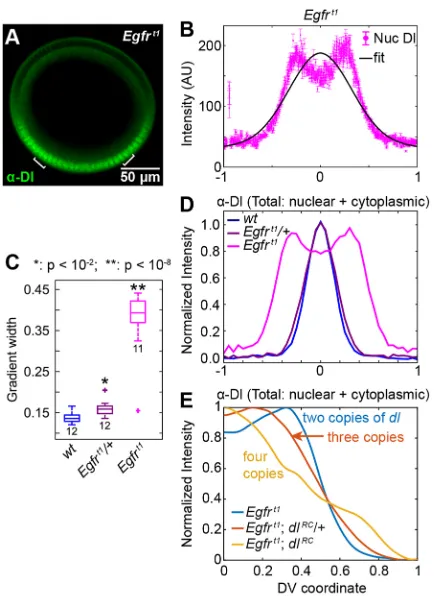
The shuttling hypothesis predicts that severe widening of the active Toll domain results in a split Dl gradient (Fig. S6A,B). As the extent of the Toll domain is controlled by Gurken/EGFR signaling during oogenesis (Sen et al., 1998; Schüpbach, 1987), we analyzed embryos from mothers carrying a hypomorphic EGFR allele (Egfrt1) (Roth
[image:6.612.90.526.54.444.2]and Schüpbach, 1994). We found that embryos from mothers heterozygous for this allele have significantly widened Dl gradients, and most (10/12) embryos from homozygous mothers have gradients Fig. 4. Embryos from mothers heterozygous for Dl have a different-shaped Dl gradient that is explained by Toll saturation.(A) Cross section of an nc 14 embryo from a mother heterozygous fordlimmunostained for Dl. (B) Plotted nuclear intensity of embryo in A as a function of DV coordinate (each magenta dot is one nucleus). Error bars indicate s.e.m. of nuclear intensity. Blue curve is the data of the individual nuclei smoothed by a sliding window of 50 nuclei; black curve is a fit of the data to a Gaussian. (C) Normalized average plots of theα-Dl nuclear fluorescence in wild-type,dl/+anddl; dl-dGFP/+embryos. Inset highlights differences in peak shape. (D) Box plot of gradient widths (σ) for the embryos in C. (E) Schematic of active Toll receptor saturation in wild-type embryos. In the edges of the Toll domain, Dl/Cact complexes can bypass active Toll receptors that are already bound by other Dl/Cact complexes. (F) Schematic of lack of active Toll saturation indl/+embryos. Fewer Dl/Cact complexes can penetrate deep into the Toll domain. (G) Extended model of the Dl/Cact system in which active Toll receptors are saturable. (H) Normalized, average profiles of totalα-Dl fluorescence in wild-type,dl/+;Tl/+anddl/+embryos. Inset reveals the difference in peak shape. The same color scheme and sets of embryos are presented in J and K. (I) Illustration of the variables compared in J and K. Blue arrow illustrates the distance from the ventral midline where the gradient peaks, and is compared across genotypes in J. Orange arrow illustrates the normalized value of the Dl gradient at the ventral midline, and is compared across genotypes in K. (J) Box plot of locations of total Dl peak (see blue arrow in I for embryos in H). (K) Box plot of normalized total Dl peak levels at the ventral midline (see orange arrow in I for embryos in H). See also Fig. S4.
DEVEL
O
so wide that the peak splits (Fig. 6A-D). This result is consistent with previous reports that variousgurkenandEgfrmutations generate a duplicated Dl gradient as measured by Dl staining, Twist staining, and sites of ventral furrow formation (Roth and Schüpbach, 1994), which are not readily explained in the absence of a shuttling phenomenon (Meinhardt, 2004; Moussian and Roth, 2005; Haskel-Ittah et al., 2012).
A model with Toll saturation predicts that the split peak can be alleviated either by decreasing the levels of active Toll receptors or by increasing the dosage of maternaldl(Fig. S6C-F). We chose to test the possibility by examining embryos from mothers carrying one (Egfrt1; dlRC/+) or two (Egfrt1; dlRC) extra copies of a Dl rescue
construct (Reeves et al., 2012) in the Egfrt1 background. These
embryos have three or four copies of full-length Dl, respectively. We found that, as our model predicts, increasing the dosage of maternaldl
reduces the severity of the split-peak phenotype (Fig. 6E; Fig. S6G-I), providing further evidence for active Toll receptor saturation.
An AP gradient of Dl supports the shuttling hypothesis It has been suggested that a shuttling phenomenon also occurs through the processing of Spätzle (Spz), the ligand for Toll signaling (Haskel-Ittah et al., 2012), which might explain some of the phenotypes described here. To determine whether the hallmark phenotypes of shuttling could occur without assistance from the protease cascade or Spz processing, we expressed a constitutively active form of Toll (Toll10b) at the anterior pole of the developing
embryo using thebicoid(bcd) 3′UTR and thebcdpromoter, similar to previously performed experiments using the stronger Hsp83
promoter (Huang et al., 1997). Embryos from mothers carrying this construct (bcd>Toll10b: bcd 3′ UTR) have an AP Dl gradient in
addition to the native DV gradient. Naïvely, one may expect that the existence of two gradients of active Toll signaling would result in higher concentrations of nuclear Dl where the two overlap. By
contrast, the shuttling hypothesis predicts that Dl nuclear concentration would be depleted in the region where both gradient tails overlap, as the two competing Dl/Cact sinks cause Cact to shuttle Dl toward both the anterior pole and the ventral midline (Fig. 7A-C). This prediction is borne out in experiments, as these embryos show a decreased intensity of the Dl gradient near the region of overlap. Furthermore, 64% (9/14) of these embryos show a visible narrowing of thesnaexpression domain at∼30% egg length, consistent with previously published data (Fig. 7B,C) (Huang et al., 1997). Our results show domain narrowing rather than a gap as previously reported, owing to the weakerbcdpromoter.
Our modeling results further support that the shuttling mechanism is responsible for this phenomenon (Fig. 7C). We expanded our model system from a one-dimensional array of nuclear compartments to a two-dimensional array, and added a second Toll signaling domain perpendicular to the first (Supplementary Materials and Methods). Our simulation results show that the overlap between the AP and DV Toll domains creates a low point in nuclear Dl concentration, similar to our experimental results. We then approximated a threshold forsna
expression based on the DV Dl gradient, and found that the local minimum in Dl concentration results in reduced or abolished sna
expression in that region (Fig. 7C′).
We also examined embryos from mothers carrying a homozygous mutation in gastrulation defective (gd7), which
eliminates the endogenous ventral-to-dorsal gradient. Swapping thebcdpromoter for the strongerHsp83promoter (Hsp83>Toll10b:
bcd 3′UTRconstruct; Huang et al., 1997), we were able to create a wider Dl gradient at the anterior pole. Half of these embryos (7/16) show two peaks of Dl (Fig. 7D-E); furthermore, they have a double peak of sna (Trisnadi et al., 2013). To determine whether this double-peak phenomenon was a result of the embryo’s geometry at the pole, we analyzed embryos with an AP Dl gradient initiated by the weakerbcdpromoter. These embryos showed no such double-Fig. 5. Simultaneous perturbations to shuttling result in defects in the Dl gradient and in thesnadomain.(A) Cross section of an nc 14dl; dl-dGFP/+ embryo immunostained for Dl. (B) Quantification ofα-Dl nuclear fluorescence of the embryo in A. Thesnadomain is also shown (gold). (C) The average snadomain from embryos in Fig. 4C,D, plusdl; dl-dGFPembryos (labeled 1× dGFP). (D,D′)snamRNA expression in wild type (D) anddl; dl-dGFP/+(D′). The embryo in D′is the same embryo as in A and B. Arrowheads indicate width ofsnadomain in wild type (gray) anddl; dl-dGFP/+(white). (E) Box plot ofsnadomain widths for the embryos in C.snadomains indl; dl-dGFPembryos are statistically narrower than those in other genotypes. See also Fig. S5.
DEVEL
O
[image:7.612.90.524.56.307.2]peak effect (Fig. S7). These results further support our shuttling hypothesis as the Dl gradient progresses from narrow (weak promoter) to double peak (strong promoter), much as it does in the native DV system when the Toll domain expands.
DISCUSSION
In this study, we investigated the embryo-scale formation of the Dl morphogen gradient in the earlyDrosophilaembryo. Based on our model and experimental verification, we conclude that establishment of peak levels of the Dl gradient is aided by a facilitated diffusion, or shuttling mechanism, in which Dl/Cact complex diffuses towards the ventral midline, where Cact is degraded. Lowering the diffusivity of either Dl or Cact widens the gradient rather than the narrowing that one might expect from a morphogen gradient established by (nonfacilitated) diffusion. The shuttling mechanism explains why Dl tagged with a weakly dimerizing GFP widens the gradient more than one tagged with monomeric GFP (Reeves et al., 2012), and also explains the related observation that one copy of Dl tagged with monomeric GFP variants complements loss of endogenous Dl while one copy of Dl-dGFP does not (Liberman et al., 2009; Reeves et al., 2012). Similarly, this mechanism makes sense of the observation that Dl tagged with β-galactosidase, which forms tetramers, is antimorphic (Govind et al., 1992), as the Dl moieties in tetramers of
Dl-βgal dimerize with endogenous Dl to disrupt the formation of the endogenous Dl gradient.
When the Toll gradient is greatly expanded, as in embryos from
Egfrt1 mothers (Roth and Schüpbach, 1994), the domains of
[image:8.612.66.283.55.353.2]saturated Toll receptors move further from the ventral midline, resulting in a split peak. This split-peak phenomenon has also been observed in the Dl gradient in abnormally large embryos (Garcia et al., 2013). This specific phenotype could be explained by the shuttling of the Toll ligand Spz (Haskel-Ittah et al., 2012); however, we observed the same phenomenon in embryos with an ectopic AP Dl gradient established by constitutively active Toll. In both cases, ventrally (anteriorly) diffusing Dl/Cact complex does not reach the Fig. 6. The hypomorphic alleleEgfrt1significantly widens the Dl gradient.
[image:8.612.327.546.55.440.2](A) Cross section of anEgfrt1embryo immunostained for Dl. Brackets indicate peaks of nuclear Dl. (B) Plottedα-Dl nuclear fluorescence of embryo in A as a function of DV coordinate (each pink dot is one nucleus). The shape has changed significantly from wild type, as the Gaussian curve (black) does not represent the gradient well. Error bars indicate s.e.m. of nuclear intensity. (C) Box plot of gradient widths (σ) for wild-type,Egfrt1/+andEgfrt1embryos. Numbers indicate sample size. (D) Normalized average plot of totalα-Dl fluorescence (nuclear+cytoplasmic) in the embryos in C, with the same color scheme. (E) Normalized totalα-Dl fluorescence (nuclear+cytoplasmic) averaged fromEgfrt1,Egfrt1; dlRC/+andEgfrt1; dlRC. See also Fig. S6.
Fig. 7. An ectopic, AP dorsal gradient exhibits shuttling phenomena. (A,A′) Dl andsnaexpression in a wild-type embryo. (B,B′) Dl andsna expression in an embryo with an AP gradient of Dl, driven by thebcdpromoter in addition to the wild-type DV gradient. White arrowheads indicate a narrowing of each domain at∼30% egg length. (C,C′) A 2D version of the model featuring AP and DV active Toll domains exhibiting a competing sink phenotype, in which a low point in the Dl gradient leads to a decrease insnaexpression. (D,D′) Dl andsnaexpression in an embryo with an AP gradient of Dl, driven by theHsp83promoter, and the DV gradient abolished by a homozygous mutation ingd. Magenta arrowheads show the second peak of expression; the peak is more clearly shown when the data are plotted in E. The definition of the AP coordinate for the plot in E is given, with 0 at the anterior pole, and ±1 at the posterior pole. (E) Plot of Dl andsnadomains from the embryo in D. Each green dot is one nucleus and the black curve is a smoothing of the Dl data. Embryo images are maximal intensity projections. See also Fig. S7.
DEVEL
O
ventral midline (anterior pole) before being dissociated, leaving the ventral (anterior)-most nuclei somewhat devoid of Dl. A similar mechanism, in which the removal rate of BMP ligands surpasses the rate of BMP flux to the dorsal midline, has been suggested to explain the computationally predicted split-peak phenotype for the BMP system in the early embryo (Umulis et al., 2006).
We further propose that active Toll receptors are saturated in the edges of the Toll domain in wild-type embryos; this saturation is not essential to the shuttling mechanism per se, though it is necessary for the mechanism to explain phenotypes associated with changing the dose of maternaldl. Under wild-type conditions, a significant flux of Dl/Cact complex can bypass the saturated active Toll receptors in the edges of the Toll domain, which results in the accumulation of a smooth, intense peak of Dl signaling at the ventral midline of the embryo. However, in dl/+ embryos, there is not enough Dl/Cact complex to saturate active Toll receptors in the tails of the gradient, leading to less accumulation of Dl at the ventral midline (Liberman et al., 2009). We have verified this assumption of active Toll saturation indl/+; Tl/+andEgfrt1; dlRCembryos.
The shuttling mechanism requires four biophysical processes: (1) Dl binding to Cact; (2) Dl/Cact complex readily diffusing through the embryo; (3) Toll signaling acting as a sink for Dl/Cact complex; and (4) Cact protecting Dl from entering the nuclei. As our experiments with Dl-paGFP show that Dl does indeed move globally through the embryo, it is now clear that Dl/Cact shuttling naturally arises in the early embryo. Indeed, our previous Dl/Cact model (O’Connell and Reeves, 2015) did not explicitly have shuttling‘programmed in’, but instead it was a natural consequence of the model equations. In the same way, shuttling naturally emerges in previous models of the Dl gradient without the authors’explicit intention (Ambrosi et al., 2014; Kanodia et al., 2009).
Given the natural emergence of the shuttling mechanism from the topology of the known Dl/Cact interactions, one may ask whether the mechanism operates at a relevant level in the fly embryo. In the Dpp system, formation of peak levels of Dpp signaling requires the BMP antagonist Sog, which binds to Dpp and facilitates its diffusion to the dorsal midline of the early embryo. Loss of zygotic Sog function results in loss of both the highest levels of Dpp signal as well as the expression of genes that require those peak levels (Ashe and Levine, 1999; Zusman et al., 1988; Rushlow et al., 2001). Cact serves as the analog to Sog in the Dl/Cact system. A seminal study of Cact showed that loss of Cact function results in an expansion of the domain of cells expressingtwi, a gene that requires peak levels of nuclear Dl (Roth et al., 1991), suggesting that there is no need for shuttling of Dl through Cact to form the peak levels of the Dl gradient. By contrast, a recent quantitative study, which analyzed embryos with varying levels of compromisedcactfunction, could support the possibility that shuttling is necessary for embryonic fitness (Cardoso et al., 2017). Embryos with stronger loss of cact function have progressively flatter Dl gradients with lower peak values and progressively narrower domains of sna, which, like twi, requires peak levels of Dl (Cardoso et al., 2017). Curiously, the same study suggests a third role for Cact in establishing the Dl gradient: Cact fragments that result from Toll-independent degradation potentiate Dl signaling, which might explain most, if not all, theircactmutant phenotypes. Even so, the authors maintain the position that the shuttling mechanism could be necessary for the formation of peak Dl levels. Further work should be done to continue to dissect novel roles Cact might have in Dl gradient formation.
Regardless of the conflicting data fromcactmutant embryos, the data presented here from thedl; dl-dGFP/+embryos show that, at minimum, the shuttling mechanism is required for formation of peak
levels of the Dl gradient (and the consequentsnaexpression) when Dl levels are compromised. Given the data presented here, the literature data explained by the shuttling hypothesis, and the fact that all biological processes required for shuttling are known to be present in the embryo, we suggest that Cact serves a dual role in establishing the Dl nuclear gradient: preventing nuclear translocation of Dl and shuttling of Dl.
MATERIALS AND METHODS
Fly lines
ywflies were used as wild type.dl-paGFP,dl-mGFPanddl-dVenuswere created by BAC recombineering. For live imaging, flies carryingdl-paGFP were crossed to flies carrying H2A-RFP on the second chromosome (BS# 23651). dl/+flies were created by cleaning updl1via two homologous recombinations withywto generatedl1.2.5.cact/+;cact-full lacZ 25flies were obtained from David Stein (Janelia Research Campus, Ashburn, VA, USA) (Fernandez et al., 2001).dl-lacZflies were obtained from Govind et al. (1992). dl-dGFPanddl-mVenusflies and the original BACs used to create them were obtained from Angela Stathopoulos (California Institute of Technology, Pasadena, CA, USA).dl-mGFP,dl1.2.5 flies were created by homologous recombination. Presence ofdl-mGFP was confirmed by w+. Presence of dl1.2.5was confirmed via sequencing by GENEWIZ.Egfrt1/CyOflies were obtained from the BloomingtonDrosophilaStock Center (# 2079). Flies carrying thebcd>Toll10b: bcd 3′UTRconstruct were obtained from Angela Stathopoulos. The plasmid carryingFRT-stop-FRT Hsp83>Toll10b: bcd 3′ UTRwas also obtained from Angela Stathopoulos. To remove the FRT-stop-FRT cassette, we crossed flies carrying this construct into a line carrying hsFLP on the X chromosome (BS# 8862). To remove the native DV Dl gradient, flies carrying theToll10b: bcd 3′UTRconstruct were crossed into a gd7background. See Supplementary Materials and Methods for more details. dl/+; Tl/+fly lines resulted from crossing thedl1.2.5/+fly line withTlrv18/+ (BS# 30913).
BAC recombineering
We followed Protocol 3 of the NCI at Frederick Recombineering website (http://ncifrederick.cancer.gov/research/brb/recombineeringinformation. aspx) to generatedl-paGFP,dl-mGFPanddl-dVenusin pACMAN (Venken et al., 2006). A proofreading Q5 DNA polymerase (New England Biolabs) was used to amplify sequences at a high level of authenticity. Using a GalK selection protocol (Warming et al., 2005), single amino acid mutations were introduced at residue 206 in previously established BACs (Reeves et al., 2012) to generatedl-mGFP(A206K) anddl-dVenus(K206A).dl-paGFP was created by adding the open reading frame ofpaGFP(Addgene plasmid #11911) (Patterson and Lippincott-Schwartz, 2002) in frame to the 3′end of thedlopen reading frame in adlrescue construct (Reeves et al., 2012) in pACMAN (Venken et al., 2006) using a 6x-Gly linker. See Table S1 for a list of primers used.
Fluorescentin situhybridization
All embryos were aged to 2-4 h, except for‘young’ywembryos, which were aged to 0-2 h, then fixed in 37% formaldehyde according to standard protocols (Kosman et al., 2004). A combination fluorescent in situ hybridization/fluorescent immunostaining was performed according to standard protocols (Kosman et al., 2004). Briefly, fixed embryos were washed in Tween/PBS buffer, then hybridized with a digoxigenin-conjugated anti-sensesnaprobe at 55°C overnight. The embryos were then washed and incubated with primary antibodies at 4°C overnight. Next, the embryos were washed and incubated for 1-2 h with fluorescent secondary antibodies at room temperature. The embryos were then washed and stored in 70% glycerol at
−20°C. Embryos were imaged within one month of completing the protocol. See Supplementary Materials and Methods for more details, including suppliers and dilutions of antibodies.
Mounting and imaging of fixed samples
Embryos were cross-sectioned and mounted in 70% glycerol as described previously (Carrell and Reeves, 2015). Briefly, a razor blade was used to remove the anterior and posterior thirds of the embryo, leaving a cross
DEVEL
O
section∼200 µm long by 200 µm in diameter. These sections were then oriented such that the cut sides became the top and bottom. They were imaged at 20× magnification on a Zeiss LSM 710 microscope. Fifteen z-slices 1.5 µm apart were analyzed. Embryos with an AP Dorsal gradient were mounted laterally in 70% glycerol using one piece of double-sided tape (weakbcdpromoter) or two pieces of double-sided tape (strong Hsp83 promoter). Images were taken at 2.5 µm intervals from just above the top of the embryo to the depth at which the embryo reached maximal size in thexy plane, which was assumed to be the midsagittal section. Stacks ranged from 15 to 25 slices.
Data analysis
Images of embryo cross sections were analyzed using previously derived code (Trisnadi et al., 2013). Briefly, the border of the embryo was found computationally, then the nuclei were segmented using a local thresholding protocol. The intensity of Dl in each segmented nucleus was calculated as the ratio between the intensity in the Dl channel divided by the intensity in the nuclear channel (Damle et al., 2006; Liberman et al., 2009). The intensity of mRNA expression and total Dl were calculated as average intensity within an annulus∼18μm wide around the perimeter of the embryo. A description of the image analysis of whole-mount embryos can be found in the Supplementary Materials and Methods and in Jermusyk et al. (2016).
All Dl gradients were fit to a Gaussian, and these fits were used to determine the width parameter,σ, and to normalize the Dl gradients. Anr2
goodness of fit was calculated for each embryo, and if this fell below 0.80, the measurement was discarded. Widths ofsnadomains were found by fitting thesnaintensity curves to canonical profiles. For further explanation of these two procedures, see the Supplementary Materials and Methods. All replicates were biological.
Statistical significance was calculated using two-tailed homoscedastic t-tests. For 80% power, minimum sample size was calculated to be <10 embryos for our measurements of the Dl gradient width.
Activating paGFP in live embryos
Embryos were dechorionated by hand or for 30 s in 100% bleach. They were then mounted laterally on a slide coated with heptane glue. Deionized water was used as a mounting medium. Two pieces of double-sided tape were used to attach the coverslip. Images were taken using a 40× water immersion objective on an LSM 710 confocal microscope. Activation box: 9000 pixels (300×30μm); number of activation passes (number of times the laser activates the region in a single cycle): 10; rest time (the length of rest time between cycles): 15 s; number of cycles: 40. Each activation session lasted
∼25 min and was followed by imaging the entire depth of the embryo. Each embryo underwent five activation sessions. Laser power was 3% for embryos activated near the ventral midline, and 2.5% for embryos activated near the dorsal midline. A 405 nm laser was used for activation and a 488 nm laser was used for excitation of the activated GFP.
Model of Dl/Cact interactions
In Figs 2 and 3, we used a simplified version of previously published models of Dl/Cact interactions to predict the effects of slowing diffusion (O’Connell and Reeves, 2015; Kanodia et al., 2009; Ambrosi et al., 2014). In brief, this model consists of four differential equations, representing nuclear and cytoplasmic Dl, and cytoplasmic Dl/Cact complex and Cact, respectively (Eqns 1-4; see also Fig. 2A for a schematic of the modeled processes).
@CDl;nuc
@t ¼kinCDl;cytkoutCDl;nuc; ð1Þ @CDl;cyt
@t ¼DDl @2C
Dl;cyt
@x2 kinCDl;cytþkoutCDl;nuckbindCDl;cytCCact
þkdissðxÞCDC; ð2Þ
@CDC @t ¼DDC
@2C
DC
@x2 þkbindCDl;cytCCactkdissðxÞCDC; ð3Þ
@CCact @t ¼DCact
@2C
Cact
@x2 þVcactkdegCCactkbindCDl;cytCCact
þkdissðxÞCDC: ð4Þ
To simplify analysis and interpretation, we simulated 100 min of development with 51 nuclei positioned along a linear (x) axis and ignore mitosis and nuclear division. The active Toll gradient was modeled phenomenologically in space askdiss(x)=kdiss,0exp (−0.5x2/φ2) (Ambrosi
et al., 2014; Kanodia et al., 2009; O’Connell and Reeves, 2015). In the Toll saturation model (Figs 4–7), activated Toll receptors were allowed to saturate (Fig. 4G), which altered the Dl/Cact complex dissociation term to a Michaelis–Menten-like saturable function (see Supplementary Materials and Methods). In other words, the final term in Eqns 2-4 becamekdissCDC/ (KR+CDC), whereKRis a Michaelis constant for Toll saturation. For more details on the model formulation, see Supplementary Materials and Methods.
To calculate flux of Dl-containing species, we applied Fick’s law, which states that diffusive flux,J, which refers to the movement of a solute from areas of high concentration to areas of low concentration, occurs at a rate proportional to its concentration gradient,dC/dx, yieldingJ=−DdC/dx. The negative sign accounts for the fact that diffusive flux is in the direction of decreasing concentration, andDis the diffusion coefficient. By convention, we define ourx-axis to be equal to 0 at the ventral midline and 1 at the dorsal midline (Fig. 2A); thus, a negative value for the flux indicates movement toward the ventral midline, and a positive value indicates movement toward the dorsal midline. Therefore, the calculation of flux of Dl-containing species (cytoplasmic Dl+Dl/Cact complex) is performed using the following equation:
Jðx;tÞ ¼ DDl@ CDl;cyt
@x DDC @CDC
@x : ð5Þ
Shuttling was defined asJ(x=0.33,t=100)<0. This occurred whenDDC/[DDl/ Keq]>1, whereKeq=kin/kout(see Fig. 2B).
To ensure our results were not the result of oversimplification, we also formulated a more complete (full) model of Dl/Cact/Toll interactions, which is an extension to the model found in O’Connell and Reeves (2015). This full model (see Supplementary Materials and Methods) consists of differential equations for three species distributed between two compartments: nuclear Dl, cytoplasmic Dl, nuclear Cact, cytoplasmic Cact, nuclear Dl/Cact complex, and cytoplasmic Dl/Cact complex, plus an equation for free active Toll receptor and one for active Toll receptor bound to Dl/Cact complex. It takes into account the mitosis and interphase associated with nc 10-14 and the possibility that Cact and Dl/Cact complex could be present in the nucleus. During mitosis, the nucleus becomes undefined, and the contents of each nucleus mix with those of the surrounding cytoplasm. At the end of mitosis, the nucleus reforms, and the concentration of Dl, Cact and Dl/Cact inside the nucleus is initialized as equal to their respective concentrations in the cytoplasm, and each species can enter and exit the nucleus. All cytoplasmic species can move between adjacent cytoplasmic compartments during interphase and mitosis.
The production rate of activated Toll receptors was modeled
phenomenologically. The eight differential equations were
nondimensionalized, resulting in a model with 20 free parameters. Optimization was performed in MATLAB (MathWorks) using an evolutionary optimization algorithm with stochastic ranking (Runarsson and Yao, 2000, 2005), yielding 114 parameter sets that were generally consistent with the spatiotemporal data published by Reeves et al. (2012). Further detail on model equations and analysis can be found in the Supplementary Materials and Methods.
We also analyzed two previous models of Dl/Cact interactions, which each have the processes necessary for shuttling, to investigate whether these processes together are sufficient for shuttling (Kanodia et al., 2009; Ambrosi et al., 2014). See Supplementary Materials and Methods for more details.
Acknowledgements
We thank Angela Stathopoulos and Leslie Dunipace for provision of Dl-dGFP and Dl-mVenus flies and BACs,bcd>Toll10b
: bcd 3′UTRflies, and the DNA used to generateHsp83>Toll10b: bcd 3′UTRflies. We are grateful to ImmunoReagents for
providing goat anti-biotin antibody, Alex Thomas for generating thesna-biotin probe, Parv Gondalia for generating thesna-FITC probe, David Stein for providing the cact-lacZfly line, and Shubba Govind for providing thedl-lacZflies.
DEVEL
O
Competing interests
The authors declare no competing or financial interests.
Author contributions
Conceptualization: S.N.C., M.D.O., G.T.R.; Methodology: G.T.R.; Validation: S.N.C., M.D.O., G.T.R.; Formal analysis: S.N.C., M.D.O., G.T.R.; Investigation: S.N.C., M.D.O., T.J., A.E.P., S.M.H., G.T.R.; Writing - original draft: S.N.C., M.D.O., G.T.R.; Writing - review & editing: S.N.C., M.D.O., G.T.R.; Supervision: G.T.R.; Project administration: G.T.R.; Funding acquisition: G.T.R.
Funding
This work was supported by the National Science Foundation [CBET-1254344 to G.T.R.] and the U.S. Department of Education [Graduate Assistance in Areas of National Need Biotechnology Fellowship to S.N.C. (P200A100004) and T.J. (P200A140020), and Scientific Computing Fellowship to M.D.O. (P200A120047)].
Data availability
All MATLAB files used for the model and data analysis are available from https:// github.ncsu.edu/ and additionally on the Reeves Lab website ( people.engr.ncsu.edu/ gtreeves/). All image files are available from the Dryad Digital Repository (Carrell et al., 2017): doi:10.5061/dryad.4h00t.
Supplementary information
Supplementary information available online at
http://dev.biologists.org/lookup/doi/10.1242/dev.155549.supplemental
References
Ali-Murthy, Z. and Kornberg, T. B.(2016). Bicoid gradient formation and function in
the Drosophila pre-syncytial blastoderm.eLife5, e13222.
Ambrosi, P., Chahda, J. S., Koslen, H. R., Chiel, H. J. and Mizutani, C. M.(2014).
Modeling of the dorsal gradient across species reveals interaction between embryo morphology and toll signaling pathway during evolution.PLoS Comput. Biol.10, e1003807.
Ashe, H. L. and Levine, M.(1999). Local inhibition and long-range enhancement of
Dpp signal transduction by Sog.Nature398, 427-431.
Belenkaya, T. Y., Han, C., Yan, D., Opoka, R. J., Khodoun, M., Liu, H. and Lin, X.
(2004). Drosophila Dpp morphogen movement is independent of Dynamin-mediated endocytosis but regulated by the glypican members of heparan sulfate proteoglycans.Cell119, 231-244.
Belvin, M. P., Jin, Y. and Anderson, K. V.(1995). Cactus protein degradation
mediates Drosophila dorsal-ventral signaling.Genes Dev.9, 783-793.
Ben-Zvi, D. Shilo, B.-Z., Fainsod, A. and Barkai, N.(2008). Scaling of the BMP
activation gradient in Xenopus embryos.Nature453, 1205-1211.
Bergmann, A., Stein, D., Geisler, R., Hagenmaier, S., Schmid, B., Fernandez,
N., Schnell, B. and Nüsslein-Volhard, C.(1996). A gradient of cytoplasmic
Cactus degradation establishes the nuclear localization gradient of the dorsal morphogen in Drosophila.Mech. Dev.60, 109-123.
Cardoso, M. A., Fontenele, M., Lim, B., Bisch, P. M., Shvartsman, S. Y. and
Araujo, H. M.(2017). A novel function for the IκB inhibitor Cactus in promoting
Dorsal nuclear localization and activity in the Drosophila embryo.Development
144, 2907-2913.
Carrell, S. N. and Reeves, G. T.(2015). Imaging the dorsal-ventral axis of live and
fixed Drosophila melanogaster embryos. InTissue Morphogenesis. Methods in
Molecular Biology(ed. C. M. Nelson), pp. 63-78. New York: Springer.
Chopra, V. S. and Levine, M.(2009). Combinatorial patterning mechanisms in the
Drosophila embryo.Brief. Funct. Genomic. Proteomic.8, 243-249.
Christian, J. L.(2012). Morphogen gradients in development: from form to function.
Wiley Interdiscip. Rev. Dev. Biol.1, 3-15.
Damle, S., Hanser, B., Davidson, E. H. and Fraser, S. E.(2006). Confocal
quantification of cis-regulatory reporter gene expression in living sea urchin.Test
299, 543-550.
Delotto, R., Delotto, Y., Steward, R. and Lippincott-Schwartz, J. (2007).
Nucleocytoplasmic shuttling mediates the dynamic maintenance of nuclear Dorsal levels during Drosophila embryogenesis.Development134, 4233-4241.
Dorfman, R. and Shilo, B. Z.(2001). Biphasic activation of the BMP pathway
patterns the Drosophila embryonic dorsal region.Development128, 965-972.
Drier, E. A., Huang, L. H. and Steward, R.(1999). Nuclear import of the Drosophila
Rel protein Dorsal is regulated by phosphorylation.Genes Dev.13, 556-568.
Drier, E. A., Govind, S. and Steward, R.(2000). Cactus-independent regulation of
Dorsal nuclear import by the ventral signal.Curr. Biol.10, 23-26.
Driever, W. and Nüsslein-Volhard, C.(1988). A gradient of bicoid protein in
Drosophila embryos.Cell54, 83-93.
Eldar, A., Dorfman, R., Weiss, D., Ashe, H., Shilo, B. Z. and Barkai, N.(2002).
Robustness of the BMP morphogen gradient in Drosophila embryonic patterning.
Nature419, 304-308.
Entchev, E. V., Schwabedissen, A. and González-Gaitán, M. A.(2000). Gradient
formation of the TGF-βhomolog Dpp.Cell103, 981-991.
Fahmy, K., Akber, M., Cai, X., Koul, A., Hayder, A. and Baumgartner, S.(2014).
αTubulin 67C and Ncd are essential for establishing a cortical microtubular network and formation of the Bicoid mRNA gradient in DrosophilaPLoS ONE9, e112053.
Fernandez, N. Q., Grosshans, J., Goltz, J. S. and Stein, D.(2001). Separable and
redundant regulatory determinants in Cactus mediate its dorsal group dependent degradation.Development128, 2963-2974.
Garcia, M. Nahmad, M., Reeves, G. T. and Stathopoulos, A. (2013).
Size-dependent regulation of dorsal–ventral patterning in the early Drosophila embryo. Dev. Biol.381, 286-299.
Govind, S., Whalen, A. M. and Steward, R.(1992). In vivo self-association of the
Drosophila rel-protein dorsal.Proc. Natl. Acad. Sci. USA89, 7861-7865.
Govind, S., Drier, E., Huang, L. H. and Steward, R.(1996). Regulated nuclear
import of the Drosophila rel protein dorsal: structure-function analysis.Mol. Cell. Biol.16, 1103-1114.
Gregor, T., Bialek, W., de Ruyter van Steveninck, R. R., Tank, D. W. and
Wieschaus, E. F.(2005). Diffusion and scaling during early embryonic pattern
formation.Proc. Natl. Acad. Sci. USA102, 18403-18407.
Gregor, T. Wieschaus, E. F., Mcgregor, A. P., Bialek, W. and Tank, D. W.(2007).
Stability and nuclear dynamics of the Bicoid morphogen gradient.Cell130, 141-152.
Guerrero, I. and Kornberg, T. B.(2014). Hedgehog and its circuitous journey from
producing to target cells.Semin. Cell Dev. Biol.33, 52-62.
Haskel-Ittah, M., Ben-Zvi, D., Branski-Arieli, M., Schejter, E. D., Shilo, B.-Z. and
Barkai, N.(2012). Self-organized shuttling: generating sharp dorsoventral polarity
in the early drosophila embryo.Cell150, 1016-1028.
Holley, S. A., Neul, J. L., Attisano, L., Wrana, J. L., Sasai, Y., O’connor, M. B., De
Robertis, E. M. and Ferguson, E. L.(1996). The Xenopus dorsalizing factor
noggin ventralizes Drosophila embryos by preventing DPP from activating its receptor.Cell86, 607-617.
Houchmandzadeh, B., Wieschaus, E. and Leibler, S.(2002). Establishment of
developmental precision and proportions in the early Drosophila embryo.Nature
415, 798-802.
Huang, A. M., Rusch, J. and Levine, M.(1997). An anteroposterior Dorsal gradient
in the Drosophila embryo.Genes Dev.11, 1963-1973.
Isoda, K., Roth, S. and Nüsslein-Volhard, C.(1992). The functional domains of the
Drosophila morphogen dorsal: evidence from the analysis of mutants.Genes Dev.6, 619-630.
Jermusyk, A. A., Murphy, N. P. and Reeves, G. T.(2016). Analyzing negative
feedback using a synthetic gene network expressed in the Drosophila melanogaster embryo.BMC Syst. Biol.10, 85.
Kanodia, J. S., Rikhy, R., Kim, Y., Lund, V. K., Delotto, R., Lippincott-Schwartz,
J. and Shvartsman, S. Y.(2009). Dynamics of the Dorsal morphogen gradient.
Proc. Natl. Acad. Sci. USA106, 21707-21712.
Kosman, D., Mizutani, C. M., Lemons, D., Cox, W. G., McGinnis, W. and Bier, E.
(2004). Multiplex detection of RNA expression in Drosophila embryos.Science
305, 846.
Lander, A. D., Nie, Q. and Wan, F. Y. M.(2002). Do morphogen gradients arise by
diffusion?Dev. Cell2, 785-796.
Liberman, L. M., Reeves, G. T. and Stathopoulos, A.(2009). Quantitative imaging
of the Dorsal nuclear gradient reveals limitations to threshold-dependent patterning in Drosophila.Proc. Natl. Acad. Sci. U.S.A.106, 22317-22322.
Little, S. C., Tkačik, G., Kneeland, T. B., Wieschaus, E. F. and Gregor, T.(2011).
The formation of the Bicoid morphogen gradient requires protein movement from anteriorly localized mRNA.PLoS Biol.9, e1000596.
Marqués, G., Musacchio, M., Shimell, M. J., Wünnenberg-Stapleton, K., Cho,
K. W. Y. and O’connor, M. B.(1997). Production of a DPP activity gradient in the
early Drosophila embryo through the opposing actions of the SOG and TLD proteins.Cell91, 417-426.
Matsuda, S., Harmansa, S. and Affolter, M., (2016). BMP morphogen gradients in
flies.Cytokine Growth Factor. Rev.27, 119-127.
Meinhardt, H.(2004). Different strategies for midline formation in bilaterians.Nat.
Rev. Neurosci.5, 502-510.
Mizutani, C. M., Nie, Q., Wan, F. Y. M., Zhang, Y.-T., Vilmos, P., Sousa-Neves, R.,
Bier, E., Marsh, J. L. and Lander, A. D.(2005). Formation of the BMP activity
gradient in the drosophila embryo.Dev. Cell8, 915-924.
Moussian, B. and Roth, S.(2005). Dorsoventral axis formation in the Drosophila
embryo–shaping and transducing a morphogen gradient. Curr. Biol. 15, R887-R899.
Müller, P. Rogers, K. W., Yu, S. R., Brand, M. and Schier, A. F.(2013). Morphogen
transport.Development140, 1621-1638.
O’Connell, M. D. and Reeves, G. T.(2015). The presence of nuclear cactus in the
early Drosophila embryo may extend the dynamic range of the dorsal gradient
PLoS Comput. Biol.11, e1004159.
Patterson, G. H. and Lippincott-Schwartz, J.(2002). A photoactivatable GFP for
selective photolabeling of proteins and cells.Science (New York, N.Y.) 297, 1873-1877.
Rahimi, N., Averbukh, I., Haskel-Ittah, M., Degani, N., Schejter, E. D., Barkai, N.
and Shilo, B.-Z.(2016). A WntD-dependent integral feedback loop attenuates
variability in drosophila toll signaling.Dev. Cell36, 401-414.
DEVEL
O
Reach, M., Galindo, R. L., Towb, P., Allen, J. L., Karin, M. and Wasserman, S. A.
(1996). A gradient of cactus protein degradation establishes dorsoventral polarity in the Drosophila embryo.Dev. Biol.180, 353-364.
Reeves, G. T. and Stathopoulos, A.(2009). Graded dorsal and differential gene
regulation in the Drosophila embryo.Cold Spring Harb. Perspect Biol.1, a000836.
Reeves, G. T., Trisnadi, N., Truong, T. V., Nahmad, M., Katz, S. and
Stathopoulos, A. (2012). Dorsal-ventral gene expression in the Drosophila
embryo reflects the dynamics and precision of the dorsal nuclear gradient.Dev. cell,22, 544-557.
Roth, S. and Schüpbach, T. (1994). The relationship between ovarian and
embryonic dorsoventral patterning in Drosophila.Development120, 2245-2257.
Roth, S., Stein, D. and Nüsslein-Volhard, C. (1989). A gradient of nuclear
localization of the dorsal protein determines dorsoventral pattern in the Drosophila embryo.Cell59, 1189-1202.
Roth, S., Hiromi, Y., Godt, D. and Nüsslein-Volhard, C.(1991). cactus, a maternal
gene required for proper formation of the dorsoventral morphogen gradient in Drosophila embryos.Development112, 371-388.
Roy, S., Huang, H., Liu, S. and Kornberg, T. B.(2014). Cytoneme-mediated
contact-dependent transport of the drosophila decapentaplegic signaling protein.
Science343, 1244624.
Runarsson, T. P. and Yao, X. (2000). Stochastic ranking for constrained
evolutionary optimization.IEEE Trans. Evol. Comput.4, 284-294.
Runarsson, T. P. and Yao, X.(2005). Search biases in constrained evolutionary
optimization.IEEE Trans. Syst. Man Cybern. C Appl. Rev.35, 233-243.
Rushlow, C. A. and Shvartsman, S. Y.(2012). Temporal dynamics, spatial range,
and transcriptional interpretation of the Dorsal morphogen gradient.Curr. Opin.
Genet. Dev.22, 542-546.
Rushlow, C., Colosimo, P. F., Lin, M. C., Xu, M. and Kirov, N. (2001).
Transcriptional regulation of the Drosophila gene zen by competing Smad and Brinker inputs.Genes Dev.15, 340-351.
Schüpbach, T.(1987). Germ line and soma cooperate during oogenesis to establish
the dorsoventral pattern of egg shell and embryo in drosophila melanogaster.Cell
49, 699-707.
Schwank, G., Dalessi, S., Yang, S. F., Yagi, R., de Lachapelle, A. M., Affolter, M.,
Bergmann, S. and Basler, K. (2011). Formation of the long range dpp
morphogen gradient.PLoS Biol.9, e1001111.
Sen, J., Goltz, J. S., Stevens, L. and Stein, D. (1998). Spatially restricted
expression of pipe in the drosophila egg chamber defines embryonic dorsal– ventral polarity.Cell95, 471-481.
Shilo, B.-Z. Haskel-Ittah, M., Ben-Zvi, D., Schejter, E. D. and Barkai, N.(2013).
Creating gradients by morphogen shuttling.Trends Genet.29, 339-347.
Shimmi, O., Umulis, D., Othmer, H. and O’Connor, M. B.(2005). Facilitated
transport of a Dpp/Scw heterodimer by Sog/Tsg leads to robust patterning of the drosophila blastoderm embryo.Cell120, 873-886.
Smith, J. C. (2009). Forming and interpreting gradients in the early Xenopus
embryo.Cold Spring Harbor Perspect. Biol.1, a002477.
Spirov, A., Fahmy, K., Schneider, M., Frei, E., Noll, M. and Baumgartner, S.
(2009). Formation of the bicoid morphogen gradient: an mRNA gradient dictates the protein gradient.Development136, 605-614.
Stathopoulos, A. and Levine, M. (2002). Dorsal gradient networks in the
Drosophila embryo.Dev. Biol.246, 57-67.
Steward, R., Zusman, S. B., Huang, L. H. and Schedl, P.(1988). The dorsal
protein is distributed in a gradient in early drosophila embryos.Cell55, 487-495.
Trisnadi, N., Altinok, A., Stathopoulos, A. and Reeves, G. T.(2013). Image
analysis and empirical modeling of gene and protein expression.Methods (San Diego, Calif.)62, 68-78.
Umulis, D. M., Serpe, M., O’connor, M. B. and Othmer, H. G.(2006). Robust,
bistable patterning of the dorsal surface of the Drosophila embryo.Proc. Natl.
Acad. Sci. USA103, 11613-11618.
Venken, K. J. T., He, Y., Hoskins, R. A. and Bellen, H. J.(2006). P[acman]: a BAC
transgenic platform for targeted insertion of large DNA fragments in D. melanogaster.Science (New York, N.Y.)314, 1747-1751.
Wang, Y.-C. and Ferguson, E. L. (2005). Spatial bistability of Dpp-receptor
interactions during Drosophila dorsal-ventral patterning.Nature434, 229-234.
Warming, S., Costantino, N., Court, D. L., Jenkins, N. A. and Copeland, N. G.
(2005). Simple and highly efficient BAC recombineering using galK selection. Nucleic Acids Res.33, e36.
Wartlick, O., Kicheva, A. and González-Gaitán, M.(2009). Morphogen gradient
formation.Spring1, a001255.
Whalen, A. M. and Steward, R.(1993). Dissociation of the Dorsal-Cactus complex
and phosphorylation of the Dorsal protein correlate with the nuclear localization of Dorsal.J. Cell Biol.123, 523-534.
Zacharias, D. A., Violin, J. D., Newton, A. C. and Tsien, R. Y.(2002). Partitioning
of lipid-modified monomeric GFPs into membrane microdomains of live cells.
Science296, 913-916.
Zusman, S. B., Sweeton, D. and Wieschaus, E. F.(1988). short gastrulation, a
mutation causing delays in stage-specific cell shape changes during gastrulation in Drosophila melanogaster.Dev. Biol.129, 417-427.