R E S E A R C H A R T I C L E
Open Access
A complex interaction between Wnt signaling
and TNF-
α
in nucleus pulposus cells
Akihiko Hiyama
1,2*, Katsuya Yokoyama
1,2, Tadashi Nukaga
1,2, Daisuke Sakai
1,2and Joji Mochida
1,2Abstract
Introduction:Increased expression of the proinflammatory cytokine TNF-αin intervertebral discs (IVDs) leads to inflammation, which results in progressive IVD degeneration. We have previously reported that activation of
Wnt-β-catenin (hereafter called Wnt) signaling suppresses the proliferation of nucleus pulposus cells and induces cell senescence, suggesting that Wnt signaling triggers the process of degeneration of the IVD. However, it is not known whether cross talk between TNF-αand Wnt signaling plays a role in the regulation of nucleus pulposus cells. The goal of the present study was to examine the effect of the interaction between Wnt signaling and the proinflammatory cytokine TNF-αin nucleus pulposus cells.
Methods:Cells isolated from rat nucleus pulposus regions of IVDs were cultured in monolayers, and the expression and promoter activity of Wnt signaling and TNF-αwere evaluated. We also examined whether the inhibition of Wnt signaling using cotransfection with Dickkopf (DKK) isoforms and Sclerostin (SOST) could block the effects of
pathological TNF-αexpression in nucleus pulposus cells.
Results:TNF-αstimulated the expression and promoter activity of Wnt signaling in nucleus pulposus cells. In addition, the activation of Wnt signaling by 6-bromoindirubin-3′-oxime (BIO), which is a selective inhibitor of glycogen synthase kinase 3 (GSK-3) activity that activates Wnt signaling, increased TNF-αexpression and promoter activity. Conversely, the suppression of TNF-αpromoter activity using aβ-catenin small interfering RNA was evident. Moreover, transfection with DKK-3, DKK-4, or SOST, which are inhibitors of Wnt signaling, blocked Wnt signaling-mediated TNF-αactivation; these effects were not observed for DKK-1 or DKK-2.
Conclusions:Here, we have demonstrated that Wnt signaling regulates TNF-αand that Wnt signaling and TNF-αform a positive-feedback loop in nucleus pulposus cells. The results of the present study provide in vitro evidence that activation of Wnt signaling upregulates the TNF-αexpression and might cause the degeneration of nucleus pulposus cells. We speculate that blocking this pathway might protect nucleus pulposus cells against degeneration.
Introduction
Wnt proteins are important intervertebral disc (IVD) cell regulatory factors. We have previously analyzed Wnt-β -catenin (hereafter called Wnt) signaling in nucleus pulpo-sus cells and reported that activation of Wnt signaling suppresses the proliferation of nucleus pulposus cells and induces cell senescence, suggesting that Wnt signaling triggers the process of degeneration of IVDs [1-3]. In-creased expression of both matrix metalloproteinase (MMP)13 and a disintegrin and metalloproteinase with
thrombospondin motifs (ADAMTS)-5 was also reported recently in the IVDs ofβ-catenin knockout mice, which is consistent with the observed IVD degeneration. Moreover, those authors found that IVD degeneration was sup-pressed when an inhibitor of MMP13 was administered to
β-catenin knockout mice. These results led to the conclu-sions thatβ-catenin is a key factor that is responsible for the maintenance of the IVD tissue structure [4]. To date, at least three intracellular signaling pathways have been shown to mediate Wnt signaling: the Wnt/β-catenin path-way, the Wnt/Ca2+ pathway, and the planar cell polarity (PCP) pathway [5,6]. As the signaling pathways that play crucial roles during embryogenesis are tightly regulated, the expression of Wnt proteins and Wnt antagonists is ex-quisitely restricted, both temporally and spatially, during
* Correspondence:[email protected]
1Department of Orthopaedic Surgery, Surgical Science, Tokai University
School of Medicine, 143 Shimokasuya, Isehara, Kanagawa 259-1193, Japan 2Research Center for Regenerative Medicine, Tokai University School of
Medicine, 143 Shimokasuya, Isehara, Kanagawa 259-1193, Japan
development [7]. Wnt signaling is activated upon binding of several members of the Wnt protein family to the Frizzled/low-density lipoprotein receptor-related protein 5 or 6 (Fz-LRP5/6) receptor complex. This causesβ-catenin stabilization and translocation to the nucleus, where it binds to the lymphoid enhancer factor and T-cell factor (LEF and TCF) transcription factors to activate Wnt target gene expression [8-11]. However, the upstream and down-stream regulatory elements of Wnt signaling in IVD cells remain unknown, and the molecular mediators in the IVD are poorly understood.
Low back pain is strongly associated with IVD degener-ation, which in turn is associated with sciatica and disc herniation [12,13]. The IVD consists of the peripheral an-nulus fibrosus that encloses a gel-like tissue, the nucleus pulposus. During development, the highly hydrated nu-cleus pulposus is populated by clusters of large vacuolated notochordal cells of distinct molecular phenotype. In humans and some other species (for example, cattle and chondrodystrophoid dogs), notochordal cells disappear be-fore maturity, to be replaced by chondrocyte-like cells [14,15]. IVD degeneration, which is linked to persistent back pain, is characterized by profound anatomical and biological changes that include a decrease in cell number and a simultaneous increase in the expression of catabolic cytokines. Elevated levels of proinflammatory cytokines and other inflammatory mediators have been reported to be present in degenerate IVDs, including TNF-α, IL-1β, IL-6, and prostaglandin E2 (PGE2) [16,17]. These cyto-kines upregulate MMPs and ADAMTs gene expression, and downregulate SOX-9, type II collagen, and aggrecan expression in articular chondrocytes [18,19]. During IVD degeneration and IVD herniation, nucleus pulposus and annulus fibrosus cells produce high levels of proinflamma-tory cytokines. Moreover, both TNF-αand IL-1βstimulate the production of nerve growth factor (NGF), brain-derived neurotrophic factor (BDNF), and vascular endo-thelial growth factor (VEGF), which are molecules that are associated with the nerve ingrowth and angiogenesis ob-served in nucleus pulposus cells [20]. Increased TNF-α levels in IVD lead to inflammation and apoptosis, which results in progressive IVD degeneration. Currently, it is not known whether cross-talk between TNF-α and Wnt signaling plays a role in the regulation of nucleus pulposus cells. Therefore, the goal of the present study was to examine the effect of the interaction between Wnt signal-ing and the proinflammatory cytokine TNF-α in nucleus pulposus cells. Here, we showed that Wnt signaling regu-lated TNF-α and that Wnt signaling and TNF-α form a positive-feedback loop in nucleus pulposus cells.
Methods
Animal experiments were performed according to a proto-col approved by the Animal Experimentation Committee of
our institution (The Institutional Animal Care and Use Committee at Tokai University, approved number: 135011).
Reagents and plasmids
To determine theβ-catenin-TCF/LEF transcriptional activ-ity, nucleus pulposus cells and annulus fibrosus cells were transiently transfected with the TCF/LEF reporter gene Topflash (optimal TCF binding site) or Fopflash (mutated TCF binding site) (Upstate Biotechnology, Lake Placid, NY, USA). The Fopflash construct is identical to the Topflash construct, with the exception that it contains mutated cop-ies of TCF/LEF binding sites and is used as a control to measure nonspecific activation of the reporter construct. K3-luc (TNF-αpromoter element; number 11110) and the SOST expression plasmid (number 10842) were purchased from Addgene (Cambridge, MA, USA). The wild-type (WT)-β-catenin expression plasmid and the backbone plasmid were provided by Dr Raymond Poon (Hospital for Sick Children, University of Toronto, Toronto, Ontario, Canada). The β-catenin small interfering RNA (siRNA) (number sc-29209) and control siRNA duplexes were pur-chased from Santa Cruz Biotechnology (Santa Cruz, CA, USA). The Dickkopf (DKK)-1, -2, -3, and −4 expression plasmids and the backbone (pMY-IRES-EGFP) plasmids were provided by Dr Siegfried Janz (Laboratory of Genet-ics, Center for Cancer Research, National Cancer Institute, National Institutes of Health, Bethesda, MD, USA) [21].
We used the vector pGL4.74 (Promega, Madison, CA, USA) containing the Renilla reniformis luciferase gene as an internal transfection control. Recombinant TNF-α was purchased from (210-TA, R & D Systems, Abing-don, UK). We used 6-bromoindirubin-3′-oxime (BIO) (number 361550; Calbiochem, San Diego, CA, USA) to examine Wnt signaling activity. BIO is a cell-permeable, highly potent, selective, reversible, and ATP-competitive specific inhibitor of glycogen synthase kinase (GSK)-3a/ b activity [22].
Isolation of IVD cells
Nucleus pulposus cells and annulus fibrosus cells were iso-lated from the lumbar discs of 11-week-old Sprague Dawley rats (n = 32) using methods reported by Hiyama et al. [3]. The isolated cells were maintained in (DMEM) and 10% fetal bovine serum (FBS) supplemented with antibiotics at 37°C in a humidified atmosphere of 5% CO2. Confluent nu-cleus pulposus and annulus fibrosus cells were harvested and subcultured in 10-cm dishes. Low-passage (<3) cells cultured in monolayers were used for all experiments, be-cause cells obtained from the rat IVD tissues exhibited vari-able morphology until passages 2 or 3.
Immunofluorescence staining
The cells were treated with 10 ng/mL TNF-α or 1.0μM BIO, fixed with 4% paraformaldehyde, permeabilized with 0.5% Triton X-100 (vol/vol) in PBS, blocked with PBS containing 10% FBS, and incubated overnight at 4°C with antibodies against TNF-α (sc-1350, 1:100 dilution; Santa Cruz Biotechnology) or β-catenin (1:200 dilution; Cell Signaling Technology, Danvers, MA, USA). The cells were washed and incubated with anti-rabbit Alexa Fluor 488 secondary (green) antibodies (Invitrogen, Carlsbad, CA, USA) at a dilution of 1:200 and 10 μM 4′,6-diamidino-2-phenylindole (DAPI) for 1 h at room temperature, for nuclear staining. The samples were ob-served under a fluorescence microscope connected to a digital imaging system. Negative controls without the primary antibody were prepared.
Real-time reverse transcription polymerase chain reaction (RT-PCR) analysis
Nucleus pulposus cells were cultured in 10-cm plates (5 × 10⁵ cells/plate) with or without TNF-α for 24 h, and the total RNA was extracted from the cells using the TRIzol RNA isolation protocol (Invitrogen). RNA was treated with RNase-free DNAse I. Total RNA (100 ng) was used as a template for the real-time PCR analyses. The cDNA was synthesized via the reverse transcription of mRNA, as de-scribed previously [3]. Reactions were set up in triplicate in 96-well plates using 1μL of cDNA with SYBR Green PCR Master Mix (Applied Biosystems, Carlsbad, CA, USA), to which gene-specific forward and reverse PCR primers for
Wnt1, Wnt3a, Wnt4, Wnt5a, Wnt5b, Wnt6, Wnt10a,
Wnt10b,Wnt16,LRP5,LRP6,β-catenin,LEF1,TCF4,
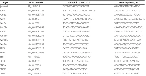
TNF-α,TNFR1andTNFR2were added. The primers were syn-thesized by (Takara Bio Inc, Otsu, Shiga, Japan) or (FAS-MAC Corp, Atsugi, Kanagawa, Japan) and are shown in Table 1. PCR reactions were performed in an Applied Bio-systems 7500 Fast system, according to the manufacturer’s instructions. A control gene, glyceraldehyde 3-phosphate dehydrogenase(GAPDH), was used to normalize each sam-ple, and the arbitrary intensity threshold (Ct) of amplifica-tion was computed. The expression scores were obtained using theΔΔCtcalculation method.
Gene-suppression studies using siRNA
We silenced β-catenin expression in nucleus pulposus cells by using siRNA technology. In brief, nucleus pulpo-sus cells were transferred into 24-well plates at a density of 6 × 10⁴ cells/well 1 day before transfection. The next day, cells were treated with β-catenin siRNA or control siRNA duplexes at a final concentration of 100 to 500 ng using Lipofectamine 2000. Cells also received TNF-α pro-moter constructs and the pGL4.74 plasmid at the time of transfection. Six hours after transfection, the medium was replaced with complete growth medium and the cells were allowed to recover for 18 h. Cells were then cultured for 24 h and luciferase activity was measured.
Western blot analysis
[image:3.595.56.544.472.725.2]Treated nucleus pulposus cells were placed immediately on ice and washed with cold PBS. Proteins were pre-pared using the CellLytic NuCLEAR extraction kit
Table 1 Primers for real-time PCR
Target NCBI number Forward primer, 5'-3' Reverse primer, 5'-3'
β-catenin AF_121265.1 GCCAGTGGATTCCGTACTGT GAGCTTGCTTTCCTGATTGC
Wnt1 NM_001105714.1 TCCTCATGAACCTTCACAATAACGA TTGCACTCTTGGCGCATCTC
Wnt3a NM_001105715.1 TGTGAGGTGAAGACCTGCTG AAAGTTGGGGGAGTTCTCGT
Wnt4 NM_053402.1 GAAACGTGCGAGAAGCTCAAAG AAAGGACTGTGAGAAGGCTACG
Wnt5a NM_022631.1 TGCCACTTGTATCAGGACCA TGTCTCTCGGCTGCCTATTT
Wnt5b NM_001100489.1 TGACTACTGCCTGCGAAATG AAAGCAACACCAGTGGAACC
Wnt6 NM_001108226.1 GTCGACTTTGGGGATGAGAA AAAGCCCATGGCACTTACAC
Wnt10a NM_001108227.1 GTTCCTAGCTCAGGCAGGTG AAGTCTGTGGAGGGGGAGAT
Wnt10b NM_001108111.1 CTGGTGCTGTTACGTGCTGT GGAGCCATGATTAACCGAAA
Wnt16 NM_001109223.1 TGGCTGTAACCTCCTCTGCT GAGGCAATCTCATGCTAGGC
LRP5 NM_001106321.2 CATCCATGCTGTGGAGGA TGTCTCGGGCACAAGGAT
LRP6 NM_001107892.1 CATGATACGAAGGCACAAGAA TCTGATTTGGAACCGAGCTT
LEF1 NM_130429.1 TGGTAAACGAGTCCGAAATCA TGTGTTTGTCCGACCACCT
TCF4 NM_053369.1 TCCAACCCTTCAACTCCTGT CGTTTCGAGACCAAACAGC
TNF-α NM_012675.3 TGAACTTCGGGGTGATCG GGGCTTGTCACTCGAGTTTT
TNFR1 NM_013091.1 AATGAGTGCACCCCTTGC CCTGGGGGTTTGTGACATT
TNFR2 NM_130426.4 GAGGCCCAAGGGTCTCAG GCTGCCATGGGAAGAATC
(Sigma-Aldrich, St Louis, MO, USA). All of the wash buffers and the final resuspension buffer included 1 × pro-tease inhibitor cocktail (Roche, Basel, Switzerland), NaF (5 mM), and Na3VO4(200μM). Nuclear or total cellular proteins were separated on a sodium dodecyl sulfate (SDS) polyacrylamide gel and were electrotransferred onto nitrocellulose membranes (Bio-Rad, Hercules, CA USA). The membranes were blocked with 5% BSA in Tris-buffered saline and Tween 20 (TBST) (50 mM Tris, pH 7.6, 150 mM NaCl, and 0.1% Tween-20) and incubated overnight at 4°C in 5% BSA in TBST with anti-TNF-α (number sc-1350. 1:200; Santa Cruz Biotechnology) or anti-β-catenin (1:1,000 dilution; Cell Signaling Technol-ogy) antibodies. Immunolabeling was detected with an ECL reagent (Amersham Bioscience, Little Chalfont, UK). The Western blot data were quantified using Image J pixel analysis (NIH Image software). Data from the western blots were presented as band density normalized to that of the loading control (actin).
Transfections and dual-luciferase™assay
Nucleus pulposus cells and annulus fibrosus cells were transferred to 24-well plates at a density of 3 × 10⁴cells/ well 1 day before transfection. Cells were co-transfected with 100 to 500 ng of expression plasmids or the back-bone vector together with the reporter plasmids. Lipo-fectamine 2000 (Invitrogen) was used as the transfection reagent. Cells were cultured for 24 h and treated with a specific concentration of TNF-α or BIO. The cells were harvested 24 h after treatment and a Dual-Luciferase™ reporter assay system (Promega) was used for the se-quential measurements of firefly and Renilla luciferase activities. The results were normalized regarding trans-fection efficiency and were expressed as a relative ratio of luciferase to pGL4.74 activities (denoted as relative activity). Nucleus pulposus cells were transfected with a plasmid encoding green fluorescent protein, to check transfection efficiency, which was 60 to 70% in nucleus pulposus cells. The luciferase activities and relative ratios were quantified using a Turner Designs Luminometer Model TD-20/20 instrument (Promega).
Statistical analyses
Typically, data were compiled from at least three inde-pendent triplicate experiments, each performed using separate cultures and on separate occasions. The re-sponses were presented as the fold change relative to the untreated control. The data were presented as the mean ± SD. Data were compared between groups using Stu-dent’s t-test or analysis of variance, to assess variance. Significance was accepted atP<0.05 and is denoted with an asterisk in the figures.
Results
Effects of Wnt signaling on TNF-αexpression in nucleus pulposus cells
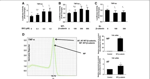
Although Wnt signaling has been shown to play an important role in nucleus pulposus cells, interactions be-tween Wnt signaling and TNF-α in these cells have not been described. Therefore, to study the relationship be-tween Wnt signaling and TNF-α, initial experiments were performed to investigate the role of Wnt signaling in the transcriptional activity of TNF-αin nucleus pulposus cells. Cells treated with different concentrations of BIO exhib-ited an increase in the activity of the TNF-α promoter (BIO 0.1μM, 1.22 ± 0.25, P= 0.2026; 0.5μM, 1.81 ± 0.37,
P <0.001; 1.0 μM, 1.81 ± 0.59, P <0.001) (Figure 1A). Nucleus pulposus cells were transiently transfected with plasmids encoding TNF-α and with a WT-β-catenin ex-pression plasmid. Figure 1B shows that forced exex-pression of WT-β-catenin significantly induced TNF-α promoter activity (WT-β-catenin 100 ng, 1.13 ± 0.26, P= 0.3801; 300 ng, 1.49 ± 0.30, P= 0.0076; 500 ng, 1.36 ± 0.30, P= 0.0304). To validate these findings, we performed loss-of-function experiments using an siRNA forβ-catenin. Sup-pression of gene exSup-pression was confirmed by real-time RT-PCR (data not shown). TNF-α promoter activity was inhibited in nucleus pulposus cells that were co-transfected with theβ-catenin siRNA (si-β-catenin 100 ng, 0.75 ± 0.20, P= 0.0041; 300 ng, 0.80 ± 0.14, P= 0.0244) (P<0.05) (Figure 1C). To confirm the reporter assay data, next we performed a real-time PCR analysis of TNF-α
mRNA expression after transfection with the WT-β -catenin expression plasmid both in nucleus pulposus cells and annulus fibrosus cells. Figure 1D shows that nucleus pulposus cells and annulus fibrosus cells transfected with WT-β-catenin exhibited a significant increase in the gene expression of TNF-α compared with that observed in untransfected control cells. The expression of TNF-αwas significantly higher in the nucleus pulposus than in the an-nulus fibrosus.
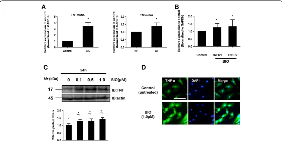
Real-time RT-PCR analysis also showed that activation of Wnt signaling by BIO increased the expression of
TNF-α mRNA 3-fold compared with untreated cells (Figure 2A). Similarly, transfection of WT-β-catenin led to an increase in the expression of the TNF-α mRNA compared with untreated cells (data not shown). The ex-pression of TNF-α mRNA was significantly higher (by 1.5-fold) in the annulus fibrosus than in the nucleus pul-posus (P <0.01, Figure 2A). In addition, activation of Wnt signaling increased the expression of TNFR1
analysis (Figure 2D). As shown in Figure 2C, immuno-blotting of nucleus pulposus cells treated with BIO (1.0μM, 24 h) showed an increased level of TNF-α pro-tein compared with control nucleus pulposus cells. Simi-larly, immunofluorescence analysis using an anti-TNF-α antibody showed that BIO treatment (1.0 μM, 24 h) promoted the nuclear translocation of TNF-α more strongly in nucleus pulposus cells than in untreated cells. TNF-αwas localized to the nucleus in cells treated with BIO (Figure 2D). To define a potential role of Wnt signaling on TNF in nucleus pulposus cells, we also used DKK-1, -2, -3, and −4 expression plasmids. No change in TNF-αpromoter activity was observed when DKK-1 or DKK-2 plasmids were used (Figure 3A and B). Conversely, transfection with DKK-3 and DKK-4 led to a dose-dependent suppression in TNF-α promoter activity (Figure 3C and D). Similarly, the BIO-mediated induction of TNF-α promoter activity was suppressed by transfection with the DKK-3 or DKK-4 plasmids (Figure 3F and G). To validate these findings, we used a Sclerostin (SOST) expression plasmid and measured the activity of the TNF-αpromoter in this condition; we detected a significant inhibition of TNF-αpromoter at a concentration of 500 ng (Figure 3E and H).
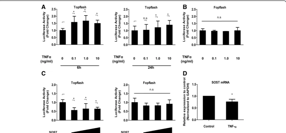
TNF-αenhanced Wnt transcriptional activity in nucleus pulposus cells
[image:5.595.58.539.90.344.2]To investigate the role of the proinflammatory cytokine TNF-αin Wnt signaling in IVDs, we first examined the soluble TNF-α protein levels after the stimulation of TNF-α (10 ng/mL, 24 h) by western blotting. The re-sults demonstrate that soluble TNF-α protein was sig-nificantly elevated after the stimulation of TNF-α compared to the control cells (Additional file 1). We next determined whether TNF-α induced Wnt tran-scriptional activity. We measured the activity of both Topflash (containing the WT TCF binding sites) and Fopflash (mutant Topflash) in nucleus pulposus cells after TNF-αtreatment. After 6 to 24 h, we measured the activity of Topflash in nucleus pulposus cells. Figure 4A shows that there was a dose-dependent increase in the activity of Topflash upon TNF-α stimulation, whereas Fopflash activity was not affected by TNF-α treatment (Figure 4B). We then co-transfected nucleus pulposus cells with plasmids expressing SOST along with both Topflash and Fopflash reporter. Figure 4C shows that overexpression of SOST results in a decrease in the ac-tivity of Topflash, whereas overexpression of SOST has no effect on Fopflash reporter activity. To explore the Figure 1Effect of Wnt signaling on TNF-αexpression in nucleus pulposus cells. (A)Nucleus pulposus cells transfected with the TNF-α reporter plasmid together with the pGL4.74 plasmid were treated with different concentrations of 6-bromoindirubin-3′-oxime (BIO) for 24 h.
(BandC)Nucleus pulposus cells were co-transfected with the TNF-αreporter plasmid together with WT-β-catenin(B),β-catenin siRNA (si-β-catenin)(C), or empty vectors and the pGL4.74 vector. Cells were cultured for 24 h and luciferase reporter activity was measured. The results were normalized for transfection efficiency and are expressed as a relative ratio of luciferase to pGL4.74 activity (denoted as relative activity).
premise that TNF-αregulatesSOSTmRNA expression, nucleus pulposus cells were treated with TNF-αand ex-pression ofSOSTmRNA analyzed using real-time PCR. Figure 4D shows that treatment with TNF-α for 24 h significantly decreases SOST mRNA levels in nucleus pulposus cells.
TNF-αenhanced Wnt related gene and protein expression in nucleus pulposus cells
We examined further the expression of Wnt-related genes via real-time PCR after treatment with TNF-α(10 ng/mL) for 6 and 24 h. Real-time PCR analysis demonstrated that TNF-αtreatment for 24 h increased the expression of the
Wnt5b,LRP6, LEF1, andTCF4mRNA, but treatment for 6 h did not (Figure 5A). TNF-αsignificantly also increased the expression of β-catenin mRNA at 6 and 24 h. In addition, we determined the expression levels of the
β-catenin protein in nucleus pulposus cells after treatment with TNF-α. Western blot analysis using an anti-β-catenin antibody demonstrated that TNF-α treatment increased the expression of the β-catenin protein (Figure 5B). Cells were evaluated by immunofluorescence analysis to deter-mine whether a concomitant elevation inβ-catenin protein expression was associated with the activation of TNF-α. Immunofluorescence analysis using an anti-β-catenin
antibody showed that TNF-αtreatment (10 ng/mL, 24 h) promoted the nuclear translocation of β-catenin more strongly in nucleus pulposus cells than in untreated con-trol cells (Figure 5C).
Discussion
[image:6.595.57.538.89.330.2]α has been associated almost exclusively with inflamma-tion or host defense. TNF-αwas first described in IVDs in association with sciatic pain. Subsequent studies showed that TNF-αis widely expressed in humans with degenera-tive disc degeneration [29], as well as in animal models. Seguinet al. reported that TNF-αreduced the synthesis of matrix molecules and upregulated the mRNA expression of MMP-1, -3, and−13 and ADAM-TS4 and ADAM-TS5 [30]. Studies performed by Alsalameh and colleagues [31]
on synovial fibroblasts from patients with rheumatoid arthritis and patients with osteoarthritis have also indi-cated there is a differential expression of the two TNF receptors (TNF-R1 and TNF-R2) in these cells and that, although both receptors can mediate the effect of TNF-α on TIMP1 expression, PGE2, the regulation of IL-6, and MMP-1 is mediated exclusively by TNF-R1.
[image:7.595.58.541.86.542.2]upregulated in the presence of degeneration [16]. TNF-α can bind to, and signal through, either TNF-R1 or TNF-R2. Although TNF binds to each with high affinity, TNF-R1 is more ubiquitously expressed and it is generally believed that TNF-R1 is responsible for the majority of bio-logical actions of TNF while TNF-R2 may function to potentiate the effects of TNF-R1. Freemontet al. have also reported that TNF-R2 is not expressed by IVD cells either in normal or degenerate IVDs [32]. In addition, they have demonstrated that human IVD cells are capable of responding to TNF-α in vivo. However, no increase in TNF-R1 synthesis was seen during IVD degeneration.
The complexity of the Wnt signaling cascade should enable the activation and/or repression of many specific signals and targets. Remarkably, we found that Wnt sig-naling, which suppresses the proliferation of nucleus pul-posus cells and induces cell senescence, activated the expression of TNF-α. Direct evidence for the activation of TNF-α by Wnt signaling was obtained from experiments such as a reporter assay, real-time PCR, western blotting, et cetera. Furthermore, Wnt signaling was modulated by several different families of secreted negative regulators. The results of this study suggest that inhibition of Wnt signaling may be able to contribute to the suppression of disc degeneration, because Wnt signaling increased the expression of TNF-αand TNF-αactivated Wnt signaling. Wnt antagonists can be divided into two functional clas-ses. These two major classes function in quite different ways: the secreted Frizzled-related protein (sFRP) class
binds to Wnt ligands, whereas the DKK or sclerostin class binds to a component of the Wnt receptor. Thus, in theory, the outcome appears to be that sFRPs inhibit canonical (β-catenin-dependent) and noncanonical (β -ca-tenin-independent) pathways, whereas DKKs or sclerostin inhibit only the canonical pathway. The DKK family com-prises four members (DKK-1 to DKK-4) in mammals [33,34]. DKK proteins have been implicated in various dis-eases, including retinal degeneration, malignancies, and cerebral ischemia [35-37].
The most-studied member of the DKK family is DKK-1. The binding of DKK-1 to the LPR5/6 receptor and a cell-surface coreceptor, Kremen-1/2, promotes internalization of the receptor complex and dampens the Wnt signal. Both DKK-1 and DKK-2 bind to the LRP5/6 receptor, which is expressed in cells and has higher affinity com-pared with Fz and Wnt. The characteristic developmental function of DKK-1 is its head-inducing activity. Previously, Yeet al. reported that TNF-αincreased the expression of
β-catenin and MMP-13, and significantly inhibited matrix synthesis, which resulted in the degeneration of rabbit IVD cells. These authors also showed that blocking Wnt/
[image:8.595.60.538.89.311.2]The Wnt-antagonizing activity of DKK-4 appears to be indistinguishable from that of DKK-1, whereas DKK-3 has distinct roles in regulating the Wnt pathway, depending on the cell types examined. For example, DKK-3 potentiates Wnt signaling in human Müller glia MIO-M1 and HEK293 cell lines [39], but inhibits Wnt signaling in PC12 and osteocarcinoma Saos-2 cells [40,41]. The biological roles of DKK-3 in Wnt signaling remain unclear, because it does not inhibit canonical Wnt signaling [33,42]. In addition, DKK-3 does not interact with LRPs or Krm1/2 [42,43]. It also remains to be determined whether DKK-3 antagonizes other growth-factor pathways via mechanisms that involve a direct association with ligands or transmem-brane receptors in a manner similar to that by which DKK-1 inhibits Wnt signaling. However, recent studies have revealed that the effect of DKK antagonists is not quite so simple. In fact, it may turn out that certain antagonists act as such only when expressed at nonphysiological levels. Therefore, there are clearly many unresolved issues regard-ing this subject. Furthermore, we checked the effect of
sclerostin, which is a different inhibitor of Wnt signaling, on TNF-αpromoter activity in nucleus pulposus cells. The results of this experiment showed significant inhibition of the TNF-αpromoter after treatment with high sclerostin. Sclerostin is the product of the SOST gene. Similar to DKK, sclerostin binds to Lrp5/6 and antagonizes canonical Wnt signaling [44-46]. Low sclerostin expression leads to bone growth, whereas high expression inhibits bone forma-tion. Recently, TNF has been identified as an inducer of sclerostin expression, but the current study showed that TNF-α suppressed the SOST gene. As a result, it might have resulted in the activation of Wnt signaling in nucleus pulposus cells.
[image:9.595.56.541.90.397.2]Overall, our results indicate that inhibition of Wnt sig-naling suppresses a catabolic response via the inhibitory action of TNF-α in nucleus pulposus cells. However, it remains to be determined which of the proposed recep-tors for DKKs or sclerostin, including LRP-4, -5, and−6, are important for the responses observed in our study and whether the effect is dependent on canonical or Figure 5TNF-αenhanced the expression of Wnt-related genes and proteins in nucleus pulposus cells. (A)Wnt-related mRNA expression after exposure of nucleus pulposus cells to TNF-α(10 ng/mL) for 6 and 24 h, as assessed by real-time PCR.(B)Western blot analysis ofβ-catenin activation after treatment of nucleus pulposus cells with TNF-α(10 ng/mL).*P< 0.05 indicates significant differences between groups. Error bars represent the SD. n.s., not significant.(C)Detection ofβ-catenin protein expression by immunofluorescence microscopy. Nucleus pulposus cells were cultured with or without 10 ng/mL TNF-αfor 24 h, fixed, and stained with an antibody against c-fos. Left: cells stained with an antibody to
β-catenin; middle: cells stained with 4′,6-diamidino-2-phenylindole (DAPI), to identify healthy nuclei; right: cells stained with an antibody to
noncanonical Wnt signaling, or on other types of signal-ing. In addition to the limitation inherent in the use of rats, because of the ambiguity of the notochordal cells in this animal, the functions of the rat disc compared with the functions of the human IVD need to be considered in the interpretation of the findings of this study. Re-garding this issue, additional studies using different species are required to evaluate and conclude whether the mechanism involving the expression of these mole-cules is specific to nucleus pulposus cells, in particular regarding the human situation.
Conclusions
Here, we have demonstrated that Wnt signaling regulates TNF-α and that Wnt signaling and TNF-α form a positive-feedback loop in nucleus pulposus cells. The re-sults of the present study provide in vitro evidence that the activation of Wnt signaling upregulates the proinflam-matory cytokine TNF-αand might cause the degeneration of nucleus pulposus cells. We speculated that blocking the Wnt signaling might protect nucleus pulposus cells against degeneration. The DKK or sclerostin families of proteins are natural regulators of Wnt signaling and can specifically block this pathway. These findings suggest that overexpression of DKK-3, DKK-4, or sclerostin inhibit TNF-α expression by specifically blocking the Wnt chan-nel. Inhibition of Wnt signaling using DKKs or sclerostin exerts a protective and reversing effect in the TNF-α -induced degeneration of IVD cells.
Additional file
Additional file 1: Figure S1.The soluble TNF-αprotein levels after the stimulation of TNF-α(10 ng/mL, 24 h) by western blotting. The results demonstrate that soluble TNF-αprotein was significantly elevated after the stimulation of TNF-αcompared to the control cells.
Abbreviations
ADAMTS:A disintegrin and metalloproteinase with thrombospondin motifs; BIO: 6-bromoindirubin-3′-oxime; BSA: Bovine serum albumin; Ct: Intensity threshold; DAPI: 4′,6-diamidino-2-phenylindole; DKK: Dickkopf;
DMEM: Dulbecco’s modified Eagle’s medium; FBS: Fetal bovine serum; GAPDH: Glyceraldehyde 3-phosphate dehydrogenase; GSK: Glycogen synthase kinase; IL-1β: Interleukin-1β; IVD: Intervertebral disc; LEF: Lymphoid enhancer factor; MMP: Matrix metalloproteinase; PBS: Phosphate-buffered saline; PCP: Planar cell polarity; PGE2: Prostaglandin E2; RT-PCR: Reverse transcription polymerase chain reaction; sFRP: Frizzled-related protein; SOST: Sclerostin; TBST: Tris-buffered saline and Tween 20; TCF: T-cell factor; TNF-α: Tumor necrosis factor-α; WT: Wild-type.
Competing interests
The authors declare that they have no competing interests.
Authors’contributions
AH participated in study design, performed experiments, analyzed data and wrote the paper. KY prepared the isolation of cells and performed experiments and the statistical analysis. TN performed experiments and analyzed data. DS participated in study design, performed experiments and performed the statistical analysis. JM participated in study design and
coordination and helped to draft the manuscript. All authors read and approved the final manuscript.
Acknowledgements
We greatly appreciate the helpful advice of Dr Tadayuki Sato and the technical assistance of Hideki Hayashi, Masayuki Tanaka, Masatoshi Ito and Mayumi Nakano. This study was supported in part by the Research and Study Program of the Tokai University Educational System General Research Organization and a Grant-in-Aid for Scientific Research (KAKENHI). The study was also supported by grants from the Nakatomi Foundation.
Received: 21 February 2013 Accepted: 24 October 2013 Published: 14 November 2013
References
1. Hiyama A, Sakai D, Tanaka M, Arai F, Nakajima D, Abe K, Mochida J:The relationship between the Wnt/beta-catenin and TGF-beta/BMP signals in the intervertebral disc cell.J Cell Physiol2011,226:1139–1148.
2. Hiyama A, Sakai D, Arai F, Nakajima D, Yokoyama K, Mochida J:Effects of a glycogen synthase kinase-3beta inhibitor (LiCl) on c-myc protein in intervertebral disc cells.J Cell Biochem2011,112:2974–2986. 3. Hiyama A, Sakai D, Risbud MV, Tanaka M, Arai F, Abe K, Mochida J:
Enhancement of intervertebral disc cell senescence by WNT/beta-catenin signaling-induced matrix metalloproteinase expression.Arthritis Rheum2010,
62:3036–3047.
4. Wang M, Tang D, Shu B, Wang B, Jin H, Hao S, Dresser KA, Shen J, Im HJ, Sampson ER, Rubery PT, Zuscik MJ, Schwarz EM, O'Keefe RJ, Wang Y, Chen D:
Conditional activation of beta-catenin signaling in mice leads to severe de-fects in intervertebral disc tissue.Arthritis Rheum2012,64:2611–2623. 5. Slusarski DC, Yang-Snyder J, Busa WB, Moon RT:Modulation of embryonic
intracellular Ca2+ signaling by Wnt-5A.Dev Biol1997,182:114–120. 6. Tree DR, Ma D, Axelrod JD:A three-tiered mechanism for regulation of
planar cell polarity.Semin Cell Dev Biol2002,13:217–224.
7. Yamaguchi TP:Heads or tails: Wnts and anterior-posterior patterning. Curr Biol2001,11:R713–R724.
8. Lee E, Salic A, Kruger R, Heinrich R, Kirschner MW:The roles of APC and Axin derived from experimental and theoretical analysis of the Wnt pathway.PLoS Biol2003,1:E10.
9. Giles RH, van Es JH, Clevers H:Caught up in a Wnt storm: Wnt signaling in cancer.Biochim Biophys Acta2003,1653:1–24.
10. Church VL, Francis-West P:Wnt signalling during limb development.Int J Dev Biol2002,46:927–936.
11. He TC, Sparks AB, Rago C, Hermeking H, Zawel L, da Costa LT, Morin PJ, Vogelstein B, Kinzler KW:Identification of c-MYC as a target of the APC pathway.Science1998,281:1509–1512.
12. Hoy D, Brooks P, Blyth F, Buchbinder R:The epidemiology of low back pain.Best Pract Res Clin Rheumatol2010,24:769–781.
13. Juniper M, Le TK, Mladsi D:The epidemiology, economic burden, and pharmacological treatment of chronic low back pain in France, Germany, Italy, Spain and the UK: a literature-based review.Expert Opin Pharmacother2009,10:2581–2592.
14. Hunter CJ, Matyas JR, Duncan NA:The three-dimensional architecture of the notochordal nucleus pulposus: novel observations on cell structures in the canine intervertebral disc.J Anat2003,202:279–291.
15. Hunter CJ, Matyas JR, Duncan NA:Cytomorphology of notochordal and chondrocytic cells from the nucleus pulposus: a species comparison. J Anat2004,205:357–362.
16. Le Maitre CL, Hoyland JA, Freemont AJ:Catabolic cytokine expression in degenerate and herniated human intervertebral discs: IL-1beta and TNFalpha expression profile.Arthritis Res Ther2007,9:R77.
17. Burke JG, Watson RW, McCormack D, Dowling FE, Walsh MG, Fitzpatrick JM:
Intervertebral discs which cause low back pain secrete high levels of proinflammatory mediators.J Bone Joint Surg Br2002,84:196–201. 18. Legendre F, Dudhia J, Pujol JP, Bogdanowicz P:JAK/STAT but not ERK1/ERK2
pathway mediates interleukin (IL)-6/soluble IL-6R down-regulation of Type II collagen, aggrecan core, and link protein transcription in articular chondrocytes. Association with a down-regulation of SOX9 expression. J Biol Chem2003,278:2903–2912.
19. Legendre F, Bogdanowicz P, Boumediene K, Pujol JP:Role of interleukin 6 (IL-6)/ IL-6R-induced signal tranducers and activators of transcription and
20. Lee JM, Song JY, Baek M, Jung HY, Kang H, Han IB, Kwon YD, Shin DE:
Interleukin-1beta induces angiogenesis and innervation in human intervertebral disc degeneration.J Orthop Res2011,29:265–269. 21. Fujita K, Janz S:Attenuation of WNT signaling by DKK-1 and−2 regulates
BMP2-induced osteoblast differentiation and expression of OPG. RANKL and M-CSF. Mol Cancer2007,6:71.
22. Sato N, Meijer L, Skaltsounis L, Greengard P, Brivanlou AH:Maintenance of pluripotency in human and mouse embryonic stem cells through activation of Wnt signaling by a pharmacological GSK-3-specific inhibitor.Nature Med2004,10:55–63.
23. Naugler WE, Karin M:NF-kappaB and cancer-identifying targets and mechanisms.Curr Opin Genet Dev2008,18:19–26.
24. Courtois G:The NF-kappaB signaling pathway in human genetic diseases. Cell Mol Life Sci2005,62:1682–1691.
25. Clevers H:Wnt/beta-catenin signaling in development and disease. Cell2006,127:469–480.
26. Baker SJ, Reddy EP:Modulation of life and death by the TNF receptor superfamily.Oncogene1998,17:3261–3270.
27. Gruss HJ, Dower SK:Tumor necrosis factor ligand superfamily: involvement in the pathology of malignant lymphomas.Blood1995,
85:3378–3404.
28. Baud V, Karin M:Signal transduction by tumor necrosis factor and its relatives.Trends Cell Biol2001,11:372–377.
29. Weiler C, Nerlich AG, Bachmeier BE, Boos N:Expression and distribution of tumor necrosis factor alpha in human lumbar intervertebral discs: a study in surgical specimen and autopsy controls.Spine (Phila Pa 1976)
2005,30:44–53. discussion 54.
30. Seguin CA, Pilliar RM, Roughley PJ, Kandel RA:Tumor necrosis factor-alpha modulates matrix production and catabolism in nucleus pulposus tissue. Spine (Phila Pa 1976)2005,30:1940–1948.
31. Alsalameh S, Amin RJ, Kunisch E, Jasin HE, Kinne RW:Preferential induction of prodestructive matrix metalloproteinase-1 and proinflammatory interleukin 6 and prostaglandin E2 in rheumatoid arthritis synovial fibroblasts via tumor necrosis factor receptor-55.J Rheumatol2003,30:1680–1690. 32. Freemont AJ, Watkins A, Le Maitre C, Baird P, Jeziorska M, Knight MT,
Ross ER, O’Brien JP, Hoyland JA:Nerve growth factor expression and innervation of the painful intervertebral disc.J Pathol2002,197:286–292. 33. Krupnik VE, Sharp JD, Jiang C, Robison K, Chickering TW, Amaravadi L,
Brown DE, Guyot D, Mays G, Leiby K, Chang B, Duong T, Goodearl AD, Gearing DP, Sokol SY, McCarthy SA:Functional and structural diversity of the human Dickkopf gene family.Gene1999,238:301–313.
34. Monaghan AP, Kioschis P, Wu W, Zuniga A, Bock D, Poustka A, Delius H, Niehrs C:Dickkopf genes are co-ordinately expressed in mesodermal lineages.Mech Dev1999,87:45–56.
35. Mastroiacovo F, Busceti CL, Biagioni F, Moyanova SG, Meisler MH, Battaglia G, Caricasole A, Bruno V, Nicoletti F:Induction of the Wnt antagonist, Dickkopf-1, contributes to the development of neuronal death in models of brain focal ischemia.J Cereb Blood Flow Metab2009,29:264–276.
36. Hsieh SY, Hsieh PS, Chiu CT, Chen WY:Dickkopf-3/REIC functions as a suppressor gene of tumor growth.Oncogene2004,23:9183–9189. 37. Hackam AS, Strom R, Liu D, Qian J, Wang C, Otteson D, Gunatilaka T, Farkas RH,
Chowers I, Kageyama M, Leveillard T, Sahel JA, Campochiaro PA, Parmigiani G, Zack DJ:Identification of gene expression changes associated with the progression of retinal degeneration in the rd1 mouse.Invest Ophthalmol Vis Sci2004,45:2929–2942.
38. Ye S, Wang J, Yang S, Xu W, Xie M, Han K, Zhang B, Wu Z:Specific inhibitory protein Dkk-1 blocking Wnt/beta-catenin signaling pathway improve protectives effect on the extracellular matrix.J Huazhong Univ Sci Technolog Med Sci2011,31:657–662.
39. Nakamura RE, Hunter DD, Yi H, Brunken WJ, Hackam AS:Identification of two novel activities of the Wnt signaling regulator Dickkopf 3 and characterization of its expression in the mouse retina.BMC Cell Biol
2007,8:52.
40. Hoang BH, Kubo T, Healey JH, Yang R, Nathan SS, Kolb EA, Mazza B, Meyers PA, Gorlick R:Dickkopf 3 inhibits invasion and motility of Saos-2 osteosarcoma cells by modulating the Wnt-beta-catenin pathway.Cancer Res2004,
64:2734–2739.
41. Caricasole A, Ferraro T, Iacovelli L, Barletta E, Caruso A, Melchiorri D, Terstappen GC, Nicoletti F:Functional characterization of WNT7A signaling in PC12 cells: interaction with A FZD5 x LRP6 receptor complex and modulation by Dickkopf proteins.J Biol Chem2003,278:37024–37031.
42. Mao B, Wu W, Li Y, Hoppe D, Stannek P, Glinka A, Niehrs C:LDL-receptor-related protein 6 is a receptor for Dickkopf proteins.Nature2001,411:321–325. 43. Mao B, Wu W, Davidson G, Marhold J, Li M, Mechler BM, Delius H, Hoppe D,
Stannek P, Walter C, Glinka A, Niehrs C:Kremen proteins are Dickkopf receptors that regulate Wnt/beta-catenin signalling.Nature2002,
417:664–667.
44. Li X, Zhang Y, Kang H, Liu W, Liu P, Zhang J, Harris SE, Wu D:Sclerostin binds to LRP5/6 and antagonizes canonical Wnt signaling.J Biol Chem
2005,280:19883–19887.
45. Ellies DL, Viviano B, McCarthy J, Rey JP, Itasaki N, Saunders S, Krumlauf R:
Bone density ligand, Sclerostin, directly interacts with LRP5 but not LRP5G171V to modulate Wnt activity.J Bone Miner Res2006,21:1738–1749. 46. Semenov M, Tamai K, He X:SOST is a ligand for LRP5/LRP6 and a Wnt
signaling inhibitor.J Biol Chem2005,280:26770–26775.
doi:10.1186/ar4379
Cite this article as:Hiyamaet al.:A complex interaction between Wnt signaling and TNF-αin nucleus pulposus cells.Arthritis Research & Therapy
201315:R189.
Submit your next manuscript to BioMed Central and take full advantage of:
• Convenient online submission
• Thorough peer review
• No space constraints or color figure charges
• Immediate publication on acceptance
• Inclusion in PubMed, CAS, Scopus and Google Scholar
• Research which is freely available for redistribution