244
Chapter 33
Histopathology of gill of Pangasius sutchi infected with Aeromonas hydrophila
and are cured using Curcumin
Morina Riauwaty S1,a
1
Fishery and Marine Science Faculty, Riau University, Pekanbaru
Kampus Bina Widya km 12,5 Simpang Baru Pekanbaru 28293, Telp. (076163275)
a
Abstract—This study aims to understand the histological structure of gill of Pangasius sutchi that is infected with
Aeromonas hydrophila and treated with curcumin has been conducted on February to April 2015. There were 3
treatments applied. The treated fishes were infected with A. hydrophila (0.1 ml of 109of A. hydrophila culture) and then were immerse in 3 different concentrations of curcumin, they were T1 (0.5 g/l); T2 (0.7 g/l) and T3 (0.9 g/l) for 5 minutes/ day for a 2 weeks period. The negative control were fishes that were not receive any treatment, while the positive control were fishes that were infected with A. hydrophila, and were not treated with curcumin. Fish organ (gill) were processed for histological studied (formalin fixed, alcohol series, HE stained and 6 sliced. The result showed various types of damage such as necrosis, hemorrhage, fused lamella, loss of epithelium on the secondary lamella and oedema. Based on data obtained, it can be concluded that immersion of fish in curcumin is able to cure A. hydrophila infection on fish.
Keywords: Gill, Aeromonas hydrophila, Pangasius sutchi, Curcumin
I.
Introduction
Aeromonas hydrophila is an apportunistic pathogen of a wide variety of hosts ( (Harikrishnan, 2009). Pangasius sutchi or“jambal fish”is freshwater fish that commonly inhabit in river in Riau Province. This fish is belonged to the members of Pangasidae family. In Riau, Pangasius sutchi has relatively high economical values. This fish commonly sold as fresh (Rp 20.000–Rp 30.000/ kg) or as smoked-fish (Rp 100.000 - Rp 120.000/ kg).
The most serious problem in the fish culture is the mass death of the fish that is caused by fish disease such as motile aeromonads septicaemia. Motile Aeromonads Septicaemia (MAS) is acute or sub acute or chronic infectious disease in all freshwater fishes caused by motile aeromonads bacteria. The disease caused about 80% mortality in fish farming especially when the fish held under stress (Austin and Austin, 1987). MAS diseases can be transmitted by discharge from the intestinal tract and external lesions on the skin. On the other hand, the parasitic damage and fungal infection of the epidemic may allow the entry and spread of infection among fish. Carrier fish also play an important role in transmission of the MAS infection. This disease is characterized by rapidly fatal septicemia with few gross signs, exophthalmia, ascitis and ulcer formation (Yardimci and Aydin, 2011).
245
Laith and Najiah (2013) reported that the clinical sign of the MAS diseases in Catfish showed symptoms of increased respiration and lethargy, skin lessions such as white discoloration, shallow hemorrhagic ulcers or deep ulcers with exposed underlying muscle. Some fish showed marked hemorrhages on the base of the fins and went. Others were dropsy, kidney congestion and enlargement, pale liver and gills, or gall-bladder, enlargement with the accumulation of yellowish fluid in the body cavity.Motile aeromonads septicaemia can be treated by using antibiotic containing 2 to 4 g of oxytetracycline/Kg of feed per day for 10 days. Sulfamerazine at 264 mg/Kg given in food for 3 days, by 154 mg/Kg/fish/day for 11 additional days is effective treatment for Motile Aeromonas Septicemia. Prolonged bath treatments with potassium permanganate at 2 to 4 mg/L will be effective (Afrianto and Liviawaty, 2001). However, the use of chemicals and antibiotics causes negative effects such as bacterial resistance and environmental pollution.
To avoid the negative effect of antibiotics, alternative treatment such as the use of traditional medicine such as turmeric can be applied. The most important chemical components of turmeric are a group of compounds called curcuminoids, which include curcumin (diferuloylmethane), demethoxycurcumin, and bisdemethoxycurcumin (Nagpal and Sood, 2013). The best studied compound is curcumin, which constitutes 3.14% of powdered turmeric (Tayyem et al., 2006). In addition, other important volatile oils include turmerone, atlantone, and zingiberene. Some general constituents are sugars, proteins, and resins.The active compound curcumin is believed to have a wide range of biological effects including anti inflammatory, antioxidant, antitumour, antibacterial, and antiviral activities, which indicate potential in clinical medicine (Aggarwald et al., 2007). Morphological changes in the gills are widely used as parameters in biomonitoring programs, for they are defense mechanisms to potential stressors of the aquatic environment. Histopathological features of the fish organ have been used as biomarkers. Research on histological structure of gill of Pangasius sutchi especially is limited. To obtain information on histological alterations of gill of Pangasius sutchi that were infected with A.
hydrophila and are cured using curcumin, this research is needed.
II.
Materials and Methods
In this study, fish samples (Pangasius sutchi) were obtained from hatchery in Bangkinang, Riau Province. Fish samples were varied from 80 - 120 mm TL and 10 to 15 g BW. The experiment were performed in the Parasite and Fish Diseases Laboratory of Fishery and Marine Science Faculty of Riau University. In this experiment, 15 aquaria (25 l) were used and fishes were adapted. Aeromonas hydrophila strain (ATCC 35654) used in this study was obtained from Fish Quarantine in Pekanbaru. The fishes were infected with A. hydrophila (0.1 ml of 109of A. hydrophila culture). Curcumin was extracted from Curcuma longa based on Harjanti (2008). There are five treatments applied e.g as negative control, the infected fishes that were not treated, while the positive control were the infected fishes that were treated with A. hydrophila. The fishes were immersed in 3 different concentrations ofcurcumin, they were T1 (0.5 g/l); T2 (0.7 g/l) and T3 (0.9 g/l) for 5 minutes/ day for a 2 weeks period. Fishes were then reared for 14 days. Gills were processed for histological study, following Darjono et
al., (2001). The tissues were alcohol series processed, paraffin embedded, 6 µ sectioned and Hematoxylin-Eosin
stained. Then the tissue were studied using a binocular microscope (Olympus CX 21), abnormalities occur in gill were observed and noted. The level of gill alteration is calculated by using Histopathologic Alteration Index (HAI) following Lopez and Thomaz (2011).
III.
Results and Discussions
246
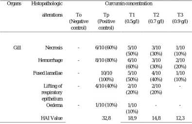
Table 1. List of histopathologic alteration observed in the gill of Pangasius sutchi in 3 different concentrationsof curcumin
Organs Histopathologic Curcumin concentration
alterations To (Negative Tp (Positive T1 (0.5g/l) T2 (0.7 g/l) T3 (0.9 g/l) control) control)
Gill Necrosis - 6/10 (60%) 5/10 3/10 1/10
(50%) (30%) (10%)
Hemorrhage - 8/10 (80%) 6/10 3/10 2/10
(60%) (30%) (20%)
Fused lamellae - 10/10 5/10 4/10 1/10
(100%) (50%) (40%) (10%)
Lifting of - 4/10 (40%) 2/10 2/10
-respiratory epithelium
Oedema - 1/10 (10%)
(20%)
1/10
(20%)
-
-(10%)
HAI Value 32,8 18,9 14,8 12,3
Histological study shown that in control fish, there were no gill abnormalities were observed. It is characterized by the primary gill lamellae are flat leaf structures with a central rod like supporting axis and a row of secondary gill lamellae side of it. Butchiram et al.,(2009) stated that histological structure of gills in control fish (Tilapia mossambica) bearing four pairs of gill lamellae and both the sides were supported by bony structure and primary lamellae. The secondary lamellae shown numerous channels of blood capillaries, each separated by single layered pillar cells when observed in vertical section (Lopes and Thomaz, 2011). The laminar epithelium was thick followed by basement membrane below which the pillar cells enclosed blood spaces, large number of mucous cells were present on the epithelial gill rackers, where as primary lamellae had comparatively small and less number of mucous cells (Saenphet et al., 2009). The value of the Histological Alteration Index (HAI) prove that the gill condition of the fish that were infected with Aeromonas hydropila was worse than that of in control fish. According to Poleksik and Mitrovic-Tutundzik in Lopez and Thomaz (2011) the HAI value of the infected fish (32,8) can be catagorized as“moderate changes in the organs”, however the HAI value of the treated fish of 0.9 g/L curcumin concentration (12,3) can be categorized as“normal”.
Yardimci and Aydin (2011) reported that focal hemorrhage and dermal lessions accompanied by ulcerative form of the desease were observed in chronic motile aeromonad infection significantly and target organs in acute septicaemeia were gill, liver and kidney. The lessions in the gill of treated fish with Aeromonas
hydrophila included necrosis (60%), hemorrhage (80%), fusion of several lamellae (100%), lifting of
respiratory epithelium (40%) and oedema (10%) (Fig. 1, A2). Rao et al.,(2004) found Astronotus ocellatus infected with motile aeromonad septicaemia contained a large amount of red-ascitic fluid accumulated in the abdominal cavity along with hemorrhages in gills. Windarti et al., (2013) stated that gill structure of Ompok
hypothalmus of the Siak River, Riau shown necrosis, hemmorhage, hyperplasia, lifting of respiratory
epithelium, fusion an disorganization of secondary gill lamellae and shortening of secondary lamellae. Harikrishnan et al., (2009) found that alteration signs such as as hyperplasia, hypertrophy and lifting of epithelial cells are present in the gill lamellae of fish that are infected with Aeromonas hydrophila. The lessions in gill of
P. sutchi that were infected with A. hydrophila in this study shown the same pattern as previous studies. A
247
The histopathology of gills of experimental fish is given in Fig. 1, A3. The gill structure of the treated fishes of 0.5 g/l curcumin concentration shown several alterations types. The most histopathological alterations observed are hemorrhages (60%), necrotic (50%), fused lamellae/lifting of respiratory epithelium (20%), while oedema (10%) was rare observed. The secondary lamellae of the treated fishes of 0.5 g/l curcumin concentration showed several damage and marked proliferation. The lifting of respiratory epithelium is the most frequent lession observed in all gills sampled followed by fused lamellae of the lamellar epithelium. Lopes and Thomaz (2011) stated that the lifting of respiratory epithelium is one of the erliest injuries found in fish. It is characterized by displacement of the klining epithelium of the secondary lamellae, in which the formation of a space called oedema occurs. Santos et al., (2014) stated that hyperplasia leads to the proliferation of adjacent lamellae cells, reducing the inter-lamellar space, which may cause a fusion of lamellae.Gill structure of the treated fishes that were cured with curcumin (0.9 g/l) showing less abnormalities (Fig. 1, A3). The abnormalities indicated hemorrhage (20%), necrosis and fused lamellae (10%). In this study, treated fish had faster regenerative responses such formation of normal gill by immersing in 0,9 g/l curcumin. Wu et al., (2001) found that weight gain of eels (A. anguilla) treated with traditional Chinese medicines (TCMs) increased significantly their resistance to common infectious diseases. Jian and Wu (1994) observed that traditional Chinese medicines had a beneficial effect on the growth and on the prevention and treatment of common diseases in C.
carpio. Dey and Chandra (1995) observed that neem leaves, garlic and turmeric powder induced diseases
resistance of fry of carp. In this study, we found that herbal medicine (curcumin) can be use as alternative medicine for practical use in diseases management strategy in fish. However, the MAS symptoms in the fish in this study was not completely cured. The gill of the fish was taken by the 14thday after being treated with curcumin. This time period may not be enough for the fish organ for recover from the damage cause by the MAS. If the fish was reared for longer time, the condition of gill structure might be better. Sukarni et al.,2012 stated that organ recovery will be completed by around 30 days. As curcumin showing great potensial for curing the symptoms, the use of curcumin for curing the MAS diseases is recommended.
Therefore,complementary studies are needed for futher evaluation of this problem.
[image:4.595.115.495.438.559.2]A1 A2 A3
Fig 2. Photomicrograph of the gill of P. sutchi (H&E, 400x). A1. Control fish, A2. Infected fish with A.
hydrophila, A3. Treated fish with 0.9 g/l of curcumin concentration
IV.
Conclusions
248
Acknowledgements
The author is grateful to the Indonesian Government for the award of Research grand (Hibah Bersaing 2015), which made this work possible.
References
[1] Afrianto, E, Liviawaty E. 2001. Pengendalian hama dan penyakit Ikan. Kanisius. Yogyakarta. 89 hal
[2] Aggarwal, BB, Sundaram C, Malani N, Ichikawa H. 2007. Curcumin: the Indian solid gold. Adv Exp Med Biol 595 (1): pp. 1–75
[3] Angka, SL. 1990. The Pathology of the Walking Catfish Clarias batrachus (L), Infected Intraperitoneally with Aeromonas hydrophila. Asian Fisheries Sci. pp. 343-351
[4] Austin, B, Austin DA. 1987. Bacterial fish pathogen in deseases farmed and wild Fish. Heriot-Watt University, Edenburgh. pp. 191-197
[5] Butchiram, MS, Tilak, KS, Raju, PW. 2009. Studies on histopathological changes in the gill, liver and kidney of Channa punctatus (Bloch) exposed to Alachlor. J Environ Biol 30 (2): pp. 303-306
[6] Darjono, CR.Tabbu, Kurniasih, R Warsito, Sutrisno. 2001. Petunjuk praktikum patologi umum (S1) Laboratorium Patologi Fakultas Kedokteran Hewan Universitas Gadjah Mada. Jogyakarta. 33 hal
[7] Dey, RK, Chandra S. 1995. Preliminary studies to raise disease resistant seed (fry) of Indian major carp, Catla catla (Ham) through herbal treatment of spown. Fish Chimes March, (5): pp. 23-25
[8] Lopes F,Thomaz, AT. 2011. Histopathologic alterations observed in fish gills as a tool in environmental monitoring. Braz j. Bio.(71), pp. 179-188
[9] Harikrishnan, R, Balasundaram C, Young-Gun Moon, Kim M-C Kim J-S, Heo. 2009. Use of herbal concoction in the therapy of Goldfish (Carassius auratus) infected with Aeromonas hydrophila. Bul Vet Inst Pulawy (53), pp. 27-36
[10] Harjanti. 2008. Pemungutan kurkumin dari kunyit (Curcuma domestica Val) dan pemakaiannya sebagai indikator analisis volumetri. Jurnal Rekayasa Proses (2), 2. 49-54 hal
[11] Jian, J, Wu Z .1994. Influences of traditional Chinese medicine on non-specific immunity of Jian carp (Cyprinus carpio) var. Jian). Fish Shellfish immunol (16), pp. 185-191
[12] Laith AR, Najiah M, 2013. Aeromonas hydrophila: Antimicrobial Susceptibility and Histopathology of Isolates from Diseased Catfish, Clarias gariepinus (Burcell). J Aquac Res Development (5): pp. 2155-9546 [13] Nagpal, M, Sood S. 2013. Role of curcumin in systemic and oral health: An overview. J Nat SciBiol
Med 4 (1): pp. 3–7
[14] Rao, CV, Ojha SK, Radhakrishnan K, Govindarajan R, Rasdtogi S, Mehrotra S, Pushpangadan P. 2004. Antiulcer activity of Utleria salicifolia rhizoma extract. I Ethopharmacol. (91): pp. 243-249
[15] Saenphet, S, Thaworn W, Saenphet, K. 2009. Histopathological alterations of the gills, liver adn kidneys in Anabas testudineus (Bloch) fish living and unused lignite mine, Li District, Lamphun Province, Thailand. Southeast Asian J Trop Med Public Health. (40), 5: pp. 1121-1126
[16] Santos, DMS, Melo, MR, Mendes CS, Rocha, IKBS, Silva, JPL, Cantanhede, SM, Meletti PC. 2014. Histological changes in gills of two fish species as indicators of water quality in Jansen lagoon (Sao Luis, Maranhao State, Brazil). Int. J. Environ.Res.Public Health 11: pp. 12927-12937
[17] Sukarni, Maftuch, Nursyam, H. 2012. Kajian Penggunaan Ciprofloxacin terhadap histologi Insang dan hati ikan Botia (Botia macrocanthus, Bleeker) yang diinfeksi bakteri Aeromonas hydrophila. J Exp Life 2 (1): pp. 2087-2852
[18] Tayyem, RF, Heath DD, Al-Delaimy WK, Rock CL. 2006. Curcumin content of turmeric and curry powders". Nutr Cancer 55 (2): pp. 126–131