Blood Group Antigen Types in Vietnamese Children
Nguyen Van Trang,aHau ThiBich Vu,aNhung ThiHong Le,aPengwei Huang,bXi Jiang,bDang Duc Anha
National Institute of Hygiene and Epidemiology, Hanoi, Vietnama; Cincinnati Children’s Hospital Medical Center, University of Cincinnati College of Medicine, Cincinnati, Ohio, USAb
Norovirus and rotavirus are the two most important causes of acute gastroenteritis in children worldwide. Both norovirus and rotavirus recognize human histo-blood group antigens (HBGAs), and multiple binding patterns for HBGAs have been reported. To explore the role of HBGAs in host susceptibility to norovirus and rotavirus, we conducted a cross-sectional study in children hospitalized with diarrhea in northern Vietnam from September 2010 through September 2012. Of 260 children with paired stool and saliva samples, 158 (61%) were classified as HBGA secretors (Leaⴚbⴙ), 31 (12%) were nonsecretors (Leaⴙbⴚ), and 71 (27%) were partial secretors (Leaⴙbⴙ). Norovirus was detected in 50 patients (19%), with viral genotypes GII.3 (nⴝ28) and GII.4
(nⴝ22) being the most common. All children infected with norovirus strains of genotype GII.4 were either HBGA secretors or partial secretors. Of the 28 GII.3 cases, 12 involved HBGA secretors, 11 partial secretors, and 5 nonsecretors. A total of 85 chil-dren tested positive for rotavirus, 74 of whom were infected with genotype P[8], 5 with P[4], and 6 with P[6]; all were HBGA se-cretors or partial sese-cretors. This is the first epidemiological study demonstrating in a population that HBGA phenotype is a key susceptibility factor for both norovirus and rotavirus infections in children.
D
iarrhea remains an important disease with high morbidity and mortality rates, especially among children in developing countries. Enteric viruses such as rotavirus (RV) and norovirus (NoV) play critical roles in severe gastroenteritis in children (1,2). Before RV vaccine implementation, RV was identified in 40 to 60% of children hospitalized with acute gastroenteritis world-wide, whereas NoV was detected in 3 to 31% of such patients (3–6). In countries in which RV vaccines are used nationwide, NoV is now the main cause of gastroenteritis in children (7,8). Thus, a successful strategy to control and to prevent acute diarrhea should focus on these two viruses.Extensive work on the genetics of these viruses has been per-formed, and both genotypes and genogroups (which include mul-tiple genotypes) have been established. NoVs can be classified into five genogroups, three of which infect humans, i.e., genogroup I (GI), GII, and GIV (9). The most commonly found NoV geno-group is GII, with the GII.4 genotype predominating worldwide. RV classification is currently based on all 11 gene segments of the virus; the genes encoding the spike protein VP4 (P-protease sen-sitive) and the coat protein VP7 (G-glycoprotein) specify the P and G genotypes, respectively. These proteins are located in the outer capsid of the virus and confer antigenic properties. Group A RVs can be classified into 27 G genotypes and 35 P genotypes, which fall within five P genogroups (10). The most commonly found RV G and P genotypes are G1, G2, G3, G4, P[4], and P[8]. It has been shown that NoV recognizes histo-blood group an-tigens (HBGAs) and that different NoV genotypes exhibit distinct HBGA binding patterns. For example, eight NoV binding pat-terns, which are dependent on the virus genotypes and are based on three major binding motifs, in the ABO, secretor (H), and Lewis families of HBGAs, have been described (11). Evidence that HBGA binding plays a role in host susceptibility to NoVs has been obtained from human volunteer challenge studies using the Nor-walk virus (12) and a GII.4 NoV (13). The detailed structures of NoV capsid protein interactions with different HBGA
oligosac-charides have been resolved using crystallography for a number of representative binding patterns (14–16).
The discovery that RVs recognize human HBGAs as well oc-curred only recently. Early studies showed that some animal RVs recognize sialic acid as a receptor, but later studies showed that human and most animal RV receptors are sialic acid independent (17,18). Following earlier research on NoV and HBGAs, Huang et al. demonstrated that the RV surface spike protein VP8* binds to human HBGAs (19). They showed that all three major human P genotypes, P[4], P[8], and P[6], recognize human HBGAs; P[4] and P[8] recognize the Lewis b and H1-type antigens, while P[6] recognizes only the H1-type antigen (19). Binding patterns in an-other RV genogroup, P[III], have also been characterized recently, revealing even greater diversity. All three genotypes in this geno-group, P[9], P[14], and P[25], recognize A antigens (10). In a separate study, Hu et al. also found that P[14] VP8* recognizes HBGAs (20). These data suggest that HBGAs may play important roles in susceptibility to RV as well as to NoV.
In this cross-sectional study, we performed a survey of children with acute gastroenteritis in a hospital in Vietnam, hypothesizing that the HBGA type would correspond to RV/NoV infection sta-tus. We observed a marked relationship between HBGA type and susceptibility to NoV and RV infection. This is the first epidemi-ological study to corroborate in a population the role of HBGAs in RV susceptibility, in a manner similar to that observed for NoV.
Received22 October 2013Returned for modification19 November 2013
Accepted29 January 2014
Published ahead of print12 February 2014
Editor:Y.-W. Tang
Address correspondence to Nguyen Van Trang, nguyenvantrang@nihe.org.vn, or Xi Jiang, jason.jiang@cchmc.org.
Copyright © 2014, American Society for Microbiology. All Rights Reserved.
doi:10.1128/JCM.02927-13
on May 16, 2020 by guest
http://jcm.asm.org/
These data also provide a baseline for future disease control and prevention efforts, including NoV and RV vaccine implementa-tion.
MATERIALS AND METHODS
Study population.The study was conducted in Thai Binh Pediatric Hos-pital (Thai Binh City, Vietnam). Thai Binh City is situated southeast of the capital city, Hanoi, and features both inland and coastal areas. The pop-ulation of the city is 1.78 million, and there are 26,639 new births each year (21). The hospital has a total of 200 beds, 45 of which are in the enteric disease department. In this region and nationwide, RV vaccine usage is restricted to the private market; less than 20% of children younger than 6 months of age receive the vaccine (L. T. Luan, personal communication). Fecal samples were collected from children less than 5 years of age who were hospitalized with acute gastroenteritis between September 2010 and September 2012. All specimens were collected with parental consent. Fe-cal specimens were collected from children who (i) had experienced diar-rhea three or more times within a 24-h period and (ii) had been admitted to the hospital within 7 days after the onset of diarrhea. Trained health care personnel used sterile stool containers to collect fecal samples within 2 days after the admission date. Samples were stored at the hospital at ⫺20°C before being transferred on ice to the National Institute of Hygiene and Epidemiology of Vietnam.
Starting in September 2011, saliva samples were also collected from children. Cotton swabs were used to collect two saliva samples from each child, and the swabs were immediately resuspended in 1 ml phosphate-buffered saline (PBS). The swabs were then removed, and samples were stored at⫺20°C. Saliva samples were transferred in the same manner as described for fecal samples. Beginning in September 2011, 260 paired saliva-stool samples were collected from children hospitalized with acute gastroenteritis. The study protocol and consent form were approved by the Medical Research Ethical Committee of Vietnam’s National Institute of Hygiene and Epidemiology.
Detection and genotyping of NoV in fecal samples.The QIAamp viral RNA extraction kit (Qiagen, Hilden, Germany) was used to extract viral RNA from fecal specimens, which were prepared as 20% suspensions in diethylpyrocarbonate (DEPC)-treated water. Subsequently, NoV geno-groups GI and GII were detected using two separate real-time reverse transcription (RT)-PCRs targeting the junction of open reading frames 1 and 2. The G1 reaction used primers COG1F and COG1R (1.5M) and a 6-carboxyfluorescein (6-FAM)-labeled RING(1a)-TP probe (0.3M), while the GII reaction used primers COG2-F1, COG2-F2, and COG2-R (1.5M) and a 6-FAM-labeled RING(2)-TP probe (0.1M) (Integrated DNA Technologies) (22). The RT-PCR was performed in one step using a Superscript III Platinum one-step quantitative RT-PCR kit (Invitrogen, Carlsbad, CA), on a Q-Rotorgene (Qiagen) platform, under the following conditions: 30 min at 50°C to reverse transcribe the RNA template, fol-lowed by 5 min at 95°C and then 45 cycles of 94°C for 20 s and 55°C for 30 s. NoV GII-positive samples with RT-PCR cycle threshold (CT) values of ⱕ35 were further genotyped using endpoint RT-PCR assays with primer sets G2SKF-G2SKR and COG2F-G2SKR, which target NoV region C (467 and 386 bp, respectively) and Cap D3/Cap C region D (252 bp) (22–24). These primers were used in sequential fashion; that is, if the first primer pair failed to amplify the gene segment, then the second pair was used, and so on. NoV GI-positive samples were subjected to nested RT-PCRs with NV33-NV35 primers followed by NV32-NV36 primers (25). Direct se-quencing of the PCR products was attempted for all samples. If it failed, then amplicons were cloned into the pGEM-T vector (Promega), trans-formed intoEscherichia coliDH5␣, and selected based on ampicillin re-sistance and blue-white selection with X-Gal and isopropyl- -thiogalac-toside (IPTG). The PCR products or pGEM-T cloned products were sequenced on a 3130xl genetic analyzer (Applied Biosystems). To avoid cross-contamination, separate rooms were used for each step of all mo-lecular procedures. In addition, Milli-Q water and negative extraction controls were used.
Sequence data were analyzed using BioEdit version 7.0.5 (Ibis Biosci-ence, Carlsbad, CA) and MEGA version 5.0 (26). A NoV genotyping tool (www.rivm.nl/mpf/norovirus/typingtool) was employed, and ClustalW was used to align each sequence with the corresponding sequences from NoV GII.4 and GII.3 strains available in GenBank. Phylogenetic analysis was carried out with MEGA 5.0 using the Kimura 2-parameter model of nucleotide substitution and the neighbor-joining method of tree con-struction, with 1,000 bootstrap replicates.
Detection of RV and P and G genotyping.RV infection was assessed in the same stool samples using Rotaclone, a qualitative enzyme immu-noassay (EIA) kit (Meridian, OH). Genotyping of RV-positive samples was carried out using published procedures (27,28). In brief, RNA sam-ples were reverse transcribed and amplified in a one-step procedure using the primer pair Beg9-End9 for the VP7 gene and the primer pair con2-con3 for the VP4 gene. Subsequently, multiplex nested PCRs were carried out using primers RVG9, aBT1, aCT2, aET3, aDT4, aAT8, and aFT9 for VP7 and primers Humcon5, P[4], P[6], P[8], and P[9] for VP4.
Determination of ABO and Lewis blood group antigens using saliva samples.HBGA A, B, O, Lea, Leb, Lex, and Leyantigenic profiles in chil-dren’s saliva samples were determined using EIAs and the corresponding monoclonal antibodies against individual HBGAs, as described previ-ously (29,30). Saliva samples were boiled at 100°C for 10 min, diluted 1:1000 in PBS, and used to coat 96-well plates overnight at 4°C. Internal positive and negative controls consisted of saliva samples with known HBGA phenotypes. After blocking of plates with 5% skim milk in PBS, 100l monoclonal antibodies specific for the A, B, and H and Lewis a, b, x, and y antigens (Covance Princeton, New Jersey) were added to each sample and incubated at 37°C for 1 h. Then, peroxidase-conjugated anti-mouse IgG, anti-anti-mouse IgM, or anti-anti-mouse IgG3 was added to the appro-priate wells, and the plates were incubated for 1 h at 37°C. After the incubation period, the plates were washed and developed using a tetram-ethylbenzidine (TMB)-H2O2system (KPL). Reactions were stopped after
3 to 5 min, and absorbance at 450 nm/620 nm was measured. The cutoff value for a positive signal was absorbance at 450 nm/620 nm⫽0.1.
Amplification of the NoV VP1 capsid gene.The complete sequence of open reading frame (ORF) 2 and a partial sequence of ORF 3 containing the P region (including P1 and P2) was amplified using the RING2PCR and PanGIIR1 primer pair in GII.4 (2006b, New Orleans 2009, and Syd-ney 2012) and GII.3 strains randomly drawn from stool samples with paired saliva samples; this yielded a 2.4-kb product (31). RT-PCR prod-ucts were sequenced (Macrogen, South Korea) using the original primer pairs and additional internal primers (32). Alignment with NoV reference strains was performed using ClustalW (26).
Statistical analysis.Data analysis was performed using SPSS software, version 20.0. Fisher’s exact test was used to compare HBGA phenotypes between the group infected with different NoV (or RV) genotypes and the NoV (or RV)-negative group, as well as between groups affected with different viral genotypes. Proportions and 95% confidence intervals (CIs) were calculated. Proportions were compared using chi-square tests.
RESULTS
Molecular epidemiology of NoV and RV gastroenteritis.In this study, 807 stool samples were collected from children hospitalized with acute gastroenteritis at Thai Binh Pediatric Hospital; 400 samples were obtained between September 2010 and August 2011, and 407 samples were obtained between September 2011 and Sep-tember 2012. Of these 807 samples (Table 1), 346 samples (43%) were positive for NoV (CTcutoff values ofⱕ40), and 311 of those samples (90%) hadCTvalues ofⱕ35. The NoV detection rate in the first period (from September 2010 through August 2011; 52% [95% CI, 47 to 57%]) was significantly higher (P⬍0.05) than that in the second period of the study (from September 2011 through September 2012; 34% [95% CI, 30 to 39%]).
NoV was detected throughout the year, but decreases in cases
on May 16, 2020 by guest
http://jcm.asm.org/
were evident in January and February, the months in which the RV detection rates were highest (Fig. 1). Interestingly, the two NoV peaks, from October to December 2010 and from June to August 2011, coincided with increased numbers of NoV GII.4 New Orleans 2009 strains. RV was detected in 321 samples (40%), peaking in December through March in both years. There was no difference in RV detection rates in the 2 study periods (39% versus 41%).
The prevalence rates of RV and NoV did not differ significantly between age groups from 6 to 47 months, while the lowest preva-lence was found in children less than 3 months of age (Table 1). The majority (⬃90%) of both NoV and RV infections occurred in children under 2 years of age. Coinfections with NoV and RV occurred in 11% of admissions, with equally high rates in age groups of 3 to 35 months.
NoV genotypes.Of the 311 NoV-positive samples withCT val-ues ofⱕ35, 287 samples (92%) were successfully genotyped. Ge-notypes GII.3 (26 to 28%) and GII.4 (55 to 62%) were the most prevalent in this sample during the 2-year surveillance period (
Ta-ble 2). In 2012, there were fewer GII.4 cases (Fig. 2), although the
decrease did not achieve statistical significance; the numbers of GII.3 cases remained constant. In 2012, the prevalence of the GII.4
2006b variant gradually declined, while the prevalence of the New Orleans 2009 variant remained stable. A single case of GII.4 Syd-ney 2012 was detected in September 2012. Codetection of differ-ent genotypes occurred in 0.6% of the NoV-positive samples genotyped. GI.8 and other GII genotypes, such as GII.2, GII.7, GII.12, GII.13, and GII.16, were detected sporadically.
Circulating RV genotypes.P[8] was the most common RV genotype, representing 79% (252 cases) of all cases. P[4] (14 cases) and P[6] (19 cases) accounted for 4% and 6% of RV cases, respec-tively (Table 3). A single case of P[9] was detected. The corre-sponding genotypes G1, G2, G3, G4, and G9 were commonly ob-served in this study.
HBGA phenotypes of NoV-infected children.Although NoV surveillance began in 2010, the collection of saliva samples was initiated only in the second study period (from September 2011). A total of 260 paired fecal-saliva samples were available for anal-ysis. Of these paired samples, 22 and 28 were found to contain NoV genotypes GII.4 and GII.3, respectively (Table 4). All 22 chil-dren infected with GII.4 were either HBGA secretors (Leb⫹and/or Ley⫹of H1-type positives) or partial secretors (both Lea⫹and Leb⫹or Lex⫹and Ley⫹); there was not a single case of a nonsecre-TABLE 1Age distributions of RV and NoV infections in infants and children⬍5 years of age hospitalized with diarrhea at Thai Binh Pediatric Hospital in Vietnam
Age (mo)
RV NoV RV-NoV coinfection
Total no. of diarrhea cases No. % (95% CI)a No. % (95% CI)a No. % (95% CI)a
⬍3 5 19 (8–38) 4 15 (5.5–34) 0 0 26
3–5 36 29 (22–38) 51 41 (33–50) 13 11 (6–17) 123
6–11 126 38 (33–44) 149 45 (40–51) 33 10 (7.2–14) 329
12–23 118 48 (42–55) 112 46 (40–52) 34 14 (10–19) 244
24–35 26 45 (33–58) 22 38 (27–51) 6 10 (4.5–21) 58
36–47 7 39 (20–61) 6 33 (16–56) 1 5.6 (0–28) 18
48–60 3 33 (12–65) 2 22 (5.3–56) 0 0 9
Total 321 40 346b 43 87 11 807
a
Percentage (95% CI) of diarrheal infections attributed to each pathogen in each age group.
bSamples withC
Tvalues of⬍40 were considered positive for NoV. Samples withCTvalues ofⱕ35 (n⫽311) were genotyped (seeTable 2).
FIG 1Monthly distributions of NoV and RV infections in infants and children⬍5 years of age hospitalized with acute diarrhea at Thai Binh Pediatric Hospital, from September 2010 through September 2012.
on May 16, 2020 by guest
http://jcm.asm.org/
[image:3.585.43.545.87.209.2] [image:3.585.111.474.507.700.2]tor (Lea⫹b⫺and/or Lex⫹y⫺). Among the 28 typed GII.3 cases, 23 were secretors or partial secretors and 5 were nonsecretors.
Most children infected with NoV GII.4 expressed high levels of Leband Ley. A few individuals also expressed Leaand/or Lex
anti-gens at low levels, indicating an overwhelming prevalence of the secretor phenotype (Fig. 3). The A antigen was coexpressed in 50% of infected individuals. Fifty percent of children infected with NoV GII.3 expressed high levels of Leband Ley. The rest, known as
partial secretors, coexpressed Leaand Lebor Lexand Ley. A greater
percentage of individuals with the partial secretor phenotype were infected with GII.3 than with GII.4.
HBGA phenotypes of RV-infected children.All 74 RV P[8]-infected and 5 P[4]-P[8]-infected individuals were HBGA secretors or
partial secretors. By comparison, the nonsecretor phenotype rep-resented 18% of RV-negative cases (P⬍0.001, compared with RV P[8]) (Table 5). All six P[6] cases involved secretors or partial secretors, although in one case the signals for Leaand Lexwere
stronger than that for Leb. Of note, among 260 children, both RV
and NoV were detected in 17 children, all of whom were secretors or partial secretors.
Most children infected with RV P[4] and P[8] expressed Leb
and Ley(Fig. 4). Low levels of Leaand Lexexpression (in addition
to Leband Leyexpression) were detected in a number of children,
providing strong confirmation of secretor/partial secretor status. In contrast, Lebexpression levels were low in children infected
with RV P[6]. H1 antigen was detected at low levels in all RV-positive individuals.
Conservation of the HBGA binding domain in Vietnamese GII.4 and GII.3 strains.A 2.4-kb segment covering the VP1 gene of selected GII.4 and GII.3 variants was amplified and sequenced
(Fig. 5AandB). Nucleotide sequence differences of 6 to 9% were
[image:4.585.296.544.87.198.2]observed among the three GII.4 variants, 2006b (n ⫽6), New Orleans 2009 (n⫽3), and Sydney 2012 (n ⫽1). Amino acid substitutions between Sydney 2012 and New Orleans 2009 (which affected⬃3% of nucleotides) occurred in 10 positions across the P2 and P1-P2 regions of the 2.4-kb sequence. The substitutions P(S)294T, A368E (epitope A) (33), S393G (epitope D), and TABLE 2Distribution of NoV genotypes detected at Thai Binh
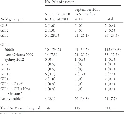
Pediatric Hospital during a 2-year period of surveillance (2010 to 2012)
NoV genotype
No. (%) of cases in:
September 2010 to August 2011
September 2011 to September
2012 Total
GI.8 2 (1.0) 0 (0) 2 (0.6)
GII.2 2 (1.0) 0 (0) 2 (0.6)
GII.3 54 (28.1) 31 (26.1) 85 (27.3)
GII.4
2006b 104 (54.2) 41 (34.5) 145 (46.6) New Orleans 2009 14 (7.3) 24 (20.2) 38 (12.2) Sydney 2012 0 (0) 1 (0.8) 1 (0.3)
GII.7 1 (0.5) 0 (0) 1 (0.3)
GII.12 1 (0.5) 0 (0) 1 (0.3)
GII.13 6 (3.1) 2 (1.7) 8 (2.6)
GII.16 2 (1.0) 0 (0) 2 (0.6)
GII.3⫹G1.8a 1 (0.5) 0 (0) 1 (0.3)
GII.3⫹GII.4 New Orleansa
1 (0.5) 0 (0) 1 (0.3)
Not typeableb 4 (2.1) 20 (16.8) 24 (7.7)
Total NoV samples typed 192 119 311
aMixed infection. b
Genotyping failed with RT-PCR amplification or sequencing.
[image:4.585.39.288.89.318.2]FIG 2Monthly distributions of NoV GII.4 and GII.3 genotypes in NoV-positive samples obtained from infants and children hospitalized with acute diarrhea at Thai Binh Pediatric Hospital, from September 2010 through September 2012.
TABLE 3Distribution of RV P and G genotypes at Thai Binh Pediatric Hospital, from September 2010 to September 2012
G genotype
No. with P genotype of:
P[8] P[4] P[6] P[9] NTa
G1 233 3 2 0 6
G2 0 8 1 0 1
G3 11 3 3 1 1
G4 1 0 13 0 0
G9 1 0 0 0 0
NTa 6 0 0 0 27
Total 252 14 19 1 35
a
NT, not typeable; these samples could not be genotyped.
on May 16, 2020 by guest
http://jcm.asm.org/
[image:4.585.114.470.519.697.2]S310D(N) (epitope F) were observed between GII.4 Sydney 2012 and New Orleans 2009. Substitutions in amino acids adjacent to these epitopes (N373H, P396H, and I413T) were also noted be-tween these two variants. Interestingly, despite the extensive se-quence variation among the three GII.4 variants, the amino acid motifs in the three HBGA binding sites described by Tan et al. (34) were conserved.
All Vietnamese GII.3 specimens analyzed fell within the GII.3c
[image:5.585.298.545.97.223.2]cluster (Fig. 5B). This cluster showed 3 to 5% amino acid sequence divergence from the GII.3b cluster. The amino acids at binding site positions 356 and 447 were conserved among GII.3 strains isolated from both nonsecretors and secretors. Interestingly, near the second binding site (386D), two amino acid variants (D385G and D385E) were found in strains from nonsecretors, whereas five of the six strains from secretors possessed the D385G substitution. TABLE 4Association between NoV infection and host HBGA types
among children hospitalized with acute diarrhea at Thai Binh Pediatric Hospital in Vietnam in 2012
NoV infection status
No.
Total A AB B O
Secretor (Leb⫹,
Ley⫹, or
H⫹)
Partial secretor (Lea⫹b⫹or
Lex⫹y⫹)
Nonsecretor (Lea⫹or
Lex⫹)
NoV-positive
GII.4 22 6 0 3 13 19 3 0
GII.3 28 6 1 1 20 12 11 5a
NoV-negative 210 49 12 34 115 127 57 26
Totalb 260 61 13 38 148 158 71 31
a
P⫽0.0588, compared with the NoV GII.4-infected group, Fisher’s exact test.
bAll children with paired stool-saliva samples were included.
[image:5.585.39.289.97.214.2]FIG 3Profiles of AB and Lewis antigen types in saliva samples from infants and children infected with NoV GII.4 (A) and GII.3 (B) genotypes. Each stacked bar represents a single case. Patient identification numbers are indicated below the bars. OD, optical density.
TABLE 5Association between RV infection and host HBGA types among children hospitalized with acute diarrhea at Thai Binh Pediatric Hospital in Vietnam in 2012
RV infection status
No.
Total A AB B O
Secretor (Leb⫹,
Ley⫹, or
H⫹)
Partial secretor (Lea⫹b⫹or
Lex⫹y⫹)
Nonsecretor (Lea⫹or
Lex⫹)
RV-positive
P[4] 5 1 1 2 1 4 1 0
P[6] 6 1 1 2 2 2 4 0
P[8] 74 20 7 16 31 51 23 0a
RV-negative 175 39 4 18 114 101 43 31
Totalb
260 61 13 38 148 158 71 31
aP⬍0.001, compared with the RV-negative group, Fisher’s exact test. b
All children with paired stool-saliva samples were included.
on May 16, 2020 by guest
http://jcm.asm.org/
[image:5.585.76.510.351.697.2]It remains unknown whether this residue is responsible for differ-ences in binding patterns for host HBGAs.
DISCUSSION
In this study, we document an association between HBGA secre-tor status in children and infection with specific RV P genotypes, a finding consistent with the recent observations that RV binds to HBGAs (10,19,20). It has been demonstrated that both P[4] and P[8] RVs recognize Leband H1-type antigens (19). The fact that
both P[4] and P[8] RVs were found only in HBGA secretors or partial secretors strongly suggests that Leband H1-type antigens
serve as host receptors or attachment factors for these two RV genotypes. Nonsecretors do not express Leband H1-type antigens,
due to a defect in theFUT2gene; they appear to be resistant to P[4] and P[8] RVs.
RV P[6] strains are genetically closely related to the P[4] and P[8] genotypes within the P[II] genogroup. In one of our previous studies, we showed that the VP8* domain of P[6] RVs recognizes only the H1-type antigen (19). In this study, all six P[6] cases involved HBGA secretors or partial secretors. This suggests that P[6] RVs preferentially infect secretors. In our previous binding assays, the H1-type antigen typically gave a weaker signal than most other HBGAs, due to the lower efficiency of the anti-H1 monoclonal antibody used. This can help explain the observation that only 2 saliva samples from P[6]-infected children were of the H1 phenotype. It is interesting that a saliva sample from one of the six P[6]-infected children gave weak signals for all HBGAs; only marginal positive findings were detected for the Leaand Lex
anti-gens, and Leblevels were detectable but below the cutoff value.
Thus, this individual was considered a partial secretor rather than a nonsecretor. A future study including more P[6]-infected chil-dren is needed to clarify whether P[6] has the ability to bind in nonsecretors.
The preferential infection of secretors by the P[4], P[8], and perhaps P[6] RV genotypes helps explain the predominance of those genotypes in cases of acute gastroenteritis around the world; because most (70 to 80%) of the people in the world are secretors (35,36), there are many susceptible hosts available to infect. Viet-nam is no exception. In the past 15 years, P[4] and P[8] strains have accounted for approximately 90% of RV gastroenteritis cases in children (37–39). Similar to findings reported from Vietnam’s RV surveillance program, which was initiated in 1998, we found that P[8] was the most common genotype, representing 79% of all RV cases. Of note, RV vaccines (Rotarix or RotaTeq) have been available in the private market only since 2007 (Rotarix) and 2009 (RotaTeq), and vaccine coverage rates among children 6 to 12 weeks of age have been less than 20% (L. T. Luan, personal com-munication). Secretor status also appears to be common in Viet-nam. In a separate study of HBGA types in Vietnamese children, 86.4% of children hospitalized with diarrhea were secretors, based onFUT2genotyping results (N. V. Trang, T. B. H. Vu, T. M. C. Nguyen, and D. D. Anh, unpublished data). It is interesting that the majority of GI and GII NoVs also recognize secretor HBGAs. The same mechanisms of selection and adaptation involving host HBGAs may be important in the evolution of both NoVs and RVs. In this study, two NoV genotypes, GII.4 and GII.3, were found FIG 4Profiles of ABH and Lewis antigens in saliva samples from infants and children infected with the three most common RV P genotypes, i.e., P[4] (5 cases) (A), P[6] (6 cases) (B), and P[8] (30 cases) (C). Patient identification numbers are indicated below the bars. OD, optical density.
on May 16, 2020 by guest
http://jcm.asm.org/
[image:6.585.77.511.63.375.2]to predominate, similar to the findings of a previous report on this region (40). Both genotypes primarily infected HBGA secretors, consistent with findings reported by many other groups (30,33). Collectively, these two genotypes represented 86.1% of the NoV cases in our study. Occasional infections of nonsecretors by NoV GII.4 and GII.3 strains have been reported (35,41). To our sur-prise, five children infected with strains belonging to the GII.3c cluster were nonsecretors. Experimental binding of GII.3 virus-like particles (VLPs), representing different strains from the GII.3 cluster (I, II, III, and NC), to the Lexantigen has been demon-strated (42). Thus, it is necessary to perform more genetic and surveillance studies to determine whether subsets of secretors and nonsecretors share HBGA epitopes for GII.3 or whether certain GII.3 sublineages have gained the ability to bind to both nonse-cretors and senonse-cretors.
We also documented genetic variation and functional site servation among major circulating NoV variants, which may con-tribute to our understanding of the evolution of NoVs. The NoV
GII.4 variants 2006b, New Orleans 2009, and Sydney 2012 emerged as major epidemic variants in Vietnam in different sea-sons, but the amino acids of the HBGA-binding interfaces were conserved across all three strains, including in all three binding sites (amino acids 344 to 347 [TRGH], 374D, and 441 to 443 [SG]) (43). This result is consistent with previous findings on the high levels of amino acid conservation found at the HBGA-binding interfaces among known GII.4 variants (15,34), indicating that strong selection pressure against genetic variation at these sites is imposed by human HBGAs. However, substitution at epitopes A, D, and F, as defined by Debbink et al. (33), and at amino acids adjacent to sites of interactions with HBGAs could affect the bind-ing affinity of the virus.
Among GII.3 NoV strains, there were no major changes in HBGA binding sites 1 (amino acids 357 to 360 [TRAH]), 2 (386D), or 3 (amino acids 448 to 450 [SSG]) between secretor-and nonsecretor-infecting variants. However, substitution of gly-cine by aspartic acid (G387E) adjacent to the second HBGA bind-FIG 5Sequences of HBGA binding sites in the VP1 capsid region of the GII.4 (A) and GII.3 (B) NoV genotypes.ⴱ, GII.3 strains from HBGA nonsecretor hosts. The conserved motifs in the HBGA binding sites (sites 1, 2, and 3, as described by Tan et al. [34]) are underlined. NoV GII.4 blocking epitopes, as described by Debbink et al. (33), are also illustrated.
on May 16, 2020 by guest
http://jcm.asm.org/
[image:7.585.46.540.68.473.2]ing site (amino acids TESG/E) was identified, which could be responsible for the nonsecretor-infecting phenotypes of the GII.3 NoVs. In addition, two amino acid variants (D385G and D385E) were found in strains from nonsecretors, whereas five of the six strains from secretors possessed the D385G substitution. It re-mains unknown whether this residue (adjacent to the HBGA binding site 386D) is responsible for differences in the HBGA binding patterns. Whether these substitutions are associated with the recently reported increase in NoV GII.3 strains in some re-gions (42,44,45) also remains unknown. A nonsecretor-infecting GII.3 variant in an outbreak in China was recently reported (41). RV surveillance in Vietnam began in 1998 and is ongoing. In our study, the majority (90%) of RV (and NoV) infections oc-curred in children younger than 2 years of age, similar to previous findings (39,40). Over the entire surveillance period, the most common genotype was G1P[8]. Strains belonging to G2P[4], G9, G5P[6], G3, and G12 appeared for short periods in 2000, 2001, 2003 to 2004, 2005, and 2008, respectively; however, the major P types are still P[8], P[4], and P[6], although a small number of P[9] strains have been documented in Vietnam (37,38,46,47). Recently, it has been shown that P[9] RVs bind type A antigen (10). Thus, HBGA binding could be a common factor for all hu-man RVs, and the [P] type could be a major determinant of RV host ranges.
Our study analyzed 260 paired stool-saliva samples, which rep-resented a subset of the total of 407 admissions during the second study period (from September 2011 through September 2012), The NoV and RV detection rates were lower in this subset of samples than in all admissions during this period. However, this reduction in detection rates is unlikely to alter our conclusions, which were based on the virus genotypes rather than virus-posi-tive detection rates.
Our findings on the distributions of different HBGA pheno-types and the relationships of HBGAs to NoV and RV infections in Vietnamese children are significant in terms of future disease con-trol. It is possible that vaccine responses by people with different HBGA types should be evaluated. For example, in a human vol-unteer challenge study, it was reported that nonsecretors were less likely to mount antibody responses upon infection, due to a lack of interactions between the NoV GII.4 virus and host HBGA recep-tors (13). The prevalence of the Lea⫹phenotype in Vietnamese children is similar to that reported for other countries from this region (48,49). The partial secretor (Lea⫹b⫹) phenotype observed in Asian populations is quite unique and might be associated with high levels of susceptibility to NoV. A study in Nicaragua showed an absence of this phenotype in both NoV-infected and control groups (50). In addition, that study found that greater propor-tions of secretors than nonsecretors developed antibodies to NoV and higher antibody titers were induced in secretors than in non-secretors, indicating that nonsecretors may develop less-robust immune responses following vaccination for the virus (50). In a study in Burkina Faso, which demonstrated an absence of Lea⫹b⫹ antigens and a high prevalence (32%) of Lea⫺b⫺antigens, a strong association between NoV infection and secretor status was iden-tified, with the exception of two cases of GII.4 and GII.7 infections in nonsecretors (35).
In conclusion, we found strong associations between infection by the predominant rotavirus and norovirus genotypes and HBGA secretor status in children. Our study could contribute to
future RV and NoV applications and vaccine strategy develop-ment.
ACKNOWLEDGMENTS
This study was sponsored by the National Foundation for Science and Technology Development (project 106.03-2010.56, to Nguyen Van Trang) and was also funded in part by grants from the National Institutes of Health and the Fogarty International Foundation (grants P01 HD13021 and 1R03TW009174-01 to Xi Jiang.). We thank Japanese Global Research in Infectious Diseases for financial support of the rotavi-rus analysis.
We thank Vega Everado (Centers for Disease Control and Prevention) for technical and scientific support. We also thank the children of Thai Binh Pediatric Hospital for participating in this study.
REFERENCES
1.Dennehy PH.2008. Rotavirus vaccines: an overview. Clin. Microbiol. Rev.21:198 –208.http://dx.doi.org/10.1128/CMR.00029-07.
2.Glass RI, Parashar UD, Estes MK.2009. Norovirus gastroenteritis. N. Engl. J. Med.361:1776 –1785.http://dx.doi.org/10.1056/NEJMra0804575. 3.Patel MM, Widdowson MA, Glass RI, Akazawa K, Vinje J, Parashar
UD.2008. Systematic literature review of role of noroviruses in sporadic gastroenteritis. Emerg. Infect. Dis. 14:1224 –1231.http://dx.doi.org/10 .3201/eid1408.071114.
4.Kawai K, O’Brien MA, Goveia MG, Mast TC, El Khoury AC. 2012. Burden of rotavirus gastroenteritis and distribution of rotavirus strains in Asia: a systematic review. Vaccine 30:1244 –1254.http://dx.doi.org/10 .1016/j.vaccine.2011.12.092.
5.Ogilvie I, Khoury H, El Khoury AC, Goetghebeur MM.2011. Burden of rotavirus gastroenteritis in the pediatric population in Central and Eastern Europe: serotype distribution and burden of illness. Hum. Vaccin.7:523– 533.http://dx.doi.org/10.4161/hv.7.5.14819.
6.Khoury H, Ogilvie I, El Khoury AC, Duan Y, Goetghebeur MM.2011. Burden of rotavirus gastroenteritis in the Middle Eastern and North Afri-can pediatric population. BMC Infect. Dis.11:9.http://dx.doi.org/10.1186 /1471-2334-11-9.
7.Hemming M, Rasanen S, Huhti L, Paloniemi M, Salminen M, Vesikari T.2013. Major reduction of rotavirus, but not norovirus, gastroenteritis in children seen in hospital after the introduction of RotaTeq vaccine into the National Immunization Programme in Finland. Eur. J. Pediatr.172:
739 –746.http://dx.doi.org/10.1007/s00431-013-1945-3.
8.Chen SY, Tsai CN, Chen CL, Chao SC, Lee YS, Lai MW, Chen CC, Huang WL, Chiu CH.2013. Severe viral gastroenteritis in children after sub-optimal rotavirus immunization in Taiwan. Pediatr. Infect. Dis. J.
32:1335–1339.http://dx.doi.org/10.1097/INF.0b013e3182a5f5b6. 9.Kroneman A, Vega E, Vennema H, Vinje J, White PA, Hansman G,
Green K, Martella V, Katayama K, Koopmans M.2013. Proposal for a unified norovirus nomenclature and genotyping. Arch. Virol.158:2059 – 2068.http://dx.doi.org/10.1007/s00705-013-1708-5.
10. Liu Y, Huang P, Tan M, Liu Y, Biesiada J, Meller J, Castello AA, Jiang B, Jiang X.2012. Rotavirus VP8*: phylogeny, host range, and interaction with histo-blood group antigens. J. Virol.86:9899 –9910.http://dx.doi.org /10.1128/JVI.00979-12.
11. Tan M, Jiang X. 2005. Norovirus and its histo-blood group antigen receptors: an answer to a historical puzzle. Trends Microbiol.13:285–293.
http://dx.doi.org/10.1016/j.tim.2005.04.004.
12. Lindesmith L, Moe C, Marionneau S, Ruvoen N, Jiang X, Lindblad L, Stewart P, LePendu J, Baric R.2003. Human susceptibility and resistance to Norwalk virus infection. Nat. Med.9:548 –553.http://dx.doi.org/10 .1038/nm860.
13. Frenck R, Bernstein DI, Xia M, Huang P, Zhong W, Parker S, Dickey M, McNeal M, Jiang X.2012. Predicting susceptibility to norovirus GII.4 by use of a challenge model involving humans. J. Infect. Dis.206:1386 – 1393.http://dx.doi.org/10.1093/infdis/jis514.
14. Tan M, Huang P, Meller J, Zhong W, Farkas T, Jiang X.2003. Muta-tions within the P2 domain of norovirus capsid affect binding to human histo-blood group antigens: evidence for a binding pocket. J. Virol.77:
12562–12571.http://dx.doi.org/10.1128/JVI.77.23.12562-12571.2003. 15. Chen Y, Tan M, Xia M, Hao N, Zhang XC, Huang P, Jiang X, Li X, Rao
Z.2011. Crystallography of a Lewis-binding norovirus, elucidation of
on May 16, 2020 by guest
http://jcm.asm.org/
strain-specificity to the polymorphic human histo-blood group antigens. PLoS Pathog. 7:e1002152. http://dx.doi.org/10.1371/journal.ppat.100 2152.
16. Hansman GS, Biertumpfel C, Georgiev I, McLellan JS, Chen L, Zhou T, Katayama K, Kwong PD.2011. Crystal structures of GII.10 and GII.12 norovirus protruding domains in complex with histo-blood group anti-gens reveal details for a potential site of vulnerability. J. Virol.85:6687– 6701.http://dx.doi.org/10.1128/JVI.00246-11.
17. Kuhlenschmidt TB, Hanafin WP, Gelberg HB, Kuhlenschmidt MS.
1999. Sialic acid dependence and independence of group A rotaviruses. Adv. Exp. Med. Biol.473:309 –317.http://dx.doi.org/10.1007/978-1-4615 -4143-1_33.
18. Isa P, Arias CF, Lopez S.2006. Role of sialic acids in rotavirus infection. Glycoconj. J.23:27–37.http://dx.doi.org/10.1007/s10719-006-5435-y. 19. Huang P, Xia M, Tan M, Zhong W, Wei C, Wang L, Morrow A, Jiang
X.2012. Spike protein VP8* of human rotavirus recognizes histo-blood group antigens in a type-specific manner. J. Virol.86:4833– 4843.http: //dx.doi.org/10.1128/JVI.05507-11.
20. Hu L, Crawford SE, Czako R, Cortes-Penfield NW, Smith DF, Le Pendu J, Estes MK, Prasad BV.2012. Cell attachment protein VP8* of a human rotavirus specifically interacts with A-type histo-blood group antigen. Na-ture485:256 –259.http://dx.doi.org/10.1038/nature10996.
21. General Statistics Office of Vietnam.2011. Results of the Vietnam house-hold living standards survey 2010. Statistical Publishing House, Hanoi, Vietnam.
22. Kageyama T, Kojima S, Shinohara M, Uchida K, Fukushi S, Hoshino FB, Takeda N, Katayama K.2003. Broadly reactive and highly sensitive assay for Norwalk-like viruses based on real-time quantitative reverse transcription-PCR. J. Clin. Microbiol.41:1548 –1557. http://dx.doi.org /10.1128/JCM.41.4.1548-1557.2003.
23. Vinje J, Hamidjaja RA, Sobsey MD.2004. Development and application of a capsid VP1 (region D) based reverse transcription PCR assay for genotyping of genogroup I and II noroviruses. J. Virol. Methods116:109 – 117.http://dx.doi.org/10.1016/j.jviromet.2003.11.001.
24. Kojima S, Kageyama T, Fukushi S, Hoshino FB, Shinohara M, Uchida K, Natori K, Takeda N, Katayama K.2002. Genogroup-specific PCR primers for detection of Norwalk-like viruses. J. Virol. Methods100:107– 114.http://dx.doi.org/10.1016/S0166-0934(01)00404-9.
25. Schreier E, Doring F, Kunkel U.2000. Molecular epidemiology of out-breaks of gastroenteritis associated with small round structured viruses in Germany in 1997/98. Arch. Virol.145(3):443– 453.http://dx.doi.org/10 .1007/s007050050038.
26. Tamura K, Peterson D, Peterson N, Stecher G, Nei M, Kumar S.2011. MEGA5: molecular evolutionary genetics analysis using maximum likeli-hood, evolutionary distance, and maximum parsimony methods. Mol. Biol. Evol.28:2731–2739.http://dx.doi.org/10.1093/molbev/msr121. 27. Gentsch JR, Glass RI, Woods P, Gouvea V, Gorziglia M, Flores J, Das
BK, Bhan MK.1992. Identification of group A rotavirus gene 4 types by polymerase chain reaction. J. Clin. Microbiol.30:1365–1373.
28. Gouvea V, Glass RI, Woods P, Taniguchi K, Clark HF, Forrester B, Fang ZY.1990. Polymerase chain reaction amplification and typing of rotavirus nucleic acid from stool specimens. J. Clin. Microbiol.28:
276 –282.
29. Huang P, Farkas T, Zhong W, Tan M, Thornton S, Morrow AL, Jiang X.2005. Norovirus and histo-blood group antigens: demonstration of a wide spectrum of strain specificities and classification of two major bind-ing groups among multiple bindbind-ing patterns. J. Virol.79:6714 – 6722.http: //dx.doi.org/10.1128/JVI.79.11.6714-6722.2005.
30. Tan M, Jin M, Xie H, Duan Z, Jiang X, Fang Z.2008. Outbreak studies of a GII-3 and a GII-4 norovirus revealed an association between HBGA phenotypes and viral infection. J. Med. Virol.80:1296 –1301.http://dx.doi .org/10.1002/jmv.21200.
31. Vega E, Vinje J.2011. Novel GII.12 norovirus strain, United States, 2009 –2010. Emerg. Infect. Dis.17:1516 –1518.http://dx.doi.org/10.3201 /eid1708.110025.
32. Vega E, Barclay L, Gregoricus N, Williams K, Lee D, Vinje J.2011. Novel surveillance network for norovirus gastroenteritis outbreaks, United States. Emerg. Infect. Dis.17:1389 –1395.
33. Debbink K, Donaldson EF, Lindesmith LC, Baric RS. 2012. Genetic mapping of a highly variable norovirus GII.4 blockade epitope: potential role in escape from human herd immunity. J. Virol.86:1214 –1226.http: //dx.doi.org/10.1128/JVI.06189-11.
34. Tan M, Xia M, Chen Y, Bu W, Hegde RS, Meller J, Li X, Jiang X.2009.
Conservation of carbohydrate binding interfaces: evidence of human HBGA selection in norovirus evolution. PLoS One4:e5058.http://dx.doi .org/10.1371/journal.pone.0005058.
35. Nordgren J, Nitiema LW, Ouermi D, Simpore J, Svensson L.2013. Host genetic factors affect susceptibility to norovirus infections in Burkina Faso. PLoS One8:e69557.http://dx.doi.org/10.1371/journal .pone.0069557.
36. Kelly RJ, Rouquier S, Giorgi D, Lennon GG, Lowe JB.1995. Sequence and expression of a candidate for the human Secretorblood group ␣(1,2)fucosyltransferase gene (FUT2): homozygosity for an enzyme-inactivating nonsense mutation commonly correlates with the non-secretor phenotype. J. Biol. Chem.270:4640 – 4649.
37. Van Man N, Luan le T, Trach DD, Thanh NT, Van Tu P, Long NT, Anh DD, Fischer TK, Ivanoff B, Gentsch JR, Glass RI.2005. Epidemiological profile and burden of rotavirus diarrhea in Vietnam: 5 years of sentinel hospital surveillance, 1998 –2003. J. Infect. Dis.192(Suppl 1):S127–S132. 38. Ngo TC, Nguyen BM, Dang DA, Nguyen HT, Nguyen TT, Tran VN, Vu TT, Ogino M, Alam MM, Nakagomi T, Nakagomi O, Yamashiro T.
2009. Molecular epidemiology of rotavirus diarrhoea among children in Haiphong, Vietnam: the emergence of G3 rotavirus. Vaccine27(Suppl 5):F75–F80.
39. Nguyen VM, Nguyen VT, Huynh PL, Dang DT, Nguyen TH, Phan VT, Nguyen TL, Le TL, Ivanoff B, Gentsch JR, Glass RI.2001. The epide-miology and disease burden of rotavirus in Vietnam: sentinel surveillance at 6 hospitals. J. Infect. Dis.183:1707–1712.http://dx.doi.org/10.1086 /320733.
40. Trang NV, Luan le T, Kim-Anh le T, Hau VT, Nhung le TH, Phasuk P, Setrabutr O, Shirley H, Vinje J, Anh DD, Mason CJ.2012. Detection and molecular characterization of noroviruses and sapoviruses in children ad-mitted to hospital with acute gastroenteritis in Vietnam. J. Med. Virol.
84:290 –297.http://dx.doi.org/10.1002/jmv.23185.
41. Jin M, He Y, Li H, Huang P, Zhong W, Yang H, Zhang H, Tan M, Duan ZJ.2013. Two gastroenteritis outbreaks caused by GII noroviruses: host susceptibility and HBGA phenotypes. PLoS One8:e58605.http://dx.doi .org/10.1371/journal.pone.0058605.
42. Boon D, Mahar JE, Abente EJ, Kirkwood CD, Purcell RH, Kapikian AZ, Green KY, Bok K.2011. Comparative evolution of GII.3 and GII.4 no-rovirus over a 31-year period. J. Virol.85:8656 – 8666.http://dx.doi.org/10 .1128/JVI.00472-11.
43. Tan M, Jiang X.2010. Norovirus gastroenteritis, carbohydrate receptors, and animal models. PLoS Pathog.6:e1000983.http://dx.doi.org/10.1371 /journal.ppat.1000983.
44. Mahar JE, Bok K, Green KY, Kirkwood CD.2013. The importance of intergenic recombination in norovirus GII.3 evolution. J. Virol.87:3687– 3698.http://dx.doi.org/10.1128/JVI.03056-12.
45. Stuart RL, Tan K, Mahar JE, Kirkwood CD, Ramsden CA, Andriano-poulos N, Jolley D, Bawden K, Doherty R, Kotsanas D, Bradford J, Buttery JP.2010. An outbreak of necrotizing enterocolitis associated with norovirus genotype GII.3. Pediatr. Infect. Dis. J.29:644 – 647.http://dx .doi.org/10.1097/INF.0b013e3181d824e1.
46. Ahmed K, Anh DD, Nakagomi O.2007. Rotavirus G5P[6] in child with diarrhea, Vietnam. Emerg. Infect. Dis.13:1232–1235.http://dx.doi.org/10 .3201/eid1308.061038.
47. Tra My PV, Rabaa MA, Vinh H, Holmes EC, Hoang NV, Vinh NT, Phuong le T, Tham NT, Bay PV, Campbell JI, Farrar J, Baker S.2011. The emergence of rotavirus G12 and the prevalence of enteric viruses in hospitalized pediatric diarrheal patients in southern Vietnam. Am. J. Trop. Med. Hyg.85:768 –775.http://dx.doi.org/10.4269/ajtmh.2011.11 -0364.
48. Chang JG, Ko YC, Lee JC, Chang SJ, Liu TC, Shih MC, Peng CT.2002. Molecular analysis of mutations and polymorphisms of the Lewis secretor type␣(1,2)-fucosyltransferase gene reveals that Taiwan aborigines are of Austronesian derivation. J. Hum. Genet.47:60 – 65.http://dx.doi.org/10 .1007/s100380200001.
49. Park KU, Song J, Han KS, Kim JQ.2005. The fusion allele of theFUT2
(secretor type␣(1,2)-fucosyltransferase) gene at a high frequency and a newse385allele in a Korean population. Ann. Hematol.84:656 – 660.http:
//dx.doi.org/10.1007/s00277-005-1041-5.
50. Bucardo F, Kindberg E, Paniagua M, Grahn A, Larson G, Vildevall M, Svensson L.2009. Genetic susceptibility to symptomatic norovirus infec-tion in Nicaragua. J. Med. Virol.81:728 –735.http://dx.doi.org/10.1002 /jmv.21426.


![FIG 4 Profiles of ABH and Lewis antigens in saliva samples from infants and children infected with the three most common RV P genotypes, i.e., P[4] (5 cases)(A), P[6] (6 cases) (B), and P[8] (30 cases) (C)](https://thumb-us.123doks.com/thumbv2/123dok_us/8288255.850352/6.585.77.511.63.375/proles-lewis-antigens-samples-infants-children-infected-genotypes.webp)

