Developmental Effects of Malathion Exposure on
Recognition Memory and Spatial Learning in
Males Wistar Rats
Pacôme Kouadio N’Go1*, Fatima-Zahra Azzaoui1, Porlo Rigobert Soro1, Majda Samih1, Ahmed Omar Touhami Ahami1, Mohamed Najimi2, Fatiha Chigr2
1Unit of Clinics and Cognitives Neurosciences, Laboratory of Biology and Health, Department of Biology,
Faculty of Sciences, University Ibn Tofail, Kénitra, Morocco
2Laboratory of Physiology Animal, Department of Biology, Faculty of Sciences and Technology,
University Sultan Moulay Slimane, Béni Mellal, Morocco Email: *[email protected]
Received March 25, 2013; revised April 26, 2013; accepted May 9, 2013
Copyright © 2013 Pacôme Kouadio N’Go et al. This is an open access article distributed under the Creative Commons Attribution License, which permits unrestricted use, distribution, and reproduction in any medium, provided the original work is properly cited.
ABSTRACT
Most cognitive effects of Organophosphate Pesticides (OP) are induced after exposure to parathion, chlorpyrifos and diazinon, which the usage has been restricted because of overt signs of their toxicities. In this study, we investigate whether developmental exposure to Malathion could impair spatial learning and recognition memory in male rats. Ani- mals exposed by intragastric route, from in utero to young adult stage, to incremental doses of Malathion dissolved in corn oil; 100, 200 and 300 mg/kg of body weight, and one control group are given corn oil. Then, cognitive and behave- ioral abilities are assessed using Barnes maze and object recognition memory task. Malathion administration at 300 mg/kg is toxic to pregnant dams, and pups are stillborns. Rats exposed to 200 mg/kg make a significant working mem- ory error, and require more time to find an escape box during the initial training phase of Barnes maze. However, fewer errors are made in rats exposed to 100 mg/kg. For reversal learning task, the high dose group shows great deficits in spatial strategy to locate the new position of the box. With respect to recognition task, both dose 100 and 200 mg/kg impair significant short-term (2 h after habituation phase) object recognition memory, but long-term (24 h after habitua-tion phase) recognihabitua-tion memory is intact in high dose group. The current study also reveals that all treatments induce high significant neocortex acetylcholinesterase (AChE) activity inhibition, but 100 mg/kg dose is not sufficient to dis- rupt great hippocampal activity alteration. These results suggest that developmental exposure to Malathion, despite low toxicity described, may induce late-emerging spatial learning and recognition memorialterations. Moreover, Cortical and hippocampal area that support strongly these behaviors remain sensitive to incremental doses of Malathion.
Keywords: AChE; Developmental Neurotoxicity; Malathion; Organophosphate Pesticide Recognition Memory; Spatial Learning
1. Introduction
Organophosphate insecticides (OP) are widely used in agriculture pest control in order to improve the quantity and quality of food production. However, they are also well-known as environmental contaminants occurred in crop products, water supplies, and in air. Thus, OPs ex-posure remains a factor of human hazard health, espe-cially in young children population [1-3]. The Malathion insecticide is one of the most used OP in United States and throughout the world. As evoked by Maroni et al.,
Malathion is used in agriculture, veterinary practice and as ectoparasiticide applied against human body lice [4]. From chemical name [S-1,2(bis-ethoxycarbonyl)ethylO, O-dimethyl phosphorodithioate], Malathion exerts, through bioactive form malaoxon, its primary effect by phosphorylating the serine residue at the active site of acetylcholinesterase (AChE) [5]. This leads to an inhibi- tion of this enzyme in neural tissues, and so resulting overstimulation of cholinergic synapse in central and pe- ripheral nervous system [6,7]. Although Malathion is considered as an OP relatively low acute toxicity, it could cause others high toxic OPs, a great risk for human
and environmental health. Several studies have examined the possible association between occupational exposure to pesticides and certain types of cancer, particularly non-Hodgkin’s lymphoma (NHL) and leukemia. It is difficult to establish association with specific pesticides because workers are exposed chronically to multiples compounds. However, exposure to Malathion was asso- ciated with increased risk of NHL in a study of men in Iowa and Minnesota [8], of women in Nebraska [9], and of men in Canada [10]. Moreover, in California, Mala- thion was the third most frequently reported pesticide and caused five times more occupational illness than the average pesticide [11]. Many biochemical and physio-logical effects of OP insecticide have been documented in man and animal. Malathion exposure has been shown to significantly decrease the sperm count of mice [12]. Aother study revealed that Malathion in subchronic ex-posure affects rat hepatic gluconeogenesis and glyco- genolysis as well as inducing hyperglycemia [13]. Fur- thermore, the main target of OPs insecticide is the nerv- ous system, and it has been suggested that chronic expo- sure to low-level of OP can interfere with normal neuro- development [14]. Clinical studies support the observa- tion that cognitive, neurobehavioral deficits, and neurol- ogy diseases occurre in agricultural communities using widely OPs insecticides [15,16]. In rats, previous study has shown that repeatedly exposure to Malathion induced depression-like behavior in Forced Swimming Test [17]. Acker, et al. [18] have also reported a decrease of motor coordination and muscular strength in rat pups exposed orally to Malathion, from post natal days (PND) 12 to 14. However, the most toxicology studies are focused on high toxic OP compounds such as Chlorpyrifos (CPF), Diazinon (DZN) and Methyl parathion (MPS). Their common effects on cholinergic system and its biomarker molecules have been demonstrated [19-21]. In addition, behavioral studies revealed that early postnatal exposure to CPF and MPS impaired memory functions [22,23]. In contrast, scarce studies in this sense have been done for Malathion compound.
The interest of the current study is to assess recogni- tion memory and spatial learning abilities in young male rats developmentally exposed to Malathion (i.e. from in utero through adolescence stage). We investigate also on AChE activity inhibition from hippocampal and neocor- tex area. Because, no studies have reported neurocogni- tive effects after developmental exposure to Malathion, discussion will made relative to high toxic OPs men- tioned.
2. Materials and Methods
2.1. ChemicalMalathion was obtained from commercial grade: Maly-
phos 50 (active ingredients 500 g by liter). The Mala- thion concentration (50% purity) in commercial grade was diluted in corn oil. Acetylthiocholine iodide, 5,5-di- thiobis-(2-nitrobenzoic acid) (DTNB) and others all che- micals were purchased from Sigma (USA).
2.2. Animals and Treatment
Twelve Wistar female rats, 4 months of age were ob- tained from a local breeding colony of Faculty of Sci- ences, Kenitra-Morocco. Rates were kept under standard condition, 12 h light/12 dark cycle, 20˚C ± 2˚C and 50% - 70% humidity). They had access to commercial diet (ALF SAHEL-Casablanca, Morocco) and tap water ad libitum. After 2 weeks of acclimation, virgin rates and one-non-pesticide exposed male were bred in propylene cage covered by stainless steel mesh (70 × 40 × 65 cm). On gestational days six (GD 6), females were randomly divided in four groups of treatment. Then, three groups received by intragastric gavage incremental doses of Malathion insecticide dissolved in corn oil; 100, 200 and 300 mg/kg of body weight per day (Mal 100, Mal 200, Mal 300), and one control group (VEH) was given corn oil daily. Gestating females were treated from GD6 though post-partum day 21, rat pups were so exposed to Malathion via their mothers. On PND 21, weaned off- springs of each experimental group were submitted to similar dose regimens of preliminary protocol, and dura- tion of treatment was extended to young adult stage (PND 45 - 60). Experimental procedures are also exam- ined and approved by the internal ethical committee for animal welfare.
2.3. Physical Parameter Measurements
Beginning on PND1, physical signs of toxicity and body weight were daily recorded during treatment.
2.4. Behavioral Assessment
On PND60, after Malathion treatment period, recognition memory, spatial and learning memory were tested using the Novel object recognition task and Barnes Maze.
2.4.1. Novel Object Recognition (NOR) Task
The apparatus and procedure has been described else- where [24,25]. The maze took place in open field box of polywood (50 × 50 × 40 cm3), illuminated with halogen
walls. In short-term memory (STM) test 2 h after famili- arization phase, the rat is allowed 5 min to explore the field with a familiar object (A) and novel object (B) at the same position. Long-term memory (LTM) study is performed 24 h after familiarization phase; rat is led to visit again the apparatus in presence of the same familiar object (A) and other novel (C). Single set of three objects (A, B, C) presented similar texture but distinctive in color, sharps and size. The arena and objects are cleaned with ethanol 70%, after each training session to remove the olfactory cues. The Recognition Index (RI) indicates the ratio of novel object exploration time to the total of both novel and familiar object exploration time. It is the main index of retention. The concept of exploration of an object is defined as directing the nose at distance ≤1 cm to the object, and/or touching it with the nose. In contrast, turning around, sitting and climbing on the object was not considered as exploratory behavior.
2.4.2. Barnes Circular Maze
Memory and spatial learning were assessed in rodents with Barnes circular maze [26]. The apparatus and pro- cedure adapted was a modification of Fedorova, et al. [27] and Greferath, et al. [28]. The maze consists of a white circular platform, 122 cm in diameter, raised to 50 cm above the floor level. The platform contains 18 holes (of 9 cm in diameter) evenly spaced around the circumfer- ence, but one of the holes was only connected to a re- movable escape box. Three visual cues on the walls, 50 cm around the maze, act as spatial cues. Each rat was preliminary given to one habituation session with the maze and existence of escape box, which the position remained constant throughout testing for a given animal. Habituation began by placing the rat in the center of the maze under a bucket in a room that was brightly lit (with halogen lamps of 500 W installed in the ceiling). After 10 s, the bucket was lifted and the rat was guided to lo- cate escape box, then the buzzer was turned off and rat was allowed to remain for 1 min. On following days the habituation phase, initial training were conducted during 4 consecutive days (2 trial per days), under the same con- dition above, except that the rats needed to locate the es- cape box by themselves. Each trial ended when the rat entered the goal tunnel or after 5min elapsed. There was a 3 min inter-trial interval for each animal. Three weeks after training phase, the rats were subjected to locate a new position of escape hole opposite to original one dur- ing 5 consecutive days; that for testing the reversal learn- ing ability. The maze was routinely cleaned with ethanol solution 20% after every trial to avoid olfactory cues. Trials were recorded with video track Logitech. Training was conducted using a working memory paradigm, that was evaluated by the total number of errors made and the escape latencies time associated [29,30]. With respect to
the reversal learning, we took also account the number of perseveration to explain deficits of news strategies elabo- ration. Perseverations were defined as repeat visits to the original hole or one of the two adjacent holes.
2.5. AChE Activity Assay
Rats are killed by decapitation 24 h after behavioral tests. Samples of brain correspondent to neocortex and hippo- campus areas are removed and homogenized in buffer Tris/HCl (50 mmol/L, pH 7.3) and Sucrose (0.32 mol/L). The homogenate is centrifuged at 1000 xg for 15 min at 4˚C. AChE activity is assayed according to Elman method [31], using acetylthiocholine iodide as a substrate. Reaction mixture contained 100 µL of supernatant, 4µL of substrate (75 mM), 15 µL of Dithiobisnitrobenzoic acid (DTNB as Ellman’s reagent, 100 mM) and 3.0 ml of phosphate buffer (pH 8.0). The rate of Acetylthiocholine iodide hydrolysis is measured at 412 nm in spectropho- tometer for 10 min. Brain AChE is expresses in percent of inhibition from control group [32].
2.6. Statistical Analysis
All data were expressed as means ±S.E.M (Standard Er- ror of Mean). Repeat measure and one-way analyses of variance (ANOVA) are used to assess difference between groups in physical and neurobehavior performances in- duced by developmental exposure to Malathion. Post hoc comparisons were made using Tukey’s HDS test, when appropriate. Statistical significant was assumed at p ˂
0.05.
3. Results
3.1. General Physical Observations
Malathion exposure at dosage 300 mg/kg (b.w) induced toxicity to pregnant dams, alls offsprings are stillborns during parturition. For others treated groups, no sign of toxicity such as body tremor, salivation, weakness and convulsion are observed. Repeated measure ANOVA shows a significant change in body weight gain between male rats study groups (p = 0.02). Tukey’s post hoc test reveals that body weight in both treated-groups, “Mal 100” and “Mal 200”, is significantly reduced (p ˂ 0.001) compared to VEH, but no difference is observed between them (data not shown).
3.2. Object Recognition Memory
ference observed is statistically significant [F(2,19) = 200, 38; p ˂ 0.001]. During LTM recognition testing, rats of Mal 100 group have enhanced their RI compared to that performed in STM phase. In contrast, the RI remains sensibly unchanged in rats of Mal 200 group, from STM to LTM session (Figure1).
3.3. Memory and Spatial Learning
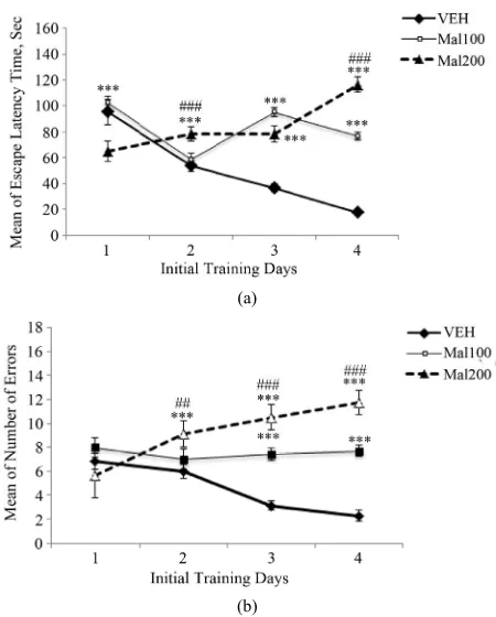
Developmental exposure to Malathion impairs signifi- cantly spatial memory in Barnes maze. During initial training period, rats of Mal 200 group perform better the first days than the others, as indicated by a reduction in latency time to find the escape box (Figure 2(a)). On 3 following days, the control rats learn the escape box po- sition faster than treated-groups. Repeated measures ANOVA shows the latencies time has increased signifi-cantly for Mal 100 and Mal 200 treated-rats [F(2,19) = 50.7; p ˂ 0.001], compared to VEH. However, no signifi- cant difference is observed between both treated groups (p > 0.05) (Figure 2(a)). The increase of latencies times in treated-groups is accompanied by significant working memory errors committed. In initial training phase, the number of errors increases during for 4 testing days, in Mal 200 group. But, errors made by Mal 100 group are remained sensibly constant during this period. Repeated measures ANOVA revealed a high significant effects of Malathion treatment on working memory [F(2,19) = 197. 80; p ˂ 0.001]. Tukey’s post hoc test confirms that these effects are significant in Malathion treated-groups (p ˂
0.001), compared to control group. It also indicates that working memory impairment is more pronounced in Mal 200 group (Figure 2(b)).
[image:4.595.313.533.84.243.2]With respect to reversal learning phase, the Malathion treated-rats express again more difficulties to learn a new position of the escape box, opposite to original one. Rats of each study group began the firth trial day by taking similar time to find rapidly the escape box. But, we ob- serve high significant increase of latencies time during the second, third and fourth days in treated-groups [F(2, 19) = 71, 58; p ˂ 0.001]. Tukey’s post hoc test detects that control group are required lesser time to find the box than treated-groups (p ˂ 0.001). However, rats of “Mal 100” group have enhanced significantly their learning abilities compared to those of “Mal 200” (p ˂ 0.01) (Fig- ure 3(a)). As working memory errors made in initial training phase, more errors are occurred in treated-groups compared to control group. There are an increasing trend of number of errors committed in reversal learning phase with the regimen dose [F(2,19) = 39, 00; p ˂ 0.001]. Al-though, both treated-groups displayed similar number of errors on trial day one, the number of errors was sig- nificantly different in mal 100 group and compared to that of Mal 200, on following days (p ˂ 0.001) (Figure 3(b)). Moreover, latency time and rate of errors number
Figure 1. Effect of developmental neurotoxicity to Mala- thion on object recognition memory (Mal100 and Mal200 represent 100 and 200 mg/kg; p.o, respectively. Data ex-press percent of object recognition index as means ± S.E.M of 7 - 8 animals per group. STM, Short Term Memory rec-ognition (2 h after habituation phase); LTM, Long Term Memory recognition (24 h after habituation phase). ***
p˂ 0.001, comparison between exposed groups and VEH; ###
p˂ 0.001 as comparison between Mal 100 and Mal 200 groups (One-way ANOVA/Tukey’s post-hoc analysis).
(a)
(b)
Figure 2. Initial training phase in Barnes maze. (a) Escape latencies times. (b) Number of Errors committed. Value are expressed in Means ± S.E.M of 7 - 8 animals per group ac-tivities (Mal100 and Mal200 represent 100 and 200 mg/kg; p.o, respectively). Developmental exposure to Malathion induced significant effects on initial spatial learning. ***
p˂ 0.001 as comparison to VEH. ##
[image:4.595.311.536.358.638.2]elevated in reversal learning have associated to high number of perseveration, in treated-group (Figure 3(c)). Repeated measure ANOVA shows a significant differ- ence in number of perseveration between all groups [F(2, 19) = 10, 85; p ˂ 0.001)]. Tukey’s post hoc testde- tectsthat the perseveration behavior increased signifi- cantly in “Mal 100” and “Mal 200” groups (p ˂ 0.001)
(a)
(b)
[image:5.595.58.289.177.614.2](c)
Figure 3. Reversal learning phase in Barnes maze. (a) Es-cape latencies times Latency time. (b) Number of Errors committed. (c) Number of Perseverations. Values are ex-pressed as Means ± S.E.M of 7 - 8 animals per group activi-ties (Mal100 and Mal200 represent 100, 200 mg/kg; p.o, respectively). Developmental exposure to Malathion in-duced significant effects on reversal spatial learning. **p˂ 0.01, ***
p˂ 0.001 as compared to VEH; #p˂ 0.05, # #p˂ 0.01; ###p ˂ 0.001 as comparison between Mal100 and Mal200 groups (Repeated measures ANOVA/Tukey’s post-hoc ana- lysis).
compared to VEH, but no difference between them (Fig- ure 3(c)).
3.4. AChE Activity Assays
Developmental exposure of Malathion to doses 100 and 200 mg/kg increases significantly neocortex AChE inhi- bition activity; +26% (p ˂ 0.001) and +46% (p ˂ 0.001) respectively, relative to control. Turkey post-hoc analysis detected that there is also great statistical difference be- tween Mal 100 and Mal 200 groups (p ˂ 0.01). We also found a significant AChE activity inhibition in hippo- campus +36% for “Mal 200” group (p ˂ 0.001) and + 20 for Mal 100 group (p ˂ 0.01), compared to the control group. However, a significant change of AChE activity is observed in Mal 200, relative to Mal 100 (p ˂ 0.01), (Figure 4).
4. Discussion
In current study, except lethal dose of 300 mg/kg, our result indicates that developmental exposure to Mala- thion does not induce general signs of systemic toxicity as weakness, tremor or convulsion. However, doses of 100 and 200 mg/kg affected body weight in treated-rats significantly compared to control. Current outcome is consistent with that of past study showing significant reduction of body weight in young rats exposed to para- thion (potential OP toxic) at PND8-20 [33]. This could probably due to the effect of Malathion as others OPs, which are caused cholinergic over stimulation, followed
Figure 4. Effect of Malathion exposure on cerebral cortex and Hippocampus AChE activity inhibition (Mal100 and Mal200 group receive 100 and 200 mg/kg; p.o, respectively). Data expressed percent of AChE change from VEH (Mean± S.E.M of 7 - 8 animals per sex’s group). **
p˂ 0.01 and ***p˂ 0.001 as compared to VEH; ##
[image:5.595.312.536.458.644.2]by an increase of gastric motility and reduction of diges-tive tract absorption [34]. In contrast, recent study has re- vealed no weight loss in rats developmentally exposed (PND11-14) to the same dose of Malathion to ours [18]. These findings suggest that prenatal phase could be a sensitive period to developmental effects of Malathion on physical appearance.
Furthermore, the current experiment has shown that developmental exposure of Malathion disrupts recogni- tion memory forming. In STM recognition task, both treated groups have spent lesser times to explore novel object than the control group. However, rats of Mal 200 group are significantly affected during long-term object recognition memory phase, relative to those of Mal 100 group. The under performances in rats receiving 200 mg/kg of Malathion are associated with high hippocam-pal AChE activity inhibition, evaluated at young adult stage. That reinforces the hypothesis that cerebral AChE inhibition caused by Malathion in rat pups may induce cognitive impairments, later in life. In fact, the object recognition test is based on the discrimination between familiar and novel stimulus and natural performance of subject who need to respond to “what” stimulus was used in experiment previously. Hippocampus plays so a piv-otal role in encoding and consolidation of novel stimulus; this process leads to integration and reorganization of the already formed memories. Thereby, when an object is previously encountered and reactivated later, the hippo- campus allows the discrimination between the old and novel object, and followed to the natural tendency of novelty preference [35]. In addition, the recognition in-dex is significantly reduced during long-term phase evaluation, in group treated with 200 mg/kg. This index reflects the main index of object’s recollection in the sense of familiarity with the feature of that particular stimulus. According to Reger, et al. [36], hippocampus is involved in long-term object recognition. Thus, the long-lasting object recognition disability could be due to the significant alteration of hippocampus AChE activity. Our findings are consistent with a recent study that re-vealed the impairment of hippocampus-dependent object recognition in neonatal rats developmentally exposed to diazinon OP [37]. Besides, short-term object recognition has been linked to other medial temporal area as the per-irhinal cortex [38]. Our results support that both level of 100 and 200 mg/kg induce significant deficits of object recognition memory, in rats evaluated 2 h after training phase. That leads to suggest that perirhinal cortex was inherently more vulnerable to developmental exposure of Malathion. In fact, perirhinal cortex is the first forebrain site of several environment information’s entrance as vis- ual, olfactory, and somatosensory stimulus, in which hippocampus receives inputs [39]. The important cho- linergic transmission within perirhinal cortex also seems
underperformances could be explained by Qiao, et al. study which reported evidences of synaptic cholinergic defects and more hippocampal cell biomarkers alteration, in neonatal rats exposed to CPF [54]. It has been also suggested that OP including Malathion exposure may change neuronal connectivity in the developing brain [52]. Previous study showed that Malathion exposure in- duces decrement effect on dendritic morphology of hip- pocampal CA1 neurons [55]. Thus, the reduction of neu- rogenesis rate could be a major explanation of disrup- tion of spatial memory consolidation, as evoked by Sark- isyan, et al [56]. We find also that Malathion level at 200 mg/kg appeared to produce greater deficits in working memory than did 100 mg/kg. Our outcomes are consis- tent with previous researches that reported only high ex- posure OP including CPF induced impairment of work- ing memory in male rats [22,57]. In animals exposed to low level of Malathion, the hippocampal AChE inhibi- tion is slower than the rats exposed to high dose. How- ever, they are performed significantly worse than control. That may suggest that other non-cholinergic mecha- nisms are involved in initial spatial learning.
After 3 weeks from initial training days, we have as- sessed rat’s ability to learn a new location of the escape, in reversal learning task. This task requires learning new search strategies by suppressing the execution of the pre- vious learning. It also evaluates also adaptive behaviors of rodents in novel environmental situation. In current study, rats developmentally exposed to both levels of Malathion have expressed great difficulties to learn new strategies (Figures 3(a) and 3(b)). That is associated to a number of errors and perseveration significantly elevated, relative to controls. However, the rats treated with low level of Malathion have performed better. In reversal learning task, spatial strategies do not require only in-volve menthippocampus functional integrity, but also prefrontal cortex and nucleus accumbens [58,59]. Pre-frontal area is an important component of Pre-frontal lobe involved in conception, choice and execution of a plan- ning. In Barnes maze, the selection of news spatial stra- tegies are based on spatial memory that requires control executive processes like attention. But, the accumbens nucleus has been identified as a critical site in the neu- ronal circuit controlling motivation and mood [60], and mediating adaptive behaviors in rats [61]. In current study, we have found that developmental exposure to Malathion elicited significant cholinergic activity chan- ges in cerebral cortex area. Aother study finds similar finding in frontal cortex in males rats exposed to DZN [62]. In additional, Campaña, et al. [55] reported evi-dences of significant decrement on dendritic morphology in prefrontal cortex neurons (PFC), also on the dendritic spine density from nucleus accumbens, in mice exposed to Malathion. The inactivation of PFC lead to impairment
of reversal learning in the rats when they ceased the us- ing of previously acquired responses, as described in visual discrimination reversal learning task [63]. Besides, other authors have reported the reduction in dopaminer- gic transmission system in ventral striatum produced reversal learning deficits. In fact, accumbens nucleus is a component of striatum area that is implicated in reward, motivation (ventral part), motor skills and cognitive con- trol (dorsal part), specifically in learning of stimulus- response association [64,65]. Repeat exposure to para- quat OP is well known to trigger neurodegeneration of dopaminergic system in nigra substantial, the principal provider of dopamine in striatum [66].
From these current findings, developmental exposure to Malathion leads to spatial learning and object recogni- tion memory impairments, in male rats’ studies. The be- havioral deficits could be explained by brain AChE ac- tivity disruption induced in treatment groups. This ani- mal model’s study may reinforce the hypothesis of rela- tionship between environmental toxicant and neurode- generative disease occurrence in agricultural communi- ties. However, some immunohistochemistry investiga- tions are required to elucidate further results obtained.
5. Acknowledgements
This study was supported by GDRI Neuro and N£uro- med consortium.
REFERENCES
[1] B. Eskenazi, A. R. Marks, A. Bradman, K. Harley, D. B. Barr, C. Johnson, et al., “Organophosphate Pesticide Ex- posure and Neurodevelopment in Young Mexican-Ame- rican Children,” Environmental Health Perspectives, Vol. 115, No. 5, 2007, pp. 792-798.
[2] C. Lu, K. Toepel, R. Irish, R. A. Fenske, D. B. Barr and R. Bravo, “Organic Diets Significantly Lower Children’s Dietary Exposure to Organophosphorus Pesticides,” En- vironmental Health Perspectives, Vol. 114, No. 2, 2006, pp. 260-263.
[3] M. K. Morgan, L. S. Sheldon, C. W. Croghan, P. A. Jones, G. L. Robertson, J. C. Chuang, N. K. Wilson and C. W. Lyu, “Exposures of Preschool Children to Chlorpyrifos and Its Degradation Product 3,5,6-trichloro-2-pyridinol in Their Everyday Environments,” Journal of Exposure Analysis and Environmental Epidemiology, Vol. 15, No. 4, 2005, pp. 297-309.
[4] M. Maroni, C. Colosio, A. Ferioli and A. Fait, “Biologi- cal Monitoring of Pesticide Exposure: A Review. Intro- duction,” Toxicology, Vol. 143, No. 1, 2000, pp. 1-118. [5] D. J. Ecobichon, “Pesticides and Neurological Diseases,”
CRC Press, Boca Raton, 1994, p. 381.
[6] L. M. Wang, W. H. Ye, S. S. Zhou, K. D. Lin, M. R. Zhao and W. P. Liu, “Acute and Chronic Toxicity of Or- ganophosphate Monocrotophos to Daphnia Magna,”
Vol. 44, No. 1, 2009, pp. 38-43.
[7] T. C. Kwong, “Organophosphate Pesticides: Biochemis- try and Clinical Toxicology,” Therapeutic Drug Monitor- ing, Vol. 24, No. 1, 2002, pp. 144-149.
doi:10.1097/00007691-200202000-00022
[8] K. P. Cantor, A. Blair, G. Everett, et al., “Pesticides and Other Agricultural Risk Factors for Non-Hodgkin’s Lym- phoma among Men in Iowa and Minnesota,” Cancer Re- search, Vol. 52, No. 9, 1992, pp. 2447-2455.
[9] S. H. Zahm, D. D. Weisenburger, R. C. Saal, et al., “The Role of Agricultural Pesticide Use in the Development of Non-Hodgkin’s Lymphoma in Women,” Archives of En-vironmental Health, Vol. 48, No. 5, 1993, pp. 353-358. doi:10.1080/00039896.1993.9936725
[10] H. H. McDuffie, P. Pahwa, J. R. McLaughlin, et al., “Non-Hodgkin’s Lymphoma and Specific Pesticide Ex-posures in Men: Cross-Canada Study of Pesticides and Health,” Cancer Epidemiology, Biomarkers & Prevention, Vol. 10, No. 11, 2001, pp. 1155-1163.
[11] L. Brenner, “Malathion,” Journal of Pesticide Reform, Vol. 12, 1992, p. 29
[12] E. Bustos-Obregón and P. Gonzáles-Hormazabal, “Effect of a Single Dose of Malathion on Spermatogenesis in Mice,” Asian Journal of Andrology, Vol. 5, No. 2, 2003, pp. 105-107.
[13] M. Abdollahi, M. Donyavi, S. Pournourmohammadi and M. Saadat, “Hyperglycemia Associated with Increased Hepatic Glycogen Phosphorylase and Phosphoenolpyru- vate Carboxykinase Activities in Rats Following Sub- chronic Exposure to Malathion,” Comparative Biochem-istry and Physiology Part C: Pharmacology, Toxicology and Endocrinology, Vol. 137, No. 4, 2004, pp. 343-347. [14] T. A. Slotkin, “Developmental Cholinotoxicants: Nico-
tine and Chlorpyrifos,” Environmental Health Perspec- tives, Vol. 107, Suppl. 1, 1999, pp. 71-80.
[15] P. Z. Ruckart, K. Kakolewski, F. J. Bove and W. E. Kaye, “Long-Term Neurobehavioral Health Effects of Methyl Nparathion Exposure in Children in Mississippi and Ohio,” Environmental Health Perspectives, Vol. 112, No. 1, 2004, pp. 46-51. doi:10.1289/ehp.6430
[16] Sanborn, Margaret, et al., “Systematic Review of Pesti-cide Human Health Effects,” Ontario College of Family Physicians Toronto, Toronto, 23 April 2004.
[17] F. L. Assini, K. D. Zanette, P. S. Brocardo, P. Pandolfo, A. L. Rodrigues and R. N. Takahashi, “Behavioral Effects and ChE Measures after Acute and Repeated Administra- tion of Malathion in Rats,” Environmental Health Per- spectives, Vol. 20, No. 3, 2005, pp. 443-449.
doi:10.1016/j.etap.2005.05.007
[18] C. I. Acker, A. C. Souza, S. Pinton, J. T. Da Rocha, C. A. Friggi, R. Zanella and C. W. Nogueira, “Repeated Ma- lathion Exposure Induces Behavioral Impairment and AChE Activity Inhibition in Brains of Rat Pups,” Eco- toxicology and Environmental Safety, Vol. 74, No. 8, 2011, pp. 2310-2315. doi:10.1016/j.ecoenv.2011.07.035 [19] T. A. Slotkin, T. L. Lassiter, I. T. Ryde, N. Wrench, E. D.
Levin and F. J. Seidler, “Consumption of a High-Fat Diet in Adulthood Ameliorates the Effects of Neonatal Para-
thion Exposure on Acetylcholine Systems in Rat Brain Regions,” Environmental Health Perspectives, Vol. 117, No. 6, 2009, pp. 916-922.
[20] S. X. Guo-Ross, , J. E. Chambers, , E. C. Meek and R. L. Carr, “Altered Muscarinic Acetylcholine Receptor Sub- type Binding in Neonatal Rat Brain Following Exposure to Chlorpyrifos or Methyl Parathion,” Toxicological Sci- ences, Vol. 100, No. 1, 2007, pp. 118-127.
doi:10.1093/toxsci/kfm195
[21] J. Tang, R. L. Carr and J. E. Chambers, “The Effects of Repeated Oral Exposures to Methyl Parathion on Rat Brain Cholinesterase and Muscarinic Receptors during Postnatal Development,” Toxicological Sciences, Vol. 76, No. 2, 2003, pp. 400-406. doi:10.1093/toxsci/kfg245 [22] F. O.Johnson, J. E. Chambers, C. A. Nail, S. Givaruang-
sawat and R. L. Carr, “Developmental Chlorpyrifos and Methyl Parathion Exposure Alters Radial Arm Maze Performance in Juvenile and Adult Rats,” Toxicological Sciences, Vol. 109, No. 1, 2009, pp. 132-142.
doi:10.1093/toxsci/kfp053
[23] L. M. Icenogle, N. C. Christopher, W. P. Blackwelder, D. P. Caldwell, D. Qiao, F. J. Seidler, T. A. Slotkin and E. D. Levin, “Behavioral Alterations in Adolescent and Adult Rats Caused by a Brief Subtoxic Exposure to Chlorpyri- fos during Neurulation,” Neurotoxicology and Teratology, Vol. 26, No. 1, 2004, pp. 95-101.
doi:10.1016/j.ntt.2003.09.001
[24] A. Ennaceur, “One-Trial Object Recognition in Rats and Mice: Methodological and Theoretical Issues,” Behav- ioural Brain Research, Vol. 215, No. 2, 2010, pp. 244- 254.
[25] A. Ennaceur and J. Delacour, “A New One-Trial Test for Neurobiological Studies of Memory in Rats. 1: Behav- ioral Data,” Behavioural Brain Research, Vol. 31, No. 1, 1988, pp. 47-59 doi:10.1016/0166-4328(88)90157-X [26] C. A. Barnes, “Memory Deficits Associated with Senes-
cence: A Neurophysiological and Behavioral Study in the Rat,” Journal of Comparative & Physiological Psychol- ogy, Vol. 93, No. 1, 1979, pp. 74-104.
[27] I. Fedorova, N. Hussein, M.H. Baumann, C. Di Martino and N. Salem, Jr. “An n-3 Fatty Acid Deficiency Impairs Rat Spatial Learning in the Barnes Maze,” Behav Neuro-sci. Vol. 123, No. 1, 2009, pp. 196-205.
doi:10.1037/a0013801
[28] U. Greferath, A. Bennie, A. Kourakis and G. L. Barrett, “Impaired Spatial Learning in Aged Rats Is Associated with Loss of p75-Positive Neurons in the Basal Fore- brain,” Neuroscience, Vol. 100, No. 2, 2000, pp. 363-373. doi:10.1016/S0306-4522(00)00260-8
[29] J. M. Daniel, A. J. Fader, A. L. Spencer and G. P. Do-hanich, “Estrogen Enhances Performance of Female Rats during Acquisition of a Radial Arm Maze,” Hormones and Behavior, Vol. 32, No. 3, 1997, pp. 217-225. [30] D. S. Olton, “The Radial Arm Maze as a Tool in Behav-
ioral Pharmacology,” Physiology & Behavior, Vol. 40, No. 6, 1987, pp. 793-797.
doi:10.1016/0031-9384(87)90286-1
Featherstone, “A New and Rapid Colorimetric Determi- nation of Acetylcholinesterase Activity,” Biochemical Pharmacology, Vol. 7, No. 2, 1961, pp. 88-95.
doi:10.1016/0006-2952(61)90145-9
[32] F.-Z. Azzaoui, H. Hami, M. El-Hioui, S. Boulbaroud and A. Ahami “Attempt at the Determination of Aluminum Nitrate LD50 and the Study of Its Neurotoxicological Ef- fect in Wistar Rat,” Biology and Medicine, Vol. 4, No. 2, 2012, pp. 89-94.
[33] H. R. Santos, W. M. Cintra, Y. Aracava, C. M. Maciel, N. G. Castro, E. X. Albuquerque, “Spine Density and Den- dritic Branching Pattern of Hippocampal CA1 Pyramidal Neurons in Neonatal Rats Chronically Exposed to the Organophosphate Paraoxon,” Neurotoxicology, Vol. 25, No. 3, 2004, pp. 481-494.
[34] A. L. Jones and L. Karalliedde, “Poisoning,” In: N. A. Boon, N. R. Colledge, S. S. Davidson and B. R. Walker, Eds., Davidson’s Principles and Practice of Medicine, 20th Edition, Churchill Livingstone, Edinburgh, 2006, pp. 203-226.
[35] M. Antunes and G. Biala. “The Novel Objects Recogni- tion Memory: Neurobiology, Test Procedure, and Its Mo- difications,” Cognitive Processing, Vol. 13, No. 2, 2012, pp. 93-110. doi:10.1007/s10339-011-0430-z
[36] M. L. Reger, D. A. Hovda and C. C. Giza, “Ontogeny of Rat Recognition Memory Measured by the Novel Object Recognition Task,” Developmental Psychobiology, Vol. 51, No. 8, 2009, pp. 672-678. doi:10.1002/dev.20402 [37] T. T. Win-Shwe, D. Nakajima, S. Ahmed and H. Fuji-
maki, “Impairment of Novel Object Recognition in Adulthood after Neonatal Exposure to Diazinon,” Ar- chives of Toxicology, Vol. 87, No. 4, 2012, pp. 753-762. [38] R. S. Hammond, L. E. Tull and R. W. Stackman, “On the
Delay-Dependent Involvement of the Hippocampus in Object Recognition Memory,” Neurobiology of Learning and Memory, Vol. 82, No. 1, 2004, pp. 26-34.
doi:10.1016/j.nlm.2004.03.005
[39] J. R. Clarke, M. Cammarota, A. Gruart, I. Izquierdo and J. M. Delgado-Garcia, “Plastic Modifications Induced by Object Recognition,” Proceedings of the National Academy of Sciences of the United States of America, Vol. 107, No. 6, 2010, pp. 2652-2657.
doi:10.1073/pnas.0915059107
[40] B. D. Winters and T. J. Bussey, “Removal of Cholinergic Input to Perirhinal Cortex Disrupts Object Recognition but Not Spatial Working Memory in the Rat,” European Journal of Neuroscience, Vol. 21, No. 8, 2005, pp. 2263- 2270.doi:10.1111/j.1460-9568.2005.04055.x
[41] C. F. Hohmann, “A Morphogenetic Role for Acetylcho- line in Mouse Cerebral Neocortex,” Neuroscience &
Biobehavioral Reviews, Vol. 27, No. 4, 2003, pp. 351-363. doi:10.1016/S0149-7634(03)00066-6
[42] J. Yanai, “Neurobehavioral Teratology,” Elsevier, Am- sterdam, 1984.
[43] T. A. Slotkin, “Cholinergic Systems in Brain Develop- ment and Disruption by Neurotoxicants: Nicotine, Envi- ronmental Tobacco Smoke, Organophosphates,” Toxi- cology and Applied Pharmacology, Vol. 198, No. 2, 2004,
pp. 132-151. doi:10.1016/j.taap.2003.06.001
[44] B. Eskenazi, A. Bradman and R. Castorina, “Exposures of Children to Organophosphate Pesticides and Their Po- tential Adverse Health Effects,” Environmental Health Perspectives, Vol. 107, Suppl. 3, 1999, pp. 409-419. doi:10.1289/ehp.99107s3409
[45] G. Sarkisyan and P. B. Hedlund, “The 5-HT7 Receptor Is Involved in Allocentric Spatial Memory Information Processing,” Behavioural Brain Research, Vol. 202, No. 1, 2009, pp. 26-31. doi:10.1016/j.bbr.2009.03.011 [46] T. A. Slotkin and F. J. Seidler, “Developmental Exposure
to Terbutaline and Chlorpyrifos, Separately or Sequen- tially, Elicits Presynaptic Serotonergic Hyperactivity in Juvenile and Adolescent Rats,” Brain Research Bulletin, Vol. 73, No. 4-6, 2007, pp. 301-309.
doi:10.1016/j.brainresbull.2007.04.004
[47] J. E. Aldridge, E. D. Levin, F. J. Seidler and T. A. Slotkin, “Developmental Exposure of Rats to Chlorpyrifos Leads to Behavioral Alterations in Adulthood, involving Sero- tonergic Mechanisms and Resembling Animal Models of Depression,” Environmental Health Perspectives, Vol. 113, No. 5, 2005, pp. 527-531.
[48] G. Koopmans, A. Blokland, P. van Nieuwenhuijzen and J. Prickaerts, “Assessment of Spatial Learning Abilities of Mice in a New Circular Maze,” Physiology & Behavior, Vol. 79, No. 4-5, 2003, pp. 683-693.
doi:10.1016/S0031-9384(03)00171-9
[49] T. Nakashiba, D. L. Buhl, T. J. McHugh and S. To- negawa, “Hippocampal CA3 Output is Crucial for Rip- ple-Associated Reactivation and Consolidation of Mem- ory,” Neuron, Vol. 62, No. 6, 2009, pp. 781-787.
[50] H. Eichenbaum, “Cortico-Hippocampal System for De- clarative Memory,” Nature Reviews Neuroscience, Vol. 1, No. 1, 2000, pp. 41-50.
[51] S. Brimijoin and C. Koenigsberger, “Cholinesterases in Neural Development: New Findings and Toxicologic Im- plications,” Environmental Health Perspectives, Vol. 107, Suppl. 1, 1999, pp. 59-64.
[52] J. W. Bigbee, K. V. Sharma, J. J. Gupta and J. L. Dupree, “Morphogenic Role for Acetylcholinesterase in Axonal Outgrowth during Neural Development,” Environmental Health Perspectives, Vol. 107, Suppl. 1, 1999, pp. 81-87. [53] M. Grifman, N. Galyam, S. Seidman and H. Soreq,
“Functional Redundancy of Acetylcholinesterase and Neuroligin in Mammalian Neuritogenesis,” Proceedings of the National Academy of Sciences of the United States of America, Vol. 95, No. 23, 1998, pp. 13935-13940. doi:10.1073/pnas.95.23.13935
[54] D. Qiao, F. J. Seidler, Y. Abreu-Villaça, C. A. Tate, M. M. Cousins and T. A. Slotkin, “Chlorpyrifos Exposure during Neurulation: Cholinergic Synaptic Dysfunction and Cellular Alterations in Brain Regions at Adolescence and Adulthood,” Brain Research. Developmental Brain Research, Vol. 148, No. 1, 2004, pp. 43-52.
Altered in Adult Male Mice Exposed to Repeated Low Dose of Malathion,” Synapse, Vol. 64, No. 4, 2008, pp. 283-290. doi:10.1002/syn.20494
[56] G. Sarkisyan and P. B. Hedlund, “The 5-HT7 Receptor Is Involved in Allocentric Spatial Memory Information Processing,” Behavioural Brain Research, Vol. 202, No. 1, 2009, pp. 26-31.doi:10.1016/j.bbr.2009.03.011 [57] E. D. Levin, N. Addy, A. Nakajima, N. C. Christopher, F.
J. Seidler and T. A. Slotkin, “Persistent Behavioral Con- sequences of Neonatal Chlorpyrifos Exposure in Rats,”
Developmental Brain Research, Vol. 130, No. 1, 2001, pp. 83-89. doi:10.1016/S0165-3806(01)00215-2
[58] R. P. Kesner, “Subregional Analysis of Mnemonic Func- tions of the Prefrontal Cortex in the Rat,” Psychobiology, Vol. 28, No. 2, 2000, pp. 219-228.
[59] A. Louilot, K. Taghzouti, H. Simon and M. Le Moal, “Limbic System, Basal Ganglia, and Dopaminergic Neu- rons. Executive and Regulatory Neurons and Their Role in the Organization of Behavior,” Brain, Behavior and Evolution, Vol. 33, No. 2-3, 1989, pp. 157-161.
doi:10.1159/000115920
[60] E. J. Nestler and W. A. Carlezon Jr., “The Mesolimbic Dopamine Reward Circuit in Depression,” Biological Psychiatry, Vol. 59, No. 12, 2006, pp. 1151-1159. doi:10.1016/j.biopsych.2005.09.018
[61] S. B. Floresco, S. Ghods-Sharifi, C. Vexelman and O. Magyar, “Dissociable Roles for the Nucleus Accumbens Core and Shell in Regulating Set Shifting,” The Journal of Neuroscience, Vol. 26, No. 9, 2006, pp. 2449-2457.
doi:10.1523/JNEUROSCI.4431-05.2006
[62] T. A. Slotkin, B. E. Bodwell, E. D. Levin and F. J. Seidler, “Neonatal Exposure to Low Doses of Diazinon: Long-Term Effects on Neural Cell Development and Acetylcholine Systems,” Environmental Health Perspec- tives, Vol. 116, No. 3, 2008, pp. 340-348.
doi:10.1289/ehp.11005
[63] S. Ghods-Sharifi, D. M. Haluk and S. B. Floresco, “Dif- ferential Effects of Inactivation of the Orbitofrontal Cor- tex on Strategy Set-Shifting and Reversal Learning,”
Neurobiology of Learning and Memory, Vol. 89, No. 4, 2008, pp. 567-573. doi:10.1016/j.nlm.2007.10.007 [64] R. N. Cardinal, J. A. Parkinson, J. Hall and B. J. Everitt,
“Emotion and Motivation: The Role of the Amygdala, Ventral Striatum, and Prefrontal Cortex,” Neuroscience &
Biobehavioral Reviews, Vol. 26, No. 3, 2002, pp. 321-352. doi:10.1016/S0149-7634(02)00007-6
[65] I. Toni and R. E. Passingham, “Prefrontal-Basal Ganglia Pathways Are Involved in the Learning of Arbitrary Visuomotor Associations: A PET Study,” Experimental Brain Research, Vol. 127, No. 1, 1999, pp. 19-32. doi:10.1007/s002210050770