Development and evaluation of the Sheffield Motor
Assessment Chart.
WHITE PARRY, Anne.
Available from Sheffield Hallam University Research Archive (SHURA) at:
http://shura.shu.ac.uk/20190/
This document is the author deposited version. You are advised to consult the
publisher's version if you wish to cite from it.
Published version
WHITE PARRY, Anne. (1982). Development and evaluation of the Sheffield Motor
Assessment Chart. Doctoral, Sheffield Hallam University (United Kingdom)..
Copyright and re-use policy
See
http://shura.shu.ac.uk/information.html
P O N D S T R E E T . ^ S H E F F I E L D SI 1 W B ] /
TELEPEN
S h e ffie ld C ity P olytec h n ic Library
ProQuest Number: 10700835
All rights reserved INFORMATION TO ALL USERS
The qu ality of this repro d u ctio n is d e p e n d e n t upon the q u ality of the copy subm itted. In the unlikely e v e n t that the a u th o r did not send a c o m p le te m anuscript and there are missing pages, these will be note d . Also, if m aterial had to be rem oved,
a n o te will in d ica te the deletion.
uest
ProQuest 10700835
Published by ProQuest LLC(2017). C op yrig ht of the Dissertation is held by the Author.
All rights reserved.
This work is protected against unauthorized copying under Title 17, United States C o d e M icroform Edition © ProQuest LLC.
ProQuest LLC.
789 East Eisenhower Parkway P.O. Box 1346
V E V E L O P M E N T A N V E V A L U A T I O N O E
T H E S H E E F J E L V M O T O R A S S E S S M E N T C H A R T
A n n e W h i t e P a r r y
Thesis submitted to the Council for National-.
Academic Awards in partial fulfilment of the
requirements for the degree of Doctor of Philosophy
Department of Health Studies, Sheffield City Polytechnic, in collaboration with
ACKNOWLEDGMENTS
Firstly, I would like to acknowledge the support for this research provided by both the Department of Health and Social Security and the Sheffield City Polytechnic.
Secondly, X would like, to thank the many people who gave their time, help and advice generously. In particular, Norma Brook, Tom Fannin and Val Reed provided valuable comments; John Mitchell gave enthusiastic supervision; and Bunny Le Roux advised about statistics. I am indebted to them and to colleagues in the Department of Health Studies for their encouragement, support and criticism.
A great deal is owed to other people in the Polytechnic also: to Ray Turner who designed the interim display; to John Dennis who did the artwork for the final display; to Geoff Wilkinson and the staff of the Print Room, Collegiate Crescent site, who printed the trial packs for the field tests; to the staff of the Television Unit who made the videotapes; to the staff of the Computer Unit who
instructed me in programming; and to the staff of the
Collegiate Crescent site and Pond Street site libraries for their unfailing good humour in dealing with several
hundred Inter-Library Loan requests.
I owe a great debt to the patients who provided the data on which the assessment is based. Additionally, the final
stage of the project could not have been undertaken without the cooperation of patients, nurses, doctors, occupational therapists, social workers and teachers of physiotherapy who agreed to be interviewed, and who are unnamed for reasons of confidentiality.
ABSTRACT
TITLE; Development and evaluation of the Sheffield Motor Assessment Chart
AUTHOR: Anne White Parry
This research was undertaken in order to develop and evaluate a physiotherapeutic assessment of hemiplegic
patients .to be called the "Sheffield Motor Assessment Chart". Specifications for its performance and appearance are
described on the basis of an extensive review of the literature. Discussion covers aspects of clinical acceptability concerning stroke and hemiplegia, and
physiotherapy and rehabilitation; criteria of scientific acceptability; and the presentation and communication of information.
Original observations are recorded which were made during development and evaluation of both the protocol of items of assessment and the graphic display of findings of
assessments. A sequence of recovery of control of movement and balance was identified using data collected from
sixty-three patients, and it was confirmed against data from one hundred and thirty-one patients. Items of assessment based on this sequence are described in two homogeneous scales (r = 0.79; p < 0.01) according to the World Health Organisation's definitions of impairment and disability'. The Guttman scalogram technique shows the items to be'a valid representation of recovery from hemiplegia (CR = 0.92; CS = 0.75). Tests of inter-observer reliability show each item to be reproducible (p<0.05). From physiotherapists' responses to questionnaires, it is estimated that 0.79 ±0.21 of the whole population of physiotherapists will find the assessment acceptable. Its potential contribution to
multidisciplinary rehabilitation of stroke patients was also investigated during interviews with practitioners and
patients.
It is concluded that the specifications have been fulfilled so that a valid and reliable physiotherapeutic assessment is available which:
(A). is suitable for routine clinical use; (B) offers an aid to communication between
physiotherapists and other practitioners; (C) is suitable for gathering data for research; (D) provides a model for other assessments so that
TABLE OF CONTENTS
page .
1. INTRODUCTION 1
REVIEW OF THE LITERATURE 6
Stroke 6
2.1.1 Classification of stroke 6
2.1.2 Epidemiology 9
Residual Hemiplegia 19
2.2.1 Mechanisms of hemiplegia 21
2.2.2 The impairment of motor function 24 2.2.3 The overall picture of motor dysfunction 27
Physiotherapy for hemiplegia 35
2.3.1 Theories of recovery related to
physiotherapy for hemiplegia 35
2.3.2 The Functional Approach 38
2.3.3 The Neurophysiological Approach 39 2.3.4 The effectiveness of physiotherapy
for hemiplegia 43
2.4 The need for a standardised physiotherapeutic
assessment of hemiplegia 49
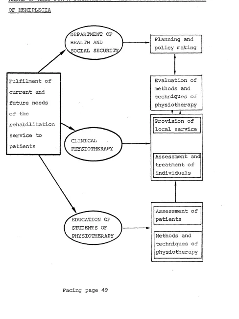
2.4.1 The interrelated needs of practitioners,
planners and policy makers 50
2.4.2 The needs of physiotherapists 52 2.4.3 Physiotherapeutic motor assessment for
hemiplegia: Criteria for acceptability 56 2.4.4 Assessments published in the literature 60 2.4.5 Procedural areas in the development of
an optimal assessment 71
2.5 Selection and presentation of items of assessment 75 2.5.1 Selection and judgment of items 75 2.5.2 Description of the procedure and findings
of the assessment 80
2.6 Measurement and scaling 10Q
2.6.1 Reliability and validity 100
2.6.2 Scales of measurement and ordering of
items of assessment 103
2.6.3 Selection of an appropriate method of
scaling 109
2.6.4 The Guttman Scalogram Technique 112 2.6.5 Application of the Guttman technique in
rehabilitation 118
2.7 Collection and analysis of data to investigate
the validity of the assessment 123 2.7.1 The survey approach to collection of data 124 2.7.2 Selection of samples for observation and
questioning 131
2.7.3 Analysis of data which has been collected
by questioning 136
Conclusions 142
3. ORIGINAL OBSERVATIONS MADE DURING THE DEVELOPMENT AND EVALUATION OF THE SHEFFIELD MOTOR ASSESSMENT
CHART 145
3.1 Introduction 145
3.1.1 Selection of samples of patients and
practitioners 147
3.1.2 Presentation of data 159
3.2 Development of the protocol of items of assessment 161
3.2.1 The prototype protocol 163
3.2.2 The preliminary protocol 166
3.2.3 The interim protocol 184
3.2.4 Tests of inter-observer reliability 201 3.2.5 The reproducibility and scalability of the
items assessing function of the upper limb 209 3.2.6 Presentation of items of assessment in the
instructional manual 213
3.2.7 The SMAC index of rate of improvement 217 3.2.8 Conclusions to the development of
3,3 The, development of the display 225 3.3.1 The semicircular display of the prototype
and preliminary assessments 226 3.3.2 The rhomboidal display of the interim
assessment 230
3.3.3 The final display of SMAC 234 3.3.4 Conclusions to the development of the
display 240
3.4 Evaluation of the chart’s acceptability to
physiotherapists 241
3.4.1 The design of the display 244 3.4.2 The utility of the items of assessment 249 3.4.3 The clarity of the manual 253 3.4.4. The methods of physiotherapy practised in
the United Kingdom 255
3.4.5 The physiotherapeutic acceptability of SMAC 258
3.4.6 Summary and conclusions 264
3.5 Evaluation of the chart's potential contribution
to the rehabilitation of stroke patients 266
3.5.1 The context of care 273
3.5.2 Reactions to SMAC 287
3.5.3 Discussion and conclusions 300
4. CONCLUSIONS 303
LIST OF REFERENCES 305
APPENDICES
1.1 Statistical tests i
1.2 The Guttman statistics v
1.3 Tests of the preliminary protocol ix 1.4 Tests of the interim and final protocols xv 11.1 Specific issues related to the design and
administration of questionnaires and
interview schedules xxv
11.2 The Questionnaires xxx
The postal questionnaire xxxi
The study day questionnaire xxxiir 11.3 Tabulation of data collected on the
questionnaires xxxv
II.5 The interview schedules liv Professional intexview schedule. lv Patients’ interview schedule lvii Physiotherapy teachers' interview schedule lix Physiotherapy clinicians' interview schedule lxi III Authors and authorities who were consulted lxiv IV The manual and the poster accompanying the
interim chart lxv
LIST OF FIGURES
These figures are on facing pages unless marked by *
1. Age-sex distribution of 379 stroke patients 2. Age-sex distribution of deaths from stroke 3. The myotatic stretch reflex
4. A typical hemiplegic stance
5. Areas of need for a standardised physiotherapeutic assessment of hemiplegia
6. An ADL assessment
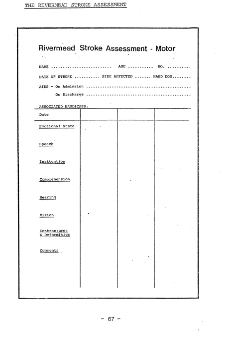
7. The Rivermead Stroke Assessment
8. Diagrammatic representation.of the relationship ‘between the position of the body and the control
of equilibrium and movement
9. The WHO's definitions of impairment, disability and handicap related to physiotherapeutic
assessment of hemiplegia
10. The model of the process of design of instructions and proformae for records
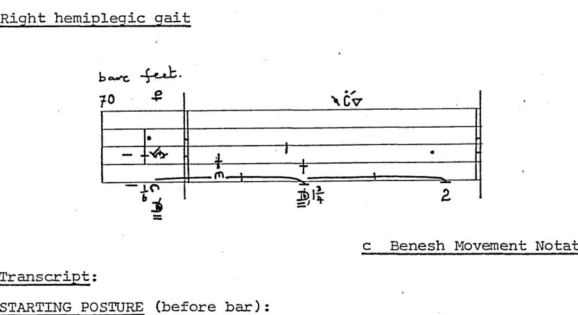
11. An example of hemiplegic gait written in. Benesh Movement Notation
12. The record card of the Riding for the Disabled s cheme
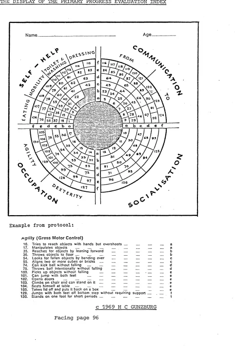
13. The display of the Primary Progress Evaluation Index
14. The Medical Research Council's scale of muscle power
15. Diagram to show matrix of a perfect Guttman scale 16. Pattern of responses for Guttman scaling
17. The time-scale of the research
18. Activities associated with the development of SMAC 19. Diagrammatic representation of the development
of SMAC
20. Summary of techniques used to collect data 21. The prototype protocol
22. The preliminary protocol
23. The apparent "recovery continuum"
24. The protocol of items described for the interim assessment
25. Headings used to standardise the Physiotherapy Items in the manual
26. The relationship of the protocol to the WHO's classification of impairments, disabilities and handicaps
27. The protocol of items of the final assessment 28. Items of assessment of the upper limb
29. The layout of items in the manual
30. Diagrammatic representation of the evolution of the display
31. The prototype display
32. Modes of presentation of patients' progress on the prototype display
33. The preliminary display
34. The general pattern of assessments recorded on the semicircular display
35. The interim display
3b. Facsimiles of patterns- of progress recorded on
the interim display 231
37. Facsimiles of records to show presentation of
status 232
38. Evolution of the interim display to the final
display 234
39. The final display of SMAC 235
LIST OF TABLES 1. 2
.
3. 4. 5. 6.
7. 8. 9. 10.
11.
12.
13. 14. 15. 16. 17. 18. 19. 20.
21.
22.
23. 24. 25. 26.Annual incidence of stroke related to age in the United Kingdom
Sizes of samples required for field tests Numbers of physiotherapists in sample. A and sample B
The samples of physiotherapists
Tabulations of losses from second field test and postal survey
Analysis of response to the field teste and the postal questionnaire
Number of categorical data for analysis from field test of preliminary protocol
The scale of twenty-nine items and their coefficients of reproducibility
Coefficients of reproducibility and scalability calculated for the scale of twenty-nine items and arbitrary subscales
Coefficients of reproducibility and scalability calculated for the scales of "Physiotherapy Items" and "Gross Functional Items"
Coefficients of reproducibility and scalability calculated for the postulated scales of the interim assessment.
Rank order correlation coefficients calculated for the interim protocol
The probability of specific items producing unreliable findings
Variance of items of final protocol
Coefficients of reproducibility and scalability obtained for items assessing function of the upper limb
Examples of SMAC Index
Number of members of each sample finding either display visually more attractive
Collation of data concerning the attractiveness and complexity of each display
Analyses of the design of the display
Proportions of respondents finding items of the interim chart easy to understand, remember, apply and record
Analyses of the utility of the items of assessment
Z statistics calculated to compare proportions of samples endorsing attributes of the interim chart
Proportions of the population of physiotherapists in the United Kingdom using various methods of physiotherapy for hemiplegia
Affirmative and negative opinions expressed on the postal questionnaire
Comparison of responses to similar questions on different questionnaires
Analyses of the physiotherapeutic acceptability of the assessment
Tables in the appendices;
1.1 1.2
1.3 1.4
1.5 II.1 II. 2
Coefficients calculated for the scale, of Physiotherapy Items
Coefficients calculated for the scale of Gross Functional Items
Coefficients calculated for the combined scale Rank order correlation coefficients of
association between performances recorded on the scale of Physiotherapy Items and on the scale of Gross Functional Items
Coefficients of reproducibility and scalability for the scale of items assessing the upper limb Data from questionnaires
Absolute numbers of physiotherapists
practising various methods of physiotherapy
xvi xvii xvii
1. INTRODUCTION
Stroke is a common occurrence, especially among the elderly Those who survive are often permanently disabled and need the support of people and institutions to enable them to lead as full lives as possible. In order to accommodate increasing demands for treatment of the physical disability physiotherapists need an assessment of stroke patients which will help them to plan and monitor their treatment more efficiently and more effectively.
Stroke has typical and easily recognisable effects on the victim*s ability to move one side of his body. This
obvious disorder of movement, posture and balance, known as hemiplegia, is not the only sequela. Depending on the side of the brain which is insulted, stroke victims may have difficulties in communicating with other people or be unaware of the position of their affected limbs without looking at them. 'They may become emotionally labile with episodes of tearfulness for no known reason; and they may have other emotional problems because they are frustrated at being unable to move and concerned about the future.
The eventual outcome for the patient will depend upon the care and rehabilitation he receives, on his capacity to respond to treatment, and on the attitudes of his family, his friends and the practitioners of several health care professions who treat him. Although physiotherapy is directed at resolving the hemiplegia, the patient's
his past experiences, and by his intellectual capacity.
For example, younger and professionally qualified people may
feel stigmatised by the hemiplegia: older and less well-
educated people are often more accepting of disability.
Additionally, while some patients enthusiastically practice
what they have been taught in treatment, some behave
aggressively towards the physiotherapist; and others are
passive and apathetic recipients of treatment.
Currently, physiotherapy for individuals is. limited by lack
of information about the process of recovery and the
effectiveness of physiotherapy for hemiplegia.
Physiotherapy for individuals would be enhanced by an
assessment which charted the sequence of recovery, allowed
the different methods of physiotherapy to be compared, and
allowed the contributions of physiotherapy to the wider
context of stroke rehabilitation to be evaluated. The
present study was designed to develop a physiotherapeutic
assessment of hemiplegic patients to be called the
Sheffield Motor Assessment Chart. Such an assessment would
provide physiotherapists with the specific information about
how a patient moves which they need to formulate aims of
treatment and to evaluate its effectiveness in enabling him
to progress towards recovery. It would also provide all
practitioners in rehabilitation teams with information about
the patient's progress and basic activities
he can perform when they need to make decisions about his
future care. The record of such an assessment needs to be
like a moving picture of the patient which demonstrates
Traditionally, assessment of stroke patients has depicted
physical progress in terms of "activities of daily living"
either to provide an overall picture of the patient's
status at one time or to show changes. Simple rating scales
have been created from "check lists" of these activities in
an attempt to introduce greater precision and objectivity.
The use of rating scales and arbitrary numbers assumes that
an absolute number is a valid indicator of recovery status
and that it has a real significance. Representing
the patient's status by an absolute number is of doubtful
value clinically and for evaluative studies. Firstly, no
two individuals with the same score on a rating scale are
likely to have achieved the same sets of assessment items.
Secondly, describing the patient by an absolute number
loses the detail of how the score was achieved. Thirdly,
the number gives no indication of the rate of progress.
A reliable assessment is needed to identify patients who
have the best chance of recovery so that limited resources
are used efficiently and physiotherapy is related both to
the severity of the patient's disability and to his
potential for recovery. Such an assessment might also
provide an index of the rate of progress in treatment which
could be used in evaluative studies of different methods of
physiotherapy.
Additionally, an optimal assessment would identify, assess
and record factors which influence recovery and treatment,
such as the patient's motivation and sensory disturbances.
seen as the first priority for a physiotherapeutic assessment for several reasons:
Firstly, physiotherapy is focussed on the patient's motor dysfunction.
Secondly, movement is directly observable.
Thirdly, from feheir experience with many hemiplegic patients, physiotherapists believe that movement is recovered in an orderly sequence.
Fourthly, influencing factors are not observed to improve in the same sequential manner.
Therefore, it was considered more feasible to concentrate on the development of a valid scale to record restitution of movement. Subsequently, this scale might provide a foundation for a "patient profile" which would demonstrate the influencing factors and include assessments made by other practitioners.
The development of the Sheffield Motor Assessment Chart is aimed at creating a motor assessment of hemiplegia which will record the patient * s recovery of movement and
functional abilities on a single-sheet graphic display. Such an assessment will provide physiotherapists with the information they need for planning and monitoring their treatment. At a glance! it will also show the patient's status and progress to other practitioners and to the patient himself. As an evaluated clinical assessment, it might also provide an instrument for gathering data for research in physiotherapy and into the associated factors.
hemiplegia. Next, the nature of the residual hemiplegia and the methods of physiotherapy will be described.
Assessments available in the literature will be discussed in relation to an optimal assessment which would fulfil the needs of physiotherapists for a standardised motor assessment of hemiplegia. Methods of scaling to create a sequence of items of assessment will also be discussed and a method appropriate to a condition characterised by progressive recovery will be selected. Finally, methods of surveying practitioners to collect data concerning the validity of the assessment will be discussed.
2o REVIEW OP THE LITERATURE 2ol STROKE
2.1,1 Classification of Stroke
Stroke is classified by the rubric of the International 1
Classification of Diseases which codes eight cerebrovascular
2 3
diseases (World Health Organisation , 1977). A universally accepted definition of CVD was agreed comparatively recently at an international conference sponsored by the WHO in 1970 in recognition of CVD as a major health problem. A detailed
classification of CVD was given in the report of the conference (WHO, 1971) and'the coding procedure has been revised with each new edition of the ICD.
4
Cerebrovascular accident describes those CVD manifest as stroke. The results of the epidemiological surveys referred to later in this review are classed according to the following common
pathological causes of CVA. The codes are those of the ninth edition of the ICD (WHO, 1977).
5
Subarachnoid haemorrhage (code 430) occurs when blood leaks from a ruptured artery into the space under one of the membranes enclosing the brain, the arachnoid mater. In the majority of individuals, SAH strikes without warning; and
it may be almost instantaneously fatal.
Cerebral haemorrhage (code 431) occurs when a blood vessel ruptures and blood lacerates the brain tissue. The clot of extravasated blood presses on brain tissue and damages it. Usually the patients are elderly and hypertensive and the stroke may occur during activity.
Hereafter the following conventional abbreviations are used: 1. ICD: International Classification of Diseases
Cerebral thrombosis (code 434.0) describes the formation, development and presence of a clot which blocks a blood vessel. The tissue distal to the blockage is starved of oxygen and nutrition.
Cerebral embolism (code 434.1) describes the sudden
blocking of a cerebral artery, by a clot of blood or other material such as fat or air carried in the blood stream, with ischaemic consequences.
Several authors have pointed out that information about these pathological subdivisions of CVA is imprecise (cf., e.g. Ford and Katz, 1966; Stallones, Dyken, Fang and Heyman, 1972).
According to Marquandson (1976), the distinction between embolic and thrombotic stroke is inadequate in epidemiological surveys. He is supported to a certain extent by the WHO. In earlier editions of the ICD, cerebral thrombosis and cerebral embolism were classified separately under codes 433 and 434 respectively. In the latest revision (1977) they are classed as subdivisions of code 434: occlusion and stenosis of cerebral arteries.
The pathological classification is also said to be inadequate "at the bedside" because it deals in static pathological states rather than in dynamic clinical phenomena (Shaw, 1972). Shaw’s criticism appears to be supported by the Hospital Activity Analysis, Area Diagnostic Index of the Sheffield Area Health Authority. For 1980 it records that forty-one residents of Sheffield were admitted to the city’s hospitals with SAH9 thirty-six with cerebral haemorrhage, seven with cerebral thrombosis and fourteen with cerebral embolus. Eight hundred and fifty residents were admitted with "acute but ill-defined CVD including apoplexy, CVA not otherwise specified, and stroke
(code 436)"*
-These figures suggest that medical practitioners in Sheffield are using a classification which is based on observable temporal patterns rather than on pathology:
Impending stroke (Millikan, Siekert and Whisnanf, 1960) described an episode of neurological dysfunction
lasting less than twenty-four hours. It is now known as TIA or transient ischaemic attack (Evans, 1981), or transient cerebral ischaemia (code 435), because it leaves no residual deficit and does not necessarily herald a major stroke.
Advancing or developing stroke describes neurological manifestations evolving with timei
Completed stroke is the evolved stroke or the abrupt onset of neurological manifestations.
Adams (1974) has observed that stroke refers to the rapidity of onset of signs and symptoms and not to the underlying cause or pathology. In reporting the result of a four year descriptive study of home and hospital care sponsored by the Department of
1
Health and Social Security , Weddell and Beresford (1979) did not distinguish between pathological types of CVA. They defined stroke as:
"A focal neurological deficit of sudden onset caused by a local disturbance in blood supply to the brain which lasted more than twentyfour hours and for which the patient was given medical care."
The majority of patients are referred for physiotherapy with a diagnosis of right or left CVA, or left or right hemiplegia, and with a report of current medical stability which might influence the inauguration or extent of active treatment.
1. Hereafter, the Department of Health and Social Security will be represented by DHSS.
-2.1.2 Epidemiology
Stroke is common historically as well as contemporaneously (cf. Sprengel, 1755, for Hippocrates; Charcot, 1881; Gowers, 1888). The current absolute incidence of stroke in the United Kingdom is not known; but a number of studies provide infor mation about people living with hemiplegia by documenting the mortality, survival and disability of particular cohorts of patients. There are no representative morbidity statistics and all rates of incidence are estimates based on the samples surveyed; the loss of quality of life for survivors cannot be quantified.
Despite the ICD, interpretation of results of epidemiological studies is complicated by the lack of a common definition of "stroke". Additionally, researchers have not used age-adjusted or sex-adjusted rates which are appropriate to disorders such as stroke which vary with age and sex. These variations will be discussed later in more detail. In general, there appears to be agreement that incidence, mortality and prevalence occur most frequently in the age group over forty years, and increase with age. There is less unanimity of opinion with regard to the contribution of such factors as race and pathological type of stroke.
-Incidence
According to WHO statistics (1971) the annual incidence of stroke in the United Kingdom lies between 1 08 and 2.0 per 1,000 population. Geographical differences are discernible which may be due to environmental factors« For example, the rate of incidence calculated for a population of 250,000 in Surrey (Weddell and Beresford, 1979) compares favourably with the rate calculated for the population of an industrial cityQ From their survey, Weddell and Beresford calculated a crude rate of incidence of l e4 per 1,000; but said that if ail those certified as dying from CVD had been included the rate would have been nearer 2.09 per 1,000. The Sheffield Area Hospital Activities Analysis shows that 948 residents from a population of 544,000 were admitted to hospital in 1980 with a primary diagnosis of CVA or stroke. There are no aggregated data from general practice; but it is likely that the number of stroke patients cared for at home or who die before admission to hospital would raise the rate of 1.7 per 1,000 to the level of the WHO estimates, and maybe beyond them.
Studies of mortality statistics based on death certification suggest that the incidence of stroke has declined since the Second World War (Acheson, 1960; Wylie, 1962; Metropolitan Life Insurance Company, 1975; Weddell and Beresford, 1979). However, Weddell and Beresford also claim that CVD is overdiagnosed; and Heasman and Lipworth (1966) in the United Kingdom and Florey, Senter and Acheson (1967, 1969) in the United States found poor correlations between death certification and findings at post mortem examinations. Wylie (1970a) expected the eight revision
-of the ICD to bring about a change in reporting due to improved specificity of diagnosis; but Israel and Klebba (1969) say that each revision of coding practice creates anomalies.
Consequently, the changing pattern of stroke is much less well documented than are the patterns of notifiable communicable diseases such as measles and poliomyelitis which are diagnosed accurately. According to Garrawav (1979) declining incidence of stroke is not accompanied by any change in prevalence; and he deduces that the probability of survival has increased. How ever, his data show an improvement of only two per cent in the number of patients who survive for 30 days. CVD is a cerebral manifestation of a vascular condition which is usually general ised and may give rise to hypertension and coronary heart
disease. Therefore, improvement in prospects for survival do not appear to be due to improved probability of surviving the acute CVA. It seems more likely that the decline in incidence and the improved prospects for survival are both due to
improvements in health which have increased longevity in the whole population.
Table 1
Annual incidence of stroke related to age in the United Kingdomo
Age Annual incidence per 1,000 population
35-44 0.25
45-54 1.00
55-64' 3.50
65-74 9.00
75-84 20.00
-84+ 40.00
-to o
c
ts n fD P' 0m
n
H-ri* P H O R fD IPO
P a o 0 n s p cr. fD W rt K Cn rt P rt H O P fD P K O Hi Hi H O fD H0 ■
P
h
i
K
fD ■ P P a fD M o P H-fa rt H* C p M ri* P a o H P* O 3 fD § CP o c ■D
Facing Page 12
> Q W I C/3 H X O H Cn H3
a
w
H O 3 o, H to <£> W rt P 0 ?r (D T5 P rt H-fD P rt M p (D iQ H-M rt (D P P CP tr fD rt £ fD P P s p K oo H M rt VD <] P P au
c v£> ^ <1 P ro• H
Incidence rises steeply with advancing age (Table 1). The age-sex distribution of 379 patients in Surrey (Figure 1) shows a preponderance of elderly women; this probably reflects the greater longevity of women. A ratio of 186 men to 145 women is obtained from the age-adjusted rates of fifteen different surveys (Dalsgaard-Neilson, 1955; SjostrcJm, 1967; Eckstrom, Brand, Edlavitch and Parrish, 1969; Alter, Christoferson, Resch, Myers and Ford, 1970; Acheson and Fairburn, 1970, 1971; Heyman, Karp, Heyden, Bartel et al, 1971; Bruun and Richter, 1973; Gordon, Sorlie & Kannel, 1973; Matsumoto, Whisnant, Kurland and Okazaki, 1973; Melamed, Cahane, Carmon and Levy, 1973; Stallones et al, 1972; Abu-Zeid, Choi and Nelsonna, 1975; Zupping and Roose, 1976). Proportionately, this ratio of 0.56 to 0.43 indicates that men are more at risk of stroke than are women to a modest degree.
Marquanason (1976) found a ratio of 121 men to 166 women in a series of patients in Denmark. When these figures were adjusted for sex rather than age they revealed that the incidence was higher for men than for women in each ten year age group over fifty-four years. This suggests that if the data from the other fifteen surveys were adjusted for sex the risk to men might be less modest than it appears.
69 per cent to thrombo-embolic pathology, and 11 per cent to ill-defined cerebrovascular disease.
Unlike other pathologies, SAH is particularly associated with younger age groups. More than half the total number of
aneurysms, or small balloon-like weaknesses of vessel walls, are said to cause symptoms first between forty and fifty-five years of age (Walton, 1977). From a survey of 135 patients in Sheffield who suffered SAH due to an aneurysm bursting, 69.6 per cent had their first symptoms after they were forty years old. Only 22.2 per cent were over fifty-five and 14.8 per cent were less that thirty years of age: 4704 per cent were in the forty to fifty-five age group (Parry, 1976).
The contraceptive pill is implicated in CVAs in women under forty-five years of age (Kannel, 1971): the case against other suspected factors is not proven. There is argument about race as a factor in the incidence of stroke in the United States. Rates for the American black population are variously reported, and in general they are higher than for the white population
(Alter et al , 1970; Wylie, 1970b; Heyman et al, 1971; Stallones et al, 1972; Bruun and Richter, 1973)» The validity of data on race has been questioned by Kurtzke (1969) who says that
differences in incidence may be explained by socio-economic and environmental factors.
S o u r c e : J M W e d d e l l an d S A A B e r e s f o r d ( 1 9 7 9 ) P l a n n i n g fo r S t r o k e P a t i en t s: A f o u r -y e a r d e s c r i t p i v e s t u d y of h o m e and h o s p i t a l c a r e . L o n d o n : H e r M a j e s t y ' s S t a t i o n e r y O f f i c e o
3 —
3 OJ Mta
o J-h •o pj rt* H-CD CS rt* in H CD IQ H* in rt* cd n CD
p.
O' CD Cj rt* c £P CD CD CD S3 hi w S rt* P> - *< hi oo VO hi
<3 in rv> rt*
ro
Facing page 14
Mortality from stroke
In 1966 CVD appeared on the list of ten leading causes of death in fifty-four out of fifty-seven countries reporting to the WHO. It ranked among the three leading causes of death in forty countries and accounted for 11.3 per cent of all deaths in the fifty-seven reporting countries.
Authors report varying mortality patterns and rates for the different pathologies: cerebral haemorrhage is identified by all as the most fatal type. While CVD in general is a common cause of death, the number of deaths registered as due to the disease is probably overestimated unless the diagnosis has been confirmed by post mortem examination (Heasman and Lipworth, 1966; Florey et al, 1967; Kurtzke, 1969).
Wylie (1970a) calculated that there was a 1:3 probability of dying from CVA. American and European authors have reported initial mortality rates as high as 50 per cent (Eisenberg et al, 1964; Drake, Hamilton and Carlsson, 1973; Pitner and Mance, 1973; Fugl-Meyer, Jaasko, Leyman, Olsson and Steglind, 1975). Langton Hewer (1976) estimated that between 35 and 40 per cent of stroke patients in the United Kingdom die within three weeks of their CVAs.
Figure 2 shows the age-sex distribution of deaths from stroke in Weddell and Beresford's study. They reported mortality rates of 61.6 per hundred for men and 57.5 per hundred for women within three months of the CVA. Age-adjusted rates of death show an increasing rate with advancing age which accelerates more than
-incidence rates. In general, the death rate doubles with each five year increase with age (Kurtzke, 1969).
-Prevalence of survivors of stroke in the United Kingdom
Prevalence of survivors with residual hemiplegia is the necessary dimension to consider in this study. Rational planning of the resources of the National Health Service, whether locally or nationally, whether fiscal or in terms of manpower, requires estimation of the number of people requiring rehabilitation at any given time. However, there are virtually no relevant data on current rehabilitation in general (Central Health Service Council, 1976). More specifically, in 1974 the Royal College of Physicians found that there were few studies to indicate the true extent of the burden of stroke on the
community-The survey of disability in Great Britain published by Harris, Cox and Smith in 1971 highlighted stroke as the single most important cause of severe disability. They estimated that there were 130,000 people in Great Britain with significant impairment from stroke and that 93,000 people were severely affected. Weddell and Beresford's study, published in 1979, found that the prevalence rate for survivors was four times the annual rate of incidence. They emphasised that this may be an underestimate because their calculations were based on the assumption that the age-sex structure of the population had remained constant from 1902 to 1971.
are under 65 years of age when they suffer their strokes, or three to four years if they are over 65. From comparison of seven studies of the level of functional ability achieved by survivors, Kottke (1974) estimated that 35 per cent of
survivors will return to "normal” or "nearly normal" status and that three to five per cent will remain totally
dependent•
Applying the WHO estimate of incidence in the United
Kingdom and Weddell and Beresford's estimate of prevalence in Surrey to the average population of 250,000 served by a district general hospital, there are likely to be 2,000 survivors in a health district at any given time. More than half of them will be over 65 years of age (Weddell and Beresford, 1979).
In order to reduce the burden on the community, elderly and retired patients will need rehabilitation to enable them to feed, bathe and dress themselves and to undertake social and domestic activities commensurate with their ages and lifestyles. More exploitive and vigorous
treatment is needed for the smaller proportion of younger survivors, to enable them to maintain their previous level of occupational activity as well as their social and
domestic activities. As younger patients have a longer life expectancy than older patients, the population of long-term survivors must include many people whose stroke occurred in middle life. Little is known about the quality of their lives (Collins, Marshall and Shaw, 1960).
-SUMMARY
"Stroke" describes the rapid onset of neurological signs and symptoms caused by a cerebrovascular accident, a sudden disturbance of the blood supply to the brain. Hemiplegia is the most common sequela of stroke: the patient is unable to move one side of his body normally.
In general, for every thousand members of the population of the United Kingdom, two new strokes can be expected each year. A..third of them is likely to end fatally within a month. The risk of stroke is higher for men than for women
and it is strongly related to age; mortality also increases with age. Less is known about the prevalence of people with residual hemiplegia.
The absolute number of people referred to the therapeutic services with hemiplegia is not precisely known. Commonly, it would include people with moderate and severe impairment and disability, and more mildly affected people who are admitted to hospital immediately after the stroke. The data indicate that a physiotherapy department in a District
2.2 RESIDUAL HEMIPLEGIA
Stroke has a catastrophic effect because the brain is
particularly vulnerable to disorders of its circulation and to metabolic deprivation. It has no means of storing energy and requires an unvarying rate of blood flow to function optimally (Keele and Neil, 1966).
Among its many functions, the brain serves as a "movement memory bank" which the individual establishes through years of practice. Each person's everyday movements, from the most mundane to the very skilful, become so automatic that he does not know how they are performed and finds it
difficult to describe them in detail (Carr and Shepherd, 1979). Additionally, the control of movement is so flexible and so adaptive that the brain can generate original and creative patterns of movement. From the sensory information it receives, it is able to represent the environment internally. Using this model, it computes the position of the body in space and the position of a target, and represents the change of position needed as a route between the two. This representation of the route is translated into a pattern of movement which will carry the limb from its initial position to the target.
Following a stroke, the control of movement on one side of the body is no longer flexible and adaptive. The
hemiplegic patient may have only a few stereotyped patterns of movement at his disposal and the internal representation of his relationship to his environment may be inaccurate.
-The motor deficit is most apparent in the limbs, because they normally move in a very wide variety of patterns, but the trunk and neck are also involved (Adams, 1974; Todd, 1974; Dardier, 1980).
-2o2.1 Mechanisms of hemiplegia
Anatomically, there are two nervous systems: the peripheral nervous system and the central nervous system. The peripheral nervous system carries sensory information from the skin, muscles and joints to the spinal cord and motor impulses from the spinal cord to the muscles.
1
The central nervous system consists of a sensory input system from the spinal cord to the brain, connections and centres for integrative activity in the brain, and a motor output system. The motor system is usually considered as two pathways which impinge on the motor nerves of the peripheral nervous system (Bowshec, 1979): (A) the pathway for willed movement from the motor cortex of the cerebrum, called the cortico-spinal tract; and (B) the extrapyramidal pathway which controls posture, muscle tone and coordination.
Coordinated movement results from harmonious activity of the components of the nervous systems. All purposive movements are initiated and guided through their execution by a constant stream of sensory information which reaches the brain from a wide variety of receptors in the skin, muscles, joints, ears and eyes. Disorders of movement and posture result from the
activity of components of the CNS which are intact combined with the defective activity of damaged and deprived areas.
1, Hereafter, the central nervous system is referred to by CNS.
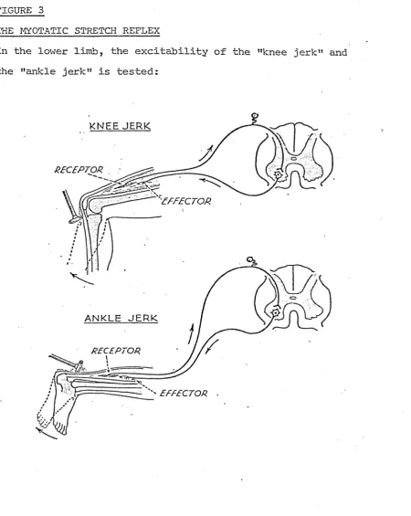
-FIGURE 3
THE MIOTATIC STRETCH REFLEX
In the lower limb, the excitability of the "knee jerk" and the "ankle jerk" is tested:
KNEE JE R K
o
A N K L E J E R K
RECEPTOR
EFFECTOR .
When the tendon is tapped sharply, the muscle is stretched. Nervous impulses pass into the spinal cord and stimulate motor fibres: the muscle 'contracts and the stretch is relieved.
Source: A B McNaught and R Callender (1967)
[image:39.617.74.525.49.615.2]According to its severity, a CVA destroys variable amounts of fibres of the pathway for willed movement and other tracts; commonly, as they are channelled through a bottleneck called the "internal capsule"e Consequently, the patient is unable to make smooth coordinated movements, even though the muscles are
capable of contracting. The CVA also releases reflexes from modification by the highest centres of the CMS. Two spinal reflexes are particularly implicated in hemiplegia: (1) the stretch reflex and (2) a modified form of the extensor thrust reflex.
1. The stretch reflex: The reflex arc of the myotatic stretch reflex connects receptors of stretch in a muscle with the
contractile muscle fibres (Figure 3). The stretch may be applied by contraction of antagonistic muscles, by the body starting to fail in any direction, or by an external force. Normally, the threshold of the receptor is controlled by higher centres of the CNS so that it is sensitive to stretch according to postural needs at any time. When the CVA releases the reflex it becomes hyperactive. The threshold is lowered and the muscle contracts inappropriately. The resulting increase in the
elastic tension of muscle is called "spasticity".
2• The positive supporting reaction (Magnus, 1926): This reaction is a modification of the extensor thrust reflex
(Sherrington, 1947). In the lower limb, it is principally evoked by pressure on the ball of the foot: the moveable limb is transformed into a rigid pillar by simultaneous contraction of the flexor and extensor muscles. When the pressure is removed, the negative supporting reaction occurs: the limb
-becomes loose at all joints and free to move again. These alternating states are inappropriate for activities in which weightbearing and movement occur simultaneously. Fixation which can only be released when pressure or weight is removed from a limb prevents the fine grading which is necessary for rising from a chair and sitting down, 'for walking, for climbing and descending stairs and for balancingo
-2.2.2 The impairment of motor function
The characteristic effects of CVA which constitute hemiplegia are due to alterations in direct and indirect influences on the motor nerves of the peripheral nervous system:
A. The patient is unable to .initiate voluntary movement. B. Immediately, the muscles lose their normal state of
tone and are flaccid. Eventually, muscle tone rises; usually to a high level of tension called spasticity. C. Postural control of the stability and balance of the
body is inadequate•
Loss of voluntary movement: Several authors have emphasised that loss of voluntary movement is not due to weakness of muscles (cf. e.g. Adams, 1974; Todd and Davies, 1977; Bobath, 1978; Carr and Shepherd, 1980). What might be called "weakness of movement" is due to lack of cortical drive, or central initiation and control of movement, due to interruption of the pathway for willed movement. Muscles may be activated in a few stereotyped mass patterns of flexion and extension by the motor centres of the brain and spinal cord. Even though they can function in these patterns, they cannot be recruited to perform with other muscles to create the almost infinite variety of patterns used in everyday life. The overall impression is of poverty of movement.
Generally, recovery of movement proceeds proximally to distally (Twitchell, 1951; Bobath, 1959; 1960; Bard and Hirschberg, 1965). More specifically, mass actions of the shoulder and hip.joints are recovered first:,
selective use of distal groups of muscles may return later allowing a clumsy grasping action of the fingers•
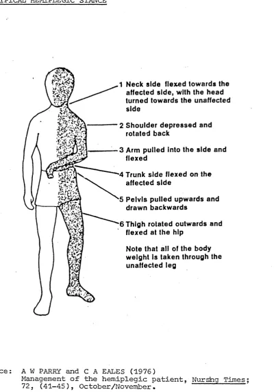
FIGURE 4
A TYPICAL HEMIPLEGIC STANCE
1 Neck side flexed towards the affected side, with the head turned towards the unaffected side
2 Shoulder depressed and rotated back
3 Arm puiied into the side and flexed
4 Trunk side flexed on the affected side
5 Pelvis pulled upwards and drawn backwards
6 Thigh rotated outwards and flexed at the hip
Note that all of the body weight is taken through the unaffected leg
Source: A W PARRY and C A EALES (1976)
Management of the hemiplegic patient, Nurshg Times 72j (41-45), October/November.
[image:43.617.96.500.73.646.2]and sufficient dorsiflexion of the foot to clear the ground in walkingo Fine skilled movements of the hand may never he recovered, unless movement of the thumb independent of the fingers is restored.
Muscle tone: Tone must be high enough to withstand gravity yet still permit movement. Immediately after the stroke the muscles may be flaccid, but tone usually rises as cerebral shock is dissipated. In some patients it never reaches the normal state of tension, and the muscles remain hypotonic. Con sequently, the patient has difficulty in shifting his weight, in moving from one point to another and in bearing weight. If hypotonia persists, he will lack stability at the pelvic girdle, which will affect the control of the pelvis on the thighs in sitting and standing, and at the shoulder girdle, which will prevent effective use of the hands.
Usually muscle tone rises above normal to a spastic state. Spasticity gives stability without mobility; and it is manifest in definite stereotyped patterns of abnormal coordination
(Bobath, 1978). On the hemiplegic side, the shoulder girdle is depressed and retracted and the pelvic girdle is elevated and retracted (Figure 4). In the upper limb, spasticity is
dominant in the muscles which draw the arm into the side, bend the elbow, wrist and fingers and pronate the forearm so that the palm of the hand faces the flooro There is usually more competition of patterns in the lower limb than in the upper limb. Typically, the lower limb is held in a mid-position, but moved by alternating patterns of total flexion and total extension.
-Jackson (1884) coined the term "clotted movement" to describe the way in which spasticity interferes with the quality of movement. This alteration in muscle tone and the loss of voluntary movement give the patient characteristic appearances in sitting and standing, and typical patterns of movement.
Postural control: Posture is the background of automatic and voluntary activity which precedes and underlies all willed movement (Critchley, 1954). Adams (1974) has referred to the
"vocabulary of posture", a collection of memories which ensure immediate and accurate response to prevent loss of balance.
Postural control requires fine changes in the distribution of muscle tone. To be safe, all movement needs to be performed on a constantly changing background of postural adjustment which is graded to the degree of voluntary control
required. Disordered postural control in hemiplegia is attributed to the distribution of spasticity (Eobath, 1965), or to persistent hypotonia, or to dense sensory deficit (Fisher, 1968).
-2„2.3 The overall picture of motor dysfunction
The loss of voluntary movement, the changes in muscle tone and the loss of .balance and postural reactions must be considered together to gain a true picture of the problems experienced by the hemiplegic patient. The net result is a person whose movement is confined to patterns of limited utility, which are also inherently unsafe. The automatic unconscious activity which controls normal movement is lost to him. Not only is he unable to move his hemiplegic side voluntarily to produce the movement he wants to make, but he is unable to make the auto matic adjustments necessary to control his posture and balance.
The physiotherapist aims to improve the patient's ability to perform his ordinary domestic and social activities by resolving the impairment of motor function. To make her aims compatible with his, usually she must link them to the activity which is probably the single most important aim of every hemiplegic patient: the ability to walk independently. To enable him to rise from a chair and walk she must counteract the effects of
spasticity and the positive supporting reaction: they interfere with the recovering patient's ability to bear weight through his
affected leg, to transfer weight over it and to balance on it.
The normal way of rising from a chair is to move the weight of the body over the feer first of all, by moving the head and the trunk forwards from the hipso Next, the flexed legs take the weight as the burrocks are raised from the seat. Finally, the
-hips, the knees and the spine are straightened so that the body becomes upright.
As the spastic hemiplegic person tries to take weight through his affected leg to stand up, the positive supporting reaction is evoked by the pressure of his forefoot on the ground. Con sequently, his foot is pushed againsr the ground; and the extension thrusts up his leg, stiffening his knee and hip. Instead of his weight being brougnt forward over his feet, his hemiplegic side is thrust further back into the chair. If he is able to haul his trunk forwards with his hands and arms, he may be able to stand up with great effort; but with little or no weight borne through his affecued leg• When he tries to sit down again, he falls back into the chair: he can neither bend the limb while he has weignt on it nor control the placing of his buttocks on the seat.
Typically, the hemiplegic patient who can walk does not strike the -ground with his heel. The ball of his foot strikes the ground first; because he can only activate mass patterns of flexion and extension, his ankle is plantarflexed as his knee extends. Again, the positive supporting reaction is evoked, and his leg becomes stiff. The rigid limb may bear the weight of his body but the joints are fixed. There is no "give
and take" between the muscles while he bears weight, and he cannot transfer his body weight over the standing leg normally. Although the rigidity may allow him to bear weight to walk, his gait is inherently unsafe: the rigidity also prevents the fine adjustments needed to maintain and regain
-balance. He has to make compensatory attempts with his unaffected side to maintain his balance.
If the hemiplegic person tries to climb and descend stairs the problems are compounded by gravity and the need for more sophisticated postural control. Normally, gradual flexion of the weightbearing leg is essential while the free leg descends to the lower step and, like rising from a chair, the flexed leg on the upper s t e p normally takes weight
and then extends to raise the body.
Many hemiplegic patients never recover- sufficiently to climb and descend stairs reciprocally; many of them are given walking aids, to compensate for loss of balance reactions, and to enable them to walk with a typical hemiplegic gait. They may still be unable to rise from a chair without assistance or great effort. Some people are readmitted to hospital because they have fallen and sustained fractures or other injuries, or because relatives and other carers are afraid that they will do > be injured if left alone for short periods.
-2.2.4 Factors influencing recovery and rehabilitation
The patient's recovery and his capacity to respond to physio therapy and rehabilitation may be affected by associated disorders of sensory appreciation or communication and by his motivation to collaborate in treatment.
Disorders of sensory appreciation: Sensory loss is common after stroke because afferent nerve fibres also pass through the internal capsule. The importance of sensory input for the control of movement was elucidated first by Mott and Sherrington in 1895. Recognition of objects, body awareness and visual-spatial orientation depend upon integration of sensation
from the skin, muscles and joints, visual information and sound. A store of sensory memories is said to exist as a basis for action in response to things perceived by sight and touch (Walton, 1977). Severe sensory defects (Garston, 1967) and persistent sensory defects (Buskirk and Webster, 1955; Hurwitz and Adams, 1971) affecting input and integration of sensory information are detrimental to further functional achievement.
The sensory loss may be slight, with minor loss of appreciation of light touch or the prick of a pin, to severe, with complete loss of joint position sense and appreciation of movement for half of the body (Macleod and Williamson, 1967). Defective proprioception denies the patient infamation which is essential for the control of movement: knowledge of the relationships of the parts of the body to each other and knowledge of the
position of his limbs in space. It also disrupts the feedback mechanisms which affect the control of balance: he may not be
-able to balance safely when he is sitting, and standing and walking may be impossible.
A large class of perceptual and cognitive disorders, called "the agnosias", is particularly associated with left-sided hemiplegia, usually caused by lesion of the non-dominant
cerehral hemisphere. (Corresponding syndromes associated with right-sided hemiplegia are less frequently seen in left-handed patients.) The patient may completely ignore objects or
activities in the left half of his field of vision; he may be unaware of his hemiplegic side or part of it and reject it
(asomatognosia); or he may deny his hemiplegia totally (anosognosia (Critchley, 1955)). Although they are able to see an object, many patients cannot assess its position, size or movement relative to themselves. Consequently, they are unable to perform simple tasks, such as placing the arm in a sleeve to dress themselves.
Currently, there is very little information on the treatment, rather than the description, of sensory disorderso Isaacs
(1962) states that widespread sensory dysfunction is difficult to rehabilitate and Marquandson (1969) says that self-care activities may be unattainable.