INTRODUCTION
Vertebrate gastrulation starts with the internalization of mesodermal and endodermal cells, which establishes the internal and external organization of the developing animal (Solnica-Krezel, 2005). During internalization, the mesodermal and endodermal cells move via the blastopore beneath the ectoderm. It is well known that after internalization, the migration of mesodermal cells is controlled by convergence and extension (C&E) movements, which narrow the field of mesodermal cells mediolaterally and elongate it along the anterior-posterior axis (Myers et al., 2002; Montero and Heisenberg, 2004). Recent comprehensive studies in Xenopusand zebrafish have demonstrated that the Wnt/planar cell polarity (PCP) signaling pathway is implicated in C&E movements (Myers et al., 2002). The internalization of endoderm is coincident with that of mesoderm and, after its migration, it gives rise to the epithelial lining of the digestive tract as well as associated organs (Warga and Nüsslein-Volhard, 1999). Although two studies concerning endoderm movements have been reported (Ober et al., 2004; Matsui et al., 2005), the molecular mechanisms underlying endodermal cell movement during gastrulation remain largely unexplored in vertebrates.
In zebrafish, a Sox-related transcription factor, Casanova (Cas; Sox32 – ZFIN), is essential for endodermal cell specification (Kikuchi et al., 2001; Fukuda and Kikuchi, 2005) and functions
to control its downstream target, sox17(Alexander and Stainier, 1999). cas-expressing endodermal cells are distributed around the blastoderm margin in a salt-and-pepper pattern before gastrulation and this distribution is maintained during the migration process (Kikuchi et al., 2001; Warga and Nüsslein-Volhard, 1999). After specification, internalized endodermal and mesodermal cells form the hypoblast layer underneath the epiblast layer, which consists of ectodermal cells. A detailed study using zebrafish embryos has revealed that within the hypoblast, the endodermal cells are internal, and are located next to the extra-embryonic yolk syncytial layer (YSL), whereas the mesodermal cells form a layer between the endoderm and the ectoderm (Warga and Nüsslein-Volhard, 1999). Endodermal cells form a noncontiguous monolayer, which is overlaid by a more coherent multilayer of mesodermal cells (Warga and Nüsslein-Volhard, 1999) (see also Fig. S1 in the supplementary material). Time-lapse video-microscopy has further shown that following internalization, endodermal cells change their morphology and become flattened with numerous filopodial processes (Warga and Nüsslein-Volhard, 1999). Since endodermal cells migrate separately from mesodermal cells within the hypoblast layer, the zebrafish gastrula provides a useful model system in which to study endodermal cell migration.
Chemokines are a large family of proteins with crucial roles not only in the immune response, but also during various developmental processes such as cell migration and the growth cone guidance of neuronal axons (Kucia et al., 2004). The chemokines, which are small secreted molecules, act through their receptors, which belong to the superfamily of G protein-coupled receptors (GPCRs) (Kucia et al., 2004). One such chemokine, stromal cell-derived factor 1 (Sdf1; Cxcl12 – ZFIN), and its receptor, Cxcr4, are well characterized in various biological processes (Busillo and Benovic, 2007). A major function of Sdf1/Cxcr4 signaling is to regulate the movement of cells, as Sdf1 attracts cxcr4-expressing cells. In zebrafish, the movement of
Sdf1/Cxcr4 signaling controls the dorsal migration of
endodermal cells during zebrafish gastrulation
Takamasa Mizoguchi1,2, Heather Verkade3,*, Joan K. Heath3, Atsushi Kuroiwa2and Yutaka Kikuchi1,2,†
During vertebrate gastrulation, both mesodermal and endodermal cells internalize through the blastopore beneath the ectoderm. In zebrafish, the internalized mesodermal cells move towards the dorsal side of the gastrula and, at the same time, they extend anteriorly by convergence and extension (C&E) movements. Endodermal cells showing characteristic filopodia then migrate into the inner layer within the hypoblast next to the yolk syncytial layer (YSL). However, little is known about how the movement of endodermal cells is regulated during gastrulation. Here we show that sdf1a- and sdf1b-expressing mesodermal cells control the movements of the cxcr4a-expressing endodermal cells. The directional migration of endodermal cells during gastrulation is inhibited by knockdown of either cxcr4a orsdf1a/sdf1b(sdf1). We also show that misexpressed Sdf1 acts as a chemoattractant for cxcr4a-expressing endodermal cells. We further found, using the endoderm-specific transgenic line Tg(sox17:EGFP), that Sdf1/Cxcr4 signaling regulates both the formation and orientation of filopodial processes in endodermal cells. Moreover, the accumulation of phosphoinositide 3,4,5-trisphosphate (PIP3), which is known to occur at the leading edge of migrating cells, is not observed at the
filopodia of endodermal cells. Based on our results, we propose that sdf1-expressing mesodermal cells, which overlie the endodermal layer, guide the cxcr4a-expressing endodermal cells to the dorsal side of the embryo during gastrulation, possibly through a PIP3-independent pathway.
KEY WORDS: Zebrafish, Endoderm, Migration, Gastrulation, Sdf1 (Cxcl12), Cxcr4a Development 135, 2521-2529 (2008) doi:10.1242/dev.020107
1Department of Biological Science, Graduate School of Science, Hiroshima University, Kagamiyama 1-3-1, Higashi-Hiroshima, Hiroshima, 739-8526 Japan. 2Division of Biological Science, Graduate School of Science, Nagoya University, Furo-cho, Chikusa-ku, Nagoya, 464-8602 Japan. 3Ludwig Institute for Cancer Research, Royal Melbourne Hospital, Parkville, VIC 3050, Australia.
*Current address: School of Biological Science, Monash University Clayton, Melbourne, VIC 3800, Australia
†Author for correspondence (e-mail: yutaka@hiroshima-u.ac.jp)
Accepted 27 May 2008
D
E
V
E
LO
P
M
E
N
various types of cells, such as primordial germ cells, olfactory placodal precursors, lateral line primordial cells and slow muscle cells, is regulated by Sdf1/Cxcr4 signaling during development (David et al., 2002; Li et al., 2004; Raz and Reichman-Fried, 2006; Chong et al., 2007; Miyasaka et al., 2007). Moreover, a recent report has shown that Sdf1/Cxcr4 signaling regulates mesendodermal cell migration during Xenopusgastrulation (Fukui et al., 2007). In Xenopusembryos, Sdf1αin the ectodermal layer attracts the Cxcr4-expressing mesendodermal cells (Fukui et al., 2007). However, the molecular mechanisms underlying mesendodermal cell migration regulated by Sdf1/Cxcr4 signaling have not been analyzed at single-cell resolution.
In this study, we have focused on the molecular mechanisms underlying the control of endoderm migration by Sdf1/Cxcr4 signaling during gastrulation in zebrafish. We show that sdf1a- and sdf1b-expressing mesodermal cells control the movement of cxcr4a-expressing endodermal cells. The directional migration of endodermal cells during gastrulation is inhibited by knockdown of either cxcr4aor sdf1a/sdf1b(collectively referred to as sdf1) using morpholino antisense oligonucleotides. We also show that misexpressed Sdf1 acts as a chemoattractant for cxcr4a-expressing endodermal cells. To further analyze endoderm migration at single-cell resolution, we generated an endoderm-specific transgenic line carrying an EGFPreporter driven by a sox17promoter fragment, Tg(sox17:EGFP). Using this system, we found that the Sdf1/Cxcr4a signaling pathway regulates both the formation and orientation of the filopodial processes in endodermal cells. The accumulation of phosphoinositide 3,4,5-trisphosphate (PIP3), which is known to
occur at the leading edge of migrating cells, is not observed at the filopodia of endodermal cells. Taken together, our results suggest that sdf1-expressing mesodermal cells, which overlie the endodermal layer, guide the cxcr4a-expressing endodermal cells to the dorsal side of the embryo during gastrulation, possibly through a PIP3-independent pathway in zebrafish.
MATERIALS AND METHODS
Zebrafish strains and generation of Tg(sox17:EGFP)transgenic line Adult zebrafish and embryos were maintained as described previously (Westerfield, 1995). Embryos were incubated in 1/3 Ringer’s solution (39 mM NaCl, 0.97 mM KCl, 1.8 mM CaCl2, 1.7 mM HEPES, pH 7.2) at 28.5°C and staged according to Kimmel et al. (Kimmel et al., 1995). The
cass4mutant allele (Kikuchi et al., 2001) was used and genotyping of homozygote casmutant embryos was performed as described previously (Kikuchi et al., 2001).
To generate the Tg(sox17:EGFP)line, a 5.0 kb region of the sox17gene promoter was cloned as described previously (Reim et al., 2004), and then subcloned into pEGFP-1 (Clontech) to generate pSox17:EGFP. The DNA fragment carrying the sox17promoter and EGFPwas isolated and diluted to 100 ng/μl in distilled water containing 0.1% Phenol Red. The resulting DNA solution was then injected into the blastomere of one-cell stage zebrafish embryos. Embryos showing fluorescent signals were raised to sexual maturity and founder fish were subsequently selected by the expression of EGFP-fluorescence in their progeny. Only one stable transgenic line was obtained using this procedure, designated
Tg(sox17:EGFP).
Whole-mount in situ hybridization, β-galactosidase staining and histological analysis
Whole-mount in situ hybridization was performed using conventional nitro blue tetrazolium (NBT)/5-bromo-4-chloro-3-indolyl phosphate (BCIP) precipitation by alkaline phosphatase (Westerfield, 1995). cDNA fragments for cxcr4a (Chong et al., 2001), sdf1a(Doitsidou et al., 2002), sdf1b (Li et al., 2004), sox17 (Alexander and Stainier, 1999), foxa3 (Odenthal and Nüsslein-Volhard, 1998), ceruloplasmin(cp) (Korzh et al., 2001), insulin
(ins), pdx1(Milewski et al., 1998), hatching gland gene 1(hgg1; ctsl1b–
ZFIN) (Thisse et al., 1994), distal-less 3(dlx3) (Akimenko et al., 1994), no tail(ntl) (Schulte-Merker et al., 1994) and myoD (myod1– ZFIN) (Weinberg et al., 1996) were utilized as templates for the antisense probes. β -Galactosidase staining and double staining with whole-mount in situ hybridization were performed as described previously (Mizoguchi et al., 2006).
For histology, the NBT/BCIP-stained in situ embryos were dehydrated, embedded in Technovit 7100 (Heraeus Kulzer, Wehrheim, Germany) and cut at 10 μm. The sectioned in situ samples were imaged by confocal laser-scanning reflection microscopy, a newly developed technique that visualizes NBT/BCIP staining by reflection of the confocal laser beam (Jékely and Arendt, 2007). The reflection images were taken with an Olympus FV1000D confocal microscope using 473 nm and 635 nm lasers, and a 40⫻ oil-immersion lens.
mRNA and morpholino injections
The pCS2+ vector carrying cDNA fragments encoding cas, nuclear lacZ
(nlacZ), sdf1aand sdf1bwere used in this study. For pleckstrin homology
(PH)-mCherrymRNA synthesis, a gene encoding the PH domain of protein kinase B (Watton and Downward, 1999) was fused to the N-terminus of the
mCherrygene (Clontech) (Shaner et al., 2004) to generate pCS2- PH-mCherry. Capped mRNA was synthesized using the SP6 mMESSAGE mMACHINE system (Ambion). Injection of nlacZmRNA (200 pg) or co-injection of cas(50 pg) and nlacZ(200 pg) mRNAs was performed into one blastomere at the 16-cell stage. PH-mCherrymRNA (250 pg) was injected into one-cell stage embryos.
Injections of morpholino antisense oligonucleotides (MOs) (Gene Tools) were carried out as described (Nasevicius and Ekker, 2000). We designed two MOs to block translation initiation of cxcr4amRNA (cxcr4aMO1, 5⬘ -CGATGTGTCCGTAATAAGCCATCTC-3⬘, sequence complementary to the predicted start codon is underlined; cxcr4aMO2, 5⬘CCTTC -AGTCTCCAGCAAGTCTTCAG-3⬘) and a control MO for cxcr4aMO1 with five mispaired bases (misMO1, 5⬘CcATGTcTCCcTAATAAc -CCATgTC-3⬘, lowercase letters indicate mispaired bases). For the double knockdown of sdf1aand sdf1b (sdf1), we co-injected the sdf1aMO (5⬘ -CTACTACGATCACTTTGAGATCCAT-3⬘) (Doitsidou et al., 2002) and the
sdf1bMO (5⬘-CGCTACTACTTTGCTATCCATGCCA-3⬘) (Knaut et al., 2003). All MOs were injected into one-cell stage embryos, 5 ng each. Injection of casmRNA (2 pg) or co-injection of casmRNA (2 pg) and
cxcr4aMO1 (0.1 ng) with dextran plus Alexa Fluor 594 (dextran-Alexa594; Invitrogen) as a lineage tracer was performed into one blastomere at the 128-cell stage.
Live imaging and confocal time-lapse imaging
Tg(sox17:EGFP)transgenic embryos were dechorionated at the one-cell stage using 0.2 mg/ml pronase in 1/3 Ringer’s solution and were raised to the three-somite stage or for 24 hours post-fertilization (hpf). The embryos were oriented in 3% methylcellulose/1/3 Ringer’s solution. Low-magnification images of Tg(sox17:EGFP) transgenic embryos were obtained with an Olympus SZX-RFL3 microscope and Penguin 600CL cooled CCD camera (Pixera).
Confocal time-lapse imaging was performed on an Olympus FV1000 confocal microscope using a 60⫻ or a 100⫻ oil-immersion lens.
Tg(sox17:EGFP)embryos were dechorionated at the one-cell stage also using 0.2 mg/ml pronase in 1/3 Ringer’s solution and were raised to 90% epiboly. These embryos were mounted in 0.7% low-melting-temperature agarose in 1/3 Ringer’s solution. We recorded 60 z-stacks (1.25 μm steps) at 30-second intervals and analyzed the images using ImageJ software (NIH). The orientation of filopodial processes was analyzed using Origin8 software (LightStone, Japan).
Cell transplantation
nlacZ (2 ng), sdf1a(3 ng) or sdf1b(2 ng) mRNA was injected together with dextran-Alexa594 into donor wild-type embryos and sdf1MOs were injected into host Tg(sox17:EGFP) embryos at the one-cell stage. Cells were extracted from donor embryos at the dome stage (4.3 hpf) and injected into the ventrolateral side, close to the margin, of shield-stage (6 hpf) hosts using standard methods (Westerfield, 1995). The time-lapse images were taken with an Olympus FV1000D confocal microscope using a 20⫻lens.
D
E
V
E
LO
P
M
E
N
RESULTS
Expression patterns of cxcr4aand sdf1during gastrulation
To elucidate the genes involved in endodermal cell migration, we compared the expression profiles of wild-type (WT) and cas morphant embryos by microarray analysis. In the course of this analysis, we found that the expression of the cxcr4a gene is significantly reduced in casmorphant embryos (data not shown). As recent studies have implicated Cxcr4 and its ligand Sdf1 in various developmental processes in zebrafish embryos (David et al., 2002; Li et al., 2004; Raz and Reichman-Fried, 2006; Chong et al., 2007; Miyasaka et al., 2007), we next examined the expression pattern of cxcr4a, sdf1a and sdf1bduring gastrulation. We confirmed that, as previously reported (Chong et al., 2001), cxcr4a is maternally expressed and its zygotic expression is restricted to a subset of blastomeres around the margin at shield stage (6 hpf) (data not shown). The restricted expression of cxcr4ais maintained during gastrulation and disappears by the three-somite stage (Chong et al., 2001) (data not shown). As this characteristic salt-and-pepper expression pattern of cxcr4ais remarkably similar to that of both cas and sox17, we next examined whether cxcr4a is expressed in endodermal cells. The salt-and-pepper expression pattern of cxcr4a was completely abolished in casmutants, whereas its expression in other tissues was unaffected in casmutants (Fig. 1A,B). Moreover, an ectopic expression pattern of cxcr4awas observed in cas mRNA-misexpressing cells (Fig. 1C,D). These results clearly demonstrate that cxcr4ais expressed in endodermal cells and is dependent on Cas.
Both sdf1aand sdf1bwere found to be expressed in the lateral region of gastrulating embryos (Fig. 1E,I,J,K,O,P). In contrast to cxcr4a, the expression patterns of both sdf1aand sdf1bdid not differ between WT and cas mutant embryos, and indicated their expression in mesodermal cells (Fig. 1E,F,K,L). In support of this, optical cross-sections and sagittal sections showed that the
expression of both genes is restricted to the hypoblast cell layer (Fig. 1G,H,M,N), which consists only of endoderm and mesoderm. Internalized mesodermal cells move animally at the beginning of gastrulation and start moving dorsally prior to mid-gastrulation (Sepich et al., 2005). Consistent with these published data, sdf1a-and sdf1b-expressing mesodermal cells in the animal-lateral region were found to move towards the dorsal midline from 80% epiboly to tail bud (TB) stages (Fig. 1E,I,J,K,O,P; red dashed lines). The sox17-expressing endodermal cells also moved towards the dorsal midline during late gastrulation (Fig. 1Q-S; red dashed lines), and this dorsal movement behavior coincided with that of the sdf1a- and sdf1b-expressing mesodermal cells, suggesting that the movements of endodermal cells might be regulated by mesodermal cells.
Endoderm migration is delayed by the inhibition ofcxcr4aor sdf1
To investigate the function of Sdf1/Cxcr4a signaling in the movement of the endodermal cells, we knocked down the corresponding genes by injecting specific MOs. The sox17-expressing endodermal cells of the animal-lateral region moved dorsally in misMO1-injected embryos at late gastrulation (Fig. 2Aa; arrowheads). By contrast, the dorsal migration of endodermal cells was delayed in cxcr4aMO1-, cxcr4aMO2- or sdf1MOs-injected embryos (Fig. 2Ab-Ac; arrowheads). Throughout this study, we compared the phenotypes of non-injected and misMO1-injected embryos, and observed no differences between them (data not shown).
[image:3.612.247.562.449.661.2]In order to visualize the movements of the endodermal cells in live zebrafish embryos, we generated the Tg(sox17:EGFP) transgenic line. Although the endodermal expression of sox17 begins soon after the onset of gastrulation (Alexander and Stainier, 1999), the earliest detectable EGFP fluorescence in the endodermal cells of the transgenic embryos is from the 60% epiboly (7 hpf) stage, and this fluorescence continues until 2 days
Fig. 1. During gastrulation in zebrafish, cxcr4ais expressed in the endoderm, and sdf1aand sdf1b are expressed in the mesoderm. (A-D) cxcr4a expression was examined by whole-mount in situ hybridization in casmutant embryos and embryos injected with either casor nlacZmRNA at the 80% epiboly stage (8.3 hpf). Lateral views, dorsal to the right (A,B), and dorsal views, anterior to the top (C,D). The salt-and-pepper endodermal expression of cxcr4ais absent in the casmutant, whereas expression in others tissues is still detectable on the dorsal side (arrow in B) and is faintly detectable (arrowheads in B) in other regions of the casmutant. Although the overexpression of nlacZmRNA does not affect the salt-and-pepper expression of cxcr4a, casmRNA injection increases the number of cxcr4a-expressing cells (arrow in D). (E-P)sdf1aand sdf1bexpression was examined in wild-type (WT) and casmutant embryos. Lateral views with dorsal to the right (except for G,H,M,N). (E,F,K,L) At the 80% epiboly stage, neither sdf1anor sdf1bexpression is altered in the casmutant embryos. (G,M) Optical cross-sections (OS) of 80% epiboly stage embryos
show that expression of both sdf1aand sdf1bis present in the hypoblast layer, but not in the epiblast layer (the epiblast layer is indicated by the red dashed lines). Dorsal views, anterior to top. (H,N) Confocal images of sagittal sections of 80% epiboly stage embryos by confocal laser-scanning reflection microscopy (Jékely and Arendt, 2007). Expression of both sdf1aand sdf1b(red) is restricted to the hypoblast layer (arrowheads). y, yolk. (I,J,O,P) The expression of sdf1aand sdf1bin WT embryos at 90% epiboly (9 hpf) and tail bud (TB; 10 hpf) stages. As gastrulation proceeds, both sdf1a- and sdf1b-expressing cells move to the dorsal side of the embryo. (Q-S) sox17expression in endodermal cells at 80% epiboly, 90% epiboly and TB stages in WT embryos. Lateral views, dorsal to the right. The regions of sox17-expressing endodermal cells from 80% epiboly to TB stages
(red dashed lines in Q-S) are comparable to those of sdf1a- and sdf1b-expressing cells (red dashed lines in E,I,J,K,O,P).
D
E
V
E
LO
P
M
E
N
post-fertilization (see Movies 1 and 2 in the supplementary material; data not shown). Using this transgenic line, the delay in the endodermal dorsal migration caused by knockdown of cxcr4a or sdf1could be clearly seen during early somitogenesis (Fig. 2Ba-Bd) and this lead to a bifurcation of the gut in the foregut region at 24 hpf (Fig. 2Ca-Cd; arrows). Since Sdf1a and Sdf1b are two distinct ligands in zebrafish, we compared the phenotypes of single- and double-knockdown embryos. Although the affect of sdf1bknockdown on the dorsal migration of endodermal cells was a little stronger than that of sdf1aknockdown, single-knockdown embryos showed only a mild phenotype compared with the double-knockdown embryos (see Fig. S2 in the supplementary material).
We next examined endodermal organ formation in embryos in which Sdf1/Cxcr4a signaling was inhibited, by observing the expression of foxa3 [a general marker of endodermal derivatives (Field et al., 2003)], cp(marker of liver), pdx1(marker of pancreas) and ins(marker of β-cells in pancreas). The anterior part of the gut was lost from cxcr4aMO1-, cxcr4aMO2- or sdf1MOs-injected embryos, as assessed by foxa3(Fig. 2Db-Dd; arrows). Consistent with this abnormal foxa3expression pattern, liver was not present in
these morphant embryos, as assessed by cp (Fig. 2Ea-Ed; arrowheads). Moreover, the expression of pdx1and insrevealed that the pancreas in these morphant embryos is small and improperly assembled, and that the endocrine cells of the pancreas do not cluster (Fig. 2Fa-Gd: black arrowheads in Fa-Fd; white arrowheads in Ga-Gd). Together, the results of these knockdown experiments suggest that Sdf1/Cxcr4a signaling is involved in the regulation of endodermal cell movement. This idea is further supported by our observation that the abnormalities in endoderm migration and organ formation are indistinguishable between the cxcr4a and sdf1 knockdown embryos. Despite the defects in endodermal cell migration, both the levels of sox17expression and the number of sox17-expressing cells at the 60% epiboly stage were found to be normal in misMO1-, cxcr4aMO1- or sdf1MOs-injected embryos by in situ analyses using sox17as a probe (data not shown), indicating that early endoderm specification is unaffected by suppression of Sdf1/Cxcr4a signaling.
Sdf1 acts as a chemoattractant for cxcr4a -expressing endodermal cells
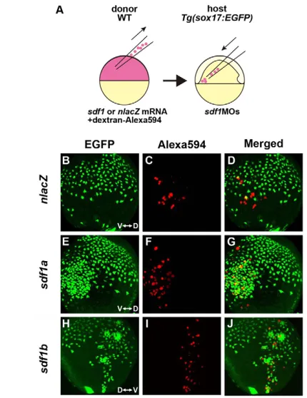
[image:4.612.49.301.53.494.2]It is now well established that Sdf1 acts as a chemoattractant for cxcr4-expressing cells (Busillo and Benovic, 2007). To test the impact of zebrafish Sdf1 on the migration of endodermal cells, we examined the consequences of transplanting nlacZ-, sdf1a- or sdf1b-expressing cells into sdf1MOs-injected embryos. The EGFP-expressing endodermal cells were found to be attracted by the cluster of sdf1a- or sdf1b-expressing cells (Fig. 3; see Movies 3, 4 and 5 in the supplementary material), indicating that both zebrafish Sdf1a and Sdf1b function as a chemoattractant for cxcr4a-expressing endodermal cells. A higher dose of sdf1amRNA (3 ng) was required to achieve an effect on the endodermal cells as compared with sdf1b mRNA (2 ng), suggesting that the Fig. 2. Sdf1/Cxcr4a signaling controls the migration of
endodermal cells during gastrulation. (Aa-Ad) sox17expression was examined by whole-mount in situ hybridization in misMO1-, cxcr4aMO1-, cxcr4aMO2- or sdf1MOs-injected zebrafish embryos at the 90% epiboly (9 hpf) stage. Lateral views, dorsal to the right. The migration of sox17-expressing endodermal cells in the animal-lateral region to the dorsal midline is delayed in the cxcr4aMO1-, cxcr4a MO2-and sdf1MOs-injected embryos (arrowheads). (Ba-Bd) Dorsal views of the mid-trunk region of Tg(sox17:EGFP)transgenic embryos at the three-somite (3s) stage. Anterior is to the top. The migration of EGFP-positive endodermal cells to the dorsal midline is delayed by the knockdown of either cxcr4aor sdf1. (Ca-Cd) Dorsal views of the pharyngeal and foregut regions of Tg(sox17:EGFP)transgenic embryos at 24 hpf. Anterior is to the top. Both the cxcr4aand sdf1 knockdown embryos show a splitting of the anterior gut (arrows). (Da-Gd) The expression of foxa3(gut and its associated organs), cp(liver), pdx1 (pancreas) and ins(β-cells in pancreas) was examined in misMO1-, cxcr4aMO1-, cxcr4aMO2- or sdf1MOs-injected embryos at 48 hpf. The expression offoxa3in the anterior part of foregut is lost in both the cxcr4a and sdf1 knockdown embryos (arrows, Db-Dd). Moreover, the liver is not formed (arrowheads, Ea-Ed), the pancreas is small and is not assembled properly (arrowheads, Fa-Fd), and insulin-producing β-cells in the pancreas do not cluster (arrowheads, Ga-Gd) in both the cxcr4a and sdf1 knockdown embryos. l, liver; p, pancreas; g, gut. Note also that no phenotypic differences in endoderm migration and organ formation among the cxcr4aMO1-, cxcr4aMO2- andsdf1MOs-injected embryos can be observed.
D
E
V
E
LO
P
M
E
N
chemoattractant effect of Sdf1b for endodermal cells is stronger than that of Sdf1a, although it is difficult to quantify such differences. Moreover, a higher density of EGFP-expressing endodermal cells was observed around the transplanted sdf1-expressing cells (Fig. 3G,J; see Movies 4 and 5 in the supplementary material). These transplantation experiments were repeated at least five times for each of the three genes (nlacZ,sdf1a and sdf1b), and we obtained consistent results for each experiment. We have already shown that sdf1a and sdf1b are expressed in mesodermal cells and that cxcr4ais expressed in endodermal cells. The endodermal cells occupy the deepest cell layer next to the extra-embryonic YSL, and the mesodermal cell layer lies above the endodermal cells, both within the hypoblast layer (see Fig. S1 in the supplementary material) (Warga and Nüsslein-Volhard, 1999). These data, combined with the results of our MO-knockdown experiments, suggest that sdf1-expressing mesodermal cells might attract endodermal cells during gastrulation.
Sdf1/Cxcr4a signaling cell-autonomously regulates the movements of endodermal cells
casmutant embryos lose the expression of cxcr4ain endodermal cells, whereas its expression is maintained in either the mesoderm or ectoderm during gastrulation (Fig. 1B). This suggests that the
Sdf1/Cxcr4a signaling pathway is also necessary for the C&E movements of mesodermal or ectodermal cells and that the inhibition of C&E movements of mesodermal cells might cause the observed delay of endodermal cell movements. To test this, we examined the position and shape of the prechordal plate, the anterior edge of the neural plate, and the body length of morphant embryos using the ntl, hgg1, dlx3and myoDmarkers (Fig. 4). Previous reports have shown that zebrafish mutants of the Wnt/PCP signaling pathway, which are defective for C&E movements, show a broadened neural plate and shortened body length (Myers et al., 2002). The shape and position of the prechordal plate and neural plate, the gap between prechordal plate and notochord, and body length were found to be normal in Sdf1/Cxcr4a signaling-inhibited embryos (Fig. 4), thereby indicating that the Sdf1/Cxcr4a signaling pathway does not govern the C&E movements of either mesodermal or ectodermal cells during gastrulation.
[image:5.612.53.268.59.338.2]To test whether Sdf1/Cxcr4a signaling directly regulates endodermal cell movements, we knocked down the function of Cxcr4a specifically within endodermal cells. This was achieved by injecting cxcr4aMO1 together with cas mRNA, because cas overexpression causes marginal blastomere cells to adopt an endodermal fate (Kikuchi et al., 2001). Using this endoderm-specific knockdown method, the influence of Cxcr4a function in mesoderm and ectoderm could be completely excluded. The endodermal cells exhibited a significant delay in their migration following the co-injection of cxcr4aMO1 and cas mRNA as compared with an injection of casmRNA alone (Fig. 5). The expression pattern and level of sdf1aand sdf1bwere unchanged in these experiments (data not shown). These data therefore suggest that the Sdf1/Cxcr4a signaling pathway cell-autonomously regulates the movements of the endodermal cells without affecting the C&E movements of the mesoderm and the ectoderm.
Fig. 3. Sdf1 acts as a chemoattractant for cxcr4a-expressing endodermal cells. (A) Schematic representation of the
[image:5.612.305.565.60.190.2]cell-transplantation approach. nlacZ,sdf1a or sdf1bmRNA was co-injected with dextran-Alexa594 into WT donor zebrafish embryos at the one-cell stage. Donor cells from dome-stage embryos were placed in the ventrolateral margin of sdf1MOs-injected host embryos at the shield stage. (B-J) Lateral views of a Tg(sox17:EGFP)transgenic embryo with anterior to the top, at TB stage. V, ventral; D, dorsal. (B-D) nlacZ -expressing donor cells do not attract the EGFP-expressing endodermal cells. (E-J)EGFP-expressing endodermal cells are attracted to the cluster of sdf1a- or sdf1b-expressing cells, which are labeled with dextran-Alexa594 (red).
Fig. 4. The C&E movements of mesoderm and ectoderm are not affected by Sdf1/Cxcr4a signaling during zebrafish gastrulation.
(A-D) The position and shape of the prechordal plate (pcp), the anterior edge of the neural plate (np) and notochord (nt) at the three-somite stage. Embryos were stained for no tail(ntl; notochord marker), hatching gland gene 1(hgg1; prechordal plate marker) and dlx3 (anterior edge of neuroectoderm) by in situ hybridization. Dorsal views, anterior to the top. No significant differences in the expression patterns of these three genes were found in either the cxcr4a or sdf1
knockdown embryos as compared with misMO1-injected embryos. White lines indicate the gap between the pcp and nt. (E-H) The body length of embryos at the three-somite stage, marked by the expression of myoD. Dorsal views, anterior to the top. The expression pattern of myoDin the cxcr4a or sdf1 knockdown embryos is indistinguishable from that in the misMO1-injected embryos.
D
E
V
E
LO
P
M
E
N
The Sdf1/Cxcr4a signaling pathway controls the morphology of endodermal cells
After involution, endodermal cells gradually flatten and extend their filopodial processes, and then form a noncontiguous inner layer of cells adjacent to the yolk (Warga and Nüsslein-Volhard, 1999). To examine whether the shape of the endodermal cells is altered when Sdf1/Cxcr4a signaling is inhibited, we carefully observed the shape and movement of individual endodermal cells in the lateral region of zebrafish embryos by confocal laser microscopy. Consistent with this previous report, the endodermal cells that we tracked exhibited many filopodial processes during their migration in live Tg(sox17:EGFP)transgenic embryos (Fig. 6A; see Movie 6 in the supplementary material). We found that the formation of these filopodia was suppressed when the function of either cxcr4aor sdf1was inhibited (Fig. 6B,C; see Movies 7 and 8 in the supplementary material). Our statistical analyses indicated that endodermal cells in either cxcr4aor sdf1knockdown embryos exhibited a more rounded shape than their counterparts in misMO1-injected embryos (Fig. 6D). We next examined the orientation of the filopodial processes in the endodermal cells and found that more than 70% of the filopodia were oriented in the direction of migration (from 0° to ±90°) in the misMO1-injected embryos (Fig. 6F). By contrast, the number of filopodial processes occurring in the direction of migration was reduced in the cxcr4aMO1- or sdf1MOs-injected embryos (Fig. 6F). In addition to the formation of filopodia, trajectory analyses of endodermal cells showed that they migrate almost straight towards the dorsal side in the misMO1-injected embryos (Fig. 6G), whereas they make many turns, and move in different (sometimes reverse) directions in both the cxcr4a and sdf1knockdown embryos (Fig. 6H,I). These results suggest that the reduced number and randomized orientation of filopodial processes that follow the inhibition of Sdf1/Cxcr4a signaling result
in a suppression of the directional migration of endodermal cells. Furthermore, we found that the total speed of migration of endodermal cells is not significantly different when Sdf1/Cxcr4a signaling is inhibited (data not shown). Therefore, it appears that the reduced level of directional migration is the major contributor to an overall delay in endodermal cell migration to the dorsal midline during gastrulation.
It has been well established that phosphoinositide 3-kinase (PI3K) plays crucial roles in filamentous actin (F-actin) assembly during chemotaxis in yeast, mammalian cells and Dictyostelium discoideum (Sasaki and Firtel, 2006). Locally activated PI3K produces PIP3at
the leading edges of the chemotactic cells, and this accumulation of PIP3leads to F-actin polymerization at these leading edges (Sasaki
and Firtel, 2006). We observed that F-actin is strongly localized at the plasma membrane and filopodia of endodermal cells in WT embryos (data not shown). We further found that co-injection of mRNA encoding a dominant-negative form ofPI3K (dnPI3K) (Montero et al., 2003) and casmRNA does not affect the migration of endodermal cells during gastrulation (see Fig. S3 in the supplementary material). We next tested whether PIP3accumulates
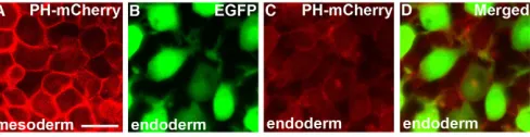
in mesendodermal cells by expressing the Pleckstrin homology (PH) domain of protein kinase B, which binds with high affinity to this molecule (Watton and Downward, 1999; Montero et al., 2003). The PH-mCherry fusion protein was strongly localized at the plasma membrane in mesodermal cells (Fig. 7A), whereas the localization of this exogenous product in endodermal cells was predominantly cytoplasmic (Fig. 7B-D). Taken together, these results suggest that the Sdf1/Cxcr4a signaling pathway controls the formation and orientation of the filopodial processes of endodermal cells, possibly through a PIP3-independent pathway, and that an abnormal filopodia
phenotype results in the impairment of the directional migration of endodermal cells.
DISCUSSION
Roles of Sdf1/Cxcr4 signaling in gastrulation and organogenesis
Our transplantation experiments showed that EGFP-expressing endodermal cells are attracted towards sdf1a- or sdf1b-expressing transplanted cells. These results, combined with the data of single-knockdown experiments of sdf1a or sdf1b, suggest that although Sdf1b has stronger effects on endodermal migration than Sdf1a, the factors are redundant chemoattractants for endodermal cells. We further found a higher density of endodermal cells around the sdf1-expressing transplanted cells. The number of endodermal cells was not altered in Sdf1/Cxcr4a signaling-inhibited embryos at the 60% epiboly stage, but it showed a ~14-24% reduction at the 90% epiboly stage by in situ hybridization using sox17as a probe (data not shown). These data suggest that Sdf1/Cxcr4a signaling might be involved in the proliferation, but not specification, of endodermal cells. Since it is well known that Sdf1/Cxcr4 signaling promotes the growth of primary tumors (Luker and Luker, 2006), excess Sdf1 secreted from the transplanted cells might enhance the proliferation of endodermal cells. It will be interesting to analyze the role of Sdf1/Cxcr4a signaling in the proliferation of endodermal cells during gastrulation.
[image:6.612.52.282.59.202.2]In Sdf1/Cxcr4a signaling-inhibited embryos, the anterior part of gut tube and liver are lost, and the pancreas is small and improperly assembled (Fig. 2). It appears that the delay of endodermal cell migration caused by inhibition of Sdf1/Cxcr4a signaling results in defects in endodermal organ patterning. Another possible explanation is that Sdf1/Cxcr4a-dependent movement might be necessary for the differentiation or patterning of the endodermal Fig. 5. Cxcr4a signaling cell-autonomously regulates the
movements of endodermal cells. (A-F) Confocal images of the endodermal cells in Tg(sox17:EGFP)transgenic zebrafish embryos at the three-somite stage. Dorsal views, anterior to the top. One marginal blastomere cell at the 128-cell stage has been transformed into an endodermal fate by the misexpression of casmRNA. The injected cell was labeled with dextran-Alexa594. (A-C) The progeny of the injected cell intermingle with EGFP-expressing endodermal cells. (D-F) The migration of both casmRNA and cxcr4aMO1 co-injected cells was significantly delayed compared with cells injected with casmRNA only (arrowheads, E,F). (C,F) Note that the width from the midline to the lateral margin of the endodermal cell layer in the casmRNA + cxcr4aMO1 co-injected embryos is broader than that of the cas mRNA-injected embryos because the movement of the endodermal cells is delayed (areas medial and lateral to the midline are indicated by the double-headed arrows).
D
E
V
E
LO
P
M
E
N
organs at the somitogenesis stage. Further analyses will be required to elucidate the function of Sdf1/Cxcr4a signaling in endodermal organ formation.
The morphology of endodermal cells during gastrulation
It has been reported that endodermal cells begin to change their morphology between 70% and 80% epiboly during gastrulation (Warga and Nüsslein-Volhard, 1999). Having first adopted a tear-drop shape, the endodermal cells gradually flatten, lose their polar appearance and develop filopodial processes (Warga and Nüsslein-Volhard, 1999). Our confocal time-lapse analysis of
[image:7.612.57.470.208.598.2]Tg(sox17:EGFP) transgenic embryos is consistent with this report. We show that endodermal cells in the zebrafish gastrula have many filopodial processes during dorsal migration, and that their morphology is regulated by Sdf1/Cxcr4a signaling. Recently, the chemokine Apelin and its receptor Agtrl1b (Aplnrb – ZFIN) were shown to regulate the protrusion of filolamellipodia and the movement of lateral mesodermal cells during zebrafish gastrulation (Scott et al., 2007; Zeng et al., 2007). Together, these results suggest that chemokine-GPCR signaling controls the movements of mesendodermal cells through regulation of their morphology. We propose that endodermal cells develop filopodia so that they can assess the correct direction in which to migrate.
Fig. 6. Sdf1/Cxcr4a signaling is required for the correct morphology of endodermal cells during zebrafish gastrulation. (A-C) Confocal images of EGFP-expressing endodermal cells at 90% epiboly from a time-lapse video. The direction of migration is towards the right. The endodermal cells show many filopodial processes in the misMO1-injected embryos, whereas both the cxcr4aMO1- and sdf1MOs-injected endodermal cells have fewer of these processes and are more rounded. (D) Cell circularity from 4-5 randomly chosen cells measured at two consecutive time points (time interval, 10 minutes) from 9-10 independent movies in misMO1- (n=100 cells), cxcr4aMO1- (n=99 cells) and sdf1MOs- (n=87 cells) injected embryos. Cell circularity is calculated as follows: circularity=4πA/p2, where A=area and p=perimeter. Note that the
differences between the misMO1 and cxcr4aMO1 values, as well as the differences between the misMO1 and sdf1MOs values, are statistically significant (*P<0.01, Student‘st-test). Error bars represent the standard error. (E) Schematic representation of the methods used to measure the angles of the filopodial processes relative to the direction of migration of the endodermal cells. ±180° indicates opposing the direction of migration. (F) Rose diagrams representing the orientation of filopodial processes relative to the direction of migration. The orientation of the filopodia from 4-5 randomly chosen cells was measured at two consecutive time points (time interval, 10 minutes) from 9-10 independent movies in misMO1- (n=100 cells), cxcr4aMO1- (n=99 cells) and sdf1MOs- (n=87 cells) injected embryos. (G-I) The migration tracks of 10-11 individual endodermal cells over a period of 30 minutes. The direction of migration is to the right and the cell position was determined every 30 seconds. Scale bars: 20 μm.
D
E
V
E
LO
P
M
E
N
This idea is supported by the fact that more than 70% of the filopodial processes are orientated in the direction of migration. It will be interesting to analyze the roles of the filopodial processes that are oriented independently of the direction of migration.
Signaling pathways controlling endodermal cell migration during gastrulation
A very recent study showed that in zebrafish embryos, internalized endodermal cells display a nonoriented/noncoordinated movement, described as a random walk, up until mid-gastrulation (75% epiboly stage), and then show directional migration from mid-gastrulation onwards (Pézeron et al., 2008). We found that the distribution of endodermal cells at 60% epiboly (identified by in situ hybridization with a cas riboprobe) is wild-type in sdf1/cxcr4a knockdown embryos (data not shown), suggesting that the random walk upon internalization is independent of Sdf1/Cxcr4a signaling. Moreover, we found that the movement and directional migration of endodermal cells in late gastrulae is impaired but not completely lost as a result of inhibition of Sdf1/Cxcr4a signaling. There are two possible explanations for this. One is that another factor shares redundancy of function with either Sdf1 or Cxcr4a. Alternatively, the migration of endodermal cells might be controlled by additional systems acting in concert with Sdf1/Cxcr4a signaling. To date, two signaling pathways, Wnt/PCP signaling and Vascular endothelial growth factor C (Vegfc) signaling, have been implicated in the regulation of endodermal cell migration (Ober et al., 2004; Matsui et al., 2005).
Genetic and molecular evidence has accumulated to demonstrate that the Wnt/PCP signaling pathway regulates the C&E movements of mesodermal cells during vertebrate gastrulation (Myers et al., 2002). Recently, the role of Wnt/PCP signaling in C&E movements in endodermal cell migration was also reported in zebrafish embryos (Matsui et al., 2005). Overexpression of a dominant-negative form of Dishevelled or combinatorial inhibition of Wnt ligands leads to a split gut and bilateral endodermal organs, suggesting that the Wnt/PCP signaling pathway is required for migration of endodermal cells toward the dorsal midline (Matsui et al., 2005). It is possible that the requirement of Wnt/PCP signaling for endodermal cell migration occurs during two developmental stages: gastrulation and somitogenesis. We found that the migration of endodermal cells is marginally delayed when the Wnt/PCP signaling pathway is inhibited exclusively in endodermal cells during gastrulation (T.M. and Y.K., unpublished). In addition to gastrulation, wnt4a and wnt11-relatedbegin to be strongly expressed during somitogenesis in the neural ectoderm and mesendoderm, and the knockdown of three Wnt ligands (wnt4a, wnt11-relatedand wnt11) inhibits the migration of endodermal cells toward the dorsal midline (Matsui et
al., 2005). These data, combined with our unpublished observations, suggest that Wnt/PCP signaling is partly involved in endodermal cell migration during gastrulation and is required for this process during somitogenesis.
A previous study revealed that the Vegfc signaling pathway regulates the migration of endodermal cells in zebrafish embryos (Ober et al., 2004). Knockdown experiments have demonstrated that Vegfc signaling is required for two distinct steps during endoderm development, the first being the initial differentiation of the dorsal endoderm, and the second involving the coalescence of the anterior endoderm with the dorsal midline (Ober et al., 2004). vegfcknockdown embryos show a splitting of the anterior gut tube, in the most severe cases accompanied by a duplication of the liver and pancreatic bud (Ober et al., 2004). Taken together, we speculate from the current evidence that either the Wnt/PCP or Vegfc signaling pathway partially compensates for the movement and directional migration of endodermal cells in sdf1/cxcr4a knockdown embryos.
Roles of PI3K and PIP3in endodermal cell
migration
Sdf1 stimulation through Cxcr4 locally activates Ras at the leading edge of the migrating cell (Busillo and Benovic, 2007). PI3K is well established as one of the downstream effectors of Ras signaling (Sasaki and Firtel, 2006). Moreover, PIP3accumulation by PI3K
activity occurs exclusively at the leading edge, and its local accumulation leads to the assembly of a branched network of actin filaments (Sasaki and Firtel, 2006). Recent reports have shown that the migration of the primordial germ cells (PGCs) is guided by Sdf1 in zebrafish (Raz and Reichman-Fried, 2006). Zebrafish PGCs exhibit a uniform distribution of PIP3on their membrane and
depletion of PIP3in these cells has no effect on their ability to
migrate in the correct direction (Dumstrei et al., 2004). In addition, a more recent study has revealed that cellular calcium is required for proper migration and formation of bleb-like protrusions in PGCs (Blaser et al., 2006). These results suggest that the PIP3
-independent mechanisms could regulate the directional migration of these cells. In addition, Dictyostelium cells in which PIP3
production is reduced remain responsive to directional cues (Sasaki and Firtel, 2006). In our current study, we find that the migration of endodermal cells is regulated by Sdf1 from mesodermal cells and that F-actin is localized to the filopodial processes in migrating endodermal cells. However, the migration of endodermal cells is not affected by misexpression of dnPI3K, and a significant accumulation of PIP3at the filopodia is not observed in endodermal
cells as compared with their mesodermal counterparts. These data suggest that F-actin polymerization might be controlled in a PIP3
-independent manner in endodermal cells. Future analyses of the F-actin polymerization mechanisms that regulate Sdf1/Cxcr4a signaling in the filopodia of endodermal cells should further our understanding of the molecular events underlying their directional migration.
We thank Tokuko Niwa and Hiroko Nishii for technical assistance and Carl-Philipp Heisenberg, John Kuwada, Wataru Shoji and Roger Y. Tsien for providing DNA templates. This work was supported by grants from the Takeda Science Foundation and a Grant-in-Aid for Scientific Research from the JSPS to Y.K., a Howard Florey Centenary Fellowship and a project grant (491087) from the NHMRC Australia to H.V., and a project grant from the NHMRC Australia (433614) to J.K.H.
Supplementary material
[image:8.612.55.299.61.124.2]Supplementary material for this article is available at http://dev.biologists.org/cgi/content/full/135/15/2521/DC1
Fig. 7. Specific subcellular localization of PH-mCherry is not observed in migrating endodermal cells. (A-D) Confocal analysis of PH-mCherry localization in mesodermal and endodermal cells at the 90% epiboly stage. In mesodermal cells, PH-mCherry is significantly localized at the cell membrane (A), but this is not observed in endodermal cells (B-D). Scale bar: 20 μm.
D
E
V
E
LO
P
M
E
N
References
Akimenko, M. A., Ekker, M., Wegner, J., Lin, W. and Westerfield, M.(1994). Combinatorial expression of three zebrafish genes related to distal-less: part of a homeobox gene code for the head. J. Neurosci.14, 3475-3486.
Alexander, J. and Stainier, D. Y.(1999). A molecular pathway leading to endoderm formation in zebrafish. Curr. Biol.9, 1147-1157.
Blaser, H., Reichman-Fried, M., Castanon, I., Dumstrei, K., Marlow, F. L., Kawakami, K., Solnica-Krezel, L., Heisenberg, C. P. and Raz, E.(2006). Migration of zebrafish primordial germ cells: a role for myosin contraction and cytoplasmic flow. Dev. Cell11, 613-627.
Busillo, J. M. and Benovic, J. L.(2007). Regulation of CXCR4 signaling. Biochim. Biophys. Acta1768, 952-963.
Chong, S. W., Emelyanov, A., Gong, Z. and Korzh, V.(2001). Expression pattern of two zebrafish genes, cxcr4aand cxcr4b. Mech. Dev.109, 347-354. Chong, S. W., Nguyet, L. M., Jiang, Y. J. and Korzh, V.(2007). The chemokine
Sdf-1 and its receptor Cxcr4 are required for formation of muscle in zebrafish. BMC Dev. Biol.7, 54.
David, N. B., Sapède, D., Saint-Etienne, L., Thisse, C., Thisse, B., Dambly-Chaudière, C., Rosa, F. M. and Ghysen, A.(2002). Molecular basis of cell migration in the fish lateral line: role of the chemokine receptor CXCR4 and of its ligand, SDF1. Proc. Natl. Acad. Sci. USA99, 16297-16302.
Doitsidou, M., Reichman-Fried, M., Stebler, J., Köprunner, M., Dörries, J., Meyer, D., Esguerra, C. V., Leung, T. and Raz, E.(2002). Guidance of primordial germ cell migration by the chemokine SDF-1. Cell111, 647-659. Dumstrei, K., Mennecke, R. and Raz, E.(2004). Signaling pathways
controlling primordial germ cell migration in zebrafish. J. Cell Sci.117, 4787-4795.
Field, H. A., Ober, E. A., Roeser, T. and Stainier, D. Y.(2003). Formation of the digestive system in zebrafish. I. Liver morphogenesis. Dev. Biol.253, 279-290.
Fukuda, K. and Kikuchi, Y.(2005). Endoderm development in vertebrates: fate mapping, induction and regional specification. Dev. Growth Differ.47, 343-355.
Fukui, A., Goto, T., Kitamoto, J., Homma, M. and Asashima, M.(2007). SDF-1αregulates mesendodermal cell migration during frog gastrulation. Biochem. Biophys. Res. Commun.354, 472-477.
Jékely, G. and Arendt, D.(2007). Cellular resolution expression profiling using confocal detection of NBT/BCIP precipitate by reflection microscopy. Biotechniques42, 751-755.
Kikuchi, Y., Agathon, A., Alexander, J., Thisse, C., Waldron, S., Yelon, D., Thisse, B. and Stainier, D. Y.(2001). casanovaencodes a novel Sox-related protein necessary and sufficient for early endoderm formation in zebrafish. Genes Dev.15, 1493-1505.
Kimmel, C. B., Ballard, W. W., Kimmel, S. R., Ullmann, B. and Schilling, T. F. (1995). Stages of embryonic development of the zebrafish. Dev. Dyn.203, 253-310.
Knaut, H., Werz, C., Geisler, R. and Nüsslein-Volhard, C.(2003). A zebrafish homologue of the chemokine receptor Cxcr4is a germ-cell guidance receptor. Nature421, 279-282.
Korzh, S., Emelyanov, A. and Korzh, V.(2001). Developmental analysis of ceruloplasmingene and liver formation in zebrafish. Mech. Dev.103, 137-139. Kucia, M., Jankowski, K., Reca, R., Wysoczynski, M., Bandura, L., Allendorf,
D. J., Zhang, J., Ratajczak, J. and Ratajczak, M. Z.(2004). CXCR4-SDF-1 signalling, locomotion, chemotaxis and adhesion. J. Mol. Histol.35, 233-245. Li, Q., Shirabe, K. and Kuwada, J. Y.(2004). Chemokine signaling regulates
sensory cell migration in zebrafish. Dev. Biol.269, 123-136.
Luker, K. E. and Luker, G. D.(2006). Functions of CXCL12 and CXCR4 in breast cancer. Cancer Lett.238, 30-41.
Matsui, T., Raya, A., Kawakami, Y., Callol-Massot, C., Capdevila, J., Rodríguez-Esteban, C. and Izpisua Belmonte, J. C.(2005). Noncanonical Wnt signaling regulates midline convergence of organ primordia during zebrafish development. Genes Dev.19, 164-175.
Milewski, W. M., Duguay, S. J., Chan, S. J. and Steiner, D. F.(1998). Conservation of PDX-1 structure, function, and expression in zebrafish. Endocrinology139, 1440-1449.
Miyasaka, N., Knaut, H. and Yoshihara, Y.(2007). Cxcl12/Cxcr4 chemokine signaling is required for placode assembly and sensory axon pathfinding in the zebrafish olfactory system. Development134, 2459-2468.
Mizoguchi, T., Izawa, T., Kuroiwa, A. and Kikuchi, Y.(2006). Fgf signaling negatively regulates Nodal-dependent endoderm induction in zebrafish. Dev. Biol.300, 612-622.
Montero, J. A. and Heisenberg, C. P.(2004). Gastrulation dynamics: cells move into focus. Trends Cell Biol.14, 620-627.
Montero, J. A., Kilian, B., Chan, J., Bayliss, P. E. and Heisenberg, C. P.(2003). Phosphoinositide 3-kinase is required for process outgrowth and cell polarization of gastrulating mesendodermal cells. Curr. Biol.13, 1279-1289.
Myers, D. C., Sepich, D. S. and Solnica-Krezel, L.(2002). Convergence and extension in vertebrate gastrulae: cell movements according to or in search of identity? Trends Genet.18, 447-455.
Nasevicius, A. and Ekker, S. C.(2000). Effective targeted gene ‘knockdown’ in zebrafish. Nat. Genet.26, 216-220.
Ober, E. A., Olofsson, B., Makinen, T., Jin, S. W., Shoji, W., Koh, G. Y., Alitalo, K. and Stainier, D. Y.(2004). Vegfc is required for vascular development and endoderm morphogenesis in zebrafish. EMBO Rep.5, 78-84.
Odenthal, J. and Nüsslein-Volhard, C.(1998). fork headdomain genes in zebrafish. Dev. Genes Evol.208, 245-258.
Pézeron, G., Mourrain, P., Courty, S., Ghislain, J., Becker, T. S., Rosa, F. M. and David, N. B.(2008). Live analysis of endodermal layer formation identifies random walk as a novel gastrulation movement. Curr. Biol.18, 276-281. Raz, E. and Reichman-Fried, M.(2006). Attraction rules: germ cell migration in
zebrafish. Curr. Opin. Genet. Dev.16, 355-359.
Reim, G., Mizoguchi, T., Stainier, D. Y., Kikuchi, Y. and Brand, M.(2004). The POU domain protein spg (pou2/Oct4) is essential for endoderm formation in cooperation with the HMG domain protein casanova. Dev. Cell 6, 91-101.
Sasaki, A. T. and Firtel, R. A.(2006). Regulation of chemotaxis by the orchestrated activation of Ras, PI3K, and TOR. Eur. J. Cell Biol. 85, 873-895. Schulte-Merker, S., van Eeden, F. J., Halpern, M. E., Kimmel, C. B. and
Nüsslein-Volhard, C.(1994). no tail(ntl) is the zebrafish homologue of the mouse T(Brachyury) gene. Development120, 1009-1015.
Scott, I. C., Masri, B., D’Amico, L. A., Jin, S.-W., Jungblut, B., Wehman, A. M., Baier, H., Audigier, Y. and Stainier, D. Y.(2007). The G protein-coupled receptor Agtrl1b regulates early development of myocardial progenitors. Dev. Cell12, 403-413.
Sepich, D. S., Calmelet, C., Kiskowski, M. and Solnica-Krezel, L.(2005). Initiation of convergence and extension movements of lateral mesoderm during zebrafish gastrulation. Dev. Dyn.234, 279-292.
Shaner, N. C., Campbell, R. E., Steinbach, P. A., Giepmans, B. N., Palmer, A. E. and Tsien, R. Y.(2004). Improved monomeric red, orange and yellow fluorescent proteins derived from Discosomasp. red fluorescent protein. Nat. Biotechnol.22, 1567-1572.
Solnica-Krezel, L.(2005). Conserved patterns of cell movements during vertebrate gastrulation. Curr. Biol.15, R213-R228.
Thisse, C., Thisse, B., Halpern, M. E. and Postlethwait, J. H.(1994). goosecoid expression in neurectoderm and mesendoderm is disrupted in zebrafish cyclops gastrulas. Dev. Biol.164, 420-429.
Warga, R. M. and Nüsslein-Volhard, C.(1999). Origin and development of the zebrafish endoderm. Development126, 827-838.
Watton, S. J. and Downward, J.(1999). Akt/PKB localisation and 3⬘
phosphoinositide generation at sites of epithelial cell-matrix and cell-cell interaction. Curr. Biol.9, 433-436.
Weinberg, E. S., Allende, M. L., Kelly, C. S., Abdelhamid, A., Murakami, T., Andermann, P., Doerre, O. G., Grunwald, D. J. and Riggleman, B.(1996). Developmental regulation of zebrafish MyoDin wild-type, no tailand spadetail embryos. Development122, 271-280.
Westerfield, M.(1995). The Zebrafish Book. University of Oregon Press, Eugene, OR.
Zeng, X.-X, Wilm, T. P., Sepich, D. S. and Solnica-Krezel, L.(2007). Apelin and its receptor control heart field formation during zebrafish gastrulation. Dev. Cell 12, 391-402.