D
E
V
E
LO
P
M
E
N
T
INTRODUCTION
Midbrain dopaminergic (mDA) neurons represent an important class of neuronal subtype that has important functional roles. These neurons are lost in Parkinson disease patients, which are characterised by loss of control of body movements (Lang and Lozano, 1998; Barzilai and Melamed, 2003). In addition, mDA neurons regulate reward-based behaviours and have been implicated in schizophrenia and attention-deficit disorders (Tzschentke and Schmidt, 2000).
mDA neurons arise from neural progenitors in the ventral midline, probably corresponding to the floor plate region. The generation of mDA neurons from a neural progenitor cell can be divided into three distinct phases based on the expression of molecular markers and the state of differentiation: regional and neuronal specification (phase I), early (phase II) and late differentiation (phase III) (reviewed by Smits et al., 2006; Ang, 2006). During phase I, sonic hedgehog (Shh), fibroblast growth factor 8 (Fgf8), Wnt1 and other extrinsic factors control regional and neuronal identity by regulating the expression of Otx2 (Ye et al., 1998; Puelles et al., 2004; Vernay et al., 2005; Prakash et al., 2006), Lmx1a (Andersson et al., 2006a), Lmx1b (Smidt et al., 2000), Msx1 and Msx2 (Andersson et al., 2006a), neurogenin 2 (Ngn2; also known as Neurog2 – Mouse Genome Informatics) and Mash1 (also known as Ascl1) (Anderson et al., 2006b; Kele
et al., 2006). Subsequently these progenitors undergo an early differentiation step to generate immature mDA neurons expressing Ngn2, Nurr1 (also known as Nr4a2), Lmx1a, Lmx1b, engrailed 1 and engrailed 2 (En1/2) and III-tubulin (Tuj1; also known as Tubb3) (reviewed by Wallen and Perlmann, 2003; Prakash and Wurst, 2006; Smits et al., 2006; Ang, 2006). Immature mDA neurons then further differentiate into mature DA neurons that express tyrosine hydroxylase (TH) and aromatic L-amino acid decarboxylase (AADC), in addition to the other markers mentioned above in immature neurons. Loss-of-function studies in mice have revealed roles for Nurr1 (Zetterstrom et al., 1997), Lmx1b (Smidt et al., 2000), Pitx3 (Hwang et al., 2003; Nunes et al., 2003; van den Munckhof et al., 2003) and En1/2 (Simon et al., 2001; Alberi et al., 2004) in late differentiation and maintenance of these neurons.
Less is known about the role of transcription factors in specification and early differentiation of mDA progenitors. The homeodomain protein Otx2 regulates mDA specification by inhibiting the expression of Nkx2.2 homeodomain protein (Puelles et al., 2004; Prakash et al., 2006) and regulating the expression of the proneural basic helix-loop-helix genes Ngn2and Mash1. Ngn2 has also been shown to be the key proneural gene regulating neurogenesis of mDA progenitors in the midbrain; Mash1 is partially able to compensate for Ngn2 function (Andersson et al., 2006b; Kele et al., 2006). Msx genes, Msx1and Msx2, are able to prematurely turn on Ngn2 expression and neurogenesis when expressed earlier in mice carrying a Shh enhancer-Msx1transgene (Andersson et al., 2006a). In addition, there is a reduction of mDA neurons in Msx1–/–mutant embryos. The LIM homeodomain protein Lmx1a has been shown to be necessary and sufficient for the generation of mDA neurons in the ventral midbrain of chick embryos and can promote the generation of mDA neurons from mouse ES cells treated with Fgf8 and Shh (Andersson et al., 2006a). However, Lmx1a is unable to generate mDA neurons in the absence of Shh or from
Foxa1
and
Foxa2
regulate multiple phases of midbrain
dopaminergic neuron development in a dosage-dependent
manner
Anna L. M. Ferri1,*, Wei Lin1,*, Yannis E. Mavromatakis1, Julie C. Wang1, Hiroshi Sasaki2, Jeffrey A. Whitsett3 and Siew-Lan Ang1,†
The role of transcription factors in regulating the development of midbrain dopaminergic (mDA) neurons is intensively studied owing to the involvement of these neurons in diverse neurological disorders. Here we demonstrate novel roles for the
forkhead/winged helix transcription factors Foxa1 and Foxa2 in the specification and differentiation of mDA neurons by analysing
the phenotype of Foxa1and Foxa2 single- and double-mutant mouse embryos. During specification, Foxa1 and Foxa2 regulate the
extent of neurogenesis in mDA progenitors by positively regulating Ngn2 (Neurog2) expression. Subsequently, Foxa1 and Foxa2
regulate the expression of Nurr1 (Nr4a2) and engrailed 1 in immature neurons and the expression of aromatic L-amino acid
decarboxylase and tyrosine hydroxylase in mature neurons during early and late differentiation of midbrain dopaminergic neurons.
Interestingly, genetic evidence indicates that these functions require different gene dosages of Foxa1and Foxa2. Altogether, our
results demonstrate that Foxa1 and Foxa2 regulate multiple phases of midbrain dopaminergic neuron development in a dosage-dependent manner.
KEY WORDS: Forkhead genes, Cell fate specification, Differentiation, Dopaminergic, Midbrain, Gene dosage
Development 134, 2761-2769 (2007) doi:10.1242/dev.000141
1Division of Developmental Neurobiology, MRC National Institute for Medical
Research, The Ridgeway, Mill Hill, London NW7 1AA, UK. 2Laboratory for Embryonic
Induction, RIKEN Center for Developmental Biology, 2-2-3 Minatojima-minamimachi, Chuo-ku, Kobe, Hyogo 650-0047, Japan.3Division of Pulmonary Biology, Cincinnati
Children’s Hospital Medical Center and University of Cincinnati College of Medicine, 3333 Burnet Avenue, Cincinnati, OH 45229-3039, USA.
*These authors contributed equally to this work
†Author for correspondence (e-mail: [email protected])
D
E
V
E
LO
P
M
E
N
T
dorsal midbrain progenitors, indicating that other molecules that contribute to the specification of mDA progenitors remain to be discovered.
In search of other factors regulating development of mDA neurons, we have focused on Foxa1 and Foxa2 (Foxa1/2), members of the forkhead/winged helix transcription factors, because Foxa1/2, but not Foxa3, are expressed in a wide domain in ventral midbrain progenitors (Ang et al., 1993; Monaghan et al., 1993; Sasaki and Hogan, 1993). Foxa2-null mutants die at embryonic day (E) 9.5 owing to gastrulation defects (Ang and Rossant, 1994; Weinstein et al., 1994). These mutant embryos lack a node and notochord; consequently, floor plate development and dorsal-ventral patterning of the neural tube are also disrupted owing to loss of Shh signalling from the notochord (Ang and Rossant, 1994). In addition, when Foxa2 is expressed ectopically in the dorsal mid-hindbrain region of En1-Foxa2 transgenic mouse embryos, ectopic floor plate-like regions develop in the dorsal hindbrain, suggesting a role for Foxa2 in the development of the floor plate in mice (Sasaki and Hogan, 1994); however, in these studies it is unclear whether Foxa2 or Shh, a downstream target, is responsible for this effect. A recent study on zebrafish foxa2 (also known as monorail and axial), has also demonstrated a role for Foxa2 in floor plate differentiation and specification of oligodendrocytes, serotonergic raphe neurons and cranial motoneurons in zebrafish embryos (Norton et al., 2005). In contrast to the early function of Foxa2, Foxa1-null embryos do not show early patterning defects, but Foxa1 is required for the regulation of pancreatic islet genes essential for glucose homeostasis (Kaestner et al., 1999). Foxa1/2 also regulate metabolic responses during gluconeogenesis (Zhang et al., 2005), and are required for the development of endodermal organs including liver (Lee et al., 2005), pancreas (Lantz et al., 2004), prostate (Gao et al., 2003) and lung (Wan et al., 2004).
In this study, we show that Foxa1/2 are continually required during multiple phases of mDA neuron development. Our data, from loss-of-function studies in mice, demonstrate that Foxa1/2 regulate the extent of neurogenesis in mDA progenitors by positively regulating Ngn2 expression. Subsequently, Foxa1/2 are required for the expression of Nurr1 and En1 in immature mDA neurons, and for the expression of TH and AADC in mature mDA neurons during early and late differentiation of mDA neurons. Genetic evidence indicates that these functions require different gene dosages of Foxa1/2. Altogether, these data demonstrate that Foxa1/2 regulate multiple phases of mDA neuron development in a dosage-dependent manner.
MATERIALS AND METHODS
Generation and genotyping of mutant embryos and animals
Nestin-Cretransgenic mice and the Foxa2flox/floxand Foxa1lacZ/lacZmouse
strains were maintained in a mixed MF1-129/SV background. To obtain conditional Foxa2mutants, we crossed Nestin-Cretransgenic mice (Isaka et al., 1999) with animals homozygous for the Foxa2floxallele (Hallonet et al.,
2002). Nestin-Cre/+;Foxa2flox/+ male animals were then mated to Foxa2flox/floxfemales. Nestin-Cre/+;Foxa2flox/+males were also mated with Foxa1lacZ/+ females (Kaestner et al., 1999) to generate Nestin-Cre/+;Foxa2flox/+;Foxa1lacZ/+male animals. Simultaneously, Foxa2flox/flox
males were mated with Foxa1lacZ/+ females to generate Foxa2flox/+;Foxa1lacZ/+female animals. Foxa2cko;Foxa1lacZ/lacZmutants were
obtained by crossing Nestin-Cre/+;Foxa2flox/+;Foxa1lacZ/+ males with Foxa2flox/flox;Foxa1lacZ/+females. The Foxa2floxand Foxa1lacZalleles were
detected by PCR (Hallonet et al., 2002; Kaestner et al., 1999), and the Cre
transgene was detected using a pair of primers and PCR conditions as described by Indra et al. (Indra et al., 1999). Shhflox/flox(Lewis et al., 2001)
animals were purchased from Jackson Laboratories. At all times, animals
were handled according to the Society of Neuroscience Policy on the Use of Animals in Neuroscience Research, as well as the European Communities Council Directive.
Immunohistochemistry of brain sections
Embryos or dissected brains were fixed overnight at 4°C in 4% paraformaldehyde in 0.1 M PBS and cryoprotected with 30% sucrose in PBS, embedded in OCT compound (VWR International, Poole, UK) and sectioned on a cryostat (CM3050S; Leica, Nussloch, Germany). A minimum of three control and three mutant embryos were analysed, except where indicated. For immunohistochemistry, sections were incubated overnight at 4°C with the appropriate primary antibody diluted in 1% BSA in PBS. Sections were then extensively washed in PBS containing 0.1% BSA and incubated 1 hour at room temperature with a secondary antibody conjugated with a fluorochrome (Molecular Probes). Sections were then washed and mounted in Vectashield H-1000 (Vector Laboratories, Burlingame, CA). The following primary antibodies were used: rabbit anti-Foxa2 (1:1000) (Filosa et al., 1997), goat Foxa2 (1:100; sc-6554, Santa Cruz), guinea-pig anti-Foxa1 (1:500) (Wan et al., 2004), rabbit anti-Lmx1a (1:1000; gift of M. German, University of California San Francisco Diabetes Center, San Francisco, CA), guinea-pig anti-Lmx1b (1:20,000; gift of T. Müller and C. Birchmeier, Max-Delbruck-Center for Molecular Medicine, Berlin, Germany), mouse anti-Nkx2.2 [1:5; 74.5A5, Developmental Studies Hybridoma Bank (DSHB)], rabbit anti-Nkx6.1 (1:1500; gift of Martyn Goulding, Salk Institute, La Jolla, CA), rabbit anti-TH (1:200; AB152, Chemicon), mouse anti-En1 (1:40; 4G11, DSHB), rabbit anti-AADC (1:200; ab3905), mouse anti-Ngn2 (1:5 ; gift of D. J. Anderson, California Institute of Technology, Pasadena, CA), rabbit anti-Shh (1:100; sc-9024, Santa Cruz), mouse Brn3a (1:100; sc-8429, Santa Cruz), mouse anti-Islet 1 (1:20; 40.2D6, DSHB), rabbit anti-Nurr1 (1:200; sc-990, Santa Cruz), and mouse anti-Tuj1+(1:1000; SDL.3D10, Sigma). In some cases, staining of nuclei with Toto-3 iodide (1:1000, Molecular Probes) was performed. All images were collected on a Leica TCS SP2 confocal microscope and processed with Adobe Photoshop 7.0 software (Adobe Systems, San Jose, CA). Quantitative immunocytochemical data represent mean±s.d. for cell counts of half the ventral midbrain in consecutive sections through the entire midbrain, every 60 m. The percentage of Nkx2.2+cells in Lmx1a+mDA progenitor domain=average percentage of midbrain Lmx1a+ Nkx2.2+ cells/number of midbrain Lmx1a+cells of mutant embryo 1 and mutant embryo 2=(24/1398⫻100+32/1871⫻100)/2.
Terminal deoxynucleotidyl transferase-mediated biotinylated UTP nick-end labelling (TUNEL)
Cryostat sections were washed once for 5 minutes in PBS containing 0.1% Triton X-100, permeabilised in ice-cold 0.01 M citrate buffer and 0.1% Triton X-100 for 2 minutes, and washed again in PBS containing 0.1% Triton X-100. The enzymatic reaction was then performed at 37°C according to the manufacturer’s protocol (1 684 795; Roche Diagnostics, Mannheim, Germany).
BrdU labelling
Pregnant females were injected intraperitoneally with a solution of BrdU (B-5002, Sigma; at 10 mg/ml in physiological serum) at 100 g per g of body weight and sacrificed 1 hour later. Proliferating cells were revealed by immunohistochemistry on frozen sections.
RESULTS
Foxa1/2 are expressed in ventral midbrain progenitors and neurons
D
E
V
E
LO
P
M
E
N
T
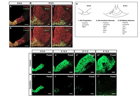
in mDA progenitors at E10.5 (Fig. 1A,B). Foxa1/2 were also expressed in Nurr1+(Fig. 1C,D) immature and TH+mature mDA neurons (Fig. 1E,F) and Brn3a+(also known as Pou4f1 – Mouse Genome Informatics) red nuclei neurons, but not in Islet 1+(Isl1) oculomotor (see Fig. S1 in the supplementary material) neurons in the ventral midbrain at E12.5. In summary, Foxa1/2 proteins are expressed in all ventral midbrain progenitors and some postmitotic neurons, including all stages of mDA cells, suggesting possible roles in regulating their specification and differentiation. A summary of the expression of Foxa1/2 proteins and other markers of mDA cells used in this study is provided in Fig. 1G.
Specific inactivation of Foxa2 in mDA progenitors from E10.5 onwards
Since Foxa2-null mutant embryos die around E9.5 owing to gastrulation defects, we used the Cre-loxP system to specifically inactivate Foxa2 in the CNS in order to study its role in the development of mDA neurons. The Nestin-Cretransgenic line, which we have previously characterised extensively (Vernay et al., 2005), was used to inactivate Foxa2 in CNS progenitors from E10.5 onwards. Analysis of Foxa2 expression by immunohistochemistry revealed that loss of Foxa2 protein in Nestin-Cre/+;Foxa2flox/flox (referred to henceforth as Foxa2cko) embryos began at E10.5. In Foxa2cko embryos, Foxa2 was only detected in some cells within the floor plate
in the midbrain at E10.5, and by E12.5 all Foxa2 expression was missing from the midbrain (Fig. 1H-H,I-I). This timing of deletion of Foxa2 by Nestin-Crecorrelates well with the timing of activation of -galactosidase expression in Nestin-Cre/+;R26Rflox/+embryos (Vernay et al., 2005). Hence, Foxa2ckoembryos allow us to study the functions of Foxa2 in mDA progenitors during neurogenesis, which occurs mainly between E10.5 and E13.5.
Foxa1/2 positively regulate the expression of Ngn2 in mDA progenitors
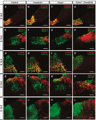
mDA progenitors are distinguishable from other progenitors in the ventral midbrain by their co-expression of Lmx1a/b at E10.5. Lmx1a and Lmx1b were found to be similarly expressed in mDA progenitors at E10.5 and E12.5, indicating that these markers are properly specified in these progenitors in Foxa2flox/flox(control), Foxa2cko, Foxa1lacZ/lacZ(henceforth referred to as Foxa1–/–) and Foxa1–/–;Foxa2cko double-mutant embryos (Fig. 2A-D,A⬘-D⬘). Msx1, a homeodomain protein, is also normally expressed in mDA progenitors in wild-type and all mutant embryos (Andersson et al., 2006a) (data not shown).
[image:3.612.51.535.55.391.2]Nkx homeodomain proteins, Nkx2.2 and Nkx6.1 (also known as Nkx2-2 and Nkx6-1, respectively – Mouse Genome Informatics), were found to be expressed in more-dorsal midbrain progenitors, and their expression patterns were not modified in
Fig. 1. Foxa1/2 are expressed in mDA progenitors and neurons and the timing of inactivation of Foxa2 in Foxa2ckoembryos.
(A-F,H-I) Coronal adjacent sections of mouse embryos. (A-F) Wild-type embryos. Co-localization of Foxa1/2 proteins with Lmx1a (A,B), Nurr1 (C,D) and TH (E,F) demonstrate that Foxa1/2 are expressed in all mDA cells in the ventral midbrain at E10.5 (A,B) and E12.5 (D-F). (G) Schematic of the ventral part of the midbrain showing the expression of markers in mDA cells at different developmental stages. For clarity, expression of Foxa1/2
D
E
V
E
LO
P
M
E
N
T
Foxa1/2single mutants (Fig. 2E-G,E⬘-G⬘) at E10.5 and E12.5. However, Foxa1–/–;Foxa2ckodouble mutants showed a ventral expansion of Nkx2.2 at E10.5. This expansion extended further into the Lmx1a+mDA progenitor domain (Fig. 2L), as 1.7% of Lmx1a+ mDA progenitors expressed Nkx2.2 in the ventral midbrain of Foxa1–/–;Foxa2ckodouble-mutant embryos at E12.5 (see Materials and methods). By contrast, Lmx1a+Nkx2.2+cells were not found in control and single Foxa1/2mutant embryos at
E12.5 (Fig. 2I-K). Nkx6.1 expression in midbrain basal plate progenitors immediately adjacent to mDA progenitors was also reduced in Foxa1–/–;Foxa2ckodouble mutants at both stages (Fig. 2H,H⬘). Since Foxa2 has previously been shown to regulate the expression of Shh and Shhpositively regulates Nkx2.2 expression (Pabst et al., 2000), we next determined whether Nkx2.2 expression is still dependent on Shh at E10.5. We therefore examined the expression of Nkx6.1 and Nkx2.2 in Nestin-Fig. 2. Nkx2.2 is abnormally expressed ventrally, including in a small number of mDA
progenitors, in Foxa1–/–;Foxa2ckobut not
Foxa2ckoor Foxa1–/–embryos.(A-H⬘) Coronal
sections of mouse embryos. (A-D,A⬘-D⬘) Lmx1a and Lmx1b are expressed normally in mDA progenitors of
Foxa1–/–, Foxa2ckoand Foxa1–/–;Foxa2ckomutants, as in control embryos. (E-H,E⬘-H⬘) Nkx2.2 expression is expanded ventrally starting at E10.5. (I-L) A small number of Lmx1a+Nkx2.2+progenitors are observed in the ventral midbrain of Foxa1–/–;Foxa2cko but not
[image:4.612.52.365.59.451.2]Foxa1–/–, Foxa2ckoand control embryos at E12.5. Scale bars: 75 m.
Fig. 3. Normal development of ventral midbrain progenitors and Nurr1+TH+mDA neurons in Shhcko embryos at E12.5.(A-H) Coronal sections of mouse embryos. (A,E) Shh expression is missing in Shhcko embyos (E), whereas it is expressed in basal midbrain progenitors in control embryos (A). (B-D,F-H) Similar expression of Foxa1/2 and Nurr1 and TH in the ventral midbrain of Shhcko
[image:4.612.51.413.565.740.2]D
E
V
E
LO
P
M
E
N
T
Cre/+;Shhflox/flox (referred to as Shhcko) mutant embryos that lacked Shh from E10.5 onwards (Fig. 3A,E and data not shown). Normal expression of Nkx2.2 and Nkx6.1 was observed in Shhcko (Fig. 3G) compared with control embryos (Fig. 3C). In addition, Foxa1/2, Nurr1 and TH were similarly expressed in mDA cells in Shhcko(Fig. 3F,H) and control (Fig. 3B,D) embryos, suggesting normal development of mDA progenitors and neurons in Shhcko embryos at E12.5. Altogether, these results suggest that Foxa1/2 act cooperatively to regulate the ventral limit of Nkx2.2 expression in the midbrain of mouse embryos from E10.5 onwards. This role of Foxa1/2 is independent of Shh signalling.
Previous studies have shown that Ngn2 is also expressed in mDA progenitors and is required for their differentiation into neurons (Andersson et al., 2006b; Kele et al., 2006). We therefore examined the expression of Ngn2 in Foxa1and Foxa2single- and double-mutant embryos at E12.5. Similar expression of Ngn2 was observed in Foxa1–/–, Foxa2ckoand control embryos (data not shown). By contrast, a 57% reduction in the number of cells expressing Ngn2 (mutant, 114±4; control, 264±9; n=3) was observed in the Lmx1a+ mDA progenitor domain in Foxa1–/–;Foxa2cko double mutant compared with control embryos (Fig. 4A-D). These results indicate that Ngn2 expression is dramatically reduced only when both Foxa1 and Foxa2 are removed, suggesting that Foxa1–/–;Foxa2ckodouble mutants may also show defects in neuronal differentiation. Altogether, these results demonstrate that whereas the majority of mDA progenitors acquire regional identity based on their expression of Lmx1a, Lmx1b and Msx1, neuronal specification is compromised because there is a 57% decrease in the number of mDA progenitors expressing Ngn2.
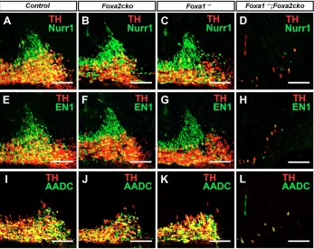
[image:5.612.336.531.56.361.2]Subsequent roles for Foxa1/2 during early and late phases of mDA neuronal differentiation We used molecular markers to determine the status of postmitotic mDA neurons in the ventral midbrain of Foxa1/2 single and double mutants at E12.5. Foxa1–/–;Foxa2ckomutants showed an
Fig. 4. Reduced expression of Ngn2 in mDA progenitors and incomplete specification of immature neurons in
Foxa1–/–;Foxa2ckodouble-mutant embryos at E12.5.(A-F) Coronal
sections of mouse embryos. (A,B) Ngn2 expression is reduced in mDA cells in mutant compared with wild-type embryos. (C-F) Lmx1a, Tuj1+ immature neurons are generated (C,D) that do not express Nurr1 and TH (E,F) in mutant as compared with control embryos. Scale bars: 75 m.
Fig. 5. Foxa1/2are required for early and late differentiation of mDA neurons at E12.5.(A-L) Coronal sections of mouse embryos. (A-H) The number of immature Nurr1+TH–(A-D) and En1+TH– (E-H) neurons (green) increases at the expense of mature Nurr1+TH+(A-D) and En1+TH+mDA (E-H) neurons (yellow) in
Foxa1/2single-mutant embryos compared with the corresponding numbers in control embryos. The total number of neurons (immature plus mature mDA neurons) is reduced only in
[image:5.612.52.399.464.740.2]D
E
V
E
LO
P
M
E
N
T
85% reduction in the total number of Lmx1a+Tuj1+mDA neurons (mutant, 121±12; control, 789±18; n=3), whereas the numbers of Lmx1a+ Sox2+ mDA progenitors was not changed in double mutant compared with control embryos (Fig. 4C-F, Fig. 5D,H,L and data not shown). The remaining Tuj1+ neurons in Foxa1–/–;Foxa2cko mutants also expressed Lmx1b (data not shown), but the majority of these cells did not express Nurr1 (Fig. 4E,F and Fig. 5D) and En1 (Fig. 5H), indicating incomplete acquisition of immature mDA neuronal properties in these Tuj1+ neurons. At E18.5, the number of Lmx1a+ Tuj1+ neurons increased to 29% (mutant, 483±8; control, 1584±3; n=3; and data not shown), whereas the number of Nurr1+TH–and Nurr1+TH+ mDA neurons did not change in Foxa1–/–;Foxa2cko mutant embryos between E12.5 and E18.5 (Fig. 6). We also found that the reduction in mDA neurons is not due to an increase in cell proliferation as determined by BrdU-labelling or to an increase in apoptosis that was analysed by TUNEL labelling in Foxa1–/–;Foxa2cko double-mutant embryos at E12.5 (see Materials and methods and Fig. S2 in the supplementary material). In addition, there was no obvious change in the number of GABAergic+neurons in Foxa1–/–;Foxa2ckodouble-mutant and wild-type embryos (see Fig. S3 in the supplementary material), suggesting that there is no transformation of mDA progenitors into GABAergic neurons at E12.5. By contrast, there was a 49% reduction in the number of Islet 1+ OCM neurons (mutant, 145±18; control, 293±37; n=3) and an almost complete loss of Brn3a+red nuclei neurons in the midbrain of Foxa1–/–;Foxa2cko double mutants when compared with control embryos (see Fig. S3 in the supplementary material). These results indicate that Foxa1/2 are also required for the development of OCM and red nuclei neurons.
Contrary to the situation in Foxa1–/–;Foxa2ckodouble-mutant embryos, the total number of mDA neurons, i.e. immature mDA neurons measured as Nurr1+TH–and En1+TH–cells (green cells in Fig. 5A-C and E-G, respectively) plus mature mDA neurons measured as Nurr1+TH+and En1+ TH+cells (yellow cells in Fig. 5A-C and E-G, respectively), did not change in Foxa1–/– and Foxa2cko mutants compared with control embryos, indicating normal neuronal differentiation (quantified in Fig. 6A). However, the number of immature Nurr1+TH–mDA neurons (Fig. 5A-C and Fig. 6A) was increased in both Foxa1and Foxa2ckosingle-mutant embryos at the expense of mature Nurr1+TH+mDA neurons (Fig. 5A-C and Fig. 6A), indicating that Foxa1/2 are required for promoting differentiation of immature into mature DA neurons.
In summary, analyses of Foxa1/2 single and double-mutant embryos revealed that Foxa1/2 proteins are required during multiple phases of mDA neuronal development. In single mutants, there is a block in the late differentiation phase, with a reduction in the number of mature mDA neurons. Foxa1–/–;Foxa2cko double-mutant embryos exhibited a more severe phenotype than Foxa1/2single mutants because neuronal specification and early differentiation of mDA progenitors were affected, leading to an almost complete loss of mDA neurons.
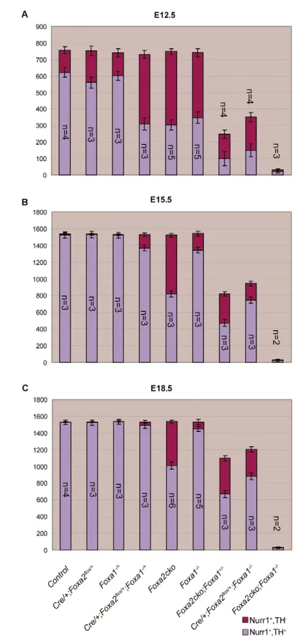
Dosage-dependent functions of Foxa1/2in mDA
cells
To better characterise similar functions of Foxa1/2in mDA cells, we compared the expression of Nurr1 and TH by double-antibody labelling experiments in the ventral midbrain of mutant embryos carrying 0-4 copies of Foxa1/2 genes in their genome at E12.5, E15.5 and E18.5. Reducing the dose of Foxa1/2 resulted in a proportionate decrease in the number of Nurr1+TH+mature mDA
neurons in mouse embryos at E12.5 (Fig. 6A and data not shown), indicating that Foxa1/2regulate differentiation of these neurons in a dose-dependent manner. Interestingly, a progressive rescue in the number of TH+mature mDA neurons was observed in all Foxa1/2 single and double mutants except in Foxa1–/–;Foxa2ckodouble mutants at E15.5 and E18.5, indicating that a single copy of Foxa1 or Foxa2 is required for this recovery (Fig. 6B,C and data not shown). It is noteworthy that Foxa2cko, Foxa1+/–;Foxa2flox/floxand Foxa1–/–;Foxa2flox/+embryos only showed partial rescue of Nurr1+ TH+mDA neurons even at E18.5 (Fig. 6C and data not shown). As these mutant embryos do not survive beyond E18.5, the status of
Fig. 6. Dosage-dependent requirement forFoxa1/2in the differentiation of mDA neurons and a progressive rescue of mDA neuron number in mutant embryos during development.
(A-C) Bar charts showing the number of immature (Nurr1+TH–) and mature (Nurr1+TH+) neurons, as determined by immunohistochemistry, in embryos carrying 0-4 copies of Foxa1 and/orFoxa2 at three different stages. The x-axis shows the genotype of the mouse embryos, and the
[image:6.612.327.538.54.508.2]D
E
V
E
LO
P
M
E
N
T
mDA neurons cannot be determined at later stages. Importantly, Foxa1+/–;Foxa2flox/+double-heterozygous embryos showed a similar reduction in the number of mature mDA neurons as Foxa1–/–and Foxa2ckosingle mutants at E12.5 (Fig. 6). This reduction was fully recovered in the double-heterozygous mutant embryos at E18.5 (Fig. 6). Altogether, these data indicate that Foxa1 and Foxa2 are functionally capable of compensating for each other in regulating the development of mDA neurons.
DISCUSSION
In this paper, we have demonstrated multiple roles for Foxa1/2 in the development of mDA neurons. Our results demonstrate that Foxa1/2 are required to regulate neuronal specification and early and late differentiation of mDA cells (summarised in Fig. 7). Genetic evidence indicates that these functions require different gene dosages of Foxa1and Foxa2. Importantly, our study shows that Foxa1/2 regulate distinct targets during multiple phases of mDA neuron development. Below, we discuss these regulatory roles of Foxa1/2 and suggest some mechanisms as to how Foxa1/2 might perform these roles in mDA cells.
Foxa1/2 regulate Ngn2 expression and
consequently neurogenesis in mDA progenitors Since Ngn2 has previously been shown to regulate neuronal differentiation of mDA progenitors, the partial inhibition of neuronal differentiation is likely to be due to reduced Ngn2 expression in Foxa1/2 double-homozygous mutant embryos. How Foxa1/2 regulates Ngn2 expression at the molecular level remains to be determined. The partial effects on Ngn2 expression could be due to the perdurance of Foxa2 protein in progenitors in mutant embryos at the beginning of mDA neurogenesis around E10.5 and/or alternative redundant mechanisms for regulating these genes in these progenitors. Support for the latter hypothesis comes from the findings that Lmx1a and Otx2 are positive regulators of Ngn2 expression (Vernay et al., 2005; Andersson et al., 2006b), and Otx2 is also required for the repression of Nkx2.2 expression in mDA progenitors (Puelles et al., 2004; Vernay et al., 2005; Prakash et al., 2006).
Our studies also showed that there is abnormal expression of Nkx2.2 in a small percentage of Lmx1a+ mDA progenitors, suggesting that Foxa1/2 might be required to prevent Nkx2.2 expression in these cells. However, the consequence of this abnormal expression in mDA progenitors remains to be determined. Previous studies have suggested that abnormal expression of Nkx2.2 in mDA progenitors may result in these progenitors acquiring a serotonergic fate (Puelles et al., 2004; Prakash et al., 2006). Since Foxa2 is also
required for the generation of serotonergic neurons in the hindbrain (J. Jacob, A.L.M.F., C. Milton, F. Prin, P. Pia, W.L., A. Gavalas, S.-L.A. and J. Briscoe, unpublished) it is unlikely that serotonergic neurons are generated in the midbrain of Foxa1–/–;Foxa2cko double-mutant embryos.
Foxa1/2 regulate the acquisition of dopaminergic properties in immature and mature midbrain neurons
Analyses of Foxa1/2 double and single mutants also revealed essential roles for Foxa1/2 in early and late differentiation phases of mDA neurons, respectively. Specifically, Foxa1/2 are required for the expression of Nurr1 and En1 in immature mDA neurons and for the expression of AADC and TH in mature mDA neurons. Altogether, these results indicate that Foxa1/2 regulate distinct genes at different phases of mDA neuron development. It is noteworthy that the neuron promoter of human AADC contains a FOXA2-binding site that overlaps with a consensus FOXA2-binding site for POU-domain transcription factors, including BRN2 (also known as POU3F2) (Raynal et al., 1998). Brn2 is widely expressed in midbrain progenitors and neurons at E12.5 (our unpublished results) and hence is available to cooperate with Foxa1/2 proteins. Whether AADC is a direct target of the combined activities of Foxa2 and a POU-domain transcription factor requires further studies.
Foxa1/2regulate mDA neuron development in a
dosage-dependent manner
Analyses of embryos with different copy numbers of Foxa1/2 indicated a dose-dependent requirement for these genes during the differentiation of mDA neurons. Foxa1/2 are required for specification and early and late differentiation steps of mDA cells; however, Foxa1/2 appear to be required at progressively higher doses as mDA cells differentiate. Loss of two copies of Foxa1/2 resulted in a late differentiation defect, whereas an early differentiation defect was only observed when three or four copies of Foxa1/2genes were removed. Moreover, a specification defect was only observed in embryos lacking all four copies of Foxa1/2. These results suggest that higher doses of Foxa1/2are required at progressively later differentiation phases and raise the intriguing possibility that the timing and duration of mDA neuron differentiation might be regulated by the concentration of Foxa1/2 in these cells.
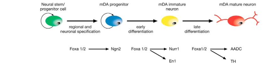
[image:7.612.64.492.57.162.2]One mechanism that could explain the requirement for higher doses of Foxa1/2 for late rather than early differentiation targets is differences in affinity of binding sites. For example, if late differentiation target genes of Foxa1/2 have lower affinity binding
Fig. 7. Schematic summarising the sequential roles of Foxa1/2 during the three different phases of development of mDA neurons from neural stem/progenitor cells.During regional and neuronal specification, Foxa1/2 positively regulates Ngn2 expression in mDA progenitors. Foxa1/2 is subsequently required for Nurr1 and En1 expression in immature mDA neurons and for the expression of aromatic L-amino acid
D
E
V
E
LO
P
M
E
N
T
sites, they might require a higher dose of Foxa1/2 transcription factors for their activation. Such a mechanism has already been shown to operate in the regulation of early and late target genes of PHA-4, the C. elegans homologue of Foxa, during pharyngeal development (Gaudet and Mango, 2002). Sequences that bind PHA-4 with high affinity in vitro are typically found in promoters of genes expressed early during pharyngeal development, whereas low-affinity sites are restricted to late promoters in C. elegans. By analogy with PHA-4, the binding site affinity for Foxa1/2 proteins might thus be a crucial determinant of gene expression in mDA neurons. However, it is also likely that additional factors function in combination with Foxa1/2 for temporal control of gene expression at different stages. Future work will concentrate on identifying molecular targets for Foxa1/2 in order to provide further understanding of molecular mechanisms of gene regulation by Foxa1/2 within the CNS. These experiments will also determine whether Foxa1/2 have identical or overlapping molecular targets. Given their important regulatory roles in mDA neuron development, further understanding of the molecular mechanisms of action of Foxa1/2 is likely to facilitate attempts to differentiate mDA neurons from stem cells.
We thank Drs D. J. Anderson, J. Briscoe, M. German, T. Müller and Carmen Birchmeier for antibodies; Drs Francois Guillemot and James Briscoe for critical reading of the manuscript; and Stella Uwabor and Wai Han Yau for help with references and illustrations, respectively. We are also grateful to Drs Klaus Kaestner and Ryoichiro Kageyama for generously providing Foxa1lacZ/+and Nestin-Cre/+mice, respectively. This work was supported by the Medical Research Council UK and by a research grant from the Parkinson’s Disease Society to S.-L.A.
Supplementary material
Supplementary material for this article is available at http://dev.biologists.org/cgi/content/full/134/15/2761/DC1
References
Alberi, L., Sgado, P. and Simon, H. H.(2004). Engrailed genes are cell-autonomously required to prevent apoptosis in mesencephalic dopaminergic neurons. Development131, 3229-3236.
Andersson, E., Tryggvason, U., Deng, Q., Friling, S., Alekseenko, Z., Robert, B., Perlmann, T. and Ericson, J.(2006a). Identification of intrinsic determinants of midbrain dopamine neurons. Cell124, 393-405.
Andersson, E., Jensen, J. B., Parmar, M., Guillemot, F. and Bjorklund, A. (2006b). Development of the mesencephalic dopaminergic neuron system is compromised in the absence of neurogenin 2. Development133, 507-516. Ang, S. L.(2006). Transcriptional control of midbrain dopaminergic neuron
development. Development 133, 3499-3506.
Ang, S. L. and Rossant, J.(1994). HNF-3 beta is essential for node and notochord formation in mouse development. Cell78, 561-574.
Ang, S. L., Wierda, A., Wong, D., Stevens, K. A., Cascio, S., Rossant, J. and Zaret, K. S.(1993). The formation and maintenance of the definitive endoderm lineage in the mouse: involvement of HNF3/forkhead proteins. Development 119, 1301-1315.
Barzilai, A. and Melamed, E.(2003). Molecular mechanisms of selective dopaminergic neuronal death in Parkinson’s disease. Trends Mol. Med.9, 126-132.
Filosa, S., Rivera-Perez, J. A., Gomez, A. P., Gansmuller, A., Sasaki, H., Behringer, R. R. and Ang, S. L.(1997). Goosecoid and HNF-3beta genetically interact to regulate neural tube patterning during mouse embryogenesis. Development124, 2843-2854.
Gao, N., Zhang, J., Rao, M. A., Case, T. C., Mirosevich, J., Wang, Y., Jin, R., Gupta, A., Rennie, P. S. and Matusik, R. J.(2003). The role of hepatocyte nuclear factor-3 alpha (Forkhead Box A1) and androgen receptor in transcriptional regulation of prostatic genes. Mol. Endocrinol.17, 1484-1507. Gaudet, J. and Mango, S. E.(2002). Regulation of organogenesis by the
Caenorhabditis elegans FoxA protein PHA-4. Science295, 821-825. Hallonet, M., Kaestner, K. H., Martin-Parras, L., Sasaki, H., Betz, U. A. and
Ang, S. L.(2002). Maintenance of the specification of the anterior definitive endoderm and forebrain depends on the axial mesendoderm: a study using HNF3beta/Foxa2 conditional mutants. Dev. Biol. 243, 20-33.
Hwang, D. Y., Ardayfio, P., Kang, U. J., Semina, E. V. and Kim, K. S.(2003). Selective loss of dopaminergic neurons in the substantia nigra of Pitx3-deficient aphakia mice. Brain Res. Mol. Brain Res.114, 123-131.
Indra, A. K., Warot, X., Brocard, J., Bornert, J. M., Xiao, J. H., Chambon, P.
and Metzger, D.(1999). Temporally-controlled site-specific mutagenesis in the basal layer of the epidermis: comparison of the recombinase activity of the tamoxifen-inducible Cre-ER(T) and Cre-ER(T2) recombinases. Nucleic Acids Res. 27, 4324-4327.
Isaka, F., Ishibashi, M., Taki, W., Hashimoto, N., Nakanishi, S. and Kageyama, R.(1999). Ectopic expression of the bHLH gene Math1 disturbs neural development. Eur. J. Neurosci. 11, 2582-2588.
Kaestner, K. H., Katz, J., Liu, Y., Drucker, D. J. and Schutz, G.(1999). Inactivation of the winged helix transcription factor HNF3alpha affects glucose homeostasis and islet glucagon gene expression in vivo. Genes Dev.13, 495-504.
Kele, J., Simplicio, N., Ferri, A. L., Mira, H., Guillemot, F., Arenas, E. and Ang, S. L.(2006). Neurogenin 2 is required for the development of ventral midbrain dopaminergic neurons. Development133, 495-505.
Lang, A. E. and Lozano, A. M.(1998). Parkinson’s disease. Second of two parts. N. Engl. J. Med.339, 1130-1143.
Lantz, K. A., Vatamaniuk, M. Z., Brestelli, J. E., Friedman, J. R., Matschinsky, F. M. and Kaestner, K. H.(2004). Foxa2 regulates multiple pathways of insulin secretion. J. Clin. Invest.114, 512-520.
Lee, C. S., Friedman, J. R., Fulmer, J. T. and Kaestner, K. H.(2005). The initiation of liver development is dependent on Foxa transcription factors. Nature 435, 944-947.
Lewis, P. M., Dunn, M. P., McMahon, J. A., Logan, M., Martin, J. F., St-Jacques, B. and McMahon, A. P.(2001). Cholesterol modification of sonic hedgehog is required for long-range signaling activity and effective modulation of signaling by Ptc1. Cell105, 599-612.
Monaghan, A. P., Kaestner, K. H., Grau, E. and Schutz, G.(1993). Postimplantation expression patterns indicate a role for the mouse forkhead/HNF-3 alpha, beta and gamma genes in determination of the definitive endoderm, chordamesoderm and neuroectoderm. Development119, 567-578.
Norton, W. H., Mangoli, M., Lele, Z., Pogoda, H. M., Diamond, B., Mercurio, S., Russell, C., Teraoka, H., Stickney, H. L., Rauch, G. J. et al.(2005). Monorail/Foxa2 regulates floorplate differentiation and specification of oligodendrocytes, serotonergic raphe neurones and cranial motoneurones. Development132, 645-658.
Nunes, I., Tovmasian, L. T., Silva, R. M., Burke, R. E. and Goff, S. P.(2003). Pitx3 is required for development of substantia nigra dopaminergic neurons. Proc. Natl. Acad. Sci. USA100, 4245-4250.
Pabst, O., Herbrand, H., Takuma, N. and Arnold, H. H.(2000). NKX2 gene expression in neuroectoderm but not in mesendodermally derived structures depends on sonic hedgehog in mouse embryos. Dev. Genes Evol. 210, 47-50.
Prakash, N. and Wurst, W.(2006). Development of dopaminergic neurons in the mammalian brain. Cell. Mol. Life Sci.63, 187-206.
Prakash, N., Brodski, C., Naserke, T., Puelles, E., Gogoi, R., Hall, A., Panhuysen, M., Echevarria, D., Sussel, L., Weisenhorn, D. M. et al.(2006). A Wnt1-regulated genetic network controls the identity and fate of midbrain-dopaminergic progenitors in vivo. Development133, 89-98.
Puelles, E., Annino, A., Tuorto, F., Usiello, A., Acampora, D., Czerny, T., Brodski, C., Ang, S. L., Wurst, W. and Simeone, A.(2004). Otx2 regulates the extent, identity and fate of neuronal progenitor domains in the ventral midbrain. Development131, 2037-2048.
Raynal, J. F., Dugast, C., Le Van Thai, A. and Weber, M. J.(1998). Winged helix hepatocyte nuclear factor 3 and POU-domain protein brn-2/N-oct-3 bind overlapping sites on the neuronal promoter of human aromatic L-amino acid decarboxylase gene. Brain Res. Mol. Brain Res.56, 227-237.
Sasaki, H. and Hogan, B. L.(1993). Differential expression of multiple fork head related genes during gastrulation and axial pattern formation in the mouse embryo. Development118, 47-59.
Sasaki, H. and Hogan, B. L.(1994). HNF-3 beta as a regulator of floor plate development. Cell76, 103-115.
Simon, H. H., Saueressig, H., Wurst, W., Goulding, M. D. and O’Leary, D. D. (2001). Fate of midbrain dopaminergic neurons controlled by the engrailed genes. J. Neurosci. 21, 3126-3134.
Smidt, M. P., Asbreuk, C. H., Cox, J. J., Chen, H., Johnson, R. L. and Burbach, J. P.(2000). A second independent pathway for development of mesencephalic dopaminergic neurons requires Lmx1b.Nat. Neurosci.3, 337-341.
Smits, S. M., Burbach, J. P. and Smidt, M. P.(2006). Developmental origin and fate of meso-diencephalic dopamine neurons. Prog. Neurobiol.78, 1-16. Tzschentke, T. M. and Schmidt, W. J.(2000). Functional relationship among
medial prefrontal cortex, nucleus accumbens, and ventral tegmental area in locomotion and reward. Crit. Rev. Neurobiol.14, 131-142.
van den Munckhof, P., Luk, K. C., Ste-Marie, L., Montgomery, J., Blanchet, P. J., Sadikot, A. F. and Drouin, J.(2003). Pitx3 is required for motor activity and for survival of a subset of midbrain dopaminergic neurons. Development130, 2535-2542.
D
E
V
E
LO
P
M
E
N
T
Wallen, A. and Perlmann, T.(2003). Transcriptional control of dopamine neuron development. Ann. N.Y. Acad. Sci.991, 48-60.
Wan, H., Xu, Y., Ikegami, M., Stahlman, M. T., Kaestner, K. H., Ang, S. L. and Whitsett, J. A.(2004). Foxa2 is required for transition to air breathing at birth. Proc. Natl. Acad. Sci. USA101, 14449-14454.
Weinstein, D. C., Ruiz i Altaba, A., Chen, W. S., Hoodless, P., Prezioso, V. R., Jessell, T. M. and Darnell, J. E., Jr(1994). The winged-helix transcription factor HNF-3 beta is required for notochord development in the mouse embryo. Cell 78, 575-588.
Ye, W., Shimamura, K., Rubenstein, J. L., Hynes, M. A. and Rosenthal, A. (1998). FGF and Shh signals control dopaminergic and serotonergic cell fate in the anterior neural plate. Cell93, 755-766.
Zetterstrom, R. H., Solomin, L., Jansson, L., Hoffer, B. J., Olson, L. and Perlmann, T.(1997). Dopamine neuron agenesis in Nurr1-deficient mice. Science276, 248-250.