The Localizing and Lateralizing Value of Auras
in Lesional Partial Epilepsy Patients
Byoung Seok Ye, Yang-Je Cho, Sang Hyun Jang, Moon Kyu Lee, Byung In Lee, and Kyoung Heo
Department of Neurology, Yonsei University College of Medicine, Seoul, Korea.
Received: June 24, 2011 Revised: August 22, 2011 Accepted: September 1, 2011 Corresponding author: Dr. Kyoung Heo, Department of Neurology, Yonsei University College of Medicine, 50 Yonsei-ro, Seodaemun-gu, Seoul 120-752, Korea. Tel: 82-2-2228-1607, Fax: 82-2-393-0705 E-mail: [email protected]
∙ The authors have no financial conflicts of interest.
© Copyright:
Yonsei University College of Medicine 2012
This is an Open Access article distributed under the terms of the Creative Commons Attribution Non-Commercial License (http://creativecommons.org/ licenses/by-nc/3.0) which permits unrestricted non-commercial use, distribution, and reproduction in any medium, provided the original work is properly cited.
Purpose: We investigated the localizing and lateralizing values of auras in patients with lesional partial epilepsy on an outpatient basis. Materials and Methods: A total of 276 subjects were retrospectively selected for this study if they had a uni-lateral single lobar lesion based on magnetic resonance image (MRI) results, and their scalp electroencephalography (EEG) findings were not discordant with the MRI-defined lobar localization and lateralization. According to the lesion loca-tions, subjects were considered as having mesial temporal (MTLE), lateral tempo-ral (LTLE), frontal (FLE), parietal (PLE), or occipital (OLE) lobe epilepsies. Auras were classified into 13 categories. Results: A hundred and seventy-six subjects (63.8%) had experienced at least one aura. FLE subjects had the fewest number of auras. Epigastric and psychic auras were frequent among MTLE subjects, while visual auras were common in those with PLE and OLE. Somatosensory auras and whole body sensations were more frequent in the subjects with PLE than those without. Autonomic auras were more common in MTLE subjects than in LTLE subjects. Dysphasic auras were more frequently found in left-sided epilepsies. Five pairs of aura categories showed concurrent tendencies, which were the epigastric and autonomic auras, autonomic and emotional auras, visual and vestibular auras, auditory and vestibular auras, and whole-body sensation and auditory auras. Auto-nomic and emotional auras had a concurrent tendency in left-sided epilepsies, but not in right-sided epilepsies. Conclusion: Our results support the previously known localizing value of auras, and suggest that dysphasic auras and the associa-tion of emoassocia-tional and autonomic auras may have a lateralizing value.
Key Words: Epilepsy, aura, lesional epilepsy, focus localization, focus lateraliza-tion
INTRODUCTION
ered as having mesial temporal lobe epilepsy (MTLE), lat-eral temporal lobe epilepsy (LTLE), frontal lobe epilepsy (FLE), parietal lobe epilepsy (PLE), or occipital lobe epi-lepsy (OLE). The subjects with lesions that are limited to the temporal lobe were regarded as having either MTLE or LTLE, according to the involvement of the mesial temporal structures. The subjects with asymmetrical hippocampal sclerosis (HS) with lateralization of a smaller hippocampus were also included in this study.
Classification of auras
Based on the well-documented history of the symptoms that the subjects recorded, we classified aura symptoms into 13 categories: epigastric auras; autonomic auras; emotional auras; vestibular auras; psychic (experiential) auras; visual auras; somatosensory auras; dysphasic auras; olfactory au-ras; auditory auau-ras; whole body sensations; cephalic sensa-tions; and a final category that includes symptoms that can-not be categorized into any of the other categories. Dysphasia cannot be a pure subjective sensation (definition of aura) but was included as aura symptom because it is not found in the majority of cases by others. Epigastric auras included nauseous feelings with or without an uprising sensation. Symptoms, including non-specific dizziness without a spin-ning sensation, headache, pressure on the head, and light-headedness, were regarded as cephalic sensations, and not as vestibular auras. An urge to urinate, shortness of breath, sialorrhea, vomiting or retching, palpitation, gooseflesh, or generalized cold or warm feeling in the body was consid-ered as an autonomic aura. Symptoms including feelings, thoughts, and distortion of memory, such as déjà vu or ja-mais vu type illusions, were classified as psychic auras. Psychic auras were subdivided into two categories, which were the memory-related and memory-unrelated catego-ries. Memory-related psychic auras included déjà vu, ja-mais vu, and flashbacks. Memory-unrelated psychic auras included forced thinking, being in a dreamy state, having the feeling of being absorbed into something, and the feel-ing of befeel-ing aware of the thoughts of another person. Symp-toms involving emotional elements, such as fear, anxiety, depression, thoughts of dying, unpleasant feelings, feelings of being tired, euphoria, elation, and pleasure, were classi-fied as emotional auras. Emotional auras were also subdi-vided into two different types of feelings, which were either good feelings or bad feelings. Euphoria, elation, or pleasure were considered as good feelings, and anxiety, depression, unpleasant feelings, the feeling of being tired, or fear were Several studies have already correlated aura symptoms with
definite seizure onset zones, especially in patients who have had prolonged EEG monitoring evaluations or surgical treatments.1,2,6-10 Since most of the patients in these studies had drug-resistant epilepsy, the generalizations that were made based on the data that were acquired in these highly selective patients become questionable when applying them to the all partial epilepsy patients, although the localization and lateralization of the epileptic focus may be accurate. The values as well as the features of auras for localizing and/or lateralizing epileptic focus in lesional epilepsy pa-tients were investigated in this study, according to the epi-lepsy localization and lateralization on an outpatient basis.
MATERIALS AND METHODS
Study population
Patients were identified who had visited the outpatient epi-lepsy clinic of Severance Hospital between 2000 and 2007 and had lesions confined to a single lobe based on MRI re-sults. All subjects had scalp EEGs and brain MRIs, and they also provided a well-documented history of their symptoms. Subjects were excluded if they met any of the following criteria: 1) were mentally retarded and could not describe their aura symptoms; 2) scalp EEG findings were discor-dant with lobar localization and lateralization of the lesion, based on the MRI results; 3) had definitely different semio-logical features according to seizure events suggesting mul-tiple lobar origins of the seizure onset; 4) seizures occurred only when they were sleeping; or 5) had arachnoid cysts in the temporal lobe or only subcortical lesions, based on the brain MRI results. After exclusions were made, a total of 276 partial epilepsy patients with single lobar lesions, based on their MRI results, were enrolled in the study. There were 180 males and 96 females.
Localization and lateralization of lesions
consid-using the MRI were as follows: HS was the most common etiology and was found in 111 (40.2%) of the subjects; atro-phic lesions, which were related to trauma, cerebrovascular accidents, central nervous system infections, or were of un-known origin, occurred in 84 (30.4%) subjects; vascular malformations were identified in 26 (9.4%) subjects; for-eign tissue in 20 (7.2%) subjects; and malformations of the cortical development in 23 (8.3%) of the subjects.
Frequencies of aura categories
There were 102 (76.1%) subjects who experienced at least one aura in MTLE, 36 (65.5%) in LTLE, 22 (33.8%) in FLE, 11 (64.7%) in PLE, and 5 (100%) in OLE. The aver-age number of aura categories per subject was 1.19±0.09 for MTLE, 1.02±0.13 for LTLE, 0.48±0.10 for FLE, 0.88±0.21 for PLE, and 1.80±0.37 for OLE, respectively. ANOVA with Bonferroni’s post-hoc analysis revealed that the number of auras in subjects with MTLE (p<0.001), LTLE (p=0.017) and OLE (p=0.024) was significantly higher than that in sub-jects with FLE.
Differences in aura characteristics among lobar epilepsies
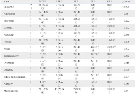
The relationship of 13 aura categories with the 5 types of lobar epilepsy was investigated by the χ2 test or Fisher’s ex-act test (Table 1). Epigastric (p<0.001), autonomic (p=0.030), psychic (p=0.008), and visual (p=0.002) auras showed dif-ferent distributions among the 5 categories of lobar epilep-sy. Olfactory auras occurred only in TLE, of which 4 were MTLE subjects and 2 LTLE subjects. Cephalic sensations, emotional auras, and dysphasic auras did not have a defi-nite localizing value in this study.
The χ2 test or Fisher’s exact test analyzing the presence/ absence of each aura category between patients with specif-ic lobar epilepsy and those without (Table 1) revealed that somatosensory auras and whole body sensations were more frequent in subjects with PLE than those without. Three (17.6%) of the 17 subjects with PLE reported having so-matosensory auras, while only 6 (2.3%) of the 259 subjects without PLE had these types of auras (p=0.013), and 2 (11.8%) of the 17 subjects with PLE had whole body sensa-tions, while only 4 (1.5%) of the 259 subjects without PLE reported having whole body sensations (p=0.046). Epigas-tric (p<0.001), autonomic (p=0.001), and psychic (p=0.002) auras were more frequent in subjects with MTLE than those without. A total of 34 (25.4%), 19 (14.2%), and 24 (17.9%) of the 134 subjects with MTLE had epigastric, autonomic, considered as bad feelings. Tingling, pain, or electrical
sen-sations that arose on either side of the body were regarded as a somatosensory aura. If these sensations diffusely arose in the body without showing clear laterality, the sensations were then regarded as whole body sensations. Visual auras included all of the visual phenomena that subjects experi-enced. Visual auras included visual illusions, elementary vi-sual hallucinations, and complex vivi-sual hallucinations. Transient deficit in reaching the target under visual guid-ance, which is termed optic ataxia, was regarded as a visual illusion. Auras that were classified into the miscellaneous category included symptoms of unexpressible feelings, fuzzy sensations, and premonitions of impending seizures. We also assessed the presence or absence of aura symptoms, as well as the number of auras reported by each subject.
Statistical analyses
We used either the Student’s t-test or the analysis of vari-ance (ANOVA) to analyze the continuous variables. The χ2 test or Fisher’s exact test was performed in order to analyze the categorical variables, which allowed us to compare the aura characteristics between different epilepsy localization and lateralization, as well as the occurrence of each aura category between the subjects with specific lobar epilepsies and those without. The Spearman’s correlation analysis was used to find pairs of closely related aura categories. Strength of correlation was estimated using the Spearman’s rank cor-relation coefficient (ρ). All statistical tests were performed with the Statistical Package for Social Sciences (SPSS) ver-sion 18 (SPSS Inc., Chicago, IL, USA). The significance level was defined as p<0.05. This study was approved by the Institutional Review Board of the Severance Hospital.
RESULTS
Of the 276 cases with partial epilepsy that were reviewed, there were 134 cases (48.6%) of MTLE, 55 cases (19.9%) of LTLE, 65 cases (23.6%) of FLE, 17 cases (6.2%) of PLE, and 5 cases (1.8%) of OLE. There were 159 cases (57.6%) of left-sided epilepsy and 117 cases (42.4%) of right-sided epilepsy. One hundred seventy-six (63.8%) sub-jects experienced at least one aura, and 67 (38.1%) of them reported having two or more aura categories. A total of 271 auras were reported. Age at seizure onset ranged from 1 to 77 years of age (mean 25.8 years).
the 134 subjects with MTLE had autonomic auras, while only 2 (3.6%) of the 55 subjects with LTLE had these au-ras. Although statistical significance was not reached, epi-gastric (p=0.079) and psychic (p=0.182) auras seemed to be more common in MTLE subjects than in LTLE subjects. A total of 34 (25.4%) and 24 (17.9%) of the 134 subjects with MTLE reported having epigastric auras and psychic auras, respectively, while 7 (12.7%) and 5 (9.1%) of 55 subjects with LTLE reported having epigastric auras and psychic au-ras, respectively. On the contrary, while statistical signifi-cance was not reached, visual (p=0.158) and vestibular (p=0.149) auras seemed to be more common in LTLE sub-jects, since 5 (9.1%) and 3 (5.5%) of 55 subjects with LTLE had visual and vestibular auras, respectively, while only 5 (3.7%) and 2 (1.5%) of the 134 subjects with MTLE had these types of auras, respectively.
and psychic auras, respectively. Ten (7.0%), 4 (2.8%), and 8 (5.6%) of the 142 subjects without MTLE had epigastric, autonomic, and psychic auras, respectively. Visual auras were more common in subjects with PLE (p=0.018) and OLE (p=0.036) than in subjects without PLE or OLE, as 4 (23.5%) of 17 with PLE and 2 (40.0%) of 5 with OLE re-ported visual auras, while only 14 (5.4%) of 259 subjects without PLE and 16 (5.9%) of 271 subjects without OLE reported having visual auras.
Comparison of aura between subjects with MTLE and those with LTLE
[image:4.595.55.528.82.409.2]We also compared auras between MTLE and LTLE sub-jects with the χ2 test or Fisher’s exact test. Autonomic auras were significantly more common in subjects with MTLE than those with LTLE (p=0.041), in which 19 (14.2%) of
Table 1. Differences of Aura Characteristics among Lobar Epilepsies
Aura MTLE LTLE FLE PLE OLE p value†
Epigastric +* –* 34 (25.4)100 ‡ 7 (12.7)48 3 (4.6)62 0 (0)17 0 (0)5 <0.001
Autonomic +– 19 (14.2)115 ‡ 2 (3.6)53 2 (3.1)63 0 (0)17 0 (0)5 0.03
Emotional +– 22 (16.4)112 7 (12.7)48 4 (6.2)61 1 (5.9)16 1 (20.0)4 0.222
Cephalic + 10 (7.5) 6 (10.9) 4 (6.2) 1 (5.9) 2 (40.0) 0.172
– 124 49 61 16 3
Vestibular +– 2 (1.5)132 3 (5.5)52 3 (4.6)62 1 (5.9)16 1 (20.0)4 0.088
Psychic + 24 (17.9)
‡ 5 (9.1) 2 (3.1) 0 (0) 1 (20.0)
0.008
– 110 50 63 17 4
Visual +– 5 (3.7)129 5 (9.1)50 2 (3.1)63 4 (23.5)13 ‡ 2 (40.0)3 ‡ 0.002
Somatosensory + 3 (2.2) 1 (1.8) 2 (3.1) 3 (17.6)
‡
0 (0) 0.063
– 131 54 63 14 5
Dysphasic +– 9 (6.7)125 2 (3.6)53 2 (3.1)63 2 (11.8)15 0 (0)5 0.518
Olfactory +– 4 (3.0)130 2 (3.6)53 0 (0)65 0 (0)17 0 (0)5 0.570
Whole body sensation + 3 (2.2) 1 (1.8) 0 (0) 2 (11.8)‡ 0 (0) 0.104
– 131 54 65 15 5
Auditory +– 1 (0.7)133 2 (3.6)53 0 (0)65 1 (5.9)16 0 (0)5 0.159
Miscellaneous +– 24 (17.9)112 13 (23.6)42 7 (10.8)59 0 (0)17 2 (40.0)3 0.04
MTLE, mesial temporal lobe epilepsy; LTLE, lateral temporal lobe epilepsy; FLE, frontal lobe epilepsy; PLE, parietal lobe epilepsy; OLE, occipital lobe epi-lepsy.
Numbers in parentheses are percentages. *+: present, –: absent.
†Results of the χ2 test or Fisher’s exact test comparing the presence/absence of 13 aura categories across the five types of lobar epilepsy.
ras were not different between the subjects with right-sided epilepsy and those with left-sided epilepsy.
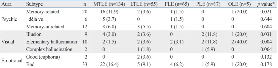
Subtype analysis of psychic, visual, and emotional auras The subtype analysis of the psychic aura showed that the memory-related psychic auras had a significantly different distribution among the 5 categories of lobar epilepsy (p=0.021), in which 16 (11.9%) of the 134 subjects with MTLE and 1 (20.0%) of 5 subjects with OLE had memory-related psychic auras, while 2 (3.6%) of 55 subjects with LTLE, 1 (1.5%) of 65 subjects with FLE, and none of 17 subjects with PLE had this type of aura (χ2 test or Fisher’s exact test) (Table 3). Among the 6 subjects who had déjà-vu illusions, 5 had lesions in the mesial temporal lobe, and 1 subject had the lesion in the frontal lobe. The frequencies of memory-unrelated type psychic auras were not different among the 5 categories of lobar epilepsy.
Differences of aura characteristics between the subjects with left-sided and right-sided epilepsy
Thirteen aura categories were compared between the sub-jects with left-sided epilepsy and those with right-sided epi-lepsy by the χ2 test or Fisher’s exact test (Table 2). Dyspha-sic auras (p<0.001) were more frequently found in subjects with left-sided epilepsy, in which 15 (9.4%) of the 159 sub-jects with left-sided epilepsy had dysphasic auras, while none of the 117 subjects with right-sided epilepsy had this type of aura. On the contrary, although statistical signifi-cance was not reached, vestibular (p=0.102) and olfactory (p=0.086) auras were more common in subjects with right-sided epilepsy, as 7 (6.0%) and 5 (4.3%) of 117 subjects with right-sided epilepsy had vestibular and olfactory auras, respectively, while only 3 (1.9%) and 1 (0.6%) of 159 sub-jects with left-sided epilepsy had these two types of auras, respectively. The frequencies of the other categories of
au-Table 2. Differences of Aura Characteristics between Left-Sided and Right-Sided Epilepsies
Aura n Right-sided (n=117) Left-sided (n=159) p value*
Epigastric 44 20 (17.1) 24 (15.1) 0.74
Autonomic 23 10 (8.5) 13 (8.2) 1.0
Emotional 35 12 (10.3) 23 (14.5) 0.362
Cephalic 23 11 (9.4) 12 (7.5) 0.661
Vestibular 10 7 (6.0) 3 (1.9) 0.102
Psychic 32 16 (13.7) 16 (10.1) 0.447
Visual 18 8 (6.8) 10 (6.3) 1.0
Somatosensory 9 4 (3.4) 5 (3.1) 1.0
Dysphasic 15 0 (0) 15 (9.4) <0.001
Olfactory 6 5 (4.3) 1 (0.6) 0.086
Whole body sensation 6 2 (1.7) 4 (2.5) 1.0
Auditory 4 2 (1.7) 2 (1.3) 1.0
Miscellaneous 46 23 (19.7) 23 (14.5) 0.258
Numbers in parentheses are percentages.
[image:5.595.70.540.331.512.2]*Results of the χ2 test or Fisher’s exact test comparing the differences in the presence/absence of aura symptoms between subjects with left-sided epi-lepsy and those with right-sided one.
Table 3. Localizing Significance of Specific Subtypes of Psychic, Visual, and Emotional Auras
Aura Subtype n MTLE (n=134) LTLE (n=55) FLE (n=65) PLE (n=17) OLE (n=5) p value*
Psychic
Memory-related 20 16 (11.9) 2 (3.6) 1 (1.5) 0 1 (20.0) 0.021
déjà vu 6 5 (3.7) 0 1 (1.5) 0 0 0.644
Memory-unrelated 12 8 (6.0) 3 (5.5) 1 (1.5) 0 0 0.604
Visual
Illusion 9 4 (3.0) 2 (3.6) 0 2 (11.8) 1 (20.0) 0.031
Elementary hallucination 10 2 (1.5) 2 (3.6) 2 (3.1) 2 (11.8) 2 (40.0) 0.004
Complex hallucination 2 0 1 (1.8) 0 1 (5.9) 0 0.064
Emotional Good (euphoria) 2 0 2 (3.6) 0 0 0 0.132
Bad 33 22 (16.4) 5 (9.1) 4 (6.2) 1 (5.9) 1 (20.0) 0.178
MTLE, mesial temporal lobe epilepsy; LTLE, lateral temporal lobe epilepsy; FLE, frontal lobe epilepsy; PLE, parietal lobe epilepsy; OLE, occipital lobe epi-lepsy.
Numbers in parentheses are percentages.
[image:5.595.73.540.570.685.2]Emotional auras were subdivided into either good feel-ings or bad feelfeel-ings. Good feelfeel-ings were observed only in those with left-sided LTLE (n=2), while bad feelings were observed in all of the lobar epilepsies studied (Table 3 and 4). No remarkable hemispheric preference was found in any of the other subtypes of psychic, visual, and emotional auras (Table 4).
Relationships between categories of auras
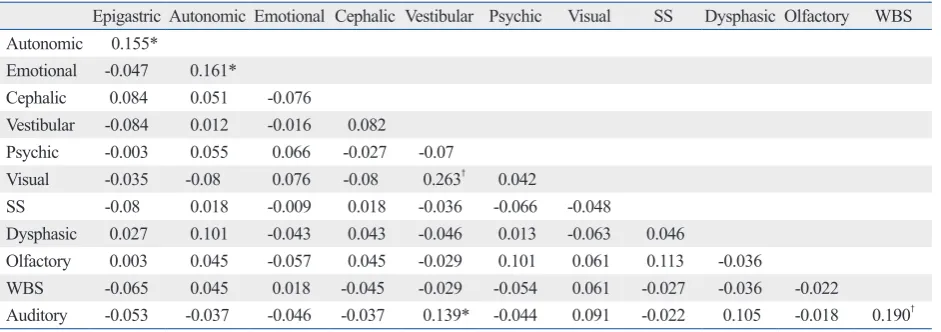
Spearman’s correlation analysis showed that 5 pairs of aura categories had concurrent tendencies among the 13 aura categories (Table 5). The pairs of aura categories were epi-gastric and autonomic auras (p=0.01, ρ=0.155), autonomic and emotional auras (p=0.007, ρ=0.161), visual and vestib-ular auras (p<0.001, ρ=0.263), auditory and vestibular au-ras (p=0.021, ρ=0.139), and whole body sensation and au-ditory auras (p=0.002, ρ=0.19). A significant association between autonomic and emotional auras was observed in left-sided epilepsy subjects (p=0.01, ρ=0.204), but not in With regard to visual auras, the elementary visual
[image:6.595.58.526.360.475.2]hallu-cinations and visual illusions both showed significantly dif-ferent distributions, although the significance of this is not clear due to too few subjects that had each subtype of visual aura. Two (40.0%) of 5 subjects with OLE and 2 (11.8%) of 17 subjects with PLE had elementary visual hallucina-tions, while 2 (1.5%) of 134 subjects with MTLE, 2 (3.6%) of 55 subjects with LTLE, and 2 (3.1%) of 65 subjects with FLE had this type of hallucinations (p=0.004). Whereas 1 (20.0%) of 5 subjects with OLE and 2 (11.8%) of 17 sub-jects with PLE had visual illusions, 4 (3.0%) of 134 subsub-jects with MTLE, 2 (3.6%) of 55 subjects with LTLE, and none of 65 subjects with FLE had them (p=0.031). Although sta-tistical significance was not reached, the frequency of com-plex visual hallucinations tended to be different among the 5 categories of lobar epilepsy, in which complex visual hallu-cinations (n=2) were observed in LTLE (n=1) and PLE (n=1), but not in MTLE, FLE, or OLE (Fisher’s exact test, p=0.064).
Table 4. Lateralizing Significance of Specific Subtypes of Psychic, Visual, and Emotional Auras
Aura Subtype n Right-sided (n=117) Left-sided (n=159) p value*
Psychic
Memory-related 20 9 (7.7) 11 (6.9) 0.818
déjà vu 6 3 (2.6) 3 (1.9) 0.701
Memory-unrelated 12 7 (6.0) 5 (3.1) 0.371
Visual
Illusion 9 6 (5.1) 3 (1.9) 0.175
Elementary hallucination 10 4 (3.4) 6 (3.8) 1.0
Complex hallucination 2 0 2 (1.3) 0.51
Emotional Good (euphoria) 2 0 2 (1.3) 0.51
Bad 33 12 (10.3) 21 (13.2) 0.574
Numbers in parentheses are percentages.
*Results of the χ2 test or Fisher’s exact test comparing the presence/absence of specific aura subtypes between subjects with left-sided epilepsy and those with right-sided one.
Table 5. Relationship between Aura Categories (Spearman’s Rank Correlation Coefficient)
Epigastric Autonomic Emotional Cephalic Vestibular Psychic Visual SS Dysphasic Olfactory WBS Autonomic 0.155*
Emotional -0.047 0.161*
Cephalic 0.084 0.051 -0.076
Vestibular -0.084 0.012 -0.016 0.082
Psychic -0.003 0.055 0.066 -0.027 -0.07
Visual -0.035 -0.08 0.076 -0.08 0.263† 0.042
SS -0.08 0.018 -0.009 0.018 -0.036 -0.066 -0.048
Dysphasic 0.027 0.101 -0.043 0.043 -0.046 0.013 -0.063 0.046
Olfactory 0.003 0.045 -0.057 0.045 -0.029 0.101 0.061 0.113 -0.036
WBS -0.065 0.045 0.018 -0.045 -0.029 -0.054 0.061 -0.027 -0.036 -0.022
Auditory -0.053 -0.037 -0.046 -0.037 0.139* -0.044 0.091 -0.022 0.105 -0.018 0.190†
SS, somatosensory aura; WBS, whole body sensation. *Correlation with p<0.05.
[image:6.595.58.529.532.698.2]agreement with our findings that epigastric auras were also observed in three FLE subjects, but not in any of the PLE or OLE subjects.
Autonomic symptoms, such as sweating, sialorrhea, pal-pitation, and respiratory difficulty, were included as symp-toms of autonomic auras, while we categorized nauseous feelings as a symptom of epigastric auras. However, epigas-tric and autonomic auras were classified as only one cate-gory of viscerosensory aura in previous studies.1,2 In this study, autonomic auras also showed a pattern of distribution similar to epigastric auras. However, the autonomic auras were more frequently reported by subjects with MTLE than those with LTLE.
Psychic auras correspond to the experiential auras dis-cussed in Palmini and Gloor’s study,2 except that, the com-plex visual and auditory hallucinations were categorized as visual and auditory auras, respectively in our study. Psychic auras auras were frequently reported by subjects with TLE. Similar to the results of Palmini and Gloor’s study, in which three patients with occipital and two patients with frontal lobe seizure focus showed experiential auras,2 two subjects with FLE and one subject with OLE in our study also had psychic auras. Considering the fact that psychic symptoms were reproduced by electrical stimulation of the temporal lobe,2,17-21 we speculate that psychic auras in the subjects with OLE and FLE may result from the spread of seizure discharge to the temporal lobe structures.
When the psychic auras were subdivided into related type and unrelated type, only the memory-related type had a tendency to be more common in MTLE subjects. This may be due to the memory-related structures, such as the hippocampus or the amygdala, which are locat-ed in the mesial temporal lobe.
The association of visual auras with PLE and OLE is consistent with what has been previously reported.2 In the present study, complex visual hallucinations were regarded as visual auras that were then subdivided into three subcate-gories; visual illusions, elementary visual hallucinations, and complex visual hallucinations. Among the three sub-types of visual auras that were studied, visual illusions and elementary visual hallucinations were associated more with PLE and OLE. Our finding that complex visual hallucina-tions were observed in LTLE and PLE subjects, but not in OLE subjects is in agreement with the findings of a previ-ous study on visual auras.9 Although elementary visual hal-lucinations were not reported in FLE in previous studies,2,22 two subjects with FLE in this study had their lesions near right-sided epilepsy subjects (p=0.292, ρ=0.098). Of the two
subtypes of emotional auras, bad feelings showed concur-rent tendency with autonomic auras (p=0.004, ρ=0.172) in left-sided epilepsies (p=0.005, ρ=0.223), but not in right-sid-ed epilepsies (p=0.292, ρ=0.098). Among the 3 subtypes of visual auras, only elementary visual hallucinations (p<0.001, ρ=0.274) and visual illusions (p=0.002, ρ=0.183) were con-current with vestibular auras. However, the strength of cor-relation of all of these items was considered weak because the correlation coefficients were all lower than 0.3.
DISCUSSION
In this study, we investigated the localizing and lateralizing values of auras in subjects with a unilateral single lobar le-sion, based on MRI results on an outpatient basis, but not in surgically treated patients. The proportion of subjects with TLE in our study (68.5%) seemed to be relatively higher than what would be expected in a general epilepsy popula-tion. This might be explained by the dominance of HS in lesional epilepsy, the small number of subjects with lesions that were confined to either the occipital or parietal lobes that were of relatively small size, and exclusion of subjects with parietal or occipital lesions (epilepsies) but temporal interictal epileptiform discharges (IEDs) on EEG.11,12
The frequency of auras in our study subjects was 64%. This percentage is lower than 81% that was reported in Palmini and Gloor’s2 study of 179 subjects with mixed lo-bar epilepsies, who underwent prolonged EEG monitoring or surgical treatment. However, our percentage is identical to 64% that was reported in a study that had investigated the relationship between auras and the lateralization of the EEG abnormalities for 290 subjects with TLE on an outpa-tient basis, which is a population that could be expected to have a higher frequency of auras.13 The mean number of auras and the proportion of subjects with at least one aura were significantly lower in subjects with FLE, and this finding was consistent with a previously published study.2
Localizing value of auras
lobar epilepsies, which is consistent with the findings of an-other previously published study.2 Vertigo is thought to be caused by the mismatch between visuospatial and vestibular information,24 which could explain the significant correla-tion between vestibular and visual auras in our study.
Lateralizing value of auras
Previous studies on the lateralizing value of auras failed to confirm their findings.1,2 Only in an EEG-clinical correla-tion study, Gupta, et al.13 were able to observe that auto-nomic and psychic auras were more frequently associated with right-sided EEG abnormalities in TLE patients. In the present study, only the dysphasic auras were found to corre-late with the side on which the lesion was present, verified by the brain MRI. The correlation of dysphasic auras with left-sided epilepsy seems to be plausible since the language centers are usually located in the left hemisphere of the brain. However, previous studies did not categorize this type of aura into its own aura categorization.1,2,13 In our study, psychic auras and déjà vu illusions did not show any significant preference for lateralization. In Palmini and Gloor’s2 study, there was a strong trend of experiential au-ras and déjà vu illusions to originate from the right tempo-ral lobe. Some stimulation studies showed that electrical stimulation of the right temporal lobe yielded more experi-ential responses and déjà vu illusions than that of the left lobe.17,18
In this study, vestibular auras tended to occur more fre-quently in subjects with right-sided epilepsy (seven out of ten subjects), although previous studies did not find lateral-izing value of vestibular auras.1,2,13 Considering the domi-nant right hemispheric influence on visuospatial informa-tion processing, our data reflect the possibility that the right hemisphere may play a significant role in the expression of the vestibular aura. Furthermore, our present study showed that the olfactory auras tended to be more common in right-sided epilepsy (p=0.087), in which five of the six subjects with olfactory auras had right-sided epilepsies. However, there has been no significant preponderance for lateraliza-tion in previous studies.2,6,10,13
In this study, there was a significant correlation between autonomic auras and the emotional auras, particularly bad feeling in left-sided epilepsy patients. Lee, et al.25 found that patients with left-sided TLE exhibited autonomic hy-perarousal when viewing negative emotional slides relative to controls and those with right-sided TLE. They suggested that the dysfunction of the left mesial temporal lobe struc-the parietal lobe and had reported nonspecific features of
el-ementary visual hallucinations, such as darkening and or-ange sunshine in their entire visual field.
In the present study, emotional auras were observed in all of the categories of lobar epilepsy. The two patients in our study who reported euphoria as their auras had left-sided LTLE. This localizing and lateralizing value requires fur-ther investigation with more patients in order to confirm this finding.
All six subjects with olfactory auras in this study had TLE. Among these subjects, two had atrophic lesions in the tem-poral pole area. The other four subjects had lesions in the mesial temporal region, which commonly involved the amygdala and the types of lesions were a benign tumor, HS, cortical dysplasia, and an atrophic lesion. Olfactory auras were produced by the stimulation of the mesial temporal structures, which include the uncus and the amygdala or the olfactory bulb,19 and are associated with MTLE and mesial temporal pathology.6,10 In this study, the olfactory auras in the subjects with LTLE may be due to the spread of the sei-zure activity to the mesial temporal lobe structure or due to the involvement of the mesial temporal lobe structure relat-ed to head trauma, which cannot be resolvrelat-ed with a MRI.
Of the four subjects with auditory auras, three had lesions in the temporal lobe, and the other one subject had a lesion in the parietal lobe. The subject with PLE had a focal infarc-tion in the left parietal lobe and an elementary auditory hal-lucination that presented itself as a buzzing sound. This find-ing is different from previous reports in that all subjects with auditory aura had TLE.1,2 Since this patient also experienced complex visual hallucinations and whole body sensations, the auditory aura may be due to the spread of seizure dis-charge from the parietal to the temporal lobe structure.
In the analysis of aura frequency between the subjects with specific lobar epilepsy and those without, whole body sensations and somatosensory auras were found to be more common in PLE subjects. This finding is consistent with previously published results that the somatosensory auras are associated with PLE patients.2
tory auras in patients with temporal lobe epilepsy. Epilepsia 2003;44:257-60.
7. Henkel A, Noachtar S, Pfänder M, Lüders HO. The localizing val-ue of the abdominal aura and its evolution: a study in focal epilep-sies. Neurology 2002;58:271-6.
8. Nair DR, Najm I, Bulacio J, Lüders H. Painful auras in focal epi-lepsy. Neurology 2001;57:700-2.
9. Bien CG, Benninger FO, Urbach H, Schramm J, Kurthen M, Elg-er CE. Localizing value of epileptic visual auras. Brain 2000; 123(Pt 2):244-53.
10. Acharya V, Acharya J, Lüders H. Olfactory epileptic auras. Neu-rology 1998;51:56-61.
11. Blume WT, Whiting SE, Girvin JP. Epilepsy surgery in the poste-rior cortex. Ann Neurol 1991;29:638-45.
12. Kim DW, Lee SK, Yun CH, Kim KK, Lee DS, Chung CK, et al. Parietal lobe epilepsy: the semiology, yield of diagnostic workup, and surgical outcome. Epilepsia 2004;45:641-9.
13. Gupta AK, Jeavons PM, Hughes RC, Covanis A. Aura in temporal lobe epilepsy: clinical and electroencephalographic correlation. J Neurol Neurosurg Psychiatry 1983;46:1079-83.
14. Currie S, Heathfield KW, Henson RA, Scott DF. Clinical course and prognosis of temporal lobe epilepsy. A survey of 666 patients. Brain 1971;94:173-90.
15. Rasmussen T. Surgical therapy of frontal lobe epilepsy. Epilepsia 1963;4:181-98.
16. Feindel W, Penfield W. Localization of discharge in temporal lobe automatism. AMA Arch Neurol Psychiatry 1954;72:603-30. 17. Mullan S, Penfield W. Illusions of comparative interpretation and
emotion; production by epileptic discharge and by electrical stim-ulation in the temporal cortex. AMA Arch Neurol Psychiatry 1959;81:269-84.
18. Penfield W, Perot P. The brain’s record of auditory and visual ex-perience. A final summary and discussion. Brain 1963;86:595-696.
19. Gloor P, Olivier A, Quesney LF, Andermann F, Horowitz S. The role of the limbic system in experiential phenomena of temporal lobe epilepsy. Ann Neurol 1982;12:129-44.
20. Bancaud J, Brunet-Bourgin F, Chauvel P, Halgren E. Anatomical origin of déjà vu and vivid ‘memories’ in human temporal lobe epilepsy. Brain 1994;117(Pt 1):71-90.
21. Vignal JP, Maillard L, McGonigal A, Chauvel P. The dreamy state: hallucinations of autobiographic memory evoked by tempo-ral lobe stimulations and seizures. Brain 2007;130(Pt 1):88-99. 22. Ludwig BI, Marsan CA. Clinical ictal patterns in epileptic patients
with occipital electroencephalographic foci. Neurology 1975;25: 463-71.
23. Penfield W, Jasper H. Epilepsy and the functional anatomy of the human brain. 1st ed. Boston: Little Brown; 1954.
24. Brandt T. Vertigo: its multisensory syndromes. 2nd ed. London: Springer; 2003.
25. Lee GP, Meador KJ, Loring DW, Bradley KP. Lateralized changes in autonomic arousal during emotional processing in patients with unilateral temporal lobe seizure onset. Int J Neurosci 2002;112: 743-57.
26. Morrow L, Vrtunski PB, Kim Y, Boller F. Arousal responses to emotional stimuli and laterality of lesion. Neuropsychologia 1981; 19:65-71.
tures may result in autonomic hyperarousal and a release of the unrestrained negative emotional tendencies of the right hemisphere. Morrow, et al.26 also reported less prominent autonomic arousal to emotionally loaded visual materials in persons with non-dominant hemisphere damage than in the control subjects or those with dominant hemisphere dam-age. Therefore, the association of autonomic auras and emo-tional auras in left-sided epilepsy patients might be ex-plained in a similar context.
There are major limitations in this study. Patients with only lesional epilepsy were selected. Therefore, without in-tracranial EEG evaluations or surgical outcomes, the possi-bility of false localization of the epileptogenic zone by le-sion exists, especially in patients with atrophic lele-sions that are related to infectious, traumatic, or unknown etiology. The exact nature of auras was not confirmed by video-EEG monitoring in most patients. Patients with an extratemporal lesion (epilepsy) who could show temporal IEDs were ex-cluded in this study, which may reflect too small number of patients with OLE, thus preventing appropriate analysis of OLE. These limitations may make the present results diffi-cult to apply to overall partial epilepsy. However, the analy-sis of auras in patients with a lesion highly suggestive of epileptogenic focus may provide some clinical value.
In conclusion, our results suggest that the previously known localizing value of auras in surgical epilepsy pa-tients can be applied to papa-tients with partial epilepsy on an outpatient basis. In addition, with a broader range of sub-jects, we were able to find the lateralizing values of dyspha-sic auras and the laterality-specific concurrent tendency of autonomic-emotional auras.
REFERENCES
1. Fried I, Spencer DD, Spencer SS. The anatomy of epileptic auras: focal pathology and surgical outcome. J Neurosurg 1995;83:60-6. 2. Palmini A, Gloor P. The localizing value of auras in partial
sei-zures: a prospective and retrospective study. Neurology 1992;42: 801-8.
3. Schäuble B, Cascino GD. Advances in neuroimaging: management of partial epileptic syndromes. Neurosurg Rev 2003;26:233-46. 4. Fish DR, Spencer SS. Clinical correlations: MRI and EEG. Magn
Reson Imaging 1995;13:1113-7.