0095-1137/05/$08.00⫹0 doi:10.1128/JCM.43.6.2656–2661.2005
Copyright © 2005, American Society for Microbiology. All Rights Reserved.
Novel PCR-Restriction Fragment Length Polymorphism Method
for Determining Serotypes or Serogroups of
Streptococcus pneumoniae
Isolates
Sarah L. Batt,
1Bambos M. Charalambous,
1Timothy D. McHugh,
1Siobhan Martin,
2and Stephen H. Gillespie
1*
Centre for Medical Microbiology, University College London, Royal Free Campus, Rowland Hill St., London NW3 2PF,1
and Specialist and Reference Microbiology Division, Health Protection Agency, 67, Colindale Avenue,
London NW9 5HT,2United Kingdom
Received 4 August 2004/Returned for modification 27 September 2004/Accepted 7 February 2005
SerotypingStreptococcus pneumoniaeis a technique generally confined to reference laboratories, as purchas-ing pneumococcal antisera is a huge investment. Many attempts have been made to modify serological agglutination techniques to make them more accessible, and more recently developments in serotyping have focused on molecular techniques. This paper describes a PCR assay which amplifies the entire capsulation locus betweendexBandaliA. Amplicons are digested to produce serotype-specific patterns. We have shown, using 81 epidemiologically unrelated strains representing 46 different serotypes, that the patterns correlate with a 90 to 100% similarity range for the same serotype or serogroup. Prospective testing of 73 isolates of unknown serotype confirmed reliable serotype attribution, and serotype profiles are reproducible on repeated testing. Once our database contains all 90 serotypes, this technique should be fully portable, cost-effective, and useful in any laboratory with sufficient molecular experience.
The speciesStreptococcus pneumoniaepossesses more than 90 serotypes defined by their polysaccharide capsule (1). Im-munologically similar serotypes, such as 19F, 19A, 19B, and 19C, are grouped together in serogroups. The immune re-sponse to capsular polysaccharide is critical for recovery from infection (17), and the first effective treatment for pneumococ-cal infection, serum therapy, depended on identifying the in-fecting serotype rapidly so that type-specific horse serum could be administered (9). The capsule is the focus for the develop-ment of an effective vaccine, initially with the 23-valent poly-saccharide and more recently the protein-polypoly-saccharide con-jugate preparations that are undergoing clinical evaluation and have entered clinical use (1, 35). Some reports on the imple-mentation of the conjugate vaccine in different communities show that there is an increase in the carriage of serotypes that are not included in the vaccine (6, 7). Additionally, serotype surveillance in developing countries and special populations, such as Aboriginal Australians, suggests that vaccine serotype coverage may be lower than for the United States and Europe, from which surveillance data were considered in vaccine for-mulations (2, 4, 25). Thus, there is a continuing need to study the epidemiology of S. pneumoniae as defined by capsular polysaccharide in monitoring the effect of conjugate vaccines and to aide in the development of new vaccine formulations for developing countries and special populations (12, 13, 16).
Effective serotyping depends on a full set of group- and type-specific antisera prepared by the Statens Serum Institut (14), an investment that is beyond the resources of many
lab-oratories. Therefore, most laboratories confine typing to the serotypes and serogroups found in the 23-valent vaccine, which represent most of the invasive types found in industrialized countries (22). As prevailing serotypes change, a wider selec-tion of sera will be required to cover common types, thus increasing the cost. In addition to the cost, equivocal results due to autoagglutination are common, and discrepancies are reported in the typing of other organisms, such asHaemophilus
influenzae, between PCR and standard slide agglutination. In
one such study, this discrepancy is thought to have been due to laboratory error, implying that PCR-based typing is more re-liable (21).
Alternative typing methods not based on the capsular poly-saccharide are described forS. pneumoniaeusing various mo-lecular methods and phenotypic methods. These include pulsed-field gel electrophoresis, restriction fragment end label-ing, BOX PCR, arbitrarily primed PCR, and multilocus se-quence typing (MLST) (8, 15a, 34, 36). Although these meth-ods prove valuable for distinguishing strains, studying epidemiology, and monitoring transmission, the importance of the capsule in pathogenesis and immunity emphasizes the fact that serotyping will remain in demand.
The genes encoding the S. pneumoniae capsule are con-tained within a gene cassette which is flanked by two genes,
dexB and aliA, in all serotypes that have been sequenced to date (28, 29, 32). Within this locus, each serotype has a set of homologous genes and a set which define serotype and thus are variable (30). This combination of homology and diversity makes the capsulation gene cassette an attractive target for a DNA amplification-based method of defining serotype. Fur-thermore, with the capsulation loci for the majority of sero-types having been sequenced, confirmation of serotype-specific patterns is possible by in silico digestions.
* Corresponding author. Mailing address: Centre for Medical Mi-crobiology, University College London, Royal Free Campus, Rowland Hill St., London NW3 2PF, United Kingdom. Phone: 44-207-794-0500, ext. 3539. Fax: 44-207-794-0433. E-mail: [email protected].
2656
on May 16, 2020 by guest
http://jcm.asm.org/
We report a PCR capable of amplifying the entire capsule locus for all serotypes tested to date, and digestion of the amplicons with HinfI yields serotype-unique patterns that dis-tinguish them from other serotypes, most of which are con-firmed in silico.
MATERIALS AND METHODS
Bacteria.Isolates ofS. pneumoniaeused in this study were obtained from clinical specimens submitted to the diagnostic microbiology laboratory of the Royal Free Hospital (RFH), a carriage survey of children collected in the Rombo district of Tanzania (Tz), and the Health Protection Agency, Colindale, United Kingdom (HPA). Table 1 contains strains used to generate the Bionumerics database of serotype-specific patterns.
Preparation of genomic DNA.Bacterial cells were harvested from a fresh overnight culture on blood agar, and DNA was extracted using the Wizard Genomic DNA purification kit (Promega, Southampton, United Kingdom) fol-lowing the manufacturer’s instructions. Briefly, a dense suspension of cells was made in 480l 0.05 M EDTA (Sigma, Poole, United Kingdom). One hundred twenty microliters of cell lysis solution and 60l lysozyme (Sigma) were added, mixed by gentle inversion, and incubated at 37°C for 30 to 60 min. The tubes were centrifuged at 13,000⫻gfor 2 min, and the supernatant was discarded. The cells were resuspended in 600l nuclei lysis solution and mixed by gentle pipetting. The tubes were incubated at 80°C for 5 min and cooled to room temperature. Three microliters of RNase solution was added, and the tubes were incubated at 37°C for 15 to 30 min and cooled to room temperature. Protein precipitation solution (200l) was added, and the tubes were vortexed for 20 seconds and then incubated on ice for 5 min. Cell debris was precipitated by centrifuging at 13,000⫻gfor 3 min and the supernatant transferred to a fresh tube containing 0.6 ml room-temperature isopropanol. The suspension was mixed by gentle inversion until a precipitate could be seen. Tubes were then centrifuged for 10 min, and the pellet was washed in 70% ethanol (vol/vol) and dried for 15 min at 37°C. The pellet was then redissolved in 100l DNA rehydration solution and incubated at 65°C in a water bath for 60 min, with mixing every 20 min. The isolated DNA was used immediately. Experiments using DNA which had been stored at 4°C for more than 24 h showed multiple PCR amplicons (data not shown).
PCR conditions.The primers used were high-performance liquid chromatog-raphy purified (Sigma Genosys, Poole, United Kingdom) and had the following sequences: for AliA2, 5⬘-ATG CAG CTA AAG TAG TCG CC-3⬘; for DexB2, 5⬘-GAC CGT CGC TTC CTA GTT GT-3⬘.
Amplification was achieved using REDAccuTaq LA DNA polymerase (Sigma). The optimal reaction tube contained 1.5l of genomic fresh DNA extract, 2lTaq(2 U), 5l buffer, deoxynucleoside triphosphates at 0.5 mM, and 0.6 mM primers, made up to 50l final reaction volume with PCR-grade water.
The optimal reaction cycle consisted of one cycle of 93°C for 2 min and 30 cycles of 93°C for 15 seconds, 50°C for 30 seconds, and 68°C for 20 min, with a hold temperature of 4°C. All reactions were performed on a Progene (Techne, United Kingdom) or GeneAmp 9700 (Applied Biosystems, Warrington, United Kingdom) machine using 0.2-ml thin-walled tubes (Alpha Laboratories, Hamp-shire, United Kingdom or Applied Biosystems). Amplimers were kept at 4°C until digests were performed and are known to be stable at this temperature for up to 1 month.
RFLP.Amplicons from the PCRs were digested using HinfI as follows. A 20-l aliquot from the PCR was incubated with 1l enzyme (10 U), 5l digestion buffer, and 0.2l bovine serum albumin (Promega) for 4 h at 37°C according to the manufacturer’s instructions. The digests were subjected immedi-ately to gel electrophoresis using a 30-ml tank (Horizon 58; Life Technologies) 3% (wt/vol) agarose gel (Bioline, London, United Kingdom) with the voltage set at 100 V (Power pack model 4001P; Life Technologies). No pretreatment of restriction fragment length polymorphism (RFLP) digests was performed before loading, and the RFLP digests are stable for storage at 4°C for at least a few days. Electrophoresis was continued until the loading dye was at the bottom of the gel. An image of the gel was taken using a stand-mounted digital camera DC 120 and Kodak Digital Science 2.0 imaging software, and the image was stored as a Tiff file.
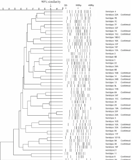
Analysis of data.The Tiff images were opened in Bionumerics software 3.5 (Applied Maths, Kortrjk, Belgium) as an RFLP experiment. The RFLP patterns were entered by a single investigator using Bionumerics software and saved into a single database. The gels were all normalized by assigning band sizes to the bands of hyperladder I (Bioline). All bands that could be seen on the image of the gel down to the 400-bp marker were selected (Fig. 1). Double bands were selected only when two distinct peaks could be seen on the gel image and the
densitometric curves (the densitometric curve is seen in the analysis window of Bionumerics). These criteria were applied to every gel.
[image:2.585.302.539.88.616.2]Cluster analysis.Cluster analysis was performed using the Dice coefficient. Similarity was calculated using parameter settings at 2% band position tolerance and optimization at 1%. The program computed a dendrogram using the un-weighted-pair group method using average linkages clustering algorithm. This algorithm links the serotype at the level of the branch so the percentage shown
TABLE 1. Isolates used to establish a Bionumerics database of serotype-specific patterns
Serotype Strain Clinical
sourcea
1 RFH 116, RFH 206 BC
3 Tz 8810-1, Tz 2604-1b, Tz 2604-1a, Tz 5809.2
TS
4 Tz 1104-2, Tz 3682-2, Tz 2706-1 TS
5 Tz 7310-1 TS
6A RFH 260, RFH 6 BC
Tz 4613-3, Tz 4202-1, Tz 1003-2, Tz 1003-3
TS
HPA 6A C
6B Tz 5303-2, Tz 7712-2b TS
RFH 200, RFH 221 BC
HPA 6B C
7F HPA 7F C
7A HPA 7A C
7B HPA 7B C
7C Tz 2514-1, Tz 4711-1, Tz 5310-2 TS
8 Tz 4603-3 TS
9A HPA 9A C
9L HPA 9L C
9N RFH 18, RFH 48, RFH 93 BC
9V RFH 78, RFH 98, RFH 120 BC
10B HPA 10B C
10C HPA 10C C
11A HPA 11A C
11B HPA 11B C
11 (subtype not known)
Tz 1101-2, Tz 3912-1, Tz 1102-1, Tz 5907-1
TS
12F/B Tz 3407-1, Tz 8103-2 TS
13 HPA 13 TS
14 RFH 16, RFH 34, RFH 40 BC
15A HPA 15A C
15C HPA 15C C
17F HPA 17F C
18F HPA 18F C
18A HPA 18A C
18B/C Tz 8715-1 TS
19A RFH 25 BC
HPA 19A C
19B HPA 19B C
19F RFH 5, RFH 32 BC
Tz 2701-2, Tz 7910-1 TS
20 Tz 1103-1 TS
21 HPA 21 C
22 RFH 33 BC
23A HPA 23A C
23F RFH 13, RFH 329, RFH 6, RFH 7 BC
24F HPA 24F C
24A HPA 24A C
24B HPA 24B C
25F HPA 25F
25A HPA 25A C
29 HPA 29 C
32F HPA 32F C
35A HPA 35A C
38 HPA 38 C
aC, control strains from a reference laboratory; TS, carriage isolates from
throat swabs; BC, isolates from blood culture.
on May 16, 2020 by guest
http://jcm.asm.org/
by clicking on the level of the branch shows the percentage similarity of the two serotypes. To set the definition of serotype and serogroup, all (n⫽81) strains for which a serotype or serogroup was known were clustered together on a single dendrogram using Bionumerics.
[image:3.585.76.516.72.602.2]Identifying the serotype of an unknown sample.To identify the serotype of an unknown sample, cluster analysis was performed blind for 73 isolates. For an unknown isolate, the normalization process described above was followed. A comparison was created with the unknown strain using one example of every type FIG. 1. Dendrogram showing one isolate for each of the 46 representative RFLP patterns. Serotypes 1, 3, 4, 5, 6A, 6B, 7F, 7A, 7B, 7C, 8, 9A, 9N, 9V, 9L, 10B, 10C, 11A, 11B, 11 (subtype not known), 12F/B, 13, 14, 15A, 15C, 17F, 18F, 18A, 18B/C, 19F, 19A, 19B, 20, 21, 22, 23A, 23F, 24F, 24A, 24B, 25F, 25A, 29, 32F, 35A, and 38 are included. The 12F and B and 18B and C patterns could not be discriminated using in silico data, since digests with HinfI were very similar. No in silico data were available for serotype 25F. “Confirmed” identifies patterns confirmed by in silico data (where available). Alignments were produced using Bionumerics software (Applied Maths).
on May 16, 2020 by guest
http://jcm.asm.org/
and group in the database. If a match with similarity greater than 90% was detected, this was defined as the serotype of the strain. If the match was less than 90%, this indicated a serotype not currently in our database (Fig. 1).
In silico digestions.The sequences from the capsulation locus were down-loaded from the Sanger website (http://www.sanger.ac.uk/Projects/S_pneumoniae /CPS/). Digestions were performed using Restrict from the UK HGMP Resource Centre (http://www.hgmp.mrc.ac.uk/) and band sizes compared to those on aga-rose gels and used to confirm experimental digestions where possible.
RESULTS
Amplifiable serotypes.The most important requirement of a molecular serotyping method is the capacity to type all of the possible serotypes ofS. pneumoniae. To test this, the PCR was performed on genomic DNA obtained from multiple isolates. PCR amplicons were obtained from all isolates, and the size range was around 14 to 23 kbp.
Serotype and serogroup identification. Isolates chosen for study were those that were likely to be epidemiologically un-related. This was achieved by selecting examples from our U.K. laboratory, isolates from field studies in Tanzania, and isolates of unusual serotypes from the Health Protection Agency. This procedure was followed to reduce the risk of false positive matches among common serotypes that were actually identical strains. Where multiple isolates of the same serotype occurred, as defined by agglutination tests, they had RFLP patterns that were at least 90% similar.
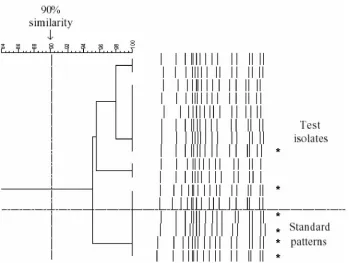
Figure 1 shows 46 serotypes and serogroups aligned using the Bionumerics software. Isolates of the same serotype showed greater than 90% similarity, and isolates of different types showed less than 90% similarity. Examples of the same serotype were tested repeatedly and shown to have an identical pattern, for example, 16 times for serotype 23F (including
standard patterns) (Fig. 2). Multiple isolates from the same patient were shown to have identical patterns.
Exceptions to the rule that only members of the same sero-type are⬎90% similar were found on four occasions. A pair of isolates was identified as serogroup 18, but the subtype could not be determined. Similar results were obtained for two strains, identified as serogroup 12 and serogroup 25, with the subtype not being distinguishable. Additionally, serotype 6B has two representative patterns.
Identification of the serotype for an unknown sample. A total of 73 unknown strains serotyped conventionally were examined in a blinded fashion. The following serotypes were correctly identified: 3 (n⫽5), 6A (n⫽5), 6B (n⫽3), 9A (n
⫽1), 11 (n⫽1), 12B/C (n⫽11), 19A (n⫽2), 19F (n⫽1), 22 (n ⫽ 1), 23F (n ⫽ 12), and 29 (n ⫽ 1). Additionally, 30 isolates representing 14 patterns not currently in the database were identified.
DISCUSSION
[image:4.585.119.468.73.336.2]Bacterial agglutination is, arguably, one of the simplest pro-cedures performed in microbiology laboratories, yet it is one where there are many pitfalls. In some cases, the agglutination may be weak, making the result difficult to see. Alternatively, some strains may autoagglutinate, making it impossible to ob-tain a typing result. Difficulties in the performance of serotyp-ing are perhaps demonstrated by the numerous modifications made to the original capsule swelling technique by different investigators, which include attempts to harness latex aggluti-nation or counterimmunoelectrophoresis (14, 15, 20, 26, 27, 33).
FIG. 2. Dendrograms of multipleS. pneumoniaeserotype 23F isolates. Four standard patterns showed greater than 94% similarity with 12 throat swab test isolates. Asterisks represent the strains that were typed using MLST.
on May 16, 2020 by guest
http://jcm.asm.org/
A method using PCR amplification of a portion within the locus betweencpsA andcpsB(located immediately upstream ofdexB) followed by restriction fragment polymorphism was described previously (23). However, the patterns produced permit discrimination only to the level of serogroup rather than serotype. This is because the PCR primers amplify a section of the capsulation locus betweencpsAandcpsB. cpsA-cpsDgenes are conserved, andcpsAis⬎90% identical in all of the gene clusters sequenced fully (11). In addition, for some serotypes there are significant sequence differences in thecpsB
homologue (11). Another drawback is that two or three en-zymes are required to generate patterns capable of discrimi-nation and add to the time taken to evaluate the strain. More recently, two groups described molecular methods that enable the identification of serotypes and serogroups using multiplex PCR (3, 24) and sequencing ofcpsA-Bgenes with additional PCRs for serotype-specific sequences (19). Lawrence et al.’s method requires the use of three multiplex PCRs and recog-nizes a limited set of serotypes (24). To include more serotypes would require additional multiplex PCRs, which would not be feasible for all 90 serotypes, and currently the method is fea-sible only where a capillary sequencer is available. Brito et al.’s method uses two successive rounds of PCR, and the choice of PCR used in the second round requires the amplicons from the first round to be electrophoresed and resolved into groups (3). The drawback of these methods is that they become more complicated as the number of serotypes to be identified in-creases.
Although we made many attempts, it was not possible to design a simple PCR system based on portions of the capsu-lation locus because of the diversity of gene arrangement in the capsulation cassettes of different serotypes. It is for this reason that we chose to develop a set of primers based on thealiAand
dexBgenes, as the method reported here has been designed to be used in routine microbiology practice.dexBandaliAgenes are found in all serotypes, and it is this homology that makes capsule switching possible forS. pneumoniae(5, 10, 11). Others used a similar PCR protocol to amplify the capsulation locus for sequencing (18). We demonstrate that the digestion of amplicons of the whole locus is capable of discriminating at least 46 types ofS. pneumoniaeto serogroup or serotype level and that the band profiles observed from the restriction digest protocol reported are robust and reproducible (Fig. 2). For serogroups 12, 18, and 25, subtypes 12F and B, 18B and C, and 25F and A could not be distinguished. The inability to distin-guish these serotypes is confirmed by the in silico digestion, as the sequences are very closely related (http://www.sanger .ac.uk/Projects/S_pneumoniae/CPS/). Types 12F and 12B can be distinguished using a second enzyme, AvaI, which cuts se-rotype 12B but not sese-rotype 12F into bands of 17,836 and 5,327 bp, as demonstrated in silico. Types 18B and 18C have whole locus sequences that differ at only two base pairs; a distinction could be made using a second PCR around one of the base pair locations and digestion with TspDTI, which will cut only 18B, as the restriction site for this enzyme is lost for the other base pair. There is no sequence available for serotype 25F, and thus it is not yet possible to suggest an enzyme to differentiate these types.
For all serotype patterns where sequences are available, the digests we obtained are matched by the in silico experiments,
during which the predicted band sizes are compared to those on the gel pattern of the corresponding type. This analysis is limited to only the sequences which are currently available.
To produce a simple and robust method, we have chosen to use a single set of primers and a single restriction enzyme. The choice ofTaqappears critical, as experiments using other en-zyme systems did not provide such reliable results (data not shown). Only the purification of high-quality DNA provided reliable amplification products. Although this is an important drawback, the technique requires only modest competence in molecular biological techniques. This is more than compen-sated for by the fact that all of the subsequent steps are iden-tical irrespective of serotype. The agglutination technique re-quires multiple reiterative steps, so that operators can process only a limited number of isolates daily. The PCR technique described here would permit a large number of isolates to be examined on a single day, the scale being limited by the equip-ment available.
Out of the 12 isolates identified as serotype 23F using this PCR method, 10 were multiple-colony picks taken from throat swabs of two children, demonstrating the reproducibility of the same and different strains. A total of six isolates of serotype 23F, including MLST types 242, 124, and 81 (two examples) and two new unique types, were tested and aligned together (Fig. 2). This indicates the robustness of the technique for strains of different genotypes but with identical serotypes. Sim-ilarly, the method was successfully applied to each of the dif-ferent PCR machines available in our laboratory, which sug-gests that the method is likely to be reproducible in other laboratories.
The application of this technique in our laboratory has im-proved the number of serotypes that can be recognized from 23 to 46. A significant number of the isolates obtained in our Tanzanian survey are nontypeable using the pneumotest 23-valent antisera kit (2). Our method was able to produce am-plicons from all of these nontypeable strains, which suggests that our PCR method will be more flexible in serotype cover-age in areas where nonvaccine types are common. In addition, it opens up the possibility that serotypes that have previously not been recognized will be identified and described more quickly, provided they share the aliA-dexB cassette arrange-ment. Once a comprehensive set of RFLP patterns is estab-lished for this PCR-RFLP protocol, an isolate that does not align with one of these can be sequenced to indicate whether it represents a new serotype or serogroup.
Due to the relative simplicity and flexibility of this technique, we believe that it will be of value to reference centers and to researchers investigating serotype replacement and the popu-lation biology ofS. pneumoniaeand that it has the potential to be a robust alternative to serological serotyping.
ACKNOWLEDGMENT
The work for this paper was funded by Pfizer Inc.
REFERENCES
1.Austrian, R.1989. Pneumococcal polysaccharide vaccines. Rev. Infect. Dis.
11(Suppl. 3):S598–S602.
2.Batt, S. L., B. M. Charalambous, A. W. Solomon, C. Knirsch, P. A. Massae, S. Safari, N. E. Sam, D. Everett, D. C. Mabey, and S. H. Gillespie.2003. Impact of azithromycin administration for trachoma control on the carriage of antibiotic-resistantStreptococcus pneumoniae. Antimicrob. Agents Che-mother.47:2765–2769.
on May 16, 2020 by guest
http://jcm.asm.org/
3.Brito, D. A., M. Ramirez, and H. de Lencastre.2003. Serotyping Streptococ-cus pneumoniaeby multiplex PCR. J. Clin. Microbiol.41:2378–2384. 4.Butler, J. C., S. Crengle, J. E. Cheek, A. J. Leach, D. Lennon, K. L. O’Brien,
and M. Santosham.2001. Emerging infectious diseases among indigenous peoples. Emerg. Infect. Dis.7:554–555.
5.Coffey, T. J., M. C. Enright, M. Daniels, J. K. Morona, R. Morona, W. Hryniewicz, J. C. Paton, and B. G. Spratt.1998. Recombinational exchanges at the capsular polysaccharide biosynthetic locus lead to frequent serotype changes among natural isolates ofStreptococcus pneumoniae. Mol. Micro-biol.27:73–83.
6.Dagan, R., N. Givon-Lavi, O. Zamir, and D. Fraser. 2003. Effect of a nonavalent conjugate vaccine on carriage of antibiotic-resistant Streptococ-cus pneumoniaein day-care centers. Pediatr. Infect. Dis. J.22:532–540. 7.Dagan, R., N. Givon-Lavi, O. Zamir, M. Sikuler-Cohen, L. Guy, J. Janco, P.
Yagupsky, and D. Fraser.2002. Reduction of nasopharyngeal carriage of Streptococcus pneumoniaeafter administration of a 9-valent pneumococcal conjugate vaccine to toddlers attending day care centers. J. Infect. Dis.
185:927–936.
8.Enright, M. C., and B. G. Spratt.1999. Multilocus sequence typing. Trends Microbiol.7:482–487.
9.Finland, M., and W. D. Sutliff.1931. Specific cutaneous reactions and cir-culating antibodies in the course of lobar pneumonia: II. Cases treated with antipneumococcic sera. J. Exp. Med.54:653–667.
10.Garcia, E., D. Llull, and R. Lopez.1999. Functional organization of the gene cluster involved in the synthesis of the pneumococcal capsule. Int. Microbiol.
2:169–176.
11.Garcia, E., D. Llull, R. Munoz, M. Mollerach, and R. Lopez.2000. Current trends in capsular polysaccharide biosynthesis ofStreptococcus pneumoniae. Res. Microbiol.151:429–435.
12.Hausdorff, W. P., J. Bryant, C. Kloek, P. R. Paradiso, and G. R. Siber.2000. The contribution of specific pneumococcal serogroups to different disease manifestations: implications for conjugate vaccine formulation and use, part II. Clin. Infect. Dis.30:122–140.
13.Hausdorff, W. P., J. Bryant, P. R. Paradiso, and G. R. Siber.2000. Which pneumococcal serogroups cause the most invasive disease: implications for conjugate vaccine formulation and use, part I. Clin. Infect. Dis.30:100–121. 14.Henrichsen, J.1979. The pneumococcal typing system. J. Infect.1(Suppl.
2):31–37.
15.Henrichsen, J., E. Berntsson, and B. Kaijser.1980. Comparison of counter-immunoelectrophoresis and the capsular reaction test for typing of pneumo-cocci. J. Clin. Microbiol.11:589–592.
15a.Hermans, P. W., M. Sluijter, T. Hoogenboezem, H. Heersma, A. van Belkum, and R. de Groot.1995. Comparative study of five different DNA fingerprint techniques for molecular typing ofStreptococcus pneumoniaestrains. J. Clin. Microbiol.33:1606–1612.
16.Jaffar, S., A. Leach, A. J. Hall, S. Obaro, K. P. McAdam, P. G. Smith, and B. M. Greenwood.1999. Preparation for a pneumococcal vaccine trial in The Gambia: individual or community randomisation? Vaccine18:633–640. 17.Janoff, E. N., C. Fasching, J. M. Orenstein, J. B. Rubins, N. L. Opstad, and
A. P. Dalmasso.1999. Killing ofStreptococcus pneumoniaeby capsular po-lysaccharide-specific polymeric IgA, complement, and phagocytes. J. Clin. Investig.104:1139–1147.
18.Jiang, S.-M., L. Wang, and P. R. Reeves.2001. Molecular characterization of Streptococcus pneumoniaetype 4, 6B, 8, and 18C capsular polysaccharide gene clusters. Infect. Immun.69:1244–1255.
19.Kong, F., and G. L. Gilbert.2003. Using cpsA-cpsB sequence polymorphisms
and serotype-/group-specific PCR to predict 51Streptococcus pneumoniae capsular serotypes. J. Med. Microbiol.52:1047–1058.
20.Kronvall, G.1973. A rapid slide-agglutination method for typing pneumo-cocci by means of specific antibody adsorbed to protein A-containing staph-ylococci. J. Med. Microbiol.6:187–190.
21.LaClaire, L. L., M. L. Tondella, D. S. Beall, C. A. Noble, P. L. Raghunathan, N. E. Rosenstein, and T. Popovic.2003. Identification ofHaemophilus influ-enzaeserotypes by standard slide agglutination serotyping and PCR-based capsule typing. J. Clin. Microbiol.41:393–396.
22.Lalitha, M. K., K. Thomas, R. S. Kumar, and M. C. Steinhoff.1999. Sero-typing ofStreptococcus pneumoniaeby coagglutination with 12 pooled anti-sera. J. Clin. Microbiol.37:263–265.
23.Lawrence, E. R., C. A. Arias, B. Duke, D. Beste, K. Broughton, A. Efstratiou, R. C. George, and L. M. Hall.2000. Evaluation of serotype prediction by cpsA-cpsBgene polymorphism inStreptococcus pneumoniae. J. Clin. Micro-biol.38:1319–1323.
24.Lawrence, E. R., D. B. Griffiths, S. A. Martin, R. C. George, and L. M. Hall.
2003. Evaluation of semiautomated multiplex PCR assay for determination ofStreptococcus pneumoniaeserotypes and serogroups. J. Clin. Microbiol.
41:601–607.
25.Leach, A. J., T. M. Shelby-James, M. Mayo, M. Gratten, A. C. Laming, B. J. Currie, and J. D. Mathews.1997. A prospective study of the impact of community-based azithromycin treatment of trachoma on carriage and re-sistance ofStreptococcus pneumoniae. Clin. Infect. Dis.24:356–362. 26.Lund, E., and J. Henrichsen.1978. Laboratory diagnosis, serology and
epi-demiology ofStreptococcus pneumoniae, p. 241–262.InT. Bergan and J. R. Norris (ed.), Methods in microbiology. Academic Press, London, United Kingdom.
27.Lund, E., and P. Rasmussen.1966. Omni-serum. A diagnostic Pneumococ-cus serum, reacting with the 82 known types of PneumococPneumococ-cus. Acta Pathol. Microbiol. Scand.68:458–460.
28.Morona, J. K., D. C. Miller, T. J. Coffey, C. J. Vindurampulle, B. G. Spratt, R. Morona, and J. C. Paton.1999. Molecular and genetic characterization of the capsule biosynthesis locus ofStreptococcus pneumoniaetype 23F. Micro-biology145:781–789.
29.Morona, J. K., R. Morona, and J. C. Paton.1997. Molecular and genetic characterization of the capsule biosynthesis locus ofStreptococcus pneu-moniaetype 19B. J. Bacteriol.179:4953–4958.
30.Morona, J. K., R. Morona, and J. C. Paton.1999. Comparative genetics of capsular polysaccharide biosynthesis inStreptococcus pneumoniaetypes be-longing to serogroup 19. J. Bacteriol.181:5355–5364.
31. Reference deleted.
32.Ramirez, M., and A. Tomasz.1999. Acquisition of new capsular genes among clinical isolates of antibiotic-resistant Streptococcus pneumoniae. Microb. Drug Resist.5:241–246.
33.Slotved, H.-C., M. Kaltoft, I. C. Skovsted, M. B. Kerrn, and F. Espersen.
2004. Simple, rapid latex agglutination test for serotyping of pneumococci (Pneumotest-Latex). J. Clin. Microbiol.42:2518–2522.
34.Sluijter, M., H. Faden, R. de Groot, N. Lemmens, W. H. Goessens, A. van Belkum, and P. W. Hermans.1998. Molecular characterization of pneumo-coccal nasopharynx isolates collected from children during their first 2 years of life. J. Clin. Microbiol.36:2248–2253.
35.World Health Organization.1999. Pneumococcal vaccines: World Health Organization position paper. Can. Commun. Dis. Rep.25:150–151. 36.Yano, H., M. Suetake, A. Kuga, K. Irinoda, R. Okamoto, T. Kobayashi, and
M. Inoue.2000. Pulsed-field gel electrophoresis analysis of nasopharyngeal flora in children attending a day care center. J. Clin. Microbiol.38:625–629.