Copyright © 2004, American Society for Microbiology. All Rights Reserved.
Identification and Molecular Characterization of Beta-Hemolytic
Streptococci Isolated from Harbor Seals (
Phoca vitulina
) and
Grey Seals (
Halichoerus grypus
) of the German North and
Baltic Seas
A. Vossen,
1,2A. Abdulmawjood,
2C. La¨mmler,
3* R. Weiß,
4and U. Siebert
1Forschungs- und Technologiezentrum Westku¨ste, Christian-Albrechts-Universita¨t Kiel, Bu¨sum,1Ißnstitut fu¨r Tiera¨rztliche
Nahrungsmittelkunde,2Institut fu¨r Pharmakologie und Toxikologie,3and Institut fu¨r Hygiene und
Infektionskrankheiten der Tiere,4Justus-Liebig-Universita¨t Gießen, Giessen, Germany
Received 3 March 2003/Returned for modification 16 May 2003/Accepted 5 October 2003
Bacteriological investigations of seals of the German North and Baltic seas resulted in the isolation of
bacteria of the genus Streptococcusbelonging to Lancefield’s serological groups C, F, and L. According to
biochemical, serological, and 16S ribosomal DNA analysis, the group C and group F streptococci were
identified as Streptococcus phocae. The group L streptococci could be classified asStreptococcus dysgalactiae
subsp.dysgalactiae.
The harbor seal (Phoca vitulina) is the most common rep-resentative of the pinnipeds in the Wadden Sea of the German North Sea (8, 12). The grey seal (Halichoerus grypus) is also resident in the Wadden Sea, but in much lower numbers than the harbor seal. Both mammals are present in the Wadden Sea for the whole year (14). Harbor and grey seals are occasionally found on the German coast of the Baltic Sea. During the seal epidemic caused by phocine distemper virus in the North and Baltic seas in 1988 and 1989, 60% of the harbor seal population died. Only a few grey seals were affected in this period. Until 2001, the number of harbor seals increased again to approxi-mately 20,000 individuals in the Wadden Sea. Grey seals could be found in numbers from 50 to 100 in the same area (3). Between May 2002 and February 2003, a new epidemic oc-curred in the North and Baltic seas, again associated with the occurrence of phocine distemper virus (11). A total of about 22,500 harbor seals and 824 grey seals died during this new epidemic (www.waddensea-secretariat.org).
However, during all these periods, beta-hemolytic strepto-cocci were isolated from seal carcasses (4, 5, 6). The present study was performed to identify and further characterize these beta-hemolytic streptococci.
A total of 72 beta-hemolytic streptococci isolated from 57 organs of 39 harbor seals and 9 organs of 4 grey seals were investigated in this study. The beta-hemolytic streptococci were isolated from approximately 30% of 226 seal organs in-vestigated between 1995 and 2000. Other bacteria isolated from these organs wereEscherichia coli(48%),Pseudomonas
spp. (17%), Neisseria spp. (13%),Staphylococcus epidermidis
(13%), and alpha-hemolytic (1%) and gamma-hemolytic (46%) streptococci. Of the 72 beta-hemolytic streptococci, 61
were isolated from 39 harbor seals of the German North Sea, 2 were from two grey seals of the North Sea, and 9 were from two grey seals from the Baltic Sea. The animal designations, the places of discovery of the seals, and the tissues from which the beta-hemolytic streptococci were isolated are shown in Table 1.
The beta-hemolytic streptococci were cultivated under mi-croaerobic conditions in a candle jar. For comparative pur-poses,Streptococcus phocaereference strains 8399 H1 (NCTC 12719) and 8190 R2, kindly provided by I. Skaar (Central Veterinary Laboratory, State Veterinary Laboratories of Nor-way, Oslo, Norway), were used.
The bacteria were investigated for serological properties by using autoclaved extracts (13) and group-specific antisera by agar gel diffusion and with a commercial grouping kit (Strep-tokokken-Identifizierungstest; Oxoid, Wesel, Germany) and for biochemical properties by using a commercial identification system (API 50 CH; bioMerieux, Laupheim, Germany).
The 16S rRNA gene ofS. phocae8399 H1 was amplified by use of the oligonucleotide primers described by Hutson et al. (10). The DNA preparation was performed as described pre-viously (1, 2). The amplicon of the 16S rRNA gene ofS. phocae
8399 H1 was sequenced using the facilities of the university (Institut fu¨r Medizinische Mikrobiologie, Justus-Liebig-Uni-versita¨t Gießen, Giessen, Germany).
A restriction fragment length polymorphism analysis of the 16S rRNA gene of the cultures was subsequently performed using the 16S ribosomal-DNA-specific oligonucleotide primer described by Bentley and Leigh (7) with the sequence 5⬘-GAG AGT TTG ATC TGG CTC AGC A-3⬘ as primer 1 and the oligonucleotide primer with the sequence 5⬘-CGG GTG TTA CAA ACT CTC GTG GT-3⬘ described previously (1, 2) as primer 2. The restriction enzymesEarI andHincII (BioLabs, Schwalbach, Germany) were selected with the computer pro-gram clone manager (version 4.1; Scientific and Educational Software) and used for restriction fragment length polymor-phism analysis. For this, 30l (EarI) and 14l (HincII) of the
* Corresponding author. Mailing address: Institut fu¨r Pharmakolo-gie und ToxikoloPharmakolo-gie, Justus-Liebig-Universita¨t Gießen, Frankfurter Str. 107, D-35392 Giessen, Germany. Phone: 49-641-99-38406. Fax: 49-641-99-38409. E-mail: Christoph.Laemmler@vetmed.uni-giessen .de.
469
on May 15, 2020 by guest
http://jcm.asm.org/
amplicons were incubated with 4l (EarI) and 3l (HincII) of the enzymes, respectively, for 2.5 h at 37°C. Before selecting the restriction enzymes the V2 region of the 16S rRNA gene of
S. phocae 8399 H1 was comparatively investigated with 16S ribosomal DNA V2 regions of various streptococcal species. The latter were obtained from Bentley and Leigh (7) and Abdulmawjood and La¨mmler (2).
Antibiotic susceptibilities were determined according to the recommendations of the Bundesinstitut fu¨r Gesundheitlichen Verbraucherschutz und Veterina¨rmedizin, Berlin, Germany.
Genomic DNA was prepared and digested with the restric-tion enzymeApaI for macrorestriction analysis of the cultures as described previously (16, 17). The restriction patterns were analyzed according to the recommendations of Tenover et al. (18).
All 72 bacteria appeared to be gram-positive,
catalase-neg-ative cocci and were surrounded by a wide zone of complete
hemolysis.
According to the serogrouping results the 72 beta-hemolytic streptococci could be classified as serogroup C (n⫽8), F (n⫽
61), and L (n⫽3).
Biochemical properties of the bacteria were determined with the commercial test system API 50 CH. The 8 group C and the 61 group F streptococci displayed almost identical biochemical properties. The group C and group F streptococci were gen-erally positive in fructose, glucose, maltose, mannose,N -acetyl-glucosamine, and ribose reactions and mostly negative for all the other substrates investigated. The three group L strepto-coccal isolates were uniformly positive in fructose, galactose, glucose, glycogen, maltose, mannose,N-acetyl-glucosamine, ri-bose, starch, sucrose, and trehalose reactions. According to cultural, serological, and biochemical properties, the 69
strep-TABLE 1. Origins of 72 beta-hemolytic streptococci isolated from 39 harbor seals and 4 grey seals of the North and Baltic seas
Animal designationa Locationb Status of animal when foundc Tissue(s) of origin
P1 Amrum K Lung
P2 Dagebu¨ll K Spleen
P3 Nordstrand K Liver, spleen
P4 St. Peter-Ording S Liver, lung, kidney, spleen
P5 Kampen on Sylt S Liver
P6 — S Intestine
P7 Pellworm — Lung
P8 St. Peter-Ording K Lung
P9 St. Peter-Ording N Lung
P10 Ulvesbu¨ll K Lung
P11 Rantum on Sylt K Articulation; lung
P12 Friedrichskoog K Lung
P13 Ho¨rnum on Sylt K Extremity
P14 Amrum S Articulation
P15 Amrum S Articulation
P16 Wenningstedt on Sylt K Articulation, liver, lung
P17 Amrum K Lung
P18 Lorenzenplate A Anus
P19 Ho¨rnum on Sylt K Lung
P20 — S Liver
P21 List on Sylt K Lung (2)
P22 Ho¨rnum on Sylt K Liver, lung, spleen
P23 Ho¨rnum on Sylt S Skin
P24 Sealstation Helgoland D Eye
P25 Pellworm S Tongue
P26 Amrum S Lung
P27 Helgoland K Blubber, eye, lung, spleen
P28 Pellworm K Eye
P29 Friedrichskoog K Eye
P30 Helgoland K Tongue
P31 Oland K Eye
P32 St. Peter-Ording K Liver
P33 Ho¨rnum on Sylt S Lung (2)
P34 Rantum on Sylt K Liver, lung, kidney
P35 List on Sylt K Liver, lung (2), skin
P36 Ho¨rnum on Sylt S Intestine
P37 Helgoland S Lung
P38 List on Sylt K Eye, liver, lung (2)
P39 Sealstation Friedrichskoog A Eye
H1 St. Peter-Ording K Lung
H2 Kleiner Haft N Intestine, liver (2), lung (2), kidney, spleen, tongue
H3 Vitt on Ru¨gen S Lung
H4 Sealstation Friedrichskoog A Mouth
aP,P. vitulina; H,H. grypus.
bAmrum, Dagebu¨ll, Friedrichskoog, Lorenzenplate, Helgoland, Ho¨rnum, Kampen, List, Nordstrand, Oland, Pellworm, Rantum, St. Peter-Ording, Ulvesbu¨ll, and
Wenningstedt are all in the North Sea. Kleiner Haft and Vitt are in the Baltic Sea. —, no information available.
cA, alive; D, died in a seal station; N, caught in fishing nets; K, killed because of poor condition; S, stranded; —, no information available.
470 NOTES J. CLIN. MICROBIOL.
on May 15, 2020 by guest
http://jcm.asm.org/
[image:2.603.44.543.78.496.2]tococci of serogroup C and F were classified asStreptococcus phocaeand the three group L streptococci were classified as
Streptococcus dysgalactiae subsp. dysgalactiae serovar L. The biochemical and serological properties of both species corre-sponded to the findings given by Skaar et al. (15) and La¨mmler and Hahn (13), respectively.
For molecular identification the 16S rRNA gene of the S. phocaereference strain 8399 H1, also including the V2 region, was sequenced (Fig. 1) and compared with 33 V2 region se-quences of different streptococcal species and subspecies. The V2 region ofS. phocae8399 H1 appeared to be unique, show-ing differences of 4 to 16 nucleotides from the V2 region sequences of the other streptococcal species and subspecies investigated (data not shown). The subsequently selected re-striction enzymeEarI specifically digested the 16S rRNA gene of all 69S. phocaeisolates, yielding three characteristic DNA fragments with sizes of 170, 380, and 840 bp (Fig. 2). HincII revealed two characteristic fragments for all 69S. phocaewith sizes of 180 and 1,230 bp (data not shown).
The species S. phocaewas first mentioned by Skaar et al. (15). These authors isolatedS. phocaefrom different organs of harbor seals. In addition, this species was detected in infections of fur seals (9). The appearance of group C- and group F-specific group antigen amongS. phocae isolates has been de-scribed as a common property of this species (15).
Determination of antibiotic resistances revealed that allS. phocae and all S. dysgalactiae subsp. dysgalactiae serovar L
isolates were uniformly sensitive to amoxicillin-clavulanic acid, bacitracin (0.04 IU), bacitracin (10 IU), cephacetrile, cefo-taxime, cefoxitin, clindamycin, erythromycin, minocycline, ofloxacin, oxacillin, piperacillin, penicillin G, sulfamethox-azole-trimethoprim, and tetracycline. Most of the S. phocae
strains showed an intermediate reaction to gentamicin, and two of theS. dysgalactiaesubsp.dysgalactiaeserovar L strains were resistant and one strain was sensitive to gentamicin. Nearly allS. phocaeand allS. dysgalactiaesubsp.dysgalactiae
serovar L isolates were resistant to kanamycin, nalidixic acid, and streptomycin. Only oneS. phocaeculture showed an
[image:3.603.334.511.575.705.2]in-FIG. 1. Sequence of the 16S rRNA gene of theS. phocaereference strain 8399 H1; the V2 region (26 nucleotides) of the 16S rRNA gene is marked separately (accession number AF235052).
FIG. 2. Amplicon of the 16S rRNA gene of S. phocae 8399 H1 before (1,390 bp) (lane 1) and after (lane 2) digestion withEarI.
on May 15, 2020 by guest
http://jcm.asm.org/
termediate reaction to streptomycin. The uniform sensitivity of the strains to almost all of the antibiotics tested could possibly be explained by a lack of contact of these animals and bacteria with the various antibiotics.
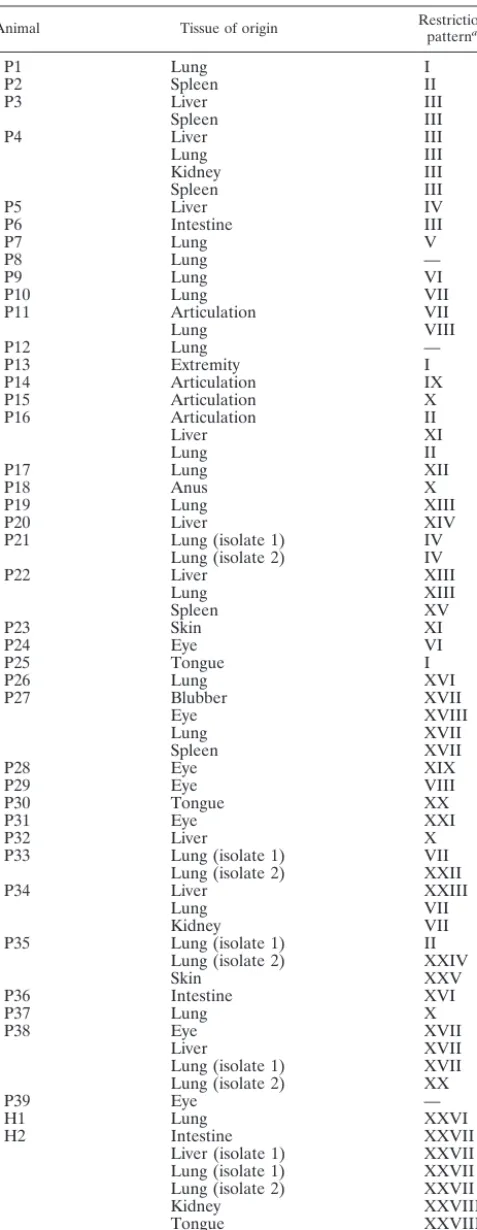
To analyze possibly existing epidemiological relations, the isolates were subjected to macrorestriction analysis of their chromosomal DNA by pulsed-field gel electrophoresis (PFGE) using the rare-cutting enzymeApaI. PFGE analysis of 66 S. phocae cultures revealed 29 different DNA patterns. There were identical as well as nonidentical DNA fragment patterns for isolates from one animal and from different ani-mals (Table 2). However, most of the DNA fingerprints were not identical, indicating that multiple bacterial clones were distributed among the harbor seal and grey seal population of the North and Baltic seas and that cross infections between animals seem to be rare. This is in contrast to previously investigated S. dysgalactiae subsp.dysgalactiae serovar L iso-lates from harbor porpoises of the North and Baltic seas. In that study, singleS. dysgalactiaesubsp.dysgalactiaeserovar L clones or at least closely related clones could be found in the various specimens (17).
Because of the occurrence of multipleS. phocaeclones ob-tained from seal organs and the isolation of the S. phocae
together with other bacterial species, the importance of this bacterial species for various health conditions remains unclear. (Parts of these results were presented at the 14th Annual Conference of the European Cetacean Society in Cork, Ire-land, 2 to 5 April 2000.)
REFERENCES
1. Abdulmawjood, A., and C. La¨mmler.1999. Amplification of 16S ribosomal RNA gene sequences for the identification of streptococci of Lancefield
group B. Res. Vet. Sci.67:159–162.
2. Abdulmawjood, A., and C. La¨mmler.2000. Determination of intraspecies
variations of the V2 region of the 16S rRNA gene ofStreptococcus equi
subsp.zooepidemicus.Res. Vet. Sci.68:33–39.
3. Abt, K. F., N. Hoyer, L. Koch, and D. Adelung.2002. The dynamics of grey
seals (Halichoerus grypus) of Amrum in south eastern North Sea—evidence
of an open population. J. Sea Res.47:55–67.
4. Bandomir, B., S. Marxen, J. Taylor, U. Siebert, and D. Adelung.1999. Totfundmonitoring von Robben in Schleswig-Holstein 1998. Bericht an das Ministerium fu¨r Umwelt, Natur und Forsten des Landes Schleswig-Holstein, Forschungs- und Technologiezentrum Westku¨ste. Universita¨t Kiel, Bu¨sum, Germany.
5. Bandomir, B., U. Siebert, and D. Adelung.2000. Untersuchungen zum Ge-sundheitszustand von Robben in Schleswig-Holstein 1999. Bericht an das Ministerium fu¨r Umwelt, Natur und Forsten des Landes Schleswig-Holstein, Forschungs- und Technologiezentrum Westku¨ste. Universita¨t Kiel, Bu¨sum, Germany.
6. Bandomir, B., U. Siebert, und D. Adelung.2000. Untersuchungen zum Ge-sundheitszustand von Robben in Schleswig-Holstein 2000. Bericht an das Ministerium fu¨r Umwelt, Natur und Forsten des Landes Schleswig-Holstein, Forschungs- und Technologiezentrum Westku¨ste. Universita¨t Kiel, Bu¨sum, Germany.
7.Bentley, R. W., and J. A. Leigh.1995. Development of PCR-based hybrid-ization protocol for identification of streptococcal species. J. Clin. Microbiol.
33:1296–1302.
8. Bonner, W. N.1989. The natural history of seals. Christopher Helm, London, Great Britain.
9. Henton, M. M., O. Zapke, and P. A. Basson.1999.Streptococcus phocae
infections associated with starvation in cape fur seals. J. S. Afr. Vet. Assoc.
70:98–99.
10. Hutson, R. A., D. E. Thompson, and M. D. Collins.1993. Genetic interre-lationships of saccharolytic Clostridium botulinum types B, E and F and related clostridia as revealed by small-subunit rRNA gene sequences.
Mi-crobiol. Lett.108:103–110.
11. Jensen, T., M. van de Bildt, H. H. Dietz, T. H. Andersen, A. S. Hammer, T. Kuiken, and A. Osterhaus.2002. Another phocine distemper outbreak in
Europe. Science297:209.
[image:4.603.48.287.91.706.2]12. King, J. E. (ed.).1983. Seals of the world. Cornell University Press, New York, N.Y.
TABLE 2. ApaI restriction patterns of 69S. phocaeisolates from 39 harbor seals and 4 grey seals from the North and Baltic seas
Animal Tissue of origin Restrictionpatterna
P1 Lung I
P2 Spleen II
P3 Liver III
Spleen III
P4 Liver III
Lung III
Kidney III
Spleen III
P5 Liver IV
P6 Intestine III
P7 Lung V
P8 Lung —
P9 Lung VI
P10 Lung VII
P11 Articulation VII
Lung VIII
P12 Lung —
P13 Extremity I
P14 Articulation IX
P15 Articulation X
P16 Articulation II
Liver XI
Lung II
P17 Lung XII
P18 Anus X
P19 Lung XIII
P20 Liver XIV
P21 Lung (isolate 1) IV
Lung (isolate 2) IV
P22 Liver XIII
Lung XIII
Spleen XV
P23 Skin XI
P24 Eye VI
P25 Tongue I
P26 Lung XVI
P27 Blubber XVII
Eye XVIII
Lung XVII
Spleen XVII
P28 Eye XIX
P29 Eye VIII
P30 Tongue XX
P31 Eye XXI
P32 Liver X
P33 Lung (isolate 1) VII
Lung (isolate 2) XXII
P34 Liver XXIII
Lung VII
Kidney VII
P35 Lung (isolate 1) II
Lung (isolate 2) XXIV
Skin XXV
P36 Intestine XVI
P37 Lung X
P38 Eye XVII
Liver XVII
Lung (isolate 1) XVII
Lung (isolate 2) XX
P39 Eye —
H1 Lung XXVI
H2 Intestine XXVII
Liver (isolate 1) XXVII
Lung (isolate 1) XXVII
Lung (isolate 2) XXVII
Kidney XXVIII
Tongue XXVIII
H3 Lung XXVII
H4 Mouth XXIX
a—, the DNA of the cultures could not be digested byApaI and separated by
PFGE.
472 NOTES J. CLIN. MICROBIOL.
on May 15, 2020 by guest
http://jcm.asm.org/
13. La¨mmler, C., and G. Hahn.1994. Streptokokken.InH. Blobel and T. Schließer (ed.), Handbuch der bakteriellen Infektionen bei Tieren, vol. 2. Gustav Fischer Verlag, Jena, Germany.
14. Schwarz, J., and G. Heidemann.1994. Zum Status der Besta¨nde der
Seehund-und Kegelrobbenpopulation im Wattenmeer, p. 296–303.InJ. L. Loza´n, E.
Rachor, K. Reise, H. Westernhagen, and W. Lenz (ed.), Warnsignale aus dem Wattenmeer. Blackwell Wissenschafts-Verlag, Berlin, Germany.
15. Skaar, I., P. Gaustad, T. Tønjum, B. Holm, and H. Stenwig.1994. Strepto-coccus phocaesp. nov., a new species isolated from clinical specimens from
seals. Int. J. Syst. Bacteriol.44:646–650.
16. Soedermanto, I., F. H. Pasaribu, I. W. T. Wibawan, and C. La¨mmler.1996.
Identification and molecular characterization of serological group C strep-tococci isolated from diseased pigs and monkeys in Indonesia. J. Clin.
Mi-crobiol.34:2201–2204.
17. Swenshon, M., C. La¨mmler, and U. Siebert.1998. Identification and molec-ular characterization of beta-hemolytic streptococci isolated from harbor
porpoises (Phocoena phocoena) of the North and Baltic seas. J. Clin.
Micro-biol.36:1902–1906.
18. Tenover, F. C., R. D. Arbeit, R. V. Goering, P. A. Mickelsen, B. E. Murray, D. H. Persing, and B. Swaminathan.1995. Interpreting chromosomal DNA restriction patterns produced by pulsed-field gel electrophoresis: criteria for
bacterial strain typing. J. Clin. Microbiol.33:2233–2239.