Copyright © 2002, American Society for Microbiology. All Rights Reserved.
Typing and Subtyping of 83 Clinical Isolates Purified from Surgically
Implanted Silicone Feeding Tubes by Random Amplified
Polymorphic DNA Amplification
Melanie P. Dautle,† Ricky L. Ulrich,† and Thomas A. Hughes*
Department of Microbiology and Molecular Medicine, Clemson University, Clemson, South Carolina Received 10 July 2001/Returned for modification 15 August 2001/Accepted 14 November 2001
In this study, 83 clinical isolates purified from biofilms colonizing 18 silicone gastrostomy devices (12 “buttons” and six tubes converted to skin level devices) were selected for subtype characterization utilizing genetic analysis. The tubes, previously used for feeding, remained in place for 3 to 47 months (mean, 20.0 months) in children ranging in age from 6 months to 17 years. Classification of specific microbes using random amplified polymorphic DNA (RAPD) analysis revealed genetic similarities and differences among isolates belonging to the same genus. Both gram-positive and -negative bacteria were investigated, including 2 isolates
ofBacillus brevis,4 isolates ofBacillus licheniformis, 2 isolates ofBacillus pumilus, 3 isolates of Enterococcus
durans, 19 isolates ofEnterococcus faecalis, 8 isolates ofEnterococcus faecium, 2 isolates ofEnterococcus hirae,
7 isolates ofEscherichia coli, 8 isolates ofLactobacillus plantarum, 19 isolates ofStaphylococcus aureus, 2 isolates
ofStaphylococcus epidermidis, and 7 isolates ofStaphylococcus saprophyticus.Amplified DNA fragments
(ampli-cons) provided species-specific fingerprints for comparison by agarose gel electrophoresis. A total of 62 distinct RAPD types were categorized from the five genera studied. Typing analysis suggested cross acquisition ofE.
coli, E. faecalis, andS. aureusin three patient pairs. Genomic polymorphism detection proved efficient and
reliable for classifying bacterial subtypes isolated from biofilms adhering to various portions of commonly employed enteral access tubes.
Long-term enteral access is a common practice associated with modern medicine, especially for direct gastric access via gastrostomy. Percutaneous endoscopic gastrostomy (PEG), first described in 1980, has facilitated direct gastric access, and skin level devices have removed many of the disadvantages associated with long enteral catheters (11). It is estimated that between 180,000 and 200,000 PEGs are performed annually in the United States (10). However, problems associated with microbial adhesion and related to patient health and device failure require further investigation.
Several studies have looked at microbial attachment on medical devices, such as catheters and prosthetic implants, while other investigations have researched fungal contamina-tion on silicone gastrostomy tubes. The following organisms
have been found associated with gastrostomy devices:Candida
tropicalis,Candida albicans,Torulopsis glabrata,Engyodontium album, Wangiella dermatitidis, Pseudomonas aeruginosa, Can-dida krusei,Escherichia coli,Enterobacter cloacae,
alpha-hemo-lytic streptococci, Lactobacillus sp., Bacteriodes sp., Bacillus
brevis,Bacillus licheniformis,Bacillus pumilus,Enterococcus du-rans,Enterococcus faecalis,Enterococcus faecium,Enterococcus hirae,Lactobacillus plantarum,Micrococcus sedentarius, Staph-ylococcus aureus,Staphylococcus epidermidis, and Staphylococ-cus saprophytiStaphylococ-cus (6, 13, 14, 16, 19). Tube deterioration in association with attached fungi, as well as gastrostomy site wound infections, were found related to these organisms.
Bacterial biofilms, defined as a structured community of sessile bacterial cells enclosed in a self-produced extracellular polysaccharide matrix adhering to inert or living surfaces, oc-cur in a wide variety of environments (5). Biofilms preferen-tially develop on inert surfaces or dead tissue, which is prob-lematic in immunocompromised individuals requiring some form of medical implant. In addition, biofilms produce local-ized environments of corrosive molecules and/or protons in excessive concentrations as well as enzymes in direct contact with the substratum (4). Molecules are produced faster than they can diffuse through the matrix, leading to device deterio-ration. It has also been reported that species of the genera
Torulopsis,Candida, andAspergilluscan catabolize intermedi-ate-chain-length hydrocarbons for cellular growth (7, 17). Based on these findings, fungi in association with biofilm bac-teria could enhance device destruction via the metabolism of silicone components.
Microbial typing is most accurately determined by genomic fingerprinting methods (21). Several methodologies are com-monly employed, including pulsed-field electrophoresis (15), ribotyping (8), restriction endonuclease analysis (18), multilo-cus enzyme electrophoresis (12), and PCR-based procedures (9). Random amplified polymorphic DNA (RAPD) analysis can be utilized to determine variations in DNA sequences among closely related species, thus allowing subtype differen-tiation (20). Single primers, usually 10 bases long, are used to amplify random domains of purified DNA, producing finger-prints characteristic of a particular strain. Single-base substi-tutions, or insertions and deletions, will alter primer annealing, causing differing RAPD profiles that can be used to estimate nucleotide diversity and divergence (3). An advantage of RAPD analysis is that it can be applied to any strain or species
* Corresponding author. Mailing address: Department of Microbi-ology and Molecular Medicine, 132 Long Hall, Clemson University, Clemson, SC 29634. Phone: (864) 656–3057. Fax: (864) 656–0435. E-mail: [email protected].
† Present address: Bacteriology Division, USAMRIID, Ft. Detrick, Frederick, MD 21702-5011.
414
on May 15, 2020 by guest
http://jcm.asm.org/
of a bacterial group without previous knowledge of that isolate (26). In addition, much smaller quantities of DNA are re-quired, which is especially important when dealing with gram-positive species. PCR-based technologies of this type produce high concentrations of DNA composing the amplicons, which eliminates the need for expensive data-imaging software. RAPD typing has been used successfully for the
characteriza-tion of numerous organisms, includingP. aeruginosa(1, 21),S.
aureus,Staphylococcus intermedius(25),Streptococcus mutans,
Streptococcus sobrinus,Streptococcus rattus(22),Salmonella en-tericasubsp.enterica(2),Lactobacillus casei,Lactobacillus cur-vatus,Lactobacillus fermentum,Lactobacillus halotolerans, Lac-tobacillus pentosus, L. plantarum, Lactobacillus rhamnosus,
Lactobacillus sake(26), andE. coli(23).
RAPD analysis was used in this study to classify 83 isolates into 62 distinct subtypes. RAPD analysis is inexpensive, effi-cient, and well suited for investigations incorporating large sample numbers (1). In the present study, isolates associated with pediatric feeding tubes were compared to determine the numerous subtypes capable of proliferating throughout the biofilm as well as proliferation of single organisms throughout
multiple areas of PEG tubes. In addition, potentially common organisms were studied in an effort to link a general localized source of contamination leading to cross acquisition of species.
MATERIALS AND METHODS
Isolation and identification of biofilm microorganisms. Low-profile PEG tubes, composed of silicone rubber, were collected from 18 pediatric patients from The Children’s Hospital of Greenville Hospital System (Greenville, S.C.). Areas of the gastrostomy tubes, including the inner and outer portions of the internal stabilizer, the inner and outer portions of the shaft, and the valve, if present, were scraped with a sterile scalpel to remove the viable biofilm (6). The biofilm cells were placed in brain heart infusion broth (Difco, Detroit, Mich.) with 0.01 g of cycloheximide (Sigma, St. Louis, Mo.)/ml (BHI-C). Aliquots were also plated in duplicate on chocolate agar (Difco) plates containing 0.01 g of
cycloheximide/ml. One set of tubes and plates was incubated under 5% CO2for
1 week at 37°C, and the other set was incubated in an anaerobic Gas-Pak 100 jar (BBL Biological Systems, Cockeysville, Md.) for 1 week at 37°C. Colonies from chocolate agar plates containing 0.01 g of cycloheximide/ml were inoculated into BHI-C and incubated either aerobically or anaerobically as previously described.
Tubes of BHI-C from the initial biofilm inoculation were diluted (10⫺5) in
phosphate-buffered saline (pH 7.2) (Sigma), plated on brain heart infusion agar (Difco) plates containing 0.01 g of cycloheximide/ml, and incubated aerobically or anaerobically as previously described. Well-isolated, morphologically distinct colonies from brain heart infusion agar plates containing 0.01 g of cyclohexi-mide/ml were inoculated into BHI/C and cultured as described above. Genus and species determinations were obtained with the BBL CRYSTAL identification systems (Becton Dickinson, Sparks, Md.). Gram-positive isolates were identified with a BBL CRYSTAL Gram-Positive identification kit (Becton Dickinson), while gram-negative isolates were identified with a BBL CRYSTAL Enteric/ Nonfermenter identification kit (Becton Dickinson). Both types of panels were inoculated according to the manufacturer’s specifications.
Chromosomal DNA preparation. For chromosomal DNA preparations, a 1.5-ml sample of the desired strain was grown in brain heart infusion broth (Difco), pelleted with a microcentrifuge (Eppendorf model 5415C; Brinkmann Instruments, Westbury, N.Y.) at 14,000 rpm for 2 to 3 min, and extracted using methods described by Ulrich and Hughes (24). The preparations were analyzed
on a 1% agarose gel containing 0.5g of ethidium bromide/ml.
[image:2.587.46.285.70.336.2]RAPD analysis.RAPD reactions were performed in duplicate using Ready-to-Go RAPD analysis beads (Amersham Pharmacia Biotech, Piscataway, N.J.). Multiple primers were tested for the capacity to amplify the target DNA from
FIG. 1. RAPD fingerprints of Bacillusisolates. Lanes 1 and 10, DirectLoad wide-range DNA markers ranging from 50 to 10,000 bp (D7058; Sigma); lanes 2 to 9,B. brevis,B. licheniformis, andB. pumilus
[image:2.587.304.544.72.306.2]isolates.
FIG. 2. RAPD fingerprints ofE. coliisolates. Lanes 1 and 9, Di-rectLoad wide-range DNA markers ranging from 50 to 10,000 bp (D7058; Sigma); lanes 2 to 8,E. coliisolates.
TABLE 1. IOS forB. licheniformis
Isolate IOS
Bl11OIS Bl11OS Bl16OIS
Bl4OIS 0.600 0.222 0.000
Bl11OIS 0.000 0.000
Bl11OS 0.800
on May 15, 2020 by guest
http://jcm.asm.org/
[image:2.587.43.283.665.728.2]each isolate. A single primer (5⬘-GTTTCGCTCC-3⬘) was chosen for its ability to produce a distinguishable RAPD profile with a limited number of amplicons
visible under UV light. Reactions were performed by combining 2l (15 pmol/
l) of primer, 1l of template DNA (approximately 50 ng), and 22l of sterile
distilled water with the reaction beads for a total volume of 25l. Amplification
was performed in a Genius thermal cycler (Techne Ltd., Cambridge, United Kingdom) programmed for one cycle of 5 min at 95°C followed by 45 cycles of 1 min at 95°C, 1 min at 36°C, and 2 min at 72°C. The amplicons were analyzed
on a 1.5% agarose gel containing 0.5g of ethidium bromide/ml at 200 V for 1 h
and 30 min.
RAPD data analysis.The RAPD patterns of individual strains were compared based on the index of similarity (IOS) between samples (2). The following
formula was used for calculations:Fxy⫽2nxy/(nx⫹ny), wherenxyis the number
of RAPD bands both samples share andnxandnyare the number of RAPD
bands in each sample. The IOS provides a mathematical model to study genetic variation, especially with difficult or closely related RAPD patterns. Computa-tional analysis of this type allows for direct comparisons without the need to count bands, which is especially important after loss of resolution resulting from manuscript duplication via photocopying.
RESULTS
Eighty-three isolates from 15 patients were classified into 62 subtypes as determined by unique banding patterns obtained from RAPD analysis and IOS. The patients ranged in age from 6 months to 17 years (mean, 93.4 months), and the implanta-tion time ranged from 3 to 47 months (mean, 20.0 months). All RAPD reactions were performed in duplicate to determine the consistency and reproducibility of the RAPD methodology. No deviations in RAPD patterns between the duplicate reactions were encountered using the selected primer discussed in Ma-terials and Methods. Isolate codes were based on organism classification, patient number, and location of the tube (IIS, inner portion of the internal stabilizer; OIS, outer portion of the internal stabilizer; IS, inner portion of the shaft; OS, outer portion of the shaft; and V, valve) and were designated with a number if multiple isolates were found in the same location. IOS values were determined by comparing two isolates and ranged from 0.000 (showing no similarity) to 1.000 (indicating identical subtypes).
Two B. brevis(Bb), four B. licheniformis (Bl), and one B. pumilus(Bp) subtypes were discerned based on unique band-ing patterns (Fig. 1) and IOS analysis of each isolate. The IOS
for the B. brevis isolates, Bb9IIS and Bb9OS, was 0.308. A
comparison ofB. licheniformisisolates can be seen in Table 1.
None of theB. licheniformisisolates were genetically identical,
and the largest IOS value was between isolates Bl11OS and
Bl16OIS. TheB. pumilusisolates, Bp1IS and Bp1OIS, had an
[image:3.587.43.282.82.175.2]IOS of 1.000.
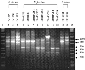
[image:3.587.125.461.430.707.2]FIG. 3. RAPD fingerprints ofEnterococcusisolates. Lanes 1 and 15, DirectLoad wide-range DNA markers ranging from 50 to 10,000 bp (D7058; Sigma); lanes 2 to 14,E. durans,E. faecium, andE. hiraeisolates.
TABLE 2. IOS forE. coli
Isolate IOS
Ec9IIS1 Ec9IIS2 Ec9IS Ec9OIS1 Ec9OIS2 Ec9OS
Ec3OS 0.571 1.000 0.800 1.000 1.000 1.000
Ec9IIS1 0.571 0.500 0.571 0.571 0.571
Ec9IIS2 0.800 1.000 1.000 1.000
Ec9IS 0.800 0.800 0.800
Ec9OIS1 1.000 1.000
Ec9OIS2 1.000
on May 15, 2020 by guest
http://jcm.asm.org/
ThreeE. coli(Ec) types were also distinguished and catego-rized as seen in Fig. 2 and Table 2. Isolates Ec3OS, Ec9IIS2, Ec9OIS1, Ec9OIS2, and Ec9OS produced identical amplicon patterns and IOS values. The least related isolates based on amplified DNA were Ec9IIS1 and Ec9IS, with an IOS of 0.500.
ThreeE. durans(Ed), sevenE. faecium(Efm), and twoE.
hirae(Eh) subtypes were also grouped using RAPD profiles
(Fig. 3) and IOS values. The IOS for theE. durans isolates
Ed10V and Ed10OS was 0.727, that for Ed10V and Ed15IIS
was 0.546, and that for Ed10OS and Ed15IIS was 0.800.E.
faeciumIOS results are reported in Table 3. Isolates Efm15IS and Efm15OIS1 produced homologous RAPD patterns. The
IOS for the E. hirae isolates, Eh15IIS1 and Eh15IIS2, was
0.833.
TwelveE. faecalis(Ef) types were differentiated and
classi-fied as reported in Fig. 4 and Table 4. Isolates Ef2OS2 and Ef5OS3 were identical in amplicon patterns and IOS values. Also, isolates Ef2OS3, Ef5IS1, Ef5IS2, and Ef5IS3; Ef5OIS and Ef5OS1; and Ef7IS1, Ef7IS2, and Ef7V were identical.
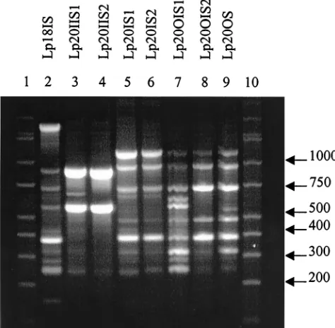
EightL. plantarum(Lp) types were also distinguished (Fig.
5) and categorized (Table 5). None of the isolates had identical banding patterns; however, Lp20IS2 and Lp20OS had the highest IOS value of 0.875, while Lp20IIS2 and Lp20OS did not share any common amplicons.
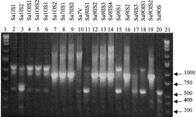
TwelveS. aureus(Sa) types were determined based on
band-ing patterns (Fig. 6) and IOS (Table 6). The identical isolates were as follows: Sa1IS1, Sa1OIS1, Sa1OIS2, and Sa1OS1; Sa1OS2 and Sa9IIS2; Sa7IIS1 and Sa7IIS2; and Sa9IIS1,
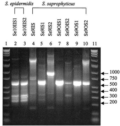
Sa9IS1, and Sa9OIS1. The twoS. epidermidisspecies analyzed
produced the same RAPD profile (Fig. 7). SevenS.
saprophy-ticustypes were classified and produced different RAPD fin-gerprints (Fig. 7) and IOS values (Table 7).
In several species, all isolates tested shared major amplicons.
The twoB. brevisisolates shared one band, the seven testedE.
colistrains shared two amplicons, three common bands were
found in the threeE. duransisolates, one amplicon was shared
by all eightE. faeciumsamples, and the two isolatedE. hirae
strains shared four bands.
Based on the RAPD fingerprints and the calculated IOS values, 62 distinct types were discerned among the 83 total isolates investigated in this study. Isolates with the same am-plicon pattern were observed in multiple patients as follows: oneE. colitype was seen in isolates Ec3OS, Ec9IIS2, Ec9OIS1,
Ec9OIS2, and Ec9OS; oneS. aureusprofile was seen in isolates
Sa1OS2 and Sa9IIS2; and twoE. faecalisprofiles were seen,
one in strains Ef2OS2 and Ef5OS3 and the other in strains
Ef2OS3, Ef5IS2, and Ef5IS3. Patients 3 and 9 had identicalE.
coli RAPD profiles and were seen in the hospital for tube
removal 1 month and 25 days apart. RAPD profiles in patients
[image:4.587.40.543.84.184.2]2 and 5 showed identicalE. faecalistypes, and these patients
TABLE 3. IOS forE. faecium
Isolate IOS
Efm15IS Efm15OIS1 Efm15OIS2 Efm15OIS3 Efm15OS1 Efm15OS2 Efm15OS3
Efm15IIS 0.667 0.667 0.600 0.750 0.500 0.400 0.444
Efm15IS 1.000 0.364 0.444 0.444 0.364 0.400
Efm15OIS1 0.182 0.222 0.444 0.546 0.400
Efm15OIS2 0.800 0.400 0.333 0.546
Efm15OIS3 0.500 0.400 0.667
Efm15OS1 0.600 0.667
Efm15OS2 0.909
FIG. 4. RAPD fingerprints ofE. faecalisisolates. Lanes 1 and 21, DirectLoad wide-range DNA markers ranging from 50 to 10,000 bp (D7058; Sigma); lanes 2 to 20,E. faecalisisolates.
on May 15, 2020 by guest
http://jcm.asm.org/
[image:4.587.123.460.508.706.2]were seen 28 days apart for device replacement. The third pair, patients 1 and 9, seen 2 months and 20 days apart, shared the sameS. aureustype.
DISCUSSION
It has been shown that RAPD fingerprints produced from arbitrarily primed PCR can be used to compare bacterial strains (27). In the present study, RAPD analysis was applied to clinical isolates for subtype differentiation and to possibly link common isolates to specific patients. The results showed that this methodology could be used to distinguish different subtypes based on the numerous fingerprints generated within a microbial genus and species group. Not only did the RAPD profiles allow for type distinction, the degree of relatedness between isolates was calculated based on band similarities.
This was apparent withB. brevis,E. coli,E. durans,E. faecium,
E. hirae, andS. epidermidis, all of which had at least one band in common within each species, indicating a potential species-specific probe for strain identification.
RAPD profiles from the genusBacillusled to several
inter-pretations. None of the isolates from the three species B.
brevis,B. licheniformis, andB. pumilusshowed identical RAPD
profiles. TheB. brevisisolates were purified from two specific
locations on the same tube from one patient, the inner portion of the internal stabilizer and the outer portion of the shaft. A hypothesis, supported by several reports (1, 22), can be drawn that the isolates came from different environmental sources and were not from one organism proliferating through various
locations of the tube.B. licheniformis was found in biofilms
isolated from three different patients, and all of the RAPD profiles were different, leading to the hypothesis that each patient encountered different environmental sources of the contaminant that produced biofilm proliferation. Patient 11
had two differentB. licheniformistypes in two different areas of
the tube, reinforcing the conclusion that multiple sources of bacterial contamination exist. In addition to these
observa-tions, the twoB. pumilusisolates had identical RAPD profiles
and were found on two areas of the same patient’s device, implying a second characteristic related to biofilm
prolifera-tion. Based on this fact, it is also possible for microorganisms to spread to multiple areas of the tube from a single source.
Of the seven E. coli isolates, five had the same amplicon
[image:5.587.41.545.89.277.2]distribution but were not from the same patient. One was from patient 3, while the other four were from patient 9. A finding of this nature could show cross acquisition between patients via direct patient-to-patient interaction or contact with a com-mon environmental source. Accessible records indicate that 1 month and 25 days elapsed between office visits and a different doctor cared for each patient. This information provides in-sight into a potential source, but further testing was restricted, so a definitive source linking the two could not be determined. Comparisons of enterococcus profiles support the previously
[image:5.587.303.540.463.695.2]FIG. 5. RAPD fingerprints ofL. plantarumisolates. Lanes 1 and 10, DirectLoad wide-range DNA markers ranging from 50 to 10,000 bp (D7058; Sigma); lanes 2 to 9,L. plantarumisolates.
TABLE 4. IOS forE. faecalis
Isolate IOS
Ef2OS2 Ef2OS3 Ef5IS1 Ef5IS2 Ef5IS3 Ef5V Ef5OIS Ef5OS1 Ef5OS2 Ef5OS3 Ef7IS1 Ef7IS2 Ef7V Ef7OS Ef14OS Ef14IIS Ef15IS Ef15OS
Ef2OS1 0.333 0.400 0.400 0.400 0.400 0.667 0.500 0.500 0.667 0.333 0.000 0.000 0.000 0.000 0.400 0.400 0.333 0.500 Ef2OS2 0.667 0.667 0.667 0.667 0.571 0.500 0.500 0.571 1.000 0.800 0.800 0.800 0.200 0.889 0.667 0.800 0.500 Ef2OS3 1.000 1.000 1.000 0.667 0.857 0.857 0.667 0.667 0.667 0.667 0.667 0.222 0.500 0.500 0.444 0.571 Ef5IS1 1.000 1.000 0.667 0.857 0.857 0.667 0.667 0.667 0.667 0.667 0.222 0.500 0.500 0.444 0.571 Ef5IS2 1.000 0.667 0.857 0.857 0.667 0.667 0.667 0.667 0.667 0.222 0.500 0.500 0.444 0.571 Ef5IS3 0.667 0.857 0.857 0.667 0.667 0.667 0.667 0.667 0.222 0.500 0.500 0.444 0.571 Ef5V 0.800 0.800 0.500 0.571 0.286 0.286 0.286 0.000 0.667 0.667 0.571 0.800
Ef5OIS 1.000 0.400 0.500 0.500 0.500 0.500 0.000 0.571 0.571 0.500 0.667
Ef5OS1 0.400 0.500 0.500 0.500 0.500 0.000 0.571 0.571 0.500 0.667
Ef5OS2 0.571 0.286 0.286 0.286 0.286 0.333 0.333 0.286 0.400
Ef5OS3 0.600 0.600 0.600 0.200 0.889 0.667 0.600 0.500
Ef7IS1 1.000 1.000 0.200 0.444 0.444 0.400 0.250
Ef7IS2 1.000 0.200 0.444 0.444 0.400 0.250
Ef7V 0.200 0.444 0.444 0.400 0.250
Ef7OS 0.000 0.000 0.000 0.000
Ef14OS 0.750 0.889 0.571
Ef14IIS 0.667 0.857
Ef15IS 0.750
on May 15, 2020 by guest
http://jcm.asm.org/
stated findings that biofilm formation occurs due to multiple sources as well as proliferation from a single contaminant. The twoE. duransisolates Ed10V and Ed10OS were from the same
patient’s tube but different locations. TwoE. faeciumisolates
from the inner portion of the shaft and the outer portion of the internal stabilizer from the same patient produced the same pattern by RAPD analysis, supporting the finding of biofilm proliferation from a single bacterium. All other isolates (Efm15IIS, Efm15OIS2, Efm15OIS3, Efm15OS1, Efm15OS2, and Efm15OS3) from the same patient were genetically differ-ent, showing that numerous microorganisms were involved in
the formation of the biofilm. TwoE. hirae isolates from the
same location were genetically different, supporting the
previ-ously stated finding.E. faecaliswas found in five patients, but
two of them showed multiple RAPD types with an IOS of 1.000. Samples Ef2OS2 and Ef5OS3 were identical, further supporting the statement that cross acquisition between pa-tients could occur. Interestingly, these two papa-tients shared an-other subtype seen in Ef2OS3, Ef5IS2, and Ef5IS3.
L. plantarumwas found in only two patients, but all eight of the isolates were genetically different based on RAPD profiles. Many of the samples were similar (Lp20IIS1 and Lp20IIS2, Lp20IS1 and Lp20IS2, and Lp20OIS2 and Lp20OS), but one or two bands that were present in one were absent in the other (Fig. 5), indicating similarity in the genetic template due to the
amplicons produced in each sample while the missing bands show the isolates were not identical. Interestingly, Lp20IIS2 and Lp20OS did not share any amplicons.
Results from the genusStaphylococcusRAPD analysis
sup-ported all three findings stated so far: that multiple sources contribute to biofilm composition, proliferation of a specific organism occurs, and patient cross acquisition is possible. A
singleS. aureus type was found associated with two patients
seen in the same office 2 months and 20 days apart, indicating the possibility of direct patient-to-patient contact or the pres-ence of a common environmental source. In patient 1, 4 of the
S. aureusisolates were identical, showing proliferation of one type over the surface of the device, as did 2 of the 3 isolates in
patient 7 and 3 of the 10S. aureus strains in patient 9. The
remaining isolates ofS. aureusas well asS. saprophyticus
[image:6.587.40.542.84.184.2]spe-cies were genetically different based on RAPD profiles. RAPD profiles, using the primer described in Materials and Methods, produced a limited number of bands, which facili-tated fingerprint analysis without the need for computer inter-vention. In addition, the relative prevalence of microbial col-onization throughout various locations of the device and potential cross acquisition among patients was determined. Identical isolates, based on RAPD profiles, were found on multiple areas of the tube, suggesting the spread of the biofilm from the initial point of attachment. In addition, numerous
TABLE 5. IOS forL. plantarum
Isolate IOS
Lp20IIS1 Lp20IIS2 Lp20IS1 Lp20IS2 Lp20OIS1 Lp20OIS2 Lp20OS
Lp18IS 0.667 0.615 0.444 0.471 0.700 0.400 0.588
Lp20IIS1 0.800 0.533 0.571 0.471 0.333 0.286
Lp20IIS2 0.308 0.167 0.400 0.200 0.000
Lp20IS1 0.824 0.700 0.800 0.706
Lp20IS2 0.737 0.857 0.875
Lp20OIS1 0.706 0.737
Lp20OIS2 0.857
FIG. 6. RAPD fingerprints ofS. aureusisolates. Lanes 1 and 21, DirectLoad wide-range DNA markers ranging from 50 to 10,000 bp (D7058; Sigma); lanes 2 to 20,S. aureusisolates.
on May 15, 2020 by guest
http://jcm.asm.org/
[image:6.587.120.463.502.707.2]subtypes were found associated with a single tube, leading to the observation that multiple bacterial subtypes are involved in the formation of gastrostomy tube-associated biofilms.
Identi-cal RAPD fingerprints for E. coli, E. faecalis, andS. aureus
were identified in three patient pairs, suggesting a potential transfer of microorganisms from patient to patient via direct interaction or contact with a common source.
Investigations of large numbers of isolates by RAPD would benefit from computer-assisted discrimination by generating a database of patterns for the comparison of present and future isolates, including antibiotic sensitivity and resistance, isolate source, and patient-associated disease. Additional studies are using plasmid analysis to investigate the transfer of antibiotic resistance genes in order to link RAPD fingerprints to antibi-otic resistance and sensitivity. This investigation supports pre-vious reports that RAPD analysis is efficient, reproducible, and capable of detecting genomic polymorphisms among various microbial species without previous knowledge of the nucleo-tide sequence on the target DNA (22), and the technique has been shown to be valuable in studies dealing with biofilm
[image:7.587.299.540.72.325.2]TABLE 6. IOS for S. aureus Isolate IOS Sa1IS2 Sa1OIS1 Sa1OIS2 Sa1OS1 Sa1OS2 Sa7IIS1 Sa7IIS2 Sa7V Sa9IIS1 Sa9IIS2 Sa9IIS3 Sa9IIS4 Sa9IS1 Sa9IS2 Sa9IS3 Sa9OIS1 Sa9OIS2 Sa9OS Sa1IS1 0.750 1.000 1.000 1.000 0.400 0.333 0.333 0.286 0.800 0.400 0.333 0.286 0.800 0.400 0.500 0.800 0.333 0.333 Sa1IS2 0.750 0.750 0.750 0.286 0.250 0.250 0.222 0.571 0.286 0.250 0.222 0.571 0.286 0.333 0.571 0.250 0.500 Sa1OIS1 1.000 1.000 0.400 0.333 0.333 0.286 0.800 0.400 0.333 0.286 0.800 0.400 0.500 0.800 0.333 0.333 Sa1OIS2 1.000 0.400 0.333 0.333 0.286 0.800 0.400 0.333 0.286 0.800 0.400 0.500 0.800 0.333 0.333 Sa1OS1 0.400 0.333 0.333 0.286 0.800 0.400 0.333 0.286 0.800 0.400 0.500 0.800 0.333 0.333 Sa1OS2 0.800 0.800 0.333 0.000 1.000 0.800 0.667 0.000 0.500 0.000 0.000 0.400 0.000 Sa7IIS1 1.000 0.286 0.000 0.800 0.667 0.857 0.000 0.400 0.500 0.000 0.333 0.000 Sa7IIS2 0.286 0.000 0.800 0.667 0.857 0.000 0.400 0.500 0.000 0.333 0.000 Sa7V 0.333 0.333 0.571 0.500 0.333 0.333 0.400 0.333 0.000 0.286 Sa9IIS1 0.000 0.000 0.000 1.000 0.000 0.667 1.000 0.000 0.400 Sa9IIS2 0.800 0.667 0.000 0.500 0.667 0.000 0.400 0.000 Sa9IIS3 0.857 0.000 0.800 0.000 0.000 0.667 0.000 Sa9IIS4 0.000 0.667 0.400 0.000 0.571 0.000 Sa9IS1 0.000 0.667 1.000 0.000 0.400 Sa9IS2 0.000 0.000 0.800 0.000 Sa9IS3 0.667 0.000 0.500 Sa9OIS1 0.000 0.400 Sa9OIS2 0.333
[image:7.587.301.541.638.729.2]FIG. 7. RAPD fingerprints ofStaphylococcusisolates. Lanes 1 and 11, DirectLoad wide-range DNA markers ranging from 50 to 10,000 bp (D7058; Sigma); lanes 2 to 10,S. epidermidisandS. saprophyticus iso-lates.
TABLE 7. IOS forS. saprophyticus
Isolate IOS
Ss9IS1 Ss9IS2 Ss9OIS1 Ss9OIS2 Ss9OS1 Ss9OS2
Ss9IIS 0.154 0.667 0.250 0.546 0.222 0.546
Ss9IS1 0.143 0.286 0.600 0.500 0.400
Ss9IS2 0.000 0.333 0.000 0.500
Ss9OIS1 0.400 0.667 0.400
Ss9OIS2 0.667 0.750
Ss9OS1 0.667
on May 15, 2020 by guest
http://jcm.asm.org/
formation on enteral access tubes. RAPD technology is an inexpensive way to type organisms without specialized equip-ment not readily available to general molecular microbiology laboratories. Although RAPD is sensitive to annealing tem-peratures, reproducibility has been achieved previously as well as in this study (22, 25). RAPD is also preferable for gram-positive species, where chromosomal preparations are difficult and low concentrations of nucleic acid are achieved. RAPD typing has proved to be efficient and cost-effective while maintaining reproducible and accurate results for analyzing large numbers of gram-positive organisms.
REFERENCES
1.Campbell, M., E. Mahenthiralingam, and D. P. Speert.2000. Evaluation of
random amplified polymorphic DNA typing of Pseudomonas aeruginosa.
J. Clin. Microbiol.38:4614–4615.
2.Chansiripornchai, N., P. Ramasoota, A. Bangtrakulnonth, J. Sasipreeyajan, and S. B. Svenson.2000. Application of randomly amplified polymorphic
DNA (RAPD) analysis for typing avianSalmonella entericasubsp.enterica.
FEMS Immunol. Med. Microbiol.29:221–225.
3.Clark, A. G., and C. M. S. Lanigan.1993. Prospects for estimating nucleotide
divergence with RAPDs. Mol. Biol. Evol.10:1096–1111.
4.Costerton, J. W.1992. The pivotal role of biofilms in the focused attack of
bacteria on soluble substrates. Int. Biodeterior. Biodegradation30:123–133.
5.Costerton, J. W., P. S. Stewart, and E. P. Greenberg.1999. Bacterial biofilm:
a common cause of persistent infections. Science284:1318–1322.
6.Dautle, M. P.1999. Isolation and identification of biofilm microorganisms from silicone gastrostomy devices. M.S. thesis. Clemson University, Clem-son, S.C.
7.Davies, J. S., and D. W. Westlake.1979. Crude oil utilization by fungi. Can.
J. Microbiol.25:146–156.
8.Denamur, E., B. Picard, P. Goullet, E. Bingen, N. Lambert, and J. Elion.
1991. Complexity ofPseudomonas aeruginosa infection in cystic fibrosis:
combined results from esterase electrophoresis and rDNA restriction
frag-ment length polymorphism analysis. Epidemiol. Infect.106:531–539.
9.Elaichouni, A., A. Vershraegen, G. Claeys, M. Devleeschouwer, C. Godard, and M. Vaneechoute.1994.Pseudomonas aeruginosaserotpye O12 outbreak
studied by arbitrary primer PCR. J. Clin. Microbiol.32:666–671.
10.Gauderer, M.1999. Twenty years of percutaneous endoscopic gastrostomy: origin and evolution of a concept and its expanded applications.
Gastroin-test. Endosc.50:879–883.
11.Gauderer, M. W. L., R. S. Abrams, and J. H. Hammond. 1998. Initial
experience with the changeable skin-level port-valve: a new concept for
long-term gastrointestinal access. J. Pediatr. Surg.33:73–75.
12.Gilmour, M. N., T. S. Whittam, M. Kilian, and R. K. Selander.1987. Genetic
relationships among the oral streptococci. J. Bacteriol.169:5247–5257.
13.Gottlieb, K., M. DeMeo, and P. Borton.1992. Gastrostomy tube
deteriora-tion and fungal colonizadeteriora-tion. Am. J. Gastroenterol.87:1683.
14.Gottlieb, K., J. Leya, P. M. Kruss, S. Mobarhan, and F. L. Iber.1993. Intraluminal fungal colonization of gastrostomy tubes. Gastrointest. Endosc.
39:413–415.
15.Grothues, D., V. Koopmann, H. van der Hardt, and B. Tümmler.1988.
Genome fingerprinting ofPseudomonas aeruginosaindicates colonization of
cystic fibrosis siblings with closely related strains. J. Clin. Microbiol.26:1973–
1977.
16.Hull, M. A., J. Rawlings, F. E. Murray, J. Field, A. S. McIntyre, and Y. R. Mahida.1993. Audit of outcome of long-term enteral nutrition by
percuta-neous endoscopic gastrostomy. Lancet341:869–872.
17.Klug, M. J., and A. J. Markovetz.1971. Utilization of aliphatic hydrocarbons
by microorganisms. Adv. Microb. Physiol.5:1–43.
18.Kulkarni, G. V., K. H. Chan, and H. J. Sandham.1989. Investigation into the use of restriction endonuclease analysis for the study of transmission of
mutans streptococci. J. Dent. Res.68:1155–1161.
19.Marcuard, S. P., J. L. Finley, and K. G. MacDonald. 1993. Large-bore
feeding tube occlusion by yeast colonies. J. Parenter. Enteral Nutr.17:187–
190.
20.Mileham, A. J.1997. Identification of microorganisms using random primed
PCR. Mol. Biotechnol.8:139–145.
21.Speijer, H., P. H. M. Sauelkoul, M. J. Bonten, E. E. Stobberingh, and J. H. T. Tjhie.1999. Application of different genotyping methods forPseudomonas aeruginosain a setting of endemicity in an intensive care unit. J. Clin.
Microbiol.37:3654–3661.
22.Truong, T. L., C. Menard, C. Mouton, and L. Trahan.2000. Identification of mutans and other oral streptococci by random amplified polymorphic DNA
analysis. J. Med. Microbiol.49:63–71.
23.Tseng, C., E. Ting, D. Johnson, M. Saluta, and R. Dunst.2001. Use of
RAPD fingerprinting for differentiatingE. coliisolates from human and
animal sources. Life Sci. News7:10–11.
24.Ulrich, R. L., and T. A. Hughes.2000. A rapid procedure for the isolation of chromosomal DNA from Gram-positive bacteria. Lett. Appl. Microbiol.
32:52–56.
25.Van Leeuwen, W., M. Sijmons, J. Sluijs, H. Verbrugh, and A. van Belkum.
1996. On the nature and use of randomly amplified DNA from
Staphylococ-cus aureus. J. Clin. Microbiol.34:2770–2777.
26.Veyrat, A., M. C. Miralles, and G. Pérez-Martinez.1999. A fast method for monitoring the colonization rate of lactobacilli in a meat model system.
J. Appl. Microbiol.87:49–61.
27.Welsh, J., and M. McClelland.1990. Fingerprinting genomes using PCR
with arbitrary primers. Nucleic Acids Res.18:7213–7218.