Detection of (1,3)-

-
D
-Glucan in Cerebrospinal Fluid in
Histoplasma
Meningitis
Thein Myint,aFelicia C. Chow,bKaren C. Bloch,cLuke Raymond-Guillen,dThomas E. Davis,ePatty W. Wright,c Laila Woc-Colburn,fRaed N. Khairy,gAlan C. Street,hTomotaka Yamamoto,iAmanda Albers,jL. Joseph Wheat,j Chadi A. Hagek
aDivision of Infectious Diseases, Department of Internal Medicine, University of Kentucky, Lexington, Kentucky, USA
bDepartment of Neurology, University of California, San Francisco, San Francisco, California, USA cDivision of Infectious Diseases, Department of Medicine, Vanderbilt University Medical Center, Nashville,
Tennessee, USA
dDivision of Infectious Diseases, Department of Internal Medicine, Indiana University, Indianapolis, Indiana, USA eDepartment of Pathology and Laboratory Medicine, Indiana University, Indianapolis, Indiana, USA
fDivision of Infectious Diseases, Department of Internal Medicine, Baylor College of Medicine, Houston, Texas, USA
gSparks Clinic, Fort Smith, Arkansas, USA
hVictorian Infectious Diseases Service, Royal Melbourne Hospital, Melbourne, Victoria, Australia iDepartment of Neurology, University of Tokyo Hospital, Tokyo, Japan
jMiraVista Diagnostics, Indianapolis, Indiana, USA
kDivision of Pulmonary and Critical Care Medicine, Thoracic Transplantation Program, Indiana University-School of Medicine, Indianapolis, Indiana, USA
ABSTRACT The diagnosis of central nervous system (CNS) histoplasmosis is often
difficult. Although cerebrospinal fluid (CSF) (1,3)--D-glucan (BDG) is available as a bi-ological marker for the diagnosis of fungal meningitis, there are limited data on its use for the diagnosis of Histoplasma meningitis. We evaluated CSF BDG detection, using the Fungitell assay, in patients with CNS histoplasmosis and controls. A total of 47 cases and 153 controls were identified. The control group included 13 patients with a CNS fungal infection other than histoplasmosis. Forty-nine percent of patients with CNS histoplasmosis and 43.8% of controls were immunocompromised. The me-dian CSF BDG level was 85 pg/ml for cases, compared to⬍31 pg/ml for all controls (P ⬍ 0.05) and 82 pg/ml for controls with other causes of fungal meningitis (P ⫽
0.27). The sensitivity for detection of BDG in CSF was 53.2%, whereas the specificity was 86.9% versus all controls and 46% versus other CNS fungal infections. CSF BDG levels of ⱖ80 pg/ml are neither sensitive nor specific to support a diagnosis of His-toplasmameningitis.
KEYWORDS (1,3)--D-glucan, cerebrospinal fluid,Histoplasma, meningitis
T
he diagnosis of central nervous system (CNS) histoplasmosis is challenging. In one large case series study (1), cerebrospinal fluid (CSF) cultures were positive for only 19.1% of patients, and culture results were often significantly delayed after clinical presentation. More rapid diagnosis could be achieved through detection of antibody (sensitivity, 82.2%) and antigen (sensitivity, 78.0%) in the CSF, with at least one of the tests being positive for 98.0% of patients with CNS histoplasmosis (1). Although a serum assay to detect (1,3)--D-glucan (BDG), a fungal cell wall polysaccharide, has been cleared by the U.S. Food and Drug Administration for serological diagnosis of invasive fungal diseases since 2004, the assay is not approved for CSF testing. Elevated levels of BDG have been detected in the CSF of patients with fungal meningitis caused byReceived19 April 2018 Returned for modification9 May 2018Accepted5 July 2018
Accepted manuscript posted online18 July 2018
CitationMyint T, Chow FC, Bloch KC, Raymond-Guillen L, Davis TE, Wright PW, Woc-Colburn L, Khairy RN, Street AC, Yamamoto T, Albers A, Wheat LJ, Hage CA. 2018. Detection of共1,3兲-β-D-glucan in cerebrospinal fluid inHistoplasmameningitis. J Clin Microbiol 56:e00663-18.https://doi.org/10 .1128/JCM.00663-18.
EditorDavid W. Warnock
Copyright© 2018 American Society for Microbiology.All Rights Reserved.
Address correspondence to Thein Myint, thein.myint3@uky.edu.
crossm
on May 16, 2020 by guest
http://jcm.asm.org/
Candidasp. (2, 3),Aspergillus(2, 4),Exserohilum(5, 6),Cryptococcus(4, 7), and Coccid-ioides (8). Data on the utility of BDG in the diagnosis ofHistoplasma meningitis are limited (4, 9). We evaluated CSF BDG detection using the Fungitell assay in the largest series of patients with CNS histoplasmosis to date.
MATERIALS AND METHODS
Cases were classified using previously defined criteria (1). Patients were categorized as CNS histo-plasmosis cases if they had clinical symptoms of meningitis and/or brain imaging abnormalities and supporting laboratory findings, as follows: confirmed CNS histoplasmosis, isolation of Histoplasma capsulatum from CSF; probable CNS histoplasmosis, detection of Histoplasma antigen by enzyme immunoassay (EIA) or anti-Histoplasmaantibodies in the CSF by immunodiffusion (ID) or complement fixation (CF); possible CNS histoplasmosis, pulmonary or disseminated histoplasmosis with CSF pleocy-tosis but without laboratory confirmation of CNS involvement (negative or absent culture findings, microscopy findings, and detection of antigen or antibody by ID or CF in the CSF) and no alternative etiology for the CSF pleocytosis. Controls included patients with pulmonary or disseminated histoplas-mosis without CNS involvement (no clinical findings for meningitis, no pleocytosis or CNS imaging abnormalities, and no diagnosis of or treatment for CNS histoplasmosis) or with negative testing for histoplasmosis (either with or without CSF pleocytosis), including patients with meningitis due to fungal pathogens other thanHistoplasma, nonfungal meningitis, and noninfectious CNS disorders.
BDG levels were measured in the remaining stored CSF specimens using the Fungitell assay, according to the methods used for serum specimens, as reported previously for CSF (4, 5). Data regarding prior treatment were not available when CSF specimens were obtained. According to the manufacturer’s guidelines for serum BDG assays, a positive CSF BDG result was defined asⱖ80 pg/ml. Chi-square analysis, Student’sttest, and the step-down Bonferroni multiple-comparison procedure were used to compare subgroups, using MedCalc software.
RESULTS
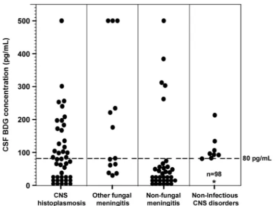
Forty-seven subjects with CNS histoplasmosis were enrolled in the study, including 9 (19.1%) confirmed, 33 (70.2%) probable, and 5 (10.6%) possible cases. A total of 153 subjects without CNS histoplasmosis were included as controls, including 13 (8.5%) with other causes of fungal meningitis, 31 (20.3%) with nonfungal meningitis, and 109 (71.2%) with noninfectious CNS disorders (e.g., encephalopathy or seizure disorder). Ten of 11 controls with pulmonary or disseminated histoplasmosis had a noninfectious CNS disorder, and 1 hadToxoplasma encephalitis. Cultures were positive for fungal pathogens for 6 (5.1%) of 117 controls for whom cultures were performed; pathogens includedCryptococcus (n⫽4),Aspergillus(n⫽1), andCandida dubliniensis(n⫽1). The other 7 fungal meningitis control cases had the following: blastomycosis (n⫽ 3) (2 controls were diagnosed by CSF antigen, one with the organism being isolated from bronchoalveolar lavage fluid and the other with characteristic large, broad-based budding yeast consistent withBlastomyces being identified with Grocott’s methena-mine silver staining of leptomeninges from a postmortem specimen; the third control had a positive urine antigen test result and a positive culture for Blastomyces from bronchoalveolar lavage fluid), cryptococcosis (n ⫽ 1, diagnosed by antigen testing), coccidioidomycosis (n ⫽ 1, diagnosed by antibody testing), aspergillosis (n ⫽ 1, diagnosed by antigen testing), and candidiasis (n ⫽1, diagnosed by blood culture). Forty-nine percent of patients with CNS histoplasmosis and 43.8% of controls were immunocompromised. BDG levels in the CSF among the different groups are shown in Fig. 1.
CSF BDG levels were not significantly different among the confirmed, probable, and possible cases ofHistoplasmameningitis (P⫽0.93) (Table 1). The median BDG level for cases was 85 pg/ml, compared to⬍31 pg/ml for all controls (P⬍0.05),⬍31 pg/ml for nonfungal meningitis (P⬍0.05), and⬍31 pg/ml for noninfectious CNS disorders (P⬍
0.05). There were no significant differences in median BDG levels between cases of
Histoplasmameningitis (85 pg/ml) and other fungal meningitis (82 pg/ml) (P⫽0.27). Twenty-five of the 47 Histoplasma meningitis cases had CSF BDG levels of ⱖ80 pg/ml, resulting in a sensitivity of 53.2%. Of the 153 controls, 133 had CSF BDG levels of⬍80 pg/ml, resulting in an overall specificity of 86.9% for detection of BDG in CSF for CNS histoplasmosis. Using the 140 controls without fungal meningitis, the specificity was 90.7%; using the 11 controls with disseminated or pulmonary histoplasmosis without CNS involvement, the specificity was 100%. Using the controls with other
on May 16, 2020 by guest
http://jcm.asm.org/
causes of fungal meningitis, however, the specificity was 46.2%. The median CSF BDG level in 6 controls with culture-positive fungal CNS infections other than histoplasmosis was 227 pg/ml, and 5 had levels ofⱖ80 pg/ml. Among the 7 controls with nonhisto-plasmosis fungal CNS infections diagnosed by antigen testing, antibody testing, or blood culture, the median BDG level was 61 pg/ml; 2 had CSF BDG levels ofⱖ80 pg/ml.
Histoplasmameningitis cases with CSF BDG levels ofⱖ80 pg/ml were older than those with BDG levels of⬍80 pg/ml (mean age, 47 years⫾7 versus 36 years⫾8;
P ⫽ 0.03). There were no statistically significant differences in sex (P ⫽ 0.12), immunocompromised status (P⫽ 0.89), positive CSF culture (P ⫽ 0.56), positive
Histoplasmaantigen testing (P⫽0.51), or the presence of highHistoplasmaantigen levels (⬎19 ng/ml) (P⫽0.22) between cases with BDG levels ofⱖ80 and those with levels of⬍80 pg/ml.
Among 37 patients with positive CSFHistoplasmaantigen testing, the median BDG level was 127.6 pg/ml; 21/37 patients (56.8%) had levels of ⱖ80 pg/ml, and 25/37 patients (67.6%) had levels of⬎31 pg/ml. Four of 25 cases with BDG levels ofⱖ80 pg/ml and 7 of 32 cases with BDG levels of⬎31 pg/ml had negative CSFHistoplasma
antigen testing. Two cases had positive CSF antigen test results that were below the detectable limit of⬍0.4 ng/ml.
[image:3.585.71.341.69.271.2]CSF BDG levels of ⱖ80 pg/ml were also detected in 13 patients with bacterial meningitis or a brain abscess (n⫽3), viral encephalitis (n⫽1), Rocky Mountain spotted fever (n⫽1), stroke (n⫽2), neurosarcoidosis (n⫽1), melanoma (n⫽1), hypoxic brain FIG 1CSF BDG levels in CNS Histoplasma meningitis, other fungal CNS infections, nonfungal CNS infections, and noninfectious CNS disorders. The dashed line represents the value of 80 pg/ml.*, There were 98 controls with noninfectious CNS disorders who had CSF BDG levels of⬍80 pg/ml.
TABLE 1CSF BDG levels in different groups ofHistoplasmameningitis cases and controls
Group No. of subjects
CSF BDG level (median [IQR]) (pg/ml)
No. (%) with BDG level
of>80 pg/ml P
All cases 47 85 (31–194) 25 (53.2) Reference
Confirmed 9 116 (62–197) 5 (55.6) 0.93a
Probable 33 85 (31–183) 18 (54.5)
Possible 5 72 (54–99) 2 (40.0)
All controls 153 ⬍31 (⬍31 to 55) 20 (13.1) ⬍0.05b
Other fungal meningitis 13 82 (61–234) 7 (53.8) 0.27b
Nonfungal meningitis 31 ⬍31 (⬍31 to 55.5) 5 (16.1) ⬍0.05b
Noninfectious CNS disorder 109 ⬍31 (⬍31 to 44) 7 (6.4) 0.05b
aPfor comparison of confirmed, probable, and possible cases. bPfor comparison with cases.
on May 16, 2020 by guest
http://jcm.asm.org/
[image:3.585.39.545.611.721.2]injury (n⫽1), acute psychosis (n⫽1), a seizure disorder (n⫽1), or an adverse reaction to medication (n⫽1). The median BDG level was 134 pg/ml (interquartile range [IQR], 93 to 303 pg/ml).
Using the Youden method (10) for receiver operating characteristic (ROC) analysis, the optimal cutoff value for CSF BDG levels was 61 pg/ml for CNS histoplasmosis versus controls, including other fungal meningitis cases, with sensitivity of 63.8%, specificity of 79.7%, and area under the curve (AUC) of 0.706 (Fig. 2). When other fungal meningitis cases were excluded, the optimal cutoff value for BDG was 58 pg/ml, yielding sensitivity and specificity of 67.8% and 83.7%, respectively, forHistoplasmameningitis, compared with nonfungal meningitis controls, and AUC of 0.767 (Fig. 3).
DISCUSSION
This is the first large case series study to evaluate the detection of BDG in the CSF of patients withHistoplasmameningitis. In this study, using the manufacturer’s recom-mended cutoff value ofⱖ80 pg/ml, the sensitivity was 53.2% and the specificity was 86.9% when all controls were used. However, the specificity fell to 46% when only controls with other types of fungal meningitis were used.
The optimal cutoff value is not well defined for CSF BDG levels. For serum BDG levels, the assay manufacturer recommends that⬍60 pg/ml be interpreted as negative, 60 pg/ml to 79 pg/ml as intermediate, andⱖ80 pg/ml as positive (11). Some authors (6, 7) usedⱖ80 pg/ml as a cutoff value for CSF BDG levels, whereas one author (8) used ⬎31 pg/ml.
FIG 2ROC curve of CSF BDG levels to distinguish histoplasmosis cases from all controls, including other fungal meningitis controls. The AUC was 0.706.
FIG 3ROC curve of CSF BDG levels to distinguish histoplasmosis cases from controls except for other fungal meningitis controls. The AUC was 0.767.
on May 16, 2020 by guest
http://jcm.asm.org/
[image:4.585.128.284.69.225.2] [image:4.585.127.284.556.712.2]The sensitivity was lower than that for other types of fungal meningitis, such as
Exserohilum meningitis (84%) (5), cryptococcal meningitis (89%) (7), and coccidioidal meningitis (96%) (8). The overall specificity in this study (87%) was comparable to that reported for Exserohilum meningitis (95%) (5), cryptococcal meningitis (85%), and coccidioidal meningitis (85%) (8), although the specificity of a diagnostic test depends on the controls selected for comparison. BDG is not specific for Histoplasma, as evidenced by the low specificity in comparison with other fungal meningitis controls. However, CSF BDG levels may help distinguish a fungal CNS process from a nonfungal process, based on the specificity of 90.7% when the cases were compared with controls with a nonfungal neurological diagnosis. In addition, CSF BDG levels below the cutoff value can help reassure clinicians that a patient with disseminated or pulmonary histoplasmosis does not have CNS involvement.
We found that the sensitivity of detection of CSF BDG inHistoplasmameningitis was lower than that of detection of BDG in other forms of fungal meningitis.Histoplasma
yeasts secrete-1,3-glucanases that remove exposed cell wall-glucans to minimize host detection ofHistoplasmayeasts (12), which may explain the lower sensitivity of CSF BDG testing inHistoplasma meningitis, compared to the other causes of fungal meningitis. Lower fungal burdens in the CSF in CNS histoplasmosis also might lead to lower sensitivity of CSF BDG testing.
CSF BDG was also detected in 13 patients without a fungal CNS infection. The reason for high CSF BDG levels in these nonfungal meningitis controls is unclear but could represent false-positive results due to cross contamination at the time of processing and testing, surgical gauze in the lumbar puncture kit (13), or the use of certain antibiotics (14). False-positive serum BDG results can also occur with a history of hemodialysis, blood transfusion, or intravenous immunoglobulin therapy.
Limitations of the study include its retrospective design and limited clinical and labo-ratory data. For some subjects, CSF fungal culture, antigen detection, and antibody detec-tion were not performed as part of clinical care, and thus results were not able to be analyzed. Paired serum BDG testing results were not available. In summary, CSF BDG levels ofⱖ80 pg/ml are not specific for a diagnosis ofHistoplasmameningitis. Furthermore, CSF BDG levels of⬍80 pg/ml cannot reliably rule out a diagnosis of CNS histoplasmosis.
ACKNOWLEDGMENT
L.J.W. is the owner of Miravista Lab.
REFERENCES
1. Bloch KC, Myint T, Raymond-Guillen L, Hage CA, Davis TE, Wright PW, Chow FC, Woc-Colburn L, Khairy RN, Street AC, Yamamoto T, Albers A, Wheat LJ. 2018. Improvement in diagnosis of histoplasma meningitis by combined testing for histoplasma antigen and immunoglobulin G and immunoglobulin M anti-histoplasma antibody in cerebrospinal fluid. Clin Infect Dis 66:89 –94.https://doi.org/10.1093/cid/cix706.
2. Salvatore CM, Chen TK, Toussi SS, DeLaMora P, Petraitiene R, Finkelman MA, Walsh TJ. 2016. (1¡3)--D-Glucan in cerebrospinal fluid as a bio-marker forCandida and Aspergillusinfections of the central nervous system in pediatric patients. J Pediatr Infect Dis Soc 5:277–286.https:// doi.org/10.1093/jpids/piv014.
3. Lyons JL, Erkkinen MG, Vodopivec I. 2015. Cerebrospinal fluid (1,3)- -D-glucan in isolated Candida meningitis. Clin Infect Dis 60:161–162. https://doi.org/10.1093/cid/ciu737.
4. Mikulska M, Furfaro E, Del Bono V, Raiola AM, Di Grazia C, Bacigalupo A, Viscoli C. 2013. (1-3)--D-Glucan in cerebrospinal fluid is useful for the diagnosis of central nervous system fungal infections. Clin Infect Dis 56:1511–1512.https://doi.org/10.1093/cid/cit073.
5. Malani AN, Singal B, Wheat LJ, Al Sous O, Summons TA, Durkin MM, Pettit AC. 2015. (1,3)--D-Glucan in cerebrospinal fluid for diagnosis of fungal meningitis associated with contaminated methylpred-nisolone injections. J Clin Microbiol 53:799 – 803.https://doi.org/10 .1128/JCM.02952-14.
6. Litvintseva AP, Lindsley MD, Gade L, Smith R, Chiller T, Lyons JL, Thakur KT, Zhang SX, Grgurich DE, Kerkering TM, Brandt ME, Park BJ. 2014.
Utility of (1-3)--D-glucan testing for diagnostics and monitoring re-sponse to treatment during the multistate outbreak of fungal meningitis and other infections. Clin Infect Dis 58:622– 630.https://doi.org/10.1093/ cid/cit808.
7. Rhein J, Bahr NC, Morawski BM, Schutz C, Zhang Y, Finkelman M, Meya DB, Meintjes G, Boulware DR. 2014. Detection of high cerebrospinal fluid levels of (1¡3)--D-glucan in cryptococcal meningitis. Open Forum Infect Dis 1:ofu105.https://doi.org/10.1093/ofid/ofu105.
8. Stevens DA, Zhang Y, Finkelman MA, Pappagianis D, Clemons KV, Mar-tinez M. 2016. Cerebrospinal fluid (1,3)-beta-D-glucan testing is useful in diagnosis of coccidioidal meningitis. J Clin Microbiol 54:2707–2710. https://doi.org/10.1128/JCM.01224-16.
9. Lyons JL, Thakur KT, Lee R, Watkins T, Pardo CA, Carson KA, Markley B, Finkelman MA, Marr KA, Roos KL, Zhang SX. 2015. Utility of measuring (1,3)--D-glucan in cerebrospinal fluid for diagnosis of fungal central nervous system infection. J Clin Microbiol 53:319 –322.https://doi.org/ 10.1128/JCM.02301-14.
10. Fluss R, Faraggi D, Reiser B. 2005. Estimation of the Youden index and its associated cutoff point. Biom J 47:458 – 472.https://doi.org/10.1002/ bimj.200410135.
11. Associates of Cape Cod Inc. 2011. Fungitell assay package insert. Asso-ciates of Cape Cod Inc., Falmouth, MA.http://www.acciusa.com/pdfs/ accProduct/Fungitell_multilang_pisheets/Fungitell%20Insert%20EN.pdf. 12. Garfoot AL, Dearing KL, VanSchoiack AD, Wysocki VH, Rappleye CA. 2017. Eng1 and Exg8 are the major-glucanases secreted by the fungal
on May 16, 2020 by guest
http://jcm.asm.org/
pathogenHistoplasma capsulatum. J Biol Chem 292:4801– 4810.https:// doi.org/10.1074/jbc.M116.762104.
13. Kanamori H, Kanemitsu K, Miyasaka T, Ameku K, Endo S, Aoyagi T, Inden K, Hatta M, Yamamoto N, Kunishima H, Yano H, Kaku K, Hirakata Y, Kaku M. 2009. Measurement of (1-3)--D-glucan derived from different gauze types. Tohoku J Exp Med 217:117–121.https://doi.org/10.1620/tjem.217.117.
14. Racil Z, Kocmanova I, Lengerova M, Weinbergerova B, Buresova L, Toskova M, Winterova J, Timilsina S, Rodriguez I, Mayer J. 2010. Difficul-ties in using 1,3--D-glucan as the screening test for the early diagnosis of invasive fungal infections in patients with haematological malignancies: high frequency of false-positive results and their analysis. J Med Microbiol 59:1016 –1022.https://doi.org/10.1099/jmm.0.019299-0.