Japanese Encephalitis Virus: A Summary
Shagun Shukla1
1
CSIR- Indian Institute of Toxicology Research, India
Abstract: Japanese encephalitis virus (JEV) is a member of Flavivirus genus. It comes under the category of viral brain infection which is spread through mosquito bites. JE virus is transmitted to humans through infected Culex species mosquitoes bite, particularly Culex tritaeniorhynchus. It is most commonly found in areas like South East Asia, the Pacific islands and the Far East. The positive sense single-stranded RNA genome is organized into a nucleocapsid formed by multiple copies of capsid (C) protein arranged in icosahedral, surrounding and anchoring RNA genome. The external envelope is made of envelope protein. It assists in entry of the virus inside of the cell. The genome encodes various nonstructural proteins (NS1, NS2a, NS2b, NS3, N4a, NS4b, and NS5). NS1 is also produced in the secretory form.
Keywords: Japanese encephalitis virus (JEV), Flavivirus, virions, nucleocapsid, non-structural protein, prM.
I. INTRODUCTION
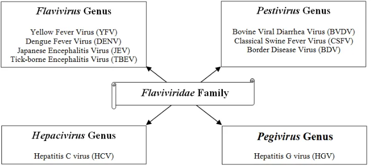
[image:2.612.125.502.422.593.2]The Flaviviridae family of viruses has been well documented. It consists of more than 73 members, many of which are important human pathogens being major cause of several hepatic and vector borne diseases [1].The virus family is presently classified into four genera: Flavivirus genus, Pestivirus genus, Hepacivirus genus and the recently proposed Pegivirus genus (fig. 1). They share significant similarities in virion morphology, life cycle and genome structure. Flaviviridae viruses have a broad host range that consists of both invertebrates and vertebrates. Ticks and mosquitoes were the only organisms recognized as Flavivirus hosts until recently. Also, arthropods were only viewed as vectors for vertebrate viruses, but this was recently challenged by the discovery of arthropod-specific viruses that are phylogenetically quite divergent. The remaining family genera i.e. Pegivirus, Hepacivirus and Pestivirus, are found in mammals alone, and their diversity has greatly widened with recent discoveries of these viruses in several mammalian species, such as dogs, pigs, bats, horses, rodents and ruminants [2].
Fig. 1 Classification of Flaviviridae family (Shi M, et al, 2015).
in humans are asymptomatic or result in non-specific flu-like illness. The ratio of apparent to inapparent infections with JEV is very low i.e., 1:100-1:300 [8].JE mostly occurs in children and young adults, but is seen to affect adults too during epidemics, like in Sri Lanka and Nepal. Also, the geographical area affected with JEV is increasing, with people living in affected areas being continually exposed to the virus [9]. Therefore, need of the hour is the development of successful vaccines with alternative strategies. In this study, JEV NS1 protein was purified to further attempt and characterize its role in the JEV infection process, which will give insights to develop strategies to prevent JEV infection.
II. FLAVIVIRUS STRUCTURE
Flavivirus virions are small and spherical in shape with diameter of 500Ǻ approximately. The Flavivirus genome is a positive-sense, unsegmented and single-stranded RNA molecule between 9.6 and 12.3 kb in length which is surrounded by an electron-dense nucleocapsid nearly 30 nm in diameter. A host cell-derived lipid bilayer further encloses the viral capsid protein. The lipid bilayer is surrounded by copies of two virally encoded membrane proteins: envelope protein (E) and membrane protein (M). The M protein is present as unprocessed prM protein, which on release from the infected cells, gets cleaved to mature M protein [2,10].The genome serves as the mRNA for all viral protein translation, a template for RNA replication, and genetic material that is packaged within new virions. It has a singular open reading frame (ORF) encoding a polyprotein. At its 5’ terminus, it has a type I cap structure (m7GpppAmp).Additional methylation has also been detected in RNA at the N2 residue, i.e. Type II cap, from infected cells [11]. Entry of flaviviruses into host cells is through receptor-mediated endocytosis via cellular receptors that are specific for the E protein. Trimerization of the E protein occurs due to acidic environment of the endosome which results in the fusion of viral and cell membranes. The nucleocapsid releases into cytoplasm after the fusion, and the capsid(C) protein and RNA dissociate, resulting in initiation of replication of RNA genome. Replication takes place in cytoplasmic replication complexes which are associated with ER membranes [12, 13]. The ORF encodes a single polyprotein with multiple transmembrane domains that is co- and post-translationally cleaved, by both viral and host proteases, into structural and nonstructural (NS) proteins. At the amino (5ˈ) terminus
are the three structural proteins- C, M, and E - that constitute the virus particle. Seven non-structural proteins (NS1, NS2A, NS2B, NS3, NS4A, NS4B and NS5) that are essential for viral replication are encoded by the remainder of the genome. Virus encoded serine protease cleaves the non-structural protein junctions, while a host signal peptidase is responsible for cleavages between the structural proteins [14].
After the proteolytic cleavages, viral RNA and the C protein are localized in the cytoplasm. The C protein remains associated with the membrane of endoplasmic reticulum (ER). The nucleocapsid obtains an envelope by budding into the lumen of ER. On the lumen side of the ER, the E and prM proteins form a stable heterodimer in no more than a few minutes after translation. The virion assembly process is quite fast. New virions assemble by budding into an intracellular membrane compartment, most likely the endoplasmic reticulum, and then they are transited via host secretory pathway, eventually being released at the cell surface.
[image:3.612.193.440.535.721.2]At first, immature particles are formed in the ER lumen. Subsequently, prM protein is cleaved in the trans-Golgi network, producing infectious and mature particles. prM protein cleavage and release leads the mature virion for acid-catalyzed rearrangements that is necessary for the entry into host cell [15].
A. Capsid Protein
The C protein is a highly basic protein of ~11 kDa (120 amino acids). Charged residues are clustered at the N- and C-termini, separated by an internal hydrophobic region that mediates membrane association. The nascent C protein also contains a hydrophobic signal sequence at its carboxyl terminus that translocates prM into the lumen of the ER from its site of synthesis on the surface of the ER. This hydrophobic domain is cleaved from mature C by the viral serine protease [16]. The C protein folds into a compact
dimer with each monomer containing four alpha helices that are connected by short loops. Helices α2 and α4 of one monomer are
anti-parallel to helices α2 and α4 of the adjoining monomer, respectively. It is not yet clear how C protein dimers are organized
within nucleocapsids, but interaction with RNA or DNA can induce isolated C protein dimers to assemble into nucleocapsid like particles. It is possible that the rather basic C protein functions like a histone [17].
B. pr Membrane/ Membrane Glycoprotein
The glycoprotein precursor of M protein, prM contains two transmembrane helices. It is ~26 kDa (165 amino acids) in size, before it is cleaved to yield the pr peptide and the M protein (~75 amino acids). The prM protein might function as a chaperone for folding and assembly of the E protein. A major function is to prevent E from undergoing acid-catalyzed rearrangement to the fusogenic form during transit through the secretory pathway. prM cleavage is delayed until late in infection and coincides with the conversion of immature virus particles to mature virions. Following cleavage, prM-E heterodimers dissociate, the pr fragment is released, and E homodimers form [18].
C. Envelope Glycoprotein
The E glycoprotein is approximately 495 amino acids in size (~53 kDa). It is the major protein on the surface of flavivirus virions and contains a cellular receptor-binding site and a fusion peptide. E is synthesized as a type I membrane protein containing 12 conserved cysteines that form disulfide bonds. The native form of E folds into an elongated structure rich in β-sheets and form head-to-tail homodimers that lie parallel with the virus envelope. Studies with monoclonal antibodies in the late 1980s suggested three antigenic domains, and these were confirmed more recently when the three dimensional structure of the ectodomain (residues 1-395) of the flavivirus tick-borne encephalitis virus (TBEV) was determined by X-Ray crystallography. The central structural domain I, which contains the N-terminus, is flanked on one side by an elongated dimerization domain (domain II), which projects along the virus surface between the transmembrane regions of the homodimer subunits and contains the fusion peptide at its distal end. On the other side of domain I is domain III, which maintains an immunoglobulin-like fold [19]. Domain III appears to be involved in receptor binding and is a major target of neutralizing antibodies. In mosquito-borne flaviviruses, domain III contains an RGD/RGE sequence, which is the recognition motif for integrin binding. The ectodomain of the E protein comprises sets of three parallel dimers that form 30 rafts over the viral surface. During maturation, 60 trimers of prM-E heterodimers that project from the virus surface dissociate and form 90 E homodimers, which lie flat on the virus surface, giving the mature virion its spike less and smooth surface [20].
D. Non-structural (NS) 1 protein
The NS1 glycoprotein (~46 kDa) is translocated into the ER during synthesis and cleaved from E protein by host signal peptidase. NS1 is largely retained within infected cells but can localize to the cell surface and is slowly secreted from mammalian cells. The protein contains two or three N-linked glycosylation sites and 12 conserved cysteines that form disulfide bonds. Around 30 minutes after synthesis, NS1 forms highly stable homodimers and acquires an affinity for membranes. NS1 localizes to sites of RNA replication and mutation of the N-linked glycosylation sites in NS1 can lead to dramatic defects in RNA replication and virus production. It elicits a humoral and cellular immune response in humans and experimental animals, and antibodies against the cell surface form can direct complement-mediated lysis of virus-infected cells [21].
E. NS2A and NS2B Proteins
F. NS3 Protein
NS3 is a large (~70 kDa) multifunctional protein, containing several activities required for polyprotein processing and RNA replication. The N-terminal third of the protein is the catalytic domain of the NS2B-NS3 serine protease complex. In addition to cleaving the NS2A/NS2B, NS2B/NS3, NS3/NS4A, and NS4B/NS5 junctions, the protease generates the C-termini of mature C protein and NS4A, and can cleave at internal sites within NS2A and NS3. RNA-stimulated nucleoside triphosphatase (NTPase) activity and RNA unwinding activity have been demonstrated for flavivirus NS3 proteins. The flavivirus NS3 protein also encodes an RNA triphosphatase activity (RTPase) proposed to dephosphorylate the 5’ end of genome RNA before cap addition. All three nucleic acid-modifying activities of NS3 (NTPase, helicase, and RTPase) rely on a common active centre. The NS3 proteins of Langat, DENV-2, and WNV have been shown to induce apoptosis, in some cases through activation of caspase-8. Although passive protection in mice by a monoclonal antibody against the non-structural protein NS3 of DENV-1 has been reported, the primary immunologic role of the non-structural proteins, with the exception of NS1, appears to be their function as targets for cytotoxic T cells [23].
G. NS4A and NS4B Proteins
The NS4A (~16 kDa) and NS4B (~27 kDa) are small, hydrophobic proteins. A role for NS4A in RNA replication is supported by the co-localization of this protein with replication complexes. Over-expression studies show that regulated NS4A/2K/4B cleavage is necessary for induction of membrane rearrangements by NS4A [24]. NS4B can associate with membranes independently of the 2K signal peptide and appears to be a polytopic membrane protein. NS4B colocalizes with NS3 and viral double stranded RNA (dsRNA) in ER-derived membrane structures presumed to be sites of RNA replication. DENV NS4A and NS4B can also block type I IFN signalling. NS4B has the strongest antagonistic effect [25].
H. NS5 Protein
NS5 is a large (~103 kDa), highly conserved, multifunctional protein with methyltransferase (MTase) and RNA dependent RNA polymerase (RdRP) activities. NS5 forms a complex with NS3 and can stimulate both the NTPase and RTPase activities of NS3. NS5 has also been shown to localize to the nucleus. New roles, other than in RNA replication, have recently emerged for flavivirus NS5. DENV-2 NS5 induces IL-8 transcription, and secretion, which may enhance viral spread or disease by recruiting inflammatory cells to the site of infection. In addition, TBEV NS5 blocks signalling of IFN- α/β and IFN-γ by binding to their receptors and inhibiting phosphorylation of both Janus kinases, JAK1 and Tyk2, and hence downstream activation of signal transducer and activator of transcription 1 (STAT1) [26].
III. JEV GLOBAL SCENARIO
JEV has a vast distribution in Asia. JE cases have been reported in Sri Lanka, India, Pakistan, Myanmar, Laos, Nepal, Cambodia, Bangladesh, Vietnam, Malaysia, Brunei, Taiwan, Thailand, Philippines, Singapore, Indonesia, China, Korea, Japan and Australia. There are two epidemiological patterns of Japanese Encephalitis that are recognized. In southern tropical countries, there is occurrence of sporadic cases all-round the year with the disease being endemic there [27]. In the subtropics and temperate zones, outbreaks have a definite seasonal pattern, commonly occurring through the rainy season.
There are total five genotypes of JEV known (GI, GII, GIII, GIV, and GV). In majority of the cases of JEV, viruses fall into the category of GIII, which suggests that this particular genotype is most widely spread. Of all the JEV genotypes, the one at the base was GIV in all phylogenetic trees. This suggests that the most ancient lineage is represented by the genotype IV that branched off before the genotypes GI, GII, and GIII [28].
The first large epidemic of Japanese Encephalitis was recognized in Japan in 1924, while it had been identified since the early 1870s. The occurrences of JE in South Korea, Taiwan and Japan have dramatically declined since the 1980s. This is because of the extensive use of vaccine in children and several other preventive measures. Even in China, the frequency of occurrence of JE annually has decreased as a result of immunization in children, but there are still cases being reported every year [29].JE was first identified in 1940 in China, but it was in 1966 that JE was recognized as a public health problem with reporting of 40,000 cases. Except for Xinjiang, Qinghai and Tibet, JEV is active in most of the provinces of China. Approximately 80% of the cases reported in the world are from China.
whereas the southern and central regions are comparatively less affected having a year round transmission of JEV [30].Similar disease patterns are observed in countries like Vietnam and Cambodia, whereas in countries like Indonesia and the Philippines, major epidemics are very infrequent as they have no subtropical zone.
IV. JEV IN INDIA
In India, the first cases of JE were identified in 1955 in Tamil Nadu and Andhra Pradesh [31]. In 1958, JEV was isolated from mosquitoes in the same area and were also isolated from the brain tissues of JEV infected patients. The first major outbreak of JE occurred in West Bengal in 1973 in the districts of Burdwan and Bankura involving 700 patients and 300 deaths; a second outbreak followed in 1976.
In India, outbreaks have appeared in 25 states. Majority of the incidences have been reported from Assam, Maharashtra, Manipur, Andhra Pradesh, Bihar, Karnataka, Uttar Pradesh, Madhya Pradesh, Tamil Nadu, Kerala, Haryana, Orissa, West Bengal and Union territories of Pondicherry and Goa. JE incidences show seasonal patterns in different parts of India. Such patterns depend on the local agricultural ecology, which again relies on the annual rainfall pattern. The outbreaks of JE in South India usually occur during the second half of the year (August to December). In West Bengal, the incidences of JE outbreaks are more between the months of May and October. During the period of September to December, epidemics are commonly reported in the states of Uttar Pradesh and Bihar. In Assam, it is more likely to occur from July to September [32].In Uttar Pradesh, JEV activity is reported every year. In 2005, the state saw one of the massive outbreaks of Japanese Encephalitis that has occurred in the recent years in northern parts of India. There were more than 6000 cases of infection and 1500 deaths reported. It was the most severe and longest epidemic seen in over three decades. Mostly the population of age group 3 months- 15 years was affected in rural areas and the overall fatality was 23 percent [33].
REFERENCES
[1] B. D. Lindenbach, C. L. Murray, H. J. Thiel and C. M. Rice, Flaviviridae, 6th ed, Fields virology, pp. 712–746, 2013.
[2] M. Shi, X. D. Lin and N. Vasilakis, “Divergent Viruses Discovered in Arthropods and Vertebrates Revise the Evolutionary History of the Flaviviridae and Related Viruses,” Journal of Virology, vol. 90, pp. 659-669, Oct. 2015.
[3] Japanese encephalitis homepage [Online]. Available: http://www.cdc.gov/japaneseencephalitis/
[4] Y. L. Koh, B. H. Tam, J. P. Loh and E. E. Ooi, “Japanese encephalitis Singapore,” Emerging Infectious Diseases, vol. 12(3), pp. 525-526, Mar. 2006. [5] M. Parida, P. K. Dash, N. K. Tripathi, S. A. Sannarangaiah, P. Saxena, S. Agarwal, A. K. Sahni, S. P. Singh, A. K. Rathi, R. Bhargava, A. Abhyankar, S. K.
Verma, P. V. L. Rao and K. Sekha, “Japanese encephalitis outbreak, India, 2005,” Emerging Infectious Diseases, vol. 12(9), pp. 1427-1430, Sept. 2006. [6] K. Bharti and S. Vrati, “Japanese encephalitis: development of new candidate vaccines,” Expert Review of Anti Infective Therapy, vol. 4(2), pp. 313-324, Apr.
2006.
[7] T. Solomon, “Control of Japanese encephalitis-within our grasp,” The New England Journal of Medicine, vol. 355(9), pp. 869-871, Aug. 2006. [8] T. J. Chambers and M. S. Diamond, “Pathogenesis of flavivirus encephalitis,” Advances in Virus Research, vol. 60, pp. 273-342, 2003. [9] T. Solomon and M. J. Cardosa, “Emerging arboviral encephalitis,” British Medical Journal, vol. 321, pp.1484-1485, Dec. 2000.
[10] B. D. Lindenbach, H. J. Thiel and C. M. Rice, “Flaviviridae: the viruses and their replication,” Fields Virology, pp. 1101-1152, Oct. 2007.
[11] T. J. Chambers, C. S. Hahn, R. Galler and C. M. Rice, “Flavivirus genome organization, expression, and replication,” Annual Review of Microbiology, vol. 44, pp. 649-688, Oct. 1990.
[12] S. L. Allison, J. Schalich, K. Stiasny, C. W. Mandl, C. Kunz and F. X. Heinz, “Oligomeric rearrangement of tick-borne encephalitis virus envelope proteins induced by an acidic pH,” Journal of Virology, vol. 69(2), pp. 695-700, Feb. 1995.
[13] S. Mukhopadhyay, R. J. Kuhn and M. G. Rossmann, “A structural perspective of the flavivirus life cycle,” Nature Reviews Microbiology, vol. 3, pp. 13-22, Jan. 2005.
[14] L. Yize, C. Dorian, L. Peng, D. Veasna, Y. Yongxin and D. Vincent, “Protective immunity to Japanese encephalitis virus associated with anti-NS1 antibodies in a mouse model,” Virology Journal, vol. 9(135), Jul. 2012.
[15] F. X. Heinz and S. L. Allison, “Flavivirus structure and membrane fusion,” Advances in Virus Research, vol.59, pp. 63-97, 2003. [16] B. D. Lindenbach and C. M. Rice, “Molecular biology of flaviviruses,” Advances in Virus Research, vol. 59, pp. 23-61, 2003.
[17] L. Ma, C. T. Jones, T. D. Groesch, R. J. Kuhn, and C. B. Post, “Solution structure of dengue virus capsid protein reveals another fold,” Proceedings of the National Academy of Sciences USA, vol. 101(10), pp. 3414-3419, Mar. 2004.
[18] R. B. Londono and T. M. Colpitts, “A Brief Review of West Nile Virus Biology,” Methods in Molecular Biology, vol.1435, pp. 1-13, 2016.
[19] J. L. S. Campos, M. Poggianella, S. Marchese, M. Bestagno and O. R. Burrone, “Secretion of dengue virus envelope protein ectodomain from mammalian cells is dependent on domain II serotype and affects the immune response upon DNA vaccination,” Journal of General Virology, vol. 96(11), pp. 3265-79, Nov. 2015.
[20] J. J. H. Chu and M-L. Ng, “Interaction of West Nile Virus with αvβ3 Integrin Mediates Virus Entry into Cells,” The Journal of Biological Chemistry, vol. 279(52), pp. 54533-54541, Dec. 2004.
[21] T. Yuki, R. Muhareva, M. Kouichi and H. Daisuke, “NS1 Protein Expression in the JaOArS982 Strain of Japanese Encephalitis Virus Does Not Enhance Virulence in Mice,” Tropical Medicine and Health, vol. 43(4), pp. 233–237, 2015.
[23] P. K. Kakumani, K. S. Rajgokul, S. S. Ponia, I. Kaur, S. Mahanty, G. R. Medigeshi, A. C. Banerjea, A. P. Chopra, P. Malhotra, S. K. Mukherjee and R. K. Bhatnagar, “Dengue NS3, an RNAi suppressor, modulates the human miRNA pathways through its interacting partner,” The Biochemical Journal, vol. 471(1), pp. 89-99, Oct. 2015.
[24] Y. Li, Y. M. Kim, J. Zou, Q. Y. Wang, S. Gayen, Y. L. Wong, L. T. Lee, X. Xie, Q. Huang, J. Lescar, P. Y. Shi and C. Kang, “Secondary structure and membrane topology of dengue virus NS4B N-terminal 125 amino acids,” Biochimica et Biophysica Acta (BBA) - Biomembranes, vol. 1848(12), pp. 3150-7, Dec. 2015.
[25] H. Khan and S. Murad, “Analysis of different HCV NS4B domains for the development of global consensus sequence,” Acta Virologica, vol. 59(3), pp. 276-83, Sep. 2015.
[26] V. J. Klema, M. Ye, A. Hindupur, T. Teramoto, K. Gottipati, R. Padmanabhan and K. H. Choi, “Dengue Virus Nonstructural Protein 5 (NS5) Assembles into a Dimer with a Unique Methyltransferase and Polymerase Interface,” PLoS Pathogens, vol. 12(2), Feb. 2016.
[27] T. Solomon, H. Ni, D. W. Beasley, M. Ekkelenkamp, M. J. Cardosa and A. D. Barrett, “Origin and evolution of Japanese encephalitis virus in southeast Asia,” Journal of Virology, vol. 77(5), pp. 3091-3098, 2003.
[28] W. R. Chen, R. R. Hesse and R. B. Tesh, “A new genotype of Japanese encephalitis virus from Indonesia,” The American Journal of Tropical Medicine and Hygiene, vol. 47(1), pp. 61-69, 1992.
[29] H. Ni and A. D. Barrett, “Nucleotide and deduced amino acid sequence of the structural protein genes of Japanese encephalitis viruses from different geographical locations,” Journal of General Virology, vol. 76(2), pp. 401-407, 1995.
[30] P. T. Nga, V. D. Cuong, S. P. Ma, F. Hasebe, S. Inoue, Y. Makino, M. Takagi, V. S. Nam and K. Morita, “Shift in Japanese encephalitis virus (JEV) genotype circulating in northern Vietnam: implications for frequent introductions of JEV from Southeast Asia to East Asia,” Journal of General Virology, vol. 85(6), pp. 1625-1631, 2004.
[31] A. Gajanana, R. Rajendran, P. P. Samuel, V. Thenmozhi, T. F. Tsai, J. K. Kuroda and R. Reuben, “Japanese encephalitis in south Arcot district, Tamil Nadu, India: a three-year longitudinal study of vector abundance and infection frequency,” Journal of Medical Entomology, vol. 34(6), pp. 651-659, 1997.
[32] L. Kabilan, R. Rajendran, N. Arunachalam, S. Ramesh, S. Srinivasan, P. P. Samuel and A. P. Dash, “Japanese encephalitis in India: an overview,” Indian Journal of Pediatrics, vol. 71(7), pp. 609-615, 2004.