comment
reviews
reports
deposited research
interactions
information
refereed research
Review
The nitrilase superfamily: classification, structure and function
Helen C Pace and Charles Brenner
Address: Structural Biology and Bioinformatics Program, Kimmel Cancer Center, 233 S Tenth Street, Philadelphia, PA 19107, USA.
Correspondence: Charles Brenner. E-mail: [email protected]
Abstract
The nitrilase superfamily consists of thiol enzymes involved in natural product biosynthesis and post-translational modification in plants, animals, fungi and certain prokaryotes. On the basis of sequence similarity and the presence of additional domains, the superfamily can be classified into 13 branches, nine of which have known or deduced specificity for specific nitrile- or hydrolysis or amide-condensation reactions. Genetic and biochemical analysis of the family members and their associated domains assists in predicting the localization, specificity and cell biology of hundreds of uncharacterized protein sequences.
Published: 15 January 2001
GenomeBiology2001, 2(1):reviews0001.1–0001.9
The electronic version of this article is the complete one and can be found online at http://genomebiology.com/2001/2/1/reviews/0001 © BioMed Central Ltd (Print ISSN 1465-6906; Online ISSN 1465-6914)
Plants, animals and fungi perform a wide variety of nonpep-tide carbon-nitrogen hydrolysis reactions using members of the nitrilase superfamily of enzymes. These nitrilase [1,2] and amidase [3,4] reactions, which produce auxin, biotin,
β-alanine and other natural products, and which result in deamination of protein and amino acid substrates, all involve attack of a cyano or carbonyl carbon by a conserved cysteine [5,6]. Many bacteria and archaea, particularly those with an ecological relationship to plants and animals, encode members of the nitrilase superfamily and utilize the enzymes for chemically similar nitrile or amide hydrolysis reactions or for condensation of acyl chains to polypeptide amino termini.
On the basis of global and structure-based sequence analysis, members of the nitrilase superfamily can now be classified into 13 branches and the substrate specificity of members of nine branches can be anticipated. Despite historical classifi-cation of all of these sequences as nitrilase-related, only one branch is known to have nitrilase activity, whereas eight branches have apparent amidase or amide-condensation activities. Members of seven branches of the nitrilase super-family have participated in domain fusion events that alter the localization of the nitrilase-related domain, link ammonia production to ammonia consumption, or potentially link pro-teins involved in cellular signaling. For example, fusion of
domains we expect to have glutamine amidohydrolase (GAT) activity to some bacterial and all eukaryotic nicotinamide adenine dinucleotide (NAD) synthetases can account for the previously unsolved problem that only some NAD syn-thetases use glutamine as a source of ammonia [7-9]. Remarkably, these fusions contain the fourth apparent GAT domain involved in coupled amide transfer reactions as they are unrelated to other GAT-domain-containing families: the amino-terminal nucleophile (Ntn) hydrolases and triad ami-dotransferases [10], and the amidase signature family [11]. Crystal structures of two nitrilase superfamily members -worm NitFhit [12] and a bacterial N-carbamyl-D-amino acid amidohydrolase [13] - reveal that nitrilase-related proteins are multimeric α-β-β-α sandwich proteins that have a con-served Glu-Lys-Cys catalytic triad responsible for covalent catalysis. Mutating catalytic triad residues may allow sub-strates to be trapped and identified for the branches that remain to be characterized biochemically.
Evolution and classification
also found in phylogenetically isolated prokaryotes that appear to have an ecological relationship to plants and animals. The nitrilase superfamily therefore probably emerged prior to the separation of plants, animals and fungi, radiated into families, and then spread laterally to bacteria and archaea. Some branches of the nitrilase superfamily are found only in prokaryotes; members of these branches may constitute rational antibiotic targets.
Automated sequence searching easily identifies predicted polypeptides as members of the nitrilase superfamily, but many database annotations have been applied haphazardly. Because members of the nitrilase superfamily are reported to be nitrilases, aliphatic amidases, β-ureidopropionases,
β-alanine synthases, N-carbamyl-D-amino acid amidohydro-lases and so on, these designations appear in the sequence-definition lines of multiple databases, often irrespective of the activity of the most closely related characterized enzyme.
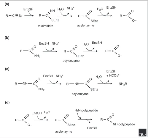
The reactions performed by nitrilases, amidases, carbamy-lases and N-acyl transferases within the nitrilase superfam-ily are shown schematically in Figure 1. It should be noted that the nitrilase branch of the nitrilase superfamily may be the only branch that contains members that perform nitrile hydrolysis (from a nitrile to the corresponding acid plus ammonia); at least eight branches appear to be either ami-dases of various specificities or enzymes that condense acyl chains to amino groups. Nitrile hydratases, metal-contain-ing enzymes that convert a nitrile to the correspondmetal-contain-ing amide [14], are not members of the nitrilase superfamily. Additionally, despite the fact that most branches of the nitrilase superfamily are actually amidases, there are many amidases including Ntn and triad hydrolases [10], amidase signature enzymes [15] and thiol proteases [16] that are unrelated to the nitrilase superfamily. Because of the histor-ical observation that aliphatic amidases are related to nitri-lases [4,6], we retain ‘nitrilase’ as the superfamily designation and as a branch designation, and embrace several families of homologous Glu-Lys-Cys amidases as branches of the nitrilase superfamily.
We performed a large number of BLASTp (version 2.1.2) [17] and manual searches to identify prototypical members of branches of the nitrilase superfamily and we currently clas-sify the superfamily as having 13 branches,shown in Table 1. For the data uniquely classifying nitrilase sequences into 13 branches, see the Additional data file available with the online version of this article. Examination of the E-values of sequences aligned with a prototype guided the classification of each of the 176 identified sequences as a member of only one branch. Within most branches, there is a relatively sharp cutoff in E-values such that sequences with E-values greater than 1 x 10-25 can be identified as belonging to another branch. In the 13th branch, definition of a prototype - a sequence to which all branch members can be easily compared - was less straightforward as the sequences are
relatively diverse. With more data, it would not be surprising to find further ways to divide and to classify members of the nitrilase superfamily.
Most members of each branch can be assigned to the branch not only by virtue of an E-value cutoff, but also by virtue of signature sequences surrounding active-site residues, pro-viding further confidence in the classification scheme. Essentially all members of the nitrilase superfamily have a conserved, apparent catalytic triad of glutamate, lysine and cysteine (only three apparently truncated sequences lack the glutamate). The motif that most highly correlates with E-value cutoffs consists of the two residues carboxy-terminal to the cysteine nucleophile. For example, members of the nitrilase branch of the nitrilase superfamily have a Cys-Trp-Glu motif at the active-site cysteine, whereas β -ureidopro-prionases have a Cys-Tyr-Gly motif. Consensus sequences for the glutamate-, lysine- and cysteine-surrounding residues of each branch of the nitrilase superfamily are shown in Figure 2.
Domain fusions in the nitrilase superfamily
In seven branches of the nitrilase superfamily, a nitrilase-related domain is fused to at least one additional conserved domain (Figure 3). In three branches, the domain fusion appears to be constitutive; that is, all members of that branch (defined by BLAST E-value and signature sequences within the nitrilase-related domains) contain the additional domain. In four branches, the additional domain(s) are not found in every member. Some of the domain-fusion events can be con-sidered ‘Rosetta Stone’ fusions, in that separate polypeptides appear to be fused to coordinate biochemical reactions or cel-lular functions [18,19]. Other domain-fusion events appear more likely to affect cellular localization. The significance of domain fusions in branches 7 and 8, the prokaryotic and eukaryotic NAD synthetases, is discussed below.Two independently derived families of GAT domains have been found in a variety of two-domain polypeptides that couple ammonia hydrolysis from glutamine to ammonia consumption at a second active site [10]. Glutamine phos-phoribosylpyrophosphate (PRPP) amidotransferase is pro-totypical of enzymes that utilize a GAT domain related to the Ntn hydrolases [20], whereas GMP synthetase is proto-typical of enzymes that use a triad amidotransferase domain to perform the GAT function [21]. The second active site of GMP synthetase contains a nucleotide-binding domain similar to that of ammonia-dependent NAD syn-thetase [22]. It has been known for more than 30 years that
comment
reviews
reports
deposited research
interactions
information
refereed research
tuberculosis, however, contains an amino-terminal domain [23] not present in the E. coli enzyme [24]. After the dis-covery that the multiprotein Bacillus glutamyl-tRNAGln amidotransferase contains yet a third type of GAT activity [11] related to the amidase signature family [15], it was hypothesized that the amino terminus of the prokaryotic
glutamine-dependent NAD synthetase is related to the amidase signature family [23].
[image:3.609.55.554.86.557.2]In contrast, we find that the amino terminus of prokaryotic glutamine-dependent NAD synthetase and the amino-termi-nal domains of all eukaryotic NAD synthetases are branches
Figure 1
Four types of reaction carried out by nitrilase superfamily members. (a)The nitrilase reaction is performed by branch 1 enzymes. In plants, the substrate is indole-3-acetonitrile and the product is indole-3-acetic acid. (b)The amidase reaction is the most frequently observed activity in the superfamily. Branch 2-4 enzymes are amidases and nitrilase-related domains of branch 7 and 8 enzymes are proposed to be amidases specific for glutamine. (c)The carbamylase reaction is a special case of the amidase reaction, carried out by branch 5 and 6 enzymes. (d)Branch 9 N-acyltransferases perform the amidase reaction in reverse, transferring a fatty acid from phospholipid (not shown) to a polypeptide amino terminus. The polypeptide acceptor usually contains an amino-terminal diacylglyceride-modified cysteine (not shown). All nitrilase-related reactions are thought to proceed through acylenzyme intermediates.
C
O
SEnz
NH
4+H
2O
EnzSH
EnzSH
+ HCO
3+NH
2R
R
R
C
O
SEnz
NH
4+H
2O
EnzSH
R
C
O
O–
C
O
NH
2EnzSH
R
C
N:
R
C
NH
SEnz
EnzSH
H
2O
R
C
O
SEnz
NH
4+H
2O
EnzSH
R
C
O
O–
(c)
acylenzyme
(b)
thioimidate
acylenzyme
(a)
acylenzyme
(d)
EnzSH
EnzSH
acylenzyme
R
C
O
O–
H
2O
R
C
O
SEnz
H
2N-polypeptide
R
C
O
NH-polypeptide
NH
R
C
O
NH
2Figure 2
The nitrilase superfamily catalytic triad motifs. Consensus sequences flanking the invariant catalytic triad residues, glutamate, lysine and cysteine, were obtained by doing multiple sequence alignments within each branch [54]. Red letters on a yellow background indicate the same residue is conserved in all branches. Dark blue letters on light blue background indicate the residue is conserved in nine or more branches. Green background shows positions in which the conserved amino acid is found in six to eight of the branches. Upper case letters indicate 90% or greater consensus levels within a branch, whereas lower case are 50% or greater. Residue numbers are shown for the prototypical members of branches 1 to 12 and for the first listed member of branch 13.
1 - Nitrilase
f
P
E
a f
h
R
K
l
. p T
l
.
C
W
E
n .
.
p
2 - Aliphatic Amidase
F
P
E
Y S
Y
R
K
i
P W c
i
I
C
d
D
G n y
P
3 - N-terminal Amidase
F
P
E
.
.
Y
r
K
. F L
.
.
I
C
M
D
.
. P
Y
4 - Biotinidase
f
P
E
d .
Y
r
K
. h L y
F
t
C
F
D
i
l
f
y
5 - Beta-ureidopropionase
.
Q
E
A W
.
R
K
N H I P
N
I
C
Y
G
R H H
P
6 - Carbamylase
F
p
E
L A
Y
R
K
i H L P
f
I
C
N
D
R R W
P
7 - Pro. NAD+ Synthetase
f
P
E
L
.
.
.
K
.
. L P
.
I
C
E
D
. w .
p
8 - Euk. NAD+Synthetase
G
P
E
L E
R
p
K
m .
l
a
E
i
C
E
E
L w .
p
9 - ALP N-acyltransferase
w
p
E
. a
.
.
K
.
.
l
v
.
i
C
y
E
.
.
f
.
10 - Nit and NitFhit
L
P
E
.
f
y
r
K
. H l
F
.
i
C
Y
D
. R F
p
11 - NB11
.
q
E
l
f
Y
R
K
. H I P
.
i
C
w
D
q w f
p
12 - NB12
F
P
E
i
F
Q
y
K
l H i T
q
I
C
Y
D
i E F
P
13 - Non-fused Outliers
l
P
E
.
.
y
r
K
. h L f
.
i
C
y
d
.
r F
p
Table 1
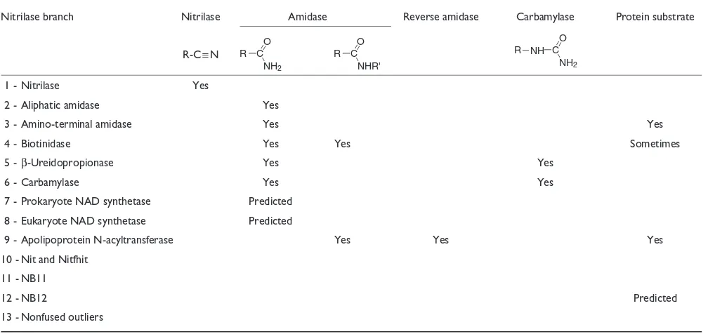
Summary of the enzyme activities of the nitrilase superfamily
Nitrilase branch Nitrilase Amidase Reverse amidase Carbamylase Protein substrate
R-C≡N
1 - Nitrilase Yes
2 - Aliphatic amidase Yes
3 - Amino-terminal amidase Yes Yes
4 - Biotinidase Yes Yes Sometimes
5 -β-Ureidopropionase Yes Yes
6 - Carbamylase Yes Yes
7 - Prokaryote NAD synthetase Predicted 8 - Eukaryote NAD synthetase Predicted
9 - Apolipoprotein N-acyltransferase Yes Yes Yes
10 - Nit and Nitfhit 11 - NB11
12 - NB12 Predicted
13 - Nonfused outliers
R C NH2
O
R C NHR' O
R C
NH2
[image:4.609.57.559.354.645.2]7 and 8 of the nitrilase superfamily, respectively. We deduce that branch 7 and 8 nitrilase-related domains have substrate specificity as glutamine amidases, and that branch 7 and 8 enzymes utilize these novel GAT domains to confer gluta-mine dependence to the associated carboxy-terminal NAD synthetase domains. We therefore expect to find that the presence of branch 7 nitrilase-related domains will correlate with the ability of purified prokaryotic NAD synthetases to use glutamine, and we expect that the glutamine dependence of prokaryotic and eukaryotic glutamine-dependent NAD synthetases will depend on nitrilase-homologous active-site residues. If this is confirmed, branch 7 and 8 nitrilase
domains will constitute the fourth independent type of GAT domain to participate in coupled amino-transfer reactions.
Enzymology
Nonenzymatic hydrolysis of a nitrile of the form R-C≡N would produce the corresponding acid amide, R-C=O(NH2), with one water addition and the corresponding acid, R-CO2-, with the second water addition. Nitrilases are interesting, however, in that the substrates are nitriles but the reaction does not involve release of, or reaction with, a substantial amount of the corresponding amide [1,25]. Nitrilases
comment
reviews
reports
deposited research
interactions
information
[image:5.609.58.555.87.452.2]refereed research
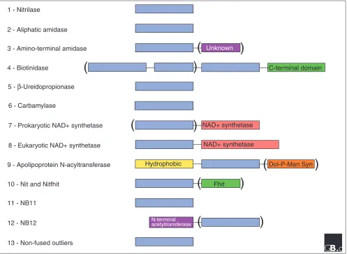
Figure 3
Domain structures for 13 branches of the nitrilase superfamily. Additional domains are found in members of seven branches. Parentheses denote domains found in only some members of the branch. In branch 4, vanins and biotinidases have carboxy-terminal domains unique to these two sub-branches and one vanin has additional full and partial nitrilase-related domains. The NAD synthetase domains of eukaryotes are always fused with a nitrilase-related domain. In contrast, only some prokaryotic NAD synthetases are fusion proteins with a nitrilase-related domain. This led to the prediction that branch 7 and 8 nitrilase domains are glutamine amidotransferases for the associated NAD synthetases (see text for details). Apolipoprotein N-acyltransferases (branch 9) always have a hydrophobic amino-terminal domain and one member is fused to an apparent dolichol phosphate mannose synthetase, which underscores the proposed function of branch 9 enzymes in post-translational modification. Nit proteins, branch 10, are found as fused Rosetta Stone proteins with Fhit in invertebrates and are
coordinately expressed with separate Fhit proteins in mammals. Branch 12 enzymes are predicted to have protein substrates as they are fused to a homolog of an amino-terminal acetyltransferase.
1 - Nitrilase
2 - Aliphatic amidase
3 - Amino-terminal amidase
4 - Biotinidase
5 -
β
-Ureidopropionase6 - Carbamylase
7 - Prokaryotic NAD+ synthetase
8 - Eukaryotic NAD+ synthetase
9 - Apolipoprotein N-acyltransferase
10 - Nit and Nitfhit
11 - NB11
12 - NB12
13 - Non-fused outliers
Unknown
C-terminal domain
NAD+ synthetase
Hydrophobic Dol-P-Man Syn
Fhit
N-terminal acetyltransferase
(
)
(
)
NAD+ synthetase
)
)
(
(
(
)
produce the acid without the production or release of an acid amide by virtue of covalent, thiol-mediated catalysis [5,25]. As illustrated in Figure 1, the enzyme attacks a nitrile sub-strate covalently, producing ammonia with the first water addition, and producing acid and a regenerated enzyme with the second water addition. The geometric constraints of this reaction suggest that nitrilase facilitates interaction with a linear (approximately 180°) substrate, planar (approxi-mately 120°) thioimidate and acylenzyme intermediates, and tetrahedral (approximately 109.5°) water-bonded intermedi-ates. In contrast, serine and thiol proteases and amidases are confined to interacting with planar substrates and tetrahe-dral intermediates. We speculate that most nitrilases bind strongly to a bulky substrate R group in a conformation that places the 2 carbon closer to 120° than to 180° from the cyano nitrogen. Fitting a distorted substrate nitrile would push the substrate toward thioimidation and would reduce the geometric sweeps required of enzyme complexes. In support of this view, most nitrilases prefer bulky substrates to nonsubstituted acetonitrile [1,25-28]. Cyanide hydratase, a member of the nitrilase branch, may be the exception that proves the rule: the R-group free substrate does not stay bound to produce acid but rather is decomposed to for-mamide after one water addition [29,30].
As we have discussed, most branches of the nitrilase super-family do not contain nitrilases but rather amide-hydrolyzing or amide-condensing enzymes. Although activation of water to attack planar intermediates is expected to be shared by all enzymatically active members of the superfamily, the bio-chemical basis for nitrile versus amide attack within the nitri-lase superfamily is not yet understood. Biotinidases, branch 4 of the nitrilase superfamily, are amidases specific for hydroly-sis of biotinamides such as biocytin to biotin plus lysine [31]. For this branch, leaving group specificity allows biotinylated peptides, biocytin, simple biotinamide and biotin esters to be substrates [31]. As alcohols are better leaving groups than amines, it would not be surprising if other members of the nitrilase superfamily have a biological function as esterases. Although no member of the nitrilase superfamily has been reported to have protease activity, members of branches 3 and 4 act on sidechains of polypeptides and members of branch 9 perform a condensation to polypeptide amino termini. Because branch 12 enzymes are fused to a probable amino-terminal acetyltransferase, they may have protein sub-strates as well. Protease activities may remain to be discov-ered in the superfamily. The enzyme activities of the nitrilase superfamily are summarized in Table 1.
Structural features
Crystal structures of an N-carbamyl-D-amino acid amidohy-drolase from Agrobacterium[13] (a carbamylase; branch 6) and the Caenorhabditis elegans NitFhit Rosetta Stone protein [12] (branch 10) have been determined. The nitrilase-homologous domain of NitFhit and the carbamylase have
similar three-dimensional structures, conserved chemical features, and were independently interpreted as utilizing the conserved glutamate residue as a general base for the cys-teine nucleophile [12,13]. The Nit domain of NitFhit and the carbamylase can be described as α-β-β-α sandwich proteins, both of which assemble as tetramers. Nit and the carbamy-lase are unrelated to other enzymes with known structures such as Ntn and triad hydrolases [10], and thiol proteases [16]. Figure 4 shows the geometry of the Nit active site, high-lighting residues that are absolutely conserved in the super-family (Glu54, Lys127 and Cys169) and residues at positions that are highly conserved (Tyr125, His129, Tyr170, Asp171, Arg173 and Phe174), as aligned in Figure 2.
Branches of the nitrilase superfamily
Branch 1: nitrilase
Members of the nitrilase branch (EC 3.5.5.1) are found in plants, animals (C. elegans), fungi (Saccharomyces cere-visiae’s frequently inactivated NIT1gene), and many types of bacteria. The best evidence that nitrilase functions in vivoto convert indoleacetonitrile to the plant growth factor indole-3-acetic acid (auxin) comes from Arabidopsis,in which it was shown that recessive mutations in a nitrilase gene resulted in reduced sensitivity to the auxin-like effects of indoleacetoni-trile and that overexpression of a nitrilase caused increased sensitivity to indoleacetonitrile [32]. Bacterial nitrilases are often exploited for biochemical syntheses and for environ-mental remediation [33]. It is not clear whether bacterial nitrilases primarily function in ecological relationships with plants or whether they benefit isolated microbes.
Branch 2: aliphatic amidase
[image:6.609.314.555.533.691.2]Aliphatic amidases (EC 3.5.1.4) [3,4] comprise a small branch of nearly identical proteins found inPseudomonas, Bacillus, Brevibacteria andHelicobacteria. They hydrolyze substrates such as the carboxamide sidechains of glutamine
Figure 4
and asparagine utilizing the conserved cysteine within the nitrilase superfamily.
Branch 3: amino-terminal amidase
The N-end rule is a means by which the rates of ubiquitin-dependent protein degradation is regulated. The S. cere-visiae Nta1 protein deaminates amino-terminal asparagine and glutamine residues to aspartate and glutamate, which lead to more rapid rates of protein turnover [34]. Nta1 has fungal homologs but mammalian amino-terminal amidases appear to be unrelated.
Branch 4: biotinidase
Biotinidases (EC 3.5.1.12) utilize specific amidase/esterase activity to release biotin from biotinamide, biotin-lysine and biotin-peptide conjugates and biotin methylester [35]. Bio-tinidase deficiency can result in an inability to recycle biotin that is manifested in neurological and cutaneous abnormali-ties in humans [36]. Biotinidases are secreted into serum and have a unique, conserved carboxy-terminal domain. Vanins [37] and GPI-80 [38] are members of the biotinidase branch that contain a similar carboxy-terminal domain containing, in addition, a GPI anchor and are involved in T-cell thymic homing and neutrophil adherence and migration. One member of this branch contains repeated nitrilase-related domains. Recently, porcine panthetheinase (EC 3.5.1.-), an amidase that converts pantetheine to panthothenate plus cys-teamine in the dissimilative pathway of CoA, was sequenced and found to be nearly identical to vanins [39]. Although the biologically important substrate of vanins remains unproven, sequence and enzymatic similarity with biotinidases suggest that an amine molecule at least the size of an amino acid (that is, bigger than ammonia) may be the leaving group. Branch 4 enzymes are the only amidases in the nitrilase superfamily known to prefer secondary amine substrates of the form R-C=O(NHR’) as opposed to simple acid amides. An exten-sive archive of vanins, including 118 expressed sequence tag (EST) sequences is available [40,41].
Branch 5: ββ-ureidopropionase
The β-ureidopropionases (EC 3.5.1.6) are enzymes involved in the catabolism of pyrimidine bases and the production of
β-alanine [42]. Substrates of this enzyme are of the carbamy-lase type (see Figure 1c) and the amine product is usually a non-standard amino acid.
Branch 6: carbamylase
A variety of bacteria express hydrolases specific for the decarbamylation of D-amino acids. These enzymes have been exploited in the production of semisynthetic β-lactam antibiotics [43] and are now represented by the structure of the Agrobacteriumenzyme [13].
Branches 7 and 8: glutamine-dependent NAD synthetase
As discussed earlier, the presence of a nitrilase-related domain appears to correlate with the ability of bacterial NAD
synthetase (EC 6.3.5.1) to utilize glutamine as an ammonia source. Eukaryotic NAD synthetases always contain this novel, putative GAT domain and exhibit glutamine depen-dence. Substrate specificity of nitrilase-related proteins as glutamine amidases is not surprising given the specificity of the branch 2 and 3 enzymes. It remains to be seen how glut-amine-dependent NAD synthetase may channel ammonia from the nitrilase-related active site to the NAD active site.
Branch 9: apolipoprotein N-acyltransferase
The modification and processing of Braun’s lipoprotein, a major component of the outer membrane of E. coli, has been studied for decades [44]. Defects in this post-translational modification pathway are associated with copper sensitivity [45]. The apolipoprotein becomes proteolized, exposing an amino-terminal cysteine. After the cysteine is modified by diacylglycerol, branch 9 enzymes condense a fatty acid to the amino terminus of the modified cysteine residue.
Branch 10: Nit
Nit was originally identified as an approximately 300 amino acid amino-terminal extension on fly and worm homologs [46] of the human [47] and murine [48] Fhit tumor suppres-sor protein. Nit homologs are found in organisms with Fhit homologs [12] and, in the mouse, Nit1and FhitmRNA levels are highly correlated in seven of eight tissues examined [46]. Satisfaction of these criteria suggested that NitFhit is a Rosetta Stone protein, whose fusion might decode a previ-ously unsuspected interaction between the proteins [18,19]. As Fhit is part of a cell-death pathway that is not clearly con-nected to known apoptotic players [49,50], identification of Nit as a Fhit-interacting protein was welcomed. The Fhit active site of NitFhit has been characterized and the struc-ture of worm NitFhit has been elucidated [12], but the Nit substrate, cell biology and relationship to tumor suppression are not known.
The most striking feature of the Nit-Fhit interaction appar-ent from the crystal structure of the worm protein is that the complex assembles with a central Nit tetramer binding two Fhit dimers [12]. The carboxy-terminal βstrands of Nit-con-served polypeptide sequences exit the compact Nit tetramer domain and physically interact with Fhit dimers. Fhit dimers are bound in a way that allows them to expose diadenosine polyphosphate-binding sites opposite from the Nit interac-tion surface [12]. Futhermore, the nucleotide kinetics of NitFhit active sites [12] were extremely similar to those of human Fhit dimers in the absence of Nit [51].
Concord between the phylogenetic profiles [52] of Fhit and Nit breaks down slightly with the discovery of Nit-related sequences in a small number of prokaryotes that have no Fhit homolog (see the Additional data file). The idea that nitrilase-related proteins spread from animals and plants to prokaryotes is, however, supported by the animal-associated ecology of these microbes.
comment
reviews
reports
deposited research
interactions
information
Branches 11-13
Branches 11 and 12 contain distinct similarity groups with no characterized member. Branch 12 may contain Rosetta Stone [18,19] proteins in that a distinctive nitrilase-related domain is found fused to an amino-terminal domain of approxi-mately 210 amino acids. The branch-12-associated domain is related to the RimI [53] superfamily of amino-terminal acetyltransferases, suggesting that branch 12 enzymes are involved in post-translational modifications. Branch 13 con-tains uncharacterized, nonfused nitrilase-related proteins that are difficult to place in a distinct similarity group.
Conclusions
On the basis of newly obtained structures of nitrilase-related proteins and the available literature, we have provided a classification of all available nitrilase-related sequences. Every activity appears to work through a thiol acylenzyme intermediate and depend on a novel Glu-Lys-Cys catalytic triad. No activity forms or hydrolyzes a peptide bond, yet several affect post-translational modifications of lysine or carboxyamide sidechains or polypeptide amino termini. Other activities are involved in natural product biosynthesis and other metabolic pathways. Activities on amide sub-strates are found in at least eight branches of the superfam-ily. Activity on nitrile substrates has only been found in one branch. Membership in branches, based on BLAST E-value and structure-based signature sequence analysis, appears to correlate well with distinct substrate specificity and biologi-cal activities in all branches for which experimental data are available. Fusions between nitrilase-related domains and other conserved sequences are extremely common in the nitrilase superfamily. Fusions with NAD synthetase domains are here interpreted as solving a 30 year old problem: two branches of the nitrilase superfamily are posited to be novel GAT domains that account for the glutamine dependence of some bacterial and all eukaryotic NAD synthetases.
Additional data
The following additional data file is available (in HTML format): links for the 176 sequences in the 13-branch classifi-cation system of the nitrilase superfamily. The additional data file can be accessed from: http://genomebiology.com/ 2001/2/1/reviews/0001/gb-2001-2-1-reviews0001-S1.asp
Acknowledgements
We thank Janet L. Smith for helpful comments. Work was supported by National Cancer Institute grant P01CA77738.
References
1. Harper DB: Characterization of a nitrilase from Nocardia sp. (Rhodochrous group) N.C.I.B. 11215, using p-hydroxy-benzonitrile as sole carbon source. Intl J Biochem 1985, 17:677-683.
2. Harper DB: Microbial metabolism of aromatic nitriles. Biochem J 1977, 165:309-319.
3. Ambler RP, Auffret AD, Clarke PH: The amino acid sequence of the aliphatic amidase from Pseudomonas aeruginosa.FEBS Lett 1987, 215:285-290.
4. Novo C, Tata R, Clemente A, Brown PR: Pseudomonas aeruginosa aliphatic amidase is related to the nitrilase/cyanide hydratase enzyme family and Cys166 is predicted to be the active site nucleophile of the catalytic mechanism.FEBS Lett 1995, 367:275-279.
5. Stevenson DE, Feng R, Storer AC: Detection of covalent enzyme-substrate complexes of nitrilase by ion-spray mass spectroscopy.FEBS Lett 1990, 277:112-114.
6. Bork P, Koonin EV: A new family of carbon-nitrogen hydro-lases.Protein Sci 1994, 3:1344-1346.
7. Spencer RL, Preiss J: Biosynthesis of diphosphopyridine nucleotide. The purification and the properties of diphos-pyridine nucleotide synthetase from Escherichia colib.J Biol Chem 1967, 242:385-392.
8. Yu CK, Dietrich LS: Purification and properties of yeast nicoti-namide adenine dinucleotide synthetase. J Biol Chem 1972, 247:4794-4802.
9. Zalkin H: NAD synthetase.Methods Enzymol 1985, 113:297-302. 10. Zalkin H, Smith JL: Enzymes utilizing glutamine as an amide
donor.Advances in Enzymology and Related Areas of Molecular Biology 1998, 72:87-144.
11. Curnow AW, Kw H, Yuan R, Si K, Martins O, Winkler W, Henkin TM, Soll D: Glu-tRNAGln amidotransferase: a novel het-erotrimeric enzyme required for correct decoding of gluta-mine codons during translation. Proc Natl Acad Sci USA 1997, 94:11819-11826.
12. Pace HC, Hodawadekar SC, Draganescu A, Huang J, Bieganowski P, Pekarsky Y, Croce CM, Brenner C: Crystal structure of the worm NitFhit Rosetta Stone protein reveals a Nit tetramer binding two Fhit dimers.Curr Biol 2000, 10:907-917.
13. Nakai T, Hasegawa T, Yamashita E, Yamamoto M, Kumasaka T, Ueki T, Nanba H, Ikenaka Y, Takahashi S, Sato M, et al.: Crystal struc-ture of N-carbamyl-D-amino acid amidohydrolase with a novel catalytic framework common to amidohydrolases. Structure 2000, 8:729-737.
14. Huang W, Jia J, Cummings J, Nelson M, Schneider G, Lindqvist Y: Crystal structure of nitrile hydratase reveals a novel iron centre in a novel fold.Structure 1997, 5:691-699.
15. Patricelli MP, Cravatt BF: Clarifying the catalytic roles of con-served residues in the amidase signature family.J Biol Chem 2000, 275:19177-19184.
16. Rawlings ND, Barrett AJ: MEROPS: the peptidase database. Nucleic Acids Res 2000, 28:323-325.
17. Altschul SF, Madden TL, Schäffer AA, Zhang J, Zhang Z, Miller W, Lipman DJ: Gapped BLAST and PSI-BLAST: a new genera-tion of protein database search programs. Nucleic Acids Res 1997, 25:3389-3402.
18. Marcotte EM, Pellegrini M, Ng HL, Rice DW, Yeates TO, Eisenberg D: Detecting protein function and protein-protein interac-tions from genome sequences.Science 1999, 285:751-753. 19. Marcotte E, Pellegrini M, Thompson M, Yeates T, Eisenberg D: A
combined algorithm for genome-wide prediction of protein function.Nature 1999, 402:83-86.
20. Smith JL, Zaluzec EJ, Wery JP, Niu L, Switzer RL, Zalkin H, Satow Y: Structure of the allosteric regulatory enzyme of purine biosynthesis.Science 1994, 264:1427-1433.
21. Tesmer JJ, Klem TJ, Deras ML, Davisson VJ, Smith JL: The crystal structure of GMP synthetase reveals a novel catalytic triad and is a structural paradigm for two enzyme families. Nat Struct Biol 1996, 3:74-86.
22. Rizzi M, Nessi C, Mattevi A, Coda A, Bolognesi M, Galizzi A: Crystal structure of NH3-dependent NAD+ synthetase from Bacillus subtilis.EMBO J 1996, 15:5125-5134.
23. Cantoni R, Branzoni M, Labo M, Rizzi M, Riccardi G: The MTCY428.08 gene of Mycobacterium tuberculosis codes for NAD+ synthetase.J Bacteriol 1998, 180:3218-3221.
24. Willison JC, Tissot G: The Escherichia coli efg gene and the Rhodobacter capsulatus adgA gene code for NH3-depen-dent NAD synthetase.J Bacteriol 1994, 176:3400-3402.
26. Stalker DM, Malyj LD, McBride KE: Purification and properties of a nitrilase specific for the herbicide bromoxynil and corre-sponding nucleotide sequence analysis of the bxn gene.J Biol Chem 1988, 263:6310-6314.
27. Kobayashi M, Nagasawa T, Yamada H: Nitrilase of Rhodococcus rhodochrous J1, Purification and characterization. Eur J Biochem 1989, 182:349-356.
28. Schmidt RC, Muller A, Hain R, Bartling D, Weiler EW: Transgenic tobacco plants expressing the Arabidopsis thaliana nitrilase II enzyme.Plant J 1996, 9:683-691.
29. Wang P, VanEtten HD: Cloning and properties of a cyanide hydratase gene from the phytopathogenic fungus Gloeocer-cospora sorghi.Biochem Biophys Res Commun 1992, 187:1048-1054. 30. Cluness MJ, Turner PD, Clements E, Brown DT, O’Reilly C:
Purifi-cation and properties of cyanide hydratase fromFusarium lateritium and analysis of the corresponding chy1 gene.J Gen Microbiol 1993; 139:1807-1815.
31. Hymes J, Wolf B: Biotinidase and its roles in biotin metabo-lism.Clinica Chimica Acta 1996, 255:1-11.
32. Normanly J, Grisafi P, Fink GR, Bartel B: Arabidopsis mutants resistant to the auxin effects of indole-3-acetonitrile are defective in the nitrilase encoded by the NIT1 gene.Plant Cell 1997, 9:1781-1790.
33. Cowan D, Cramp R, Pereira R, Graham D, Almatawah Q: Bio-chemistry and biotechnology of mesophilic and ther-mophilic nitrile metabolizing enzymes. Extremophiles 1998, 2:207-216.
34. Baker RT, Varshavsky A: Yeast N-terminal amidase. A new enzyme and component of the N-end rule pathway. J Biol Chem 1995, 270:12065-12074.
35. Cole H, Reynolds TR, Lockyer JM, Buck GA, Denson T, Spence JE, Hymes, Wolf B: Human serum biotinase: cDNA cloning, sequence, and characterization. J Biol Chem 1994, 269: 6566-6570.
36. Pomponio RJ, Hymes J, Reynolds TR, Meyers GA, Fleischhauer K, Buck GA, Wolf B: Mutations in the human biotinidase gene that cause profound biotinidase deficiency in symptomatic children: molecular, biochemical, and clinical analysis. Pedi-atric Research 1997, 42:840-848.
37. Aurrand-Lions M, Galland F, Bazin H, Zakharyev VM, Imhof BA, Naquet P: Vanin-1, a novel GPI-linked perivascular molecule involved in thymus homing.Immunity 1996, 5:391-405.
38. Suzuki K, Watanabe T, Sakurai S, Ohtake K, Kinoshita T, Araki A, Fujita T, Takei H, Takeda Y, Sato Y, Yamashita T, Araki Y, Sendo F: A novel glycosylphosphatidyl inositol-anchored protein on human leukocytes: a possible role for regulation of neutrophil adherence and migration.J Immunol 1999, 162:4277-4284. 39. Maras B, Barra D, Dupre S, Pitari G: Is pantetheinase the actual
identity of mouse and human vanin-1 proteins? FEBS Lett 1999, 461:149-152.
40. Granjeaud S, Naquet P, Galland F: An ESTs description of the new Vanin gene family conserved from fly to human. Immunogenetics 1999, 49:964-972.
41. Vanin project[http://tagc.univ-mrs.fr/pub/vanin]
42. Kvalnes-Krick KL, Traut TW: Cloning, sequencing, and expres-sion of a cDNA encoding beta-alanine synthase from rat liver.J Biol Chem 1993, 268:5686-5693.
43. Louwrier A, Knowles CJ: The aim of industrial enzymic amoxy-cillin production: characterization of a novel carbamoylase enzyme in the form of a crude, cell-free extract.Biotechnol Appl Biochem 1997, 25:143-149.
44. Tokunaga M, Tokunaga H, Wu HC: Post-translational modifica-tion and processing of Eschericia coliprolipoprotein in vitro. Proc Natl Acad Sci USA 1982, 79:2255-2259.
45. Rogers SD, Bhave MR, Mercer JFB, Camakaris J, Lee BTO: Cloning and characterization of cutE, a gene involved in copper transport in Escherichia coli.J Bacteriol 1991, 173:6742-6748. 46. Pekarsky Y, Campiglio M, Siprashvili Z, Druck T, Sedkov Y, Tillib S,
Draganescu A, Wermuth P, Rothman JH, Huebner K, Buchberg AM, Mazo A, Brenner C, Croce CM: Nitrilase and Fhit homologs are encoded as fusion proteins in Drosophila melanogaster and Caenorhabditis elegans.Proc Natl Acad Sci USA 1998, 95:8744-8749. 47. Ohta M, Inoue H, Cotticelli MG, Kastury K, Baffa R, Palazzo J, Siprashvili Z, Mori M, McCue P, Druck T, et al.: The FHITgene, spanning the chromosome 3p14.2 fragile site and renal car-cinoma-associated t(3;8) breakpoint, is abnormal in diges-tive tract cancers.Cell 1996, 84:587-597.
48. Fong LYY, Fidanza V, Zanesi N, Lock LF, Siracusa LD, Mancini R, Siprashvili Z, Ottey M, Martin SE, Dolsky R, Druck T, McCue PA, Croce CM, Huebner K: Muir-Torre-like syndrome in FHIT defi-cient mice.Proc Natl Acad Sci USA 2000, 97:4742-4747.
49. Sard L, Accornero P, Tornielli S, Delia D, Bunone G, Campiglio M, Colombo MP, Gramegna M, Croce CM, Pierotti MA, et al.: The tumor-suppressor gene FHITis involved in the regulation of apoptosis and in cell cycle control.Proc Natl Acad Sci USA 1999, 96:8489-8492.
50. Ji L, Fang B, Yeh N, Fong K, Minna JD, Roth JA: Induction of apop-tosis and inhibition of tumorigenicity and tumor growth by adenovirus vector-mediated fragile histidine triad (FHIT) gene overexpression.Cancer Res 1999, 59:3333-3339.
51. Draganescu A, Hodawadekar SC, Gee KR, Brenner C: Fhit-nucleotide specificity probed with novel fluorescent and flu-origenic substrates.J Biol Chem 2000, 275:4555-4560.
52. Pellegrini M, Marcotte EM, Thompson MJ, Eisenberg D, Yeates TO: Assigning protein functions by comparative genome analy-sis: protein phylogenetic profiles.Proc Natl Acad Sci USA 1999, 96:4285-4288.
53. Yoshikawa A, Isono S, Sheback A, Isono K: Cloning and nucleotide sequencing of the genes rimI and rimJ which encode enzymes acetylating ribosomal proteins S18 and S5 of Esherichia coli K12.Mol Gen Genet1987, 209:481-488. 54. Corpet F: Multiple sequence alignment with hierarchical
clus-tering.Nucleic Acids Res 1988, 16:10881-10890.
comment
reviews
reports
deposited research
interactions
information
![Figure 4Nitrilase-related active site of C. elegans NitFhit. Stereoviewof sidechains of invariant and highly conserved residues fromthe crystal structure of NitFhit [12]](https://thumb-us.123doks.com/thumbv2/123dok_us/8596000.864535/6.609.314.555.533.691/nitrilase-stereoviewof-sidechains-invariant-conserved-residues-structure-nitfhit.webp)
