R E S E A R C H A R T I C L E
Open Access
Growth of epiphysis after
epiphyseal-preservation surgery for childhood
osteosarcoma around the knee joint
Akihiko Takeuchi
*, Norio Yamamoto, Katsuhiro Hayashi, Hidenori Matsubara, Hiroaki Kimura, Shinji Miwa,
Takashi Higuchi, Kensaku Abe, Yuta Taniguchi and Hiroyuki Tsuchiya
Abstract
Background:Epiphyseal-preservation surgery for osteosarcoma is an alternative method which has been indicated carefully to selected patients. The tumor-devitalised autograft treated with liquid nitrogen procedure is one of the biological reconstruction method to reconstruct the defect after tumor excision. The limb length discrepancy is usually appeared in children with their growth after limb-sparing surgery. This study was aimed to investigated the growth of residual epiphysis following epiphyseal-preservation surgery for childhood osteosarcoma around the knee joint.
Methods:We retrospectively reviewed 12 patients with osteosarcoma who underwent epiphysis preserving tumor excision (8 in distal femur and 4 in proximal tibia) and reconstructed by using tumor-devitalized autograft treated with liquid nitrogen. The mean patient age was 11 (range, 6 to 14) years. The mean follow-up period were 63 (range, 41 to 90) months. Epiphysis transverse growth rate, epiphysis-width discrepancy (EWD) and collapse of epiphysis were evaluated by using pre- and post-operative whole standing leg radiographs. A retrospective chart review was performed to investigate functional outcome, complications and oncological status.
Results:The mean growth of epiphysis rate was 12.6% (range, 3.3 to 28.0%) of affected side and 12.7% (range, 3.8
to 28.9%) of contralateral side, mean EWD was 0.1 mm (range,−1.0 to 1.7 mm), mean LLD was + 26.1 mm (range,
+ 1 to + 48 mm) and two patients with distal femoral reconstruction underwent limb lengthening of tibia. There was no collapse of the residual epiphysis. The mean MSTS score was 27.7 (range, 18 to 30).
Conclusions:Epiphysis transverse growth was not diminished, and there was absence of epiphyseal collapse even after epiphyseal-preservation surgery in this small series of childhood osteosarcoma around the knee. With careful assessment for epiphyseal tumor involvement, epiphyseal-preservation surgery shall be possible, and could be an alternative method worth considering.
Keywords:Pediatric osteosarcoma, Epiphysis growth, Epiphyseal-preservation surgery
Background
The advances in imaging modalities, multi-agent chemo-therapy and surgical procedures have made limb-sparing surgery more common in the treatment of osteosar-coma, which is the most common malignant bone tumor [1]. However, osteosarcoma usually occurs in metaphy-seal locations and must be excised with the adjacent joint and replaced by an endoprosthesis. Moreover,
treatment of skeletally immature children should con-sider any growth-related complications following tumor surgery. Limb-length discrepancy is a major complica-tion [2], and extendable endoprostheses [3] or distrac-tion osteogenesis [4] have been applied to address this problem.
Recently, the advanced imaging [5], accurate tumor excision [6] and rigid fixation using a locking plate [7]
have made it possible to perform the
epiphyseal-preservation surgery for selected patients who responded to chemotherapy without tumor * Correspondence:a_take@med.kanazawa-u.ac.jp
Department of Orthopaedic Surgery, Kanazawa University Graduate School of Medical Sciences, 13-1 Takara-machi, Kanazawa 920-8641, Japan
© The Author(s). 2018Open AccessThis article is distributed under the terms of the Creative Commons Attribution 4.0
International License (http://creativecommons.org/licenses/by/4.0/), which permits unrestricted use, distribution, and
extension to the epiphysis [8]. This procedure is ex-pected to preserve excellent limb function. To recon-struct the defect after tumor excision, various methods, including allograft [8], distraction osteogenesis [9], tumor-devitalised autograft [10], vascularised fibular graft [11] and custom-made implants [6], have been ap-plied. Tumor-devitalised autograft treated with liquid ni-trogen procedure was introduced in 1999 [10] and its usefulness has since been reported [12]. The advantages of frozen autografts include simplicity and the possibility of preserving proteins, including bone morphogenetic protein (BMP) [13]. However, the growth of residual epiphysis in children after epiphyseal-preservation sur-gery has not been fully studied. The purpose of this study was to investigate the growth of residual epiphysis, limb function, complications and oncological status after epiphyseal-preservation surgery for childhood osteosar-coma around the knee.
Methods
After the approval of our institutional review board, the authors retrospectively reviewed consecutive cases of osteosarcoma in our hospital treated with epiphysis pre-serving tumor excision and reconstion by using the tumor-devitalised autograft treated with liquid nitrogen procedure at our hospital between 2009 and 2013. All patients received neoadjuvant chemotherapy following our base protocol of five pre-operative courses of intra-arterial or intra-venous cisplatin (120 mg/m2) and doxorubicin (30 mg/m2/day × 2 days) [14]. The indica-tions for this procedure were: 1) good radiological re-sponses to neoadjuvant chemotherapy without extend to the physis, described as findings of sclerotic changes or good margination of the tumor observed on plain radio-graphs, marked shrinkage of tumors extending into soft
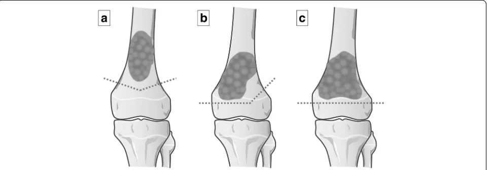
tissue on MR images, or the disappearance of abnormal accumulation on TI 201 scintigrams [15]; 2) a thickness of their residual epiphysis > 1 cm; 3) a surgical margin of ≥10 mm. When tumor was located within 2 cm of the physis, the osteotomy line was transepiphyseal or passed through the physis, resulted in complete or partial abla-tion of the physis [16], and 4) either the absence of (Enneking stages IIA and IIB), or a resectable lung me-tastasis (Enneking stage III) [17]. The inclusion criteria of the present study was patients < 15 years old with open epiphyseal plate, follow-up of > 24 months and who had a tumor located in the distal femur or the prox-imal tibia. The patients with a diaphyseal osteosarcoma for which the distance of the osteotomy to the epiphys-eal plate was < 1 cm were also included. The tumor exci-sion was classified into three types: transmetaphyseal excision (epiphyseal plate preserved), transphyseal exci-sion (epiphyseal plate partially sacrificed) and transepi-physeal excision (epitransepi-physeal plate totally sacrificed) (Fig. 1). Transphyseal and transepiphyseal excision were indicated for tumors that did not extend to the epiphysis [16], as determined by magnetic resonance imaging (MRI) T1-weighted and short tau inversion recovery (STIR) images [5].
Surgical procedure
Two different techniques of tumor-devitalised autograft treated with liquid nitrogen—free freezing [10] and ped-icle freezing [18]—were used depending on the location
of the tumor.
Free freezing technique
A K-wire was inserted into the osteotomy line under fluoroscopy. The tumor was then excised en bloc using a microsurgical saw along the K-wire. The specimen’s soft
[image:2.595.59.540.526.694.2]tissues were removed, and the tumor curetted before freezing. The excised portion was frozen in liquid nitro-gen that was stored in sterilized flask right before freez-ing for 20 min, thawed at room temperature for 15 min, then in distilled water for another 15 min. The frozen autograft was then fixed to the residual limb with double or triple locking plates, and the epiphysis stabilized with two or three screws applied either through the plate or separately (Fig.2a). For cases of proximal tibial osteosar-coma, the patella tendon was reattached to the frozen autograft by using a screw and spike washer.
Pedicle freezing technique
After the osteotomy of the proximal side of the tumor with adequate surgical margin, the surrounding soft tis-sues were carefully protected by using surgical sheets and the proximal part of tumor was elevated with isola-tion by using cotton for cast padding, an Esmarch ban-dage, and three-layer surgical sheets to prevent tumor contamination and damages to the normal tissue during freezing. The intramedullary canal is subsequently cu-retted to remove the bone marrow and contents of the
tumor in order to prevent a graft-related fracture due to water expansion during freezing. The isolated bony spe-cimen, still in continuity with the distal residual limb, was then carefully rotated and put in a container filled with liquid nitrogen for 20 min. After thawing, recon-struction was performed similarly as for the free freezing technique (Fig.2b).
The length of resection for free freezing, or of the treated bone for pedicle freezing, was recorded as the length of graft. Additional chemotherapy was adminis-tered during the postoperative period. Passive range of motion (ROM) therapy was initiated after the early post-operative period, and active ROM therapy was initiated at 6 weeks postoperatively when the muscle or tendon had to be reattached. The authors encouraged full weight-bearing mobilization after evidence of union. The follow-up protocol consisted of whole standing leg ra-diographs after 1.5, 3, 6, 9, 12, 16, 20 and 24 months and then every 6 months up to 5 years. Yearly examination was performed up to 10 years thereafter. Graft union was confirmed when the osteotomy line had disappeared in both anteroposterior (AP) and lateral views on
[image:3.595.58.539.362.674.2]radiography or computed tomography. When it was dif-ficult to evaluate bone union because of the implant shadow, confirmation was achieved by using only one view (either AP or lateral) or computed tomographic scan. Graft union was not evaluated for cases of pedicle freezing for distal femoral tumors because the osteotomy line was only on the proximal side.
The width of the epiphysis of the distal femur was measured with reference to the method of Souryal TO et al. [19]. The distance between the intersection of a line parallel to the joint surface and passing through the medial and lateral cortex at the level of the intercondylar fossa was measured by using whole standing leg radio-graphs with a 1-mm precision scale. The width of the epiphysis of the proximal tibia was measured from a line perpendicular to the tibial anatomical axis that passed through the medial edge of the medial condyle. At the time of surgery, a locking plate was placed at the lateral side hence the lateral end of the lateral condyle was diffi-cult to detect. So, the distance between the medial end of the medial condyle and the cross point to the mech-anical axis was measured on the proximal tibia (Fig.3). Epiphysis-width discrepancy (EWD) was calculated by comparing the width of the affected side and the width
of the unaffected side on radiographs at the latest exam-ination. The epiphysis growth rate was calculated by comparing the width at the latest examination with the width immediately before surgery. To analyse the col-lapse of the residual epiphysis, the anatomical lateral dis-tal femoral angle (aLDFA) for the disdis-tal femoral tumors and medial proximal tibial angle (MPTA) for the prox-imal tibial tumors at the latest examination were com-pared with those after surgery (ΔaLDFA/MPTA) [20]. Limb-length discrepancy (LLD) was measured at the lat-est follow-up. All parameters were measured by using computer software (EV Insite Version 3.1.1.218; PSP Corporation, Tokyo, Japan), which was performed inde-pendently by two assessors (AT and TH) blinded to pa-tient information and the assessment was duplicated. Status of the epiphyseal plate at the latest examination, the Musculoskeletal Tumor Society (MSTS) scores [21], complications and oncological statuses were also recorded.
Results
Epiphyseal preservation surgery was performed in 18 pa-tients. Six patients were excluded; five were older than 15 years and the other one patient was excluded because the frozen autograft was retrieved at 6 months after sur-gery due to the deep infection. Twelve patients met our inclusion criteria. The demographics and results are shown in Table 1. The mean patient age was 11 years (range, 6 to 14 years). The mean follow-up period was 61 months (range, 32 to 90 months). Eight tumors were located in the distal femur, and four were in the prox-imal tibia. Among the 12 patients, 11 (91.7%) were Enneking stage IIB and 1 (8.3%) was Enneking stage III with resectable lung metastases at the time of diagnosis.
Five (41.7%) patients underwent reconstruction using the pedicle-freezing technique, with 2 for distal femoral tumors and 3 for proximal tibial tumors (Fig.4). A vas-cularised fibular graft was used in one patient, while two patients underwent cancellous allograft over the osteot-omy site. Tumor excision was transmetaphyseal for 3 distal femoral cases, transphyseal for 3 distal femoral cases and 1 proximal tibial cases, and transepiphyseal for 2 distal femoral and 3 proximal tibial cases. The mean length of tumor-devitalised autograft treated with liquid nitrogen was 14.4 cm (range, 8 to 29 cm), and the mean graft union time was 9 months (range, 3 to 35 months). The mean epiphysis growth was 12.6% (range, 3.3 to 28.0%) of affected side and 12.7% (range, 3.8 to 28.9%) of contralateral side, and EWD was 0.1 mm (range,−1.0 to 1.7 mm). The mean ΔaLDFA/MPTA was 0.6° (range, − 2.4 to 4.0°). These results indicated that the epiphysis transverse growth was preserved without residual epi-physeal collapse, and the preservation or sacrifice of the epiphyseal plate did not influence the epiphysis Fig. 3The width of epiphysis was measured by using whole
[image:4.595.57.290.390.582.2]Table 1 Dempgrapic data and outcomes Patien t
No. /Sex /Age,
y Site Enne king st age Type of tumor exci sion Free zing method Graft lengt h, cm Bone graft
Union time, mo
Ep iphysis growth, % EW D LLD, mm
Δ aLDFA /MPTA
[image:5.595.175.418.82.733.2]transverse growth. The mean limb shortening was 26 mm (range, 1 to 48 mm).
Complications that needed additional surgery were ob-served in 8 (61.5%) patients, that included one deep infec-tion at 6 months after surgery requiring removal of the frozen autograft, and three (25%) fractures. Two of the fracture complications followed pedicle-freezing (Patient No. 1 and 2), and these occurred at the border between the frozen and host bone. One fracture occurred in the frozen bone and was accompanied by plate breakage (Patient No. 7). All three fractures were treated with open reduction and internal fixation combined with allograft or iliac autograft. One local recurrence (8.3%) developed in the surrounding soft tissue in a patient with pathological fracture at the initial presentation, and was treated with re-wide excision including the frozen autograft and recon-struction using an endoprosthesis (Patient No. 8). One superficial infection (8.3%) was managed by irrigation and administration of antibiotics (Patient No. 9). Limb short-ening (> 3 cm) was observed in six patients, and two pa-tients (16.7%) (distal femur, 38 mm and 48 mm) underwent limb lengthening of their tibia with three-dimensional external fixation (Taylor Spatial Frame,
Smith and Nephew, Memphis, USA) (Patient No. 3 and
4). Four cases were managed with shoe lifts. One patient with diaphyseal osteosarcoma underwent initial stabilization of the epiphysis. The screws were removed from the epiphysis after 1.5 years, with subsequent longitudinal growth of epiphysis was observed (Patient
No. 5, Fig. 5). The mean MSTS score was 28 (range, 18 to 30). Oncological outcomes were continuous dis-ease free (CDF) in 11 patients and no evidence of disease (NED) in 1 patient at 41 to 90 months follow-up (mean 63 months).
Discussion
In the present study, we found that the transverse growth of the residual epiphysis was observed with al-most same rate with contralateral side (12.6% of affected side and 12.7% of contralateral side), and no apparent discrepancy in epiphysis widths was detected even though the epiphyseal plate was totally sacrificed.
[image:6.595.58.536.88.312.2]final follow up, and no recurrence in residual epiphyses were described. However, a second surgical procedure was performed in 19 patients (54.3%), three to treat oncologic complications (three local recurrences) and 16 to treat orthopaedic complications, including 11 fractures, three diaphyseal nonunions, and two deep infections. The epiphysis was eventually removed in 5 patients (14.3%) be-cause of fracture (3 patients), amputation (1 patient) or in-fection (1e patient). Their study included 12 paediatric patients (< 15 years old); however, nothing was mentioned about the growth of the residual epiphysis [8]. In another study, Henderson ER et al. reported thirty-eight patients who were performed lower-limb preservation with an ex-pandable endoprosthesis after bone tumor resection in-cluding replacement of distal femur in twenty-five and proximal tibia in five patients. Limb length discrepancy was managed by the lengthening of prosthesis (mean, 4.5 cm) and the mean limb-length discrepancy was 0.7 cm at the latest examination. However, sixteen patients (42%) experienced one or more complications: superficial infec-tion without sequelae in five, soft-tissue failures in four, aseptic loosening in four, structural failure in two and fail-ure due to infection in three cases at a mean of twenty-eight months. Ten patients (26%) required pros-thesis revision, and two patients required amputation. The mean MSTS score was 26.4 in distal femoral and 26.7 in proximal tibial replacement [23]. Complications in the present study was occurred in eight of thirteen patients (61.5%), however two limb-shortening which needed the additional lengthening was inevitable. Therefore, the au-thors considered that the incidence of remaining 6 com-plications (46.2%) including 3 fractures, 1 soft tissue recurrence, 1 superficial infection and 1 deep infection were comparable with those of allograft (54.3%) and ex-pandable prosthesis (42%). Limb reconstruction using tumor-devitalised autograft treated with liquid nitrogen procedure was introduced in 1999 [10], with advantages reported as follows: simplicity, low cost, preservation of osteoinductive [24] and osteoconductive properties, per-fect fit between graft and host bone, sufficient biomechan-ical strength, avoidance of disease transmission, avoidance of immunological rejection, nondependent of bone bank, nondependent of special equipment and strict thermal control, easy attachment of tendons and ligaments to bone, preserving of bone morphogenetic proteins (BMP) [13]. The pedicle freezing technic has the following add-itional reported advantages as well: shorter operating time, maintaining of joint continuity in selected patients, de-creased osteotomy sites, and a lower rate of graft healing complications. The impossibility of histological analysis of the whole specimen (tumor necrosis analysis) and compli-cations similar to those related to allograft implantation are noted disadvantages. Moreover, because a frozen auto-graft is a tumor-devitalised bone, the strength of the
autograft depends on the integrity of the remaining struc-tures following cryogenic sterilization. For this reason, prerequisites for our frozen autograft methods are an osteoblastic lesion and that massive osteolytic destruction has not taken place [18]. Although Yamamoto N, et al. re-ported that the compression strength of frozen autograft immediately after the treatment was similar to intact bone in animal model [25], the biomechanical strength will be reduced by devitalized tissue with time.
Wong KC et al. reported eight cases of
joint-preserving tumor resection in which image-guided computer navigation and reconstruction using a computer-aided design (CAD) prosthesis were per-formed with excellent limb function (mean MSTS score, 29). They presented a case of continuous growth of the remaining distal femur epiphysis [6]. Puhaindran ME et al. reported nine cases of paediatric osteosarcoma treated by epiphysis preserving tumor excision and re-constructed with fibula grafts supplemented by auto-claved bone grafts infused with bone marrow. They showed one case of latitudinal residual epiphysis growth following distal femur osteosarcoma excision and recon-struction [26]. The epiphysis in a child is composed of articular cartilage, epiphyseal cartilage, a secondary centre of ossification and the physis, all of which are re-ferred to as the articular-epiphyseal cartilage complex [27]. The secondary centre of ossification is supplied by the epiphyseal artery, branches of which end in the pro-liferating cartilage zone [28]. The groove of Ranvier and the perichondrial ring of LaCroix, both of which support latitudinal growth of the physis, surround the latter. Al-though the latitudinal growth arrest after damage to the groove of Ranvier and the perichondrial ring of LaCroix caused by physeal injury has been well discussed [29], the latitudinal growth after epiphyseal-preservation sur-gery has not been fully clarified.
tibia (Patients No. 3 and 4). Four cases were managed with shoe lifts. In lieu of expandable prostheses, exces-sive limb shortening shall require additional salvage sur-gery such as distraction osteogenesis. However, we believe our biological reconstruction has several benefits that help preserve excellent limb function as shown by the MSTS score.
The long-term safety of close surgical margin resection including epiphyseal preservation surgery in the treat-ment of osteosarcoma is yet to be clarified. The most important consideration is to evaluate tumor extension to the epiphysis accurately. Jesus-Garcia R, et al. re-ported the epiphyseal plate invasion was detected 44% radiologically in their study, whereas histological exam-ination detected 84%. However, the epiphyseal involve-ment was evaluated by using the routine radiographs [30]. Hoffer FA et al. reported that epiphyseal extension of an osteosarcoma could be detected on MRI as a dark signal on T1 and a bright signal on STIR in MRI. [5] These findings have led to a consensus that epiphyseal-preservation surgery can be performed [11]. Before this consensus, a few studies have reported use of epiphyseal-preservation surgery, including physeal dis-traction, when removing the tumor [31]. Andreou D et al. reported that the close margins did not lead to in-creased local recurrence in their 123 of 1355 cases of osteosarcoma treated with close surgical margin resec-tion [32]. In the present study, our method was indi-cated for those patients who responded to neoadjuvant chemotherapy and with no tumor extension to the epiphy-sis. All patients were alive within the mean follow-up
period of 61 months (range, 32 to 93 months).,However one (8.3%) of 12 patients underwent removal of the re-sidual epiphysis due to soft tissue recurrence at 20 months after the initial surgery. Careful assessment of tumor exten-sion to the epiphysis by using MRI and more long-term analyses are mandatory to evaluate the safety of this method.
This study had some limitations. The number of pa-tients was small; the patient age at the time of sur-gery, type of excision used for preservation of the epiphysis and the follow-up term varied and the follow-up periods were relatively short. The growth of residual epiphysis was evaluated by measuring the two-dimensional latitudinal growth only by using plain radiography. Moreover, the epiphysis of the proximal tibia was investigated only by examining half of the entire epiphysis width due to the shadow of the plate. Although Computed tomography (CT) scan is more accurate, we did not routinely preform CT scan to reduce radiation exposures. We did not ana-lyse the longitudinal growth of the residual epiphysis because this study contained three types of osteot-omy, and it was also difficult to measure the longitu-dinal growth after achieving bony union with a transepiphyseal excision. Despite its limitations related to its retrospective nature, the present study provides the possibility of continued residual growth of epiphy-sis after epiphyseal-preservation surgery in childhood osteosarcomas around the knee. Further prospective studies in a larger series of patients with long-term follow-up should be conducted.
[image:8.595.57.538.87.294.2]Conclusion
Epiphysis transverse growth was not diminished, and there was no collapse following epiphyseal-preservation surgery in this limited series of childhood osteosarcoma around the knee. Although careful assessment is neces-sary to evaluate the extent of tumor involvement of the epiphysis, epiphyseal-preservation surgery has the ad-vantage of preserving adjacent joint function with excel-lent limb function. Epiphysis-preservation surgery should be considered in children.
Abbreviations
aLDFA:Anatomical lateral distal femoral angle; EWD: Epiphysis-width discrepancy; LLD: Limb-length discrepancy; MPTA: Medial proximal tibial angle; MRI: Magnetic resonance imaging; MSTS: the Musculoskeletal Tumor Society
Acknowledgements
The authors would like to thank Dr. Mamer Rosario for review and comment on the manuscript prior to submission. The authors also thank Mr. Toshiya Nomura for drawing the figures.
Availability of data and materials
The datasets supporting the conclusions of this article are included within the article. If you wish to obtain access for the underlying material please contact the corresponding author to discuss your request in detail.
Authors’contributions
AT performed the study design, data collection, analysis of data, paper drafting. TH carried out the analysis of the radiogram. NY, KH, HM, SM, KA and YT analyzed data and edited the manuscript. HT performed the study design and manuscript review. All authors approved the final manuscript.
Ethics approval and consent to participate
This study protocol was approved by The Medical Ethics Committee of Kanazawa University, Kanazawa, Japan (Application number 1784). This study complied with ethical standards outlined in the Declaration of Helsinki. Informed concent was obtained from the patient’s parents or gurdian.
Competing interests
The authors declare that they have no competing interests.
Publisher’s Note
Springer Nature remains neutral with regard to jurisdictional claims in published maps and institutional affiliations.
Received: 6 May 2017 Accepted: 23 May 2018
References
1. Ayerza MA, Farfalli GL, Aponte-Tinao L, Luis Muscolo D. Does increased rate of limb-sparing surgery affect survival in osteosarcoma? Clin Orthop Relat Res. 2010;468:2854–9.
2. Kang S, Lee JS, Park J, Park S-S. Staged lengthening and reconstruction for children with a leg-length discrepancy after excision of an osteosarcoma around the knee. Bone Joint J. 2017;99-B:401–8.
3. Pala E, Henderson ER, Calabrò T, Angelini A, Abati CN, Trovarelli G, et al. Survival of current production tumor endoprostheses: complications, functional results, and a comparative statistical analysis. J Surg Oncol. 2013; 108:403–8.
4. Han C, Chung D, Lee J, Jeong B. Lengthening of intercalary allograft combined with free vascularized fibular graft after reconstruction in pediatric osteosarcoma of femur. J Pediatr Orthop B. 2010:61–5.
5. Hoffer FA, Nikanorov AY, Reddick WE, Bodner SM, Xiong X, Jones-Wallace D, et al. Accuracy of MR imaging for detecting epiphyseal extension of osteosarcoma. Pediatr Radiol. 2000;30:289–98.
6. Wong KC, Kumta SM. Joint-preserving tumor resection and reconstruction using image-guided computer navigation tumor. Clin Orthop Relat Res. 2013;471:762–73.
7. Mei J, Ni M, Jia GY, Chen YX, Zhu XZ. Intermittent internal fixation with a locking plate to preserve epiphyseal growth function during limb-salvage surgery in a child with osteosarcoma of the distal femur. Medicine (Baltimore). 2015;94:1–4.
8. Aponte-tinao L, Ayerza MA, Muscolo DL, Farfalli GL. Survival, recurrence, and function after epiphyseal preservation and allograft reconstruction in osteosarcoma of the knee. Clin Orthop Relat Res. 2015:1789–96. 9. Tsuchiya H, Abdel-Wanis ME, Sakurakichi K, Yamashiro T, Tomita K.
Osteosarcoma around the knee. Intraepiphyseal excision and biological reconstruction with distraction osteogenesis. J Bone Joint Surg Br. 2002;84: 1162–6.
10. Tsuchiya H, Wan SL, Sakayama K, Yamamoto N, Nishida H, Tomita K. Reconstruction using an autograft containing tumour treated by liquid nitrogen. J Bone Joint Surg Br. 2005;87:218–25.
11. Kiss S, Terebessy T, Szöke G, Kiss J, Antal I, Szendröi M. Epiphysis preserving resection of malignant proximal tibial tumours. Int Orthop. 2013;37:99–104. 12. Igarashi K, Yamamoto N, Shirai T, Hayashi K, Nishida H, Kimura H, et al. The
long-term outcome following the use of frozen autograft treated with liquid nitrogen in the management of bone and soft-tissue sarcomas. Bone Joint J. 2014;96 B:555–61.
13. Takata M, Sugimoto N, Yamamoto N, Shirai T, Hayashi K, Nishida H, et al. Activity of bone morphogenetic protein-7 after treatment at various temperatures: freezing vs. pasteurization vs. allograft. Cryobiology. 2011;63: 235–9.
14. Tsuchiya H, Tomita K, Mori Y, Asada N, Yamamoto N. Marginal excision for osteosarcoma with caffeine assisted chemotherapy. Clin Orthop Relat Res. 1999;358:27–35.
15. Miwa S, Takeuchi A, Shirai T, Taki J, Yamamoto N, Nishida H, et al. Prognostic value of radiological response to chemotherapy in patients with osteosarcoma. PLoS One. 2013;8
16. Kumta SM, Chow TC, Griffith J, Li CK, Kew J, Leung PC. Classifying the location of osteosarcoma with reference to the epiphyseal plate helps determine the optimal skeletal resection in limb salvage procedures. Arch Orthop Trauma Surg. 1999;119:327–31.
17. Enneking WF, Spanier SS, Goodman MA. A system for the surgical staging of musculoskeletal sarcoma. Clin Orthop Relat Res. 1980;(153):106–20. 18. Tsuchiya H, Nishida H, Srisawat P, Shirai T, Hayashi K, Takeuchi A, et al.
Pedicle frozen autograft reconstruction in malignant bone tumors. J Orthop Sci. 2010;15:340–9.
19. Souryal TO, Freeman TR. Intercondylar notch size and anterior cruciate ligament injuries in athletes. A prospective study. Am J Sports Med. 1993;21: 535–9.
20. Paley D, Herzenberg JE, Tetsworth K, McKie J, Bhave A. Deformity planning for frontal and sagittal plane corrective osteotomies. Orthop Clin North Am. 1994;25:425–65.
21. Enneking WF, Dunham W, Gebhardt MC, Malawar M, Pritchard DJ. A system for the functional evaluation of reconstructive procedures after surgical treatment of tumors of the musculoskeletal system. Clin Orthop Relat Res. 1993;(286):241–6.
22. Muscolo DL, Ayerza MA, Aponte-Tinao LA, Ranalletta M. Partial epiphyseal preservation and intercalary allograft reconstruction in high-grade metaphyseal osteosarcoma of the knee. J Bone Joint Surg Am. 2005;87 Suppl 1 (Pt 2):226–36.
23. Henderson ER, Pepper AM, Marulanda G, Binitie OT, Cheong D, Letson GD. Outcome of lower-limb preservation with an expandable endoprosthesis after bone tumor resection in children. J Bone Joint Surg Am. 2012;94:537–47.
24. Tanzawa Y, Tsuchiya H, Shirai T, Hayashi K, Yo Z, Tomita K. Histological examination of frozen autograft treated by liquid nitrogen removed after implantation. J Orthop Sci. 2009;14:761–8.
25. Yamamoto N, Tsuchiya H, Tomita K. Effects of liquid nitrogen treatment on the proliferation ofosteosarcoma and the biomechanical properties of normal bone. J Orthop Sci. 2003;8:374–80.
26. Puhaindran ME, Pho RWH. Biological reconstruction for children with osteosarcoma around the knee. Ann Acad Med Singap. 2014;43:499–505. 27. Carlson CS, Hilley HD, Henrikson CK. Ultrastructure of normal epiphyseal
28. Chung SM. The arterial supply of the developing proximal end of the human femur. J Bone Joint Surg Am. 1976;58:961–70.
29. Peterson HA. Anatomy and Growth. In: Peterson HA, editor. Epiphyseal growth plate fractures. Heidelberg: Springer; 2007. p. 7–17.
30. Jesus-Garcia R, Seixas MT, Costa SR, Petrilli AS, Filho JL. Epiphyseal plate involvement in osteosarcoma. Clin Orthop Relat Res. 2000;373:32–8. 31. Cañadell J, Forriol F, Cara JA. Removal of metaphyseal bone tumours with
preservation of the epiphysis. Physeal distraction before excision. J Bone Joint Surg Br. 1994;76:127–32.