S T U D Y P R O T O C O L
Open Access
Medium-sized posterior fragments in AO
Weber-B fractures, does open reduction
and fixation improve outcome? the
POSTFIX-trial protocol, a multicenter
randomized clinical trial
Sander Verhage
1*, Peer van der Zwaal
1, Maarten Bronkhorst
1, Huub van der Meulen
2, Sanne Kleinveld
2,
Sven Meylaerts
1, Steven Rhemrev
1, Pieta Krijnen
3, Inger Schipper
3and Jochem Hoogendoorn
1Abstract
Background:Guidelines for treatment of the posterior fracture fragment in trimalleolar fractures are scarce and show varying advices. Did the increasing size of the posterior fragment seem to relate to worse outcome in the past, nowadays this has changed to the amount of dislocation of the posterior fragment post-operatively. Despite many retrospective cohort studies and some prospective cohort studies, no consistent guideline could be derived from the current literature.
Methods:The POSTFIX-study is designed as a multicenter randomized clinical trial to analyse the effects of anatomical reduction and fixation of the posterior fragment in AO 44-B3 fractures with medium-sized posterior fragment. A total of 84 patients will be included and online allocated to either anatomical reduction and fixation of the posterior fragment via the posterolateral approach (n= 42) or no fixation of the posterior fragment (n= 42). The concomitant fractured medial and lateral malleoli are treated according to the AO-principles. Functionality of the ankle as measured by the AAOS-questionnaire (American Association of Orthopaedic Surgeons) 1 year post-operatively was set as primary outcome. Main secondary outcome measures are the AAOS-questionnaire 5 years postoperatively and osteoarthritis as measured on plain radiographs 1 year and 5 years post-operatively. The Olerud and Molander score, the AOFAS-score, the VAS-pain, the Euroqol-5D and Range of Motion by physical examination will also be evaluated during the follow-up period. Discussion:The POSTFIX-trial is the first high quality multicenter randomized clinical trial worldwide to analyse the effects of anatomical fixation of the posterior fragment in trimalleolar fractures. New guidelines on anatomical reduction and fixation of the posterior fragment can in future be based on the results of this trial.
Trial registration:This trial is registered on ClinicalTrials.gov with reference number: NCT02596529. Registered 3 November 2015, retrospectively registered.
Keywords:Trimalleolar fracture, Posterior malleolar fracture, Posterior fragment fixation, Ankle fracture, Posterolateral approach
* Correspondence:sanverhage@hotmail.com 1
Trauma-unit Haaglanden MC, The Hague, The Netherlands Full list of author information is available at the end of the article
Background
The optimal treatment of ankle fractures with involve-ment of the posterior malleolus remains a subject of debate. Despite a large amount of literature on the role of the posterior malleolus in a so-called trimalleolar frac-ture, there are no clear guidelines for its treatment. Its size was taught to be the leading indicator for the need of fixation of the fragment [1, 2]. Most orthopaedic surgeons consider a posterior malleolar fracture frag-ment larger than 25 to 33% of the joint surface an indi-cation for fixation. Interestingly, after careful evaluation of the available literature [3–8], there does not seem to be solid substantiation of this assumption.
Theories about the relation between the size of the posterior fracture fragment and outcome are partially based on biomechanical studies. First, a large posterior fragment was thought to lead to posterior instability and therefore to worse outcome on the long term. Several cadaveric ankle studies however, showed different cutoff values ranging from 25 to 33% of the involved articular surface [9, 10]. Other studies could not prove the theory of posterior instability in cadaveric ankles [11–14]. Later cadaveric studies suggested a shift of contact pressure pattern in case of a posterior malleolar fracture and there-fore the early induction of increased in post-traumatic osteoarthritis [15, 16].
Several retrospective cohort studies showed no clear re-lation between fragment size and functional outcome. Jaskulka et al. and Langenhuijsen et al. found a worse long-term outcome (follow-up 5.7 and 6.9 years) in frag-ments larger than 5 and 10% of the involved articular surface respectively (3,5]. Broos et al. found a worse out-come after 1 year in patients with posterior fragments larger than 33% of the involved articular surface [6]. De Vries et al. found no relation between size and functional outcome but he suggested a worse functional outcome in case of a posterior malleolar fracture dislocation [4].
More recently two large retrospective cohort studies on the influence of joint congruency in posterior malleo-lar fractures were published. The first was performed by Xu et al., who studied 102 trimalleolar fractures with a mean follow up of 2.8 years [8]. There was no relation between fragment size and radiological or functional outcome (AOFAS). However, they found a worse functional outcome if a persisting tibiotalar step-off was present after open reduction and internal fixation. Therefore they advised to anatomically restore the articular surface, especially when fragment size involves than 25% of the joint surface [8].
Drijfhout et al. performed a retrospective cohort study of 131 trimalleolar fractures with a mean follow-up of 7.3 years [7]. Functional outcome was worse in trimal-leolar fractures with a medium-sized (5–25%) or large (>25%) posterior fragment compared to small fragments
(<5%). A postoperatively persisting step-off ≥1 mm of medium-sized or large posterior fragments showed to be the most important predictor for the development of osteoarthritis [7]. Restoration of the tibiotalar articular surface therefore seems essential in medium-sized and large fragments.
As shown, no consistent advice is found in the litera-ture as to which fragment size of the posterior malleolus should be internally fixed. Currently, according to AO guidelines [17] fixation of posterior fragments is indi-cated if there is a displaced fragment larger than 25% of the involved articular surface or if instability is persistent after reduction of the lateral injury [17]. Traditionally, reduction of these larger fragments is performed indirectly, followed by percutaneous screw fixation in anterior-posterior direction. It is often challenging to achieve an anatomical reduction and fixation of smaller fragments using the percutaneous method. Recently, direct exposure of the posterior tibia via a posterolateral approach, followed by open reduction and fixation with screws in posterior-anterior direction or an antiglide plate, is advocated by several authors [18–21]. This approach, with the patient in prone position, allows good visualization of the fracture, ar-ticular anatomical reduction, and solid fixation. Another advantage is that even small posterior fragments can be addressed. Several case series published favourable results; few major wound complications, good functional out-comes, and rarely a need for reoperation [22, 23].
To test the hypothesis that anatomical reduction and fixation of medium-sized posterior fragments via the posterolateral approach is meaningful, this multicenter randomized clinical trial was designed. As far as we know, this is the first randomized controlled trial on fixation of the posterior fragment in trimalleolar frac-tures worldwide.
Methods/design
Aim of the study
Anatomical reduction and fixation of medium-sized posterior malleolar fractures leads to a stable and anatom-ical fixation [22, 23]. In this study, we hope to prove that anatomical reduction and fixation of medium sized posterior fragments leads to less osteoarthritis and better functional outcome in patients having a trimal-leolar fracture.
Study design and setting
fixation of the posterior malleolus via the posterolateral approach or 2. Open reduction and fixation without fix-ation of the posterior fragment. Two level-1 traumacen-ters (MCH Westeinde, Haga ziekenhuis) and one level-2 traumacenter (Bronovo hospital) participate, which are all centers in The Hague. The study will be open for in-clusion from 2015 till 2019, the follow up will be com-pleted in 2024. The Medical Ethics Committee South West Netherlands approved of the study protocols (protocol number 15-040). The study is registered at ClinicalTrials.gov (Number NCT02596529).
Study population
All patients presenting at the emergency department with an AO-44B3 trimalleolar fracture with a medium-sized (5–25%) posterior fragment are asked to partici-pate in the study. The initial decision on inclusion is based on review of pre-operative radiographs since this best resembles daily practice. The pre-operative X-rays are judged by 2 observers to confirm eligibility for the study. Fragment size is measured on plain lateral radio-graphs at tibiotalar joint level. A computed tomography scan is made in order to assess intra-articular fragments and correlation with pre-operative X-rays and posterior fragment size.
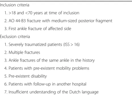
The inclusion criteria are: age between 18 and 75 years, trimalleolar AO-44B3 fracture with medium sized (5–25% of the distal tibial articular surface) posterior fragment and first ankle fracture of affected side. Excluded are multitraumatized patients (ISS > 16), patients with multiple fractures or open fractures, ankle fracture of same side in medical history, with pre-existent disability or mobility problems (need of walking gait), where follow-up takes place in another hospital and with insufficient understanding of the Dutch language. A detailed list of in- and exclusion criteria is presented in Table 1.
Recruitment, informed consent and randomization
Patients are first seen at the Emergency Department and will receive the study information from the attending surgeon or surgical resident. All patients will be in-formed about the potential risks and complications of both fix and non-fix management of the posterior malle-olus. If the patient is eligible for inclusion (based on fragment size measured on plain lateral X-ray) and willing to participate, the research coordinator includes the patient within 1 week. All participants provide writ-ten informed consent. After inclusion, participants are allocated to one of the two randomisation groups by an online randomization program in blocks of 6 or 8 patients. A flow-chart of inclusion and randomization is shown in Fig. 1. Blinding is not possible because the two different soft tissue approaches in the treatment protocols indicate which type of surgical fixation is performed.
Treatment protocol
All operative interventions are performed by experi-enced surgeons familiar with both treatment protocols and fixation techniques. Pre-operatively 1 gram Cefazo-line® prophylaxis is administered. Dependent form surgeon’s preference, patients will be operated with a thigh tourniquet. In case of non-fixation of the posterior fragment, patients are operated in supine position. The lateral malleolus is fixed with two lag screws and/or lat-eral plate fixation. The medial malleolus is fixed with two cancellous screws or tension band wiring. In case of allocation to the fixation-group, the participant is oper-ated in prone position. The posterolateral approach is used for fixation of both the lateral malleolus and the posterior fragment. The posterior fragment is fixed with lag screws or a buttress or antiglide plate. The lateral and medial malleolus are fixed in the same manner as in the first group. After fixation the syndesmosis is tested by a bone hook or external rotatory stress under fluoro-scopic control. If the syndesmosis is unstable, two transsyndesmotic positioning screws are placed. Non-weightbearing mobilization is instructed for 6 weeks after operation. After 6 weeks gradual weight-bearing mobilization is allowed and physiotherapy is started. Low-molecular weight heparin 2850 international units was administered daily as long as patients are immobilized in cast.
End points and follow-up
[image:3.595.57.290.561.729.2]The primary outcome of this study is the functional out-come after 1 year assessed by the AAOS (American Association of Orthopaedic Surgeons) Foot and Ankle-score. Secondary outcome parameters include functional outcome measured with the AOFAS-questionnaire (American Orthopaedic Foot and Ankle Society) and with the Olerud and Molander functional score and Table 1Inclusion and exclusion criteria
Inclusion criteria
1. >18 and <70 years at time of inclusion
2. AO 44-B3 fracture with medium-sized posterior fragment 3. First ankle fracture of affected side
Exclusion criteria
1. Severely traumatized patients (ISS > 16) 2. Multiple fractures
3. Ankle fractures of the same ankle in the history 4. Patients with pre-existent mobility problems 5. Pre-existent disability
osteoarthritis on plain radiographs 1 and 5 years postop-eratively. Pain measured on a Visual Analogue Scale (VAS-pain), range of motion in the upper ankle joint (dorsal and plantarflexion) measured using a goniometer and general health measured using the Euroqol-5D are assessed at each visit. The total study period for a participant is 5 years. The entire study will be performed in approximately 10 years. An overview of all measure-ments during the follow-up period is provided in Table 2. Patients visit the outpatient clinic at 6 weeks, 12 weeks, 26 weeks, 1 year and 5 years after surgery. Deviations of the treatment protocol will be reported.
Sample size
In this study functional outcome as measured by the AAOS score will be used as the primary outcome parameter. Up to now the minimal clinically important difference has not been determined and published for the AAOS score. Recently, a Cochrane Review suggested a difference of 10 points to be clinically relevant [24].
For the sample size calculation we adopted this 10 point difference, with a standard deviation of 15 points and a significance level of 5%. To achieve 80% power, group samples of at least 36 patients are needed. To account for 15% drop-out, group samples of 42 patients are needed (84 in total).
Statistical analysis
The analysis will be performed on the basis of the intention-to-treat principle. Baseline characteristics (age, gender, fragment size, dislocation posterior fragment etc.) of the study groups will be described using summary statistics. Continuous outcome measures (Olerud and Molander ankle score, VAS-pain, AAOS, range of motion) will be reported as mean and standard deviation and will be compared between the treatment groups by an unpaired t-test. Multiple imputation for missing data will be performed. Linear mixed models will be used to compare the functional outcome of the two groups during the follow-up.
[image:4.595.57.543.87.309.2]Fig. 1Flow-chart of inclusion and randomisation
Table 2Measurements during follow-up
Post-operative 6 weeks 12 weeks 26 weeks 1 year 5 years
X-ray × × × ×
CT-scan ×
Olerud & Molander × × × ×
AAOS × × ×
AOFAS × × ×
Euroqol-5D × × × × ×
VAS-pain × × × × ×
[image:4.595.63.536.605.732.2]Data will be analyzed using the“Statistical Package for the Sciences” version 22.0 or higher. Statistical testing will be 2-tailed and a p-value <0.05 will be used as threshold for statistical significance.
Discussion
This study seeks answers to the question whether or not open anatomical reduction and fixation of medium-sized posterior malleolar fractures via a posterolateral approach leads to better functional outcome than when the fragment is left untouched.
Since the treatment involves two different surgical procedures with different scars from which the treat-ment can be deduced, randomisation status will not be blinded. Also post-operative X-rays will show different implants and therefore partial blinding in the post-operative treatment phase will not be possible.
For the pre-operative measurement of posterior frag-ment size and therefore patient selection, we use the Picture Achieving and Communication System (PACS) which is standard in all participating hospitals. Two ob-servers measure the fragment size in PACS on plain X-ray. However, in a discussion amongst clinicians the preferred device for posterior fracture fragment was recently debated [25, 26]. The most widely used and currently most reproducible one, is the method de-scribed above in PACS, which we use for this study.
Functional outcome is measured with three question-naires. The Olerud and Molander ankle score is best available for short term functional outcome and there-fore used to evaluate during the first post-operative year. The AAOS ankle questionnaire is best available func-tional outcome score on long term and therefore used to evaluate 1 and 5 years post-operatively. The AOFAS ankle questionnaire is build up from a questionnaire and a limited range of motion and therefore also used to evaluate 1 and 5 years post-operatively.
The first patient was included in January 2014. We expect to include the last patient in 2018. Latest follow-up visit to our outpatient clinic and the conclusion of the data-acquisition will therefore be in 2023.
Conclusion
The POSTFIX-trial is the first multicenter randomized clinical trial worldwide to analyse the effects of open anatomical fixation of the posterior fragment in trimal-leolar fractures. New guidelines on anatomical reduction and fixation of the posterior fragment can in future be based on the results of this trial.
Abbreviations
AAOS:American academy of orthopaedic surgeons; AO: Arbeitsgemeinschaft für osteosynthesefragen; AOFAS: American orthopaedic foot and ankle society; VAS: Visual analogue scale
Acknowledgements No acknowledgements.
Funding
This study is possible by a local research grant of the Haaglanden Medical Center.
Availability of data and materials Not applicable.
Authors contributions
SV Study design, data collection, writing the paper, study coordinator. PZ, MB, HM, SK, SM, SR, PK, IS Study design, data collection, correcting the paper. JH Study design, data collection, correcting the paper, research coordinator.
Competing interests
The authors declare that they have no competing interests.
Consent for publication Not applicable.
Ethics approval and consent to participate
The Medical Ethics Committee South West Netherlands approved of the study protocols (protocol number 15-040). Informed Consent was taken from all participants.
Study status
Active and recruiting patients.
Author details
1Trauma-unit Haaglanden MC, The Hague, The Netherlands.2Department of
Traumasurgery, Haga Ziekenhuis, The Hague, The Netherlands.3Department
of Traumasurgery, Leiden University Medical Center, Leiden, The Netherlands.
Received: 13 July 2016 Accepted: 7 February 2017
References
1. Irwin TA, Lien J, Kadakia AR. Posterior malleolus fracture. J Am Acad Orthop Surg. 2014;22(2):67.
2. Odak S, Ahluwalia R, Unnikrishnan P, Hennessey M, Platt S. Management of the posterior malleolar fractures: a systematic review. J Foot Ankle Surg. 2016;55(1):140–5.
3. Jaskulka RA, Ittner G, Schedl R. Fractures of the posterior tibial margin: their role in the prognosis of malleolar fractures. J Trauma. 1989;29(11):1565–70. 4. De Vries JS, Wijgman AJ, Sierevelt IN, Schaap GR. Long-term results of ankle
fractures with a posterior malleolar fragment. J Foot Ankle Surg. 2005;44(3):211–7.
5. Langenhuijsen JF, Heetveld MJ, Ultee JM, Steller EP, Butzelaar RM. Results of ankle fractures with involvement of the posterior tibial margin. J Trauma. 2002;53(1):55–60.
6. Broos PL, Bisschop AP. Operative treatment of ankle fractures in adults: correlation between types of fracture and final results. Injury. 1991;22(5):403–6. 7. Drijfhout van Hooff CC, Verhage SM, Hoogendoorn JM. Influence of
fragment size and postoperative joint congruency on long-term outcome of posterior malleolar fractures. Foot Ankle Int. 2015;36(6):673–8. 8. Xu HL, Li X, Zhang DY, Fu ZG, Wang TB, Zhang PX, Jiang BG, Shen HL,
Wang G, Wang GL, Wu XB. A retrospective study of posterior malleolus fractures. Int Orthop. 2012;36(9):1929–36.
9. Macko VW, Matthews LS, Zwirkoski P, Goldstein SA. The joint-contact area of the ankle. The contribution of the posterior malleolus. J Bone Joint Surg Am. 1991;73(3):347–51.
10. Hartford JM, Gorczyca JT, McNamara JL, Mayor MB. Tibiotalar contact area. Contribution of posterior malleolus and deltoid ligament. Clin Orthop Relat Res. 1995;320:182–7.
11. Harper MC. Posterior instability of the talus: an anatomic evaluation. Foot Ankle. 1989;10(1):36–9.
12. Harper MC, Hardin G. Posterior malleolar fractures of the ankle associated with external rotation-abduction injuries. Results with and without internal fixation. J Bone Joint Surg Am. 1988;70(9):1348–56.
14. Raasch WG, Larkin JJ, Draganich LF. Assessment of the posterior malleolus as a restraint to posterior subluxation of the ankle. J Bone Joint Surg Am. 1992;74(8):1201–6.
15. Vrahas M, Fu F, Veenis B. Intraarticular contact stresses with simulated ankle malunions. J Orthop Trauma. 1994;8(2):159–66.
16. Fitzpatrick DC, Otto JK, McKinley TO, Marsh JL, Brown TD. Kinematic and contact stress analysis of posterior malleolus fractures of the ankle. J Orthop Trauma. 2004;18(5):271–8.
17. P Barbosa, F Bonnaire, K Kojima. AO foundation. http://www2.aofoundation. org. 2013.
18. Talbot M, Steenblock TR, Cole PA. Posterolateral approach for open reduction and internal fixation of trimalleolar ankle fractures. Can J Surg. 2005;48(6):487–90.
19. Tornetta 3rd P, Ricci W, Nork S, Collinge C, Steen B. The posterolateral approach to the tibia for displaced posterior malleolar injuries. J Orthop Trauma. 2011;25(2):123–6.
20. Miller AN, Carroll EA, Parker RJ, Helfet DL, Lorich DG. Posterior malleolar stabilization of syndesmotic injuries is equivalent to screw fixation. Clin Orthop Relat Res. 2010;468(4):1129–35.
21. Gardner MJ, Brodsky A, Briggs SM, Nielson JH, Lorich DG. Fixation of posterior malleolus fractures provides greater syndesmotic stability. Clin Orthop Relat Res. 2006;447:165–71.
22. Forberger J, Sabandal PV, Dietrich M, Gralla J, Lattmann T, Platz A. Posterolateral approach to the displaced posterior malleolus: functional outcome and local morbidity. Foot Ankle Int. 2009;30(4):309–14. 23. Verhage SM, Boot F, Schipper IB, Hoogendoorn JM. Open reduction and
internal fixation of posterior malleolar fractures using the posterolaterale approach. Bone Joint J. 2016;98-B(6):812–7.
24. Donken CCMA, Al-Khateeb H, Verhofstad MHJ, van Laarhoven CJHM. Surgical versus conservative interventions for treating ankle fractures in adults. Cochrane Database Syst Rev. 2012, Issue 8.
25. Meijer DT, de Muinck Keizer RJ, Doornberg JN, Sierevelt IN, Stufkens SA, Kerkhoffs GM, van Dijk CN. Ankle platform study collaborative—science of variation group. Diagnostic accuracy of 2-dimensional computed tomography for articular involvement and fracture pattern of posterior malleolar fractures. Foot Ankle Int. 2016;37(1):75–82.
26. Meijer DT, Doornberg JN, Sierevelt IN, Mallee WH, van Dijk CN, Kerkhoffs GM, Stufkens SA. AnklePlatform study collaborative—science of variation group; ankle platform study collaborative—science of variation group. Guesstimation of posterior malleolar fractures on lateral plain radiographs. Injury. 2015;46(10):2024–9.
• We accept pre-submission inquiries
• Our selector tool helps you to find the most relevant journal
• We provide round the clock customer support
• Convenient online submission
• Thorough peer review
• Inclusion in PubMed and all major indexing services
• Maximum visibility for your research
Submit your manuscript at www.biomedcentral.com/submit