Clinical Evaluation of a Self-Etching Adhesive for
All-Ceramic Indirect Restorations
Augusto Robles, D.D.S.
A thesis submitted in partial fulfillment of the requirements
for the degree of Master of Science in Restorative Dentistry
Horace H. Rackham School of Graduate Studies
The University of Michigan
Ann Arbor, Michigan
2007
Thesis Committee Members:
Peter Yaman, D.D.S., M.S. (Chairman)
Joseph B. Dennison, D.D.S., M.S.
Michael Razzoog, D.D.S., M.S., M.P.H.
Gisele Neiva, D.D.S., M.S.
To my wife Monica and my son, Rodrigo.
To my parents, Augusto and Zoila.
To my Lord and Savior Jesus Christ for blessing me abundantly.
To my parents, Augusto and Zoila, for giving me the opportunity to further my professional
training in the United States of America.
To my wife and son, Monica and Rodrigo, for their love and support throughout this period
of training.
To the members of my thesis committee for guidance and direction in the design and
completion of this project.
To all the staff and faculty of the Graduate General Dentistry Clinic for their assistance.
To Carol Stamm and Michelle Hughes for being involved with patient care.
To Dr. Joseph Dennison for statistical assistance.
To the Graduate Restorative Dentistry residents for their friendship and camaraderie.
Title page 1
Dedication 2 Acknowledgements 3
Table of contents 4
List of Figures and Tables 6
Chapter 1 1.1 Background and Significance 8
1.2 Hypothesis 11
1.3 Review of the Literature 12
1.3.1 Adhesives 12
1.3.2 Total-etch vs. Self-etch 17
1.3.3 Sensitivity 24
1.3.4 Visual Analog Scales 28
1.3.5 Ceramic Restorations 33
1.3.6 Clinical Evaluation 39
1.4 References 43 Chapter 2 2.1 Abstract 52
2.2 Introduction 53
2.3 Research Design and Methods 56
2.3.1 Patient Recruitment 56
2.3.2 Selection Criteria 56
2.3.2a Inclusion Criteria 56
2.3.2b Exclusion Criteria 56
2.3.3 Materials and Methods 57
2.3.4 Evaluation 58
2.3.4a Direct Evaluation 58
2.3.4b Indirect Evaluation 59
2.3.5 Data Analysis 60
2.4 Results 61
2.4.1 Sensitivity 62
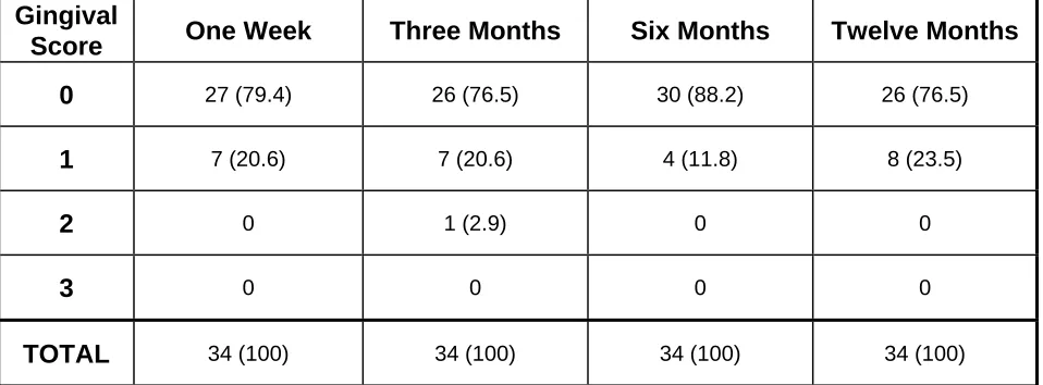
2.4.2 Gingival Index 63
2.4.3 Color Match 64 2.4.4 Margin Discoloration 65
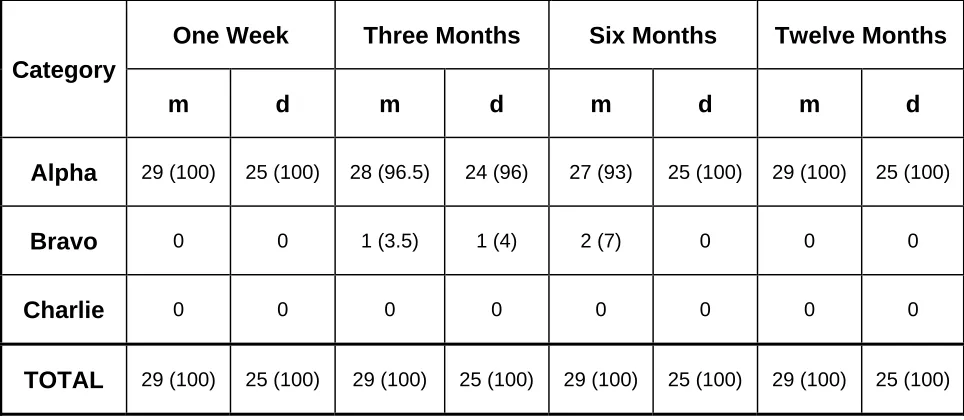
2.4.5 Margin Integrity 65
2.4.6 Restoration Integrity 66
2.4.7 Caries 66 2.4.8 Proximal Contact 67
2.4.9 Failure Mode 67
2.5.2 Gingival Index 71
2.5.3 Margin Integrity 72
2.5.4 Restoration Integrity 73
2.5.5 Caries 74
2.5.6 Proximal Contact 74
2.5.7 Failure Mode 75
2.5.8 Indirect Evaluation 76
2.6 Conclusion 81
2.7 Recommendations 82
2.8 References 83
Appendices 86
1.SEM photomicrograph showing the resin-dentin interdiffusion zone 15 2.Schematic representation explaining the ultrastructure of the resin-dentin
interdiffusion zone at the resin impregnation phase 16 3.Lip of over contoured porcelain 77
4.Polishing of over contoured margin 77
5.Final ideal margin 77
6.Margin defects observed clinically 77 7.Margin defects observed clinically 78 8.Intact inlay margin at baseline 79 9.The restoration in Figure 8 at the 1 year recall with well preserved margins 79 10.Inlay margin at baseline showing an area of exposed cement 79 11.Restoration in Figure 9 at 1 year 80 12.Intact inlay margin at baseline 80 13.The restoration in Figure 11 at 6 months 80
1. Distribution of Restorations by Tooth Type 61 2. Distribution of Restorations per Patient by Gender 62 3. Mean Sensitivity Ratings Taken from a VAS 62 4. Analysis of Variance of the Measured Values for Sensitivity 63 5. Measured Values of Sensitivity and Differences from Baseline 63 6. Analysis of Variance of the Differences from Baseline 63
7. Gingival Index 64
8. Color Match 64
9. Margin Discoloration 65
10. Margin Integrity 66
11. Restoration Integrity 67
12. Recurrent Caries 67
13. Proximal Contact 68
14. Failure Mode for Inlays and Onlays 69
Clinical Evaluation of a Self-Etching Adhesive for All-Ceramic
Indirect Restorations
1.1 Background and Significance
Introduction
Adhesive bonding has changed the practice of dentistry. This revolution of
adhesion, started by Buonocuore in 1955, has evolved through many generations in which
adhesives have become stronger, easier to use, and have gained wide acceptance by the
profession.1
Despite their great popularity among clinicians as well as researchers, some
problems cloud the success of adhesion in dentistry. The most persistent problem with
adhesives is post-operative sensitivity. Christensen states that the problem of sensitivity
keeps resurfacing, because class I, II and V restorations are among the most common
procedures dentists accomplish, and many of the dentin-bonding concepts are over
promoted in terms of preventing post-operative tooth sensitivity.2 Since the method for bonding indirect restorations (i.e. crowns, inlays, onlays, etc) is similar to the method for
bonding direct restorations, post-cementation sensitivity is also a problem with indirect
restorations. In 1995, Trowbridge stated that luting cements are still a source of frustration
to the dentist and that none of the cements currently available satisfies everyone, including
the patient.3 The author concluded that the cause of post-cementation sensitivity continues to be a perplexing problem.
It is still not clear what causes post-operative sensitivity and hence, several theories
explain dentin sensitivity.4 Their study in 1970 stated that the movement of fluids within the dentinal tubule caused by thermal or concentration changes, activated receptors related to
the odontoblasts. This is probably the most widely accepted explanation for sensitivity
physiology. Eick et al, in 1986, proposed the polymerization shrinkage of composite
restorations as the causative factor for post-operative sensitivity.5 In 1990, Kanca proposed an alternative explanation for the post-operative sensitivity.6 He presented the possibility that the inflammatory response in pulpal tissues noted in early studies when
dentin was treated with phosphoric acid was not caused by the acid. He suggested that the
inflammatory response was caused by the prolonged exposure to zinc oxide-eugenol and
documented many reports showing ZOE to be a relatively toxic material. In 2000,
Bergenholtz presented another explanation for dentin sensitivity: the presence of bacterial
leakage at the restoration-tooth interface.7 Modern restorative procedures involving resin and resin-bonded restoratives must still rely on the ability of the pulp to cope with the
injurious elements to which it may be exposed during and after the procedure. The review
examined factors that may govern the pulp's response to restorative procedures that
involve adhesive technologies. It was concluded that an intact, although thin, wall of
primary dentin often enables the pulp to overcome both toxic material effects and the
influences of bacterial leakage. A lack of controlled clinical studies in this area of dentistry
calls for confirmation that pulpal health prevails over the long term following the use of
total-etch and resin-bonding techniques.
The most likely explanation for post-operative sensitivity is a combination of factors:
1) pulpal inflammation due to the carious extension or the cavity preparation procedures,
removal, 4) the inability to properly seal the dentin tubules and 5) hyperfunctional occlusal
contacts developed by the restoration.
All the theories were accompanied consequently with a philosophy of treatment or
at least a technique to overcome the causative factor. The different techniques to “direct”
polymerization shrinkage, the development of non-eugenol materials, and the use of
antibacterial solutions prior to the bonding procedure, have been studied and tried, but the
problem persists.
The development of self-etch adhesives makes the bonding procedure less
aggressive to the pulp as it obviates the use of a strong acid to etch the tooth structure.
Self-etching adhesives are believed to prevent postoperative sensitivity when used under
posterior resin-based composite restorations. Swift stated in a review article published in
2001 that the self-etch approach reduces the incidence of postoperative sensitivity.8 However, the long-term clinical performance of self-etch materials, particularly those that
use a single solution to etch, prime, and bond, is not yet proven.
Assessment of Sensitivity
Only a few publications regarding post-operative sensitivity exist; probably because
it is difficult to assess sensitivity (pain). Sensitivity or pain are subjective experiences and
therefore cannot be objectively measured. The use of assessment tools such as the Visual
Analog Scale (VAS), Pain Questionnaire and Self-report have been tried in an attempt to
measure sensitivity (or pain). The simplest and most widely used tool is the VAS because
it allows the possibility of assigning numeric values to the responses, which can be
statistically analyzed and conclusions can be made. It is also very simple for patients to
article in 1997 reporting the consensus of a committee that convened to discuss the
subject of clinical trials on dentin hypersensitivity and stated that sensitivity may be
assessed either in terms of the stimulus intensity required to evoke pain or the subjective
evaluation of pain produced by a stimulus using a visual analog or other appropriate
scale.9 Other authors also recommend the use of an analog scale.10-17
1.2 Objectives and Hypotheses
Primary Objective
The primary objective of the proposed research is to evaluate the sensitivity in
teeth receiving indirect restorations cemented with a new self-etch, self-cure adhesive
(XENO IV/ SCA). The sensitivity will be determined as a change in the response to thermal
stimuli from baseline (pre-operative) to every recall (post-operatively).
Secondary Objective
Evaluate the clinical performance of cemented all-ceramic indirect restorations
using seven categories of the modified USPHS clinical evaluation criteria.
Hypotheses:
Ho1: There is no significant tooth sensitivity using the new self-etch, self-cure adhesive
for all-ceramic indirect restorations.
Ha1: There is a significant tooth sensitivity using the new self-etch, self-cure adhesive for
all-ceramic indirect restorations.
Ho2: There is no loss in restoration retention using the new self-etch, self-cure adhesive
Ha2: There is a significant loss in restoration retention using the new self-etch, self-cure
adhesive for all-ceramic indirect restorations.
1.3 Review of the Literature
1.3.1 Adhesives
Buonocuore introduced the concept of enamel etching in 1955 after observing the
way ships were treated prior to painting. The metal surfaces are etched with an acid
solution to provide more retention for the acrylic paint.1 He tried this idea on enamel (mineralized tissue) to provide better retention for the acrylic restorations that were placed
at that time. An acid solution was applied to the enamel and the demineralized surface
provided microretention where the restorative material would be locked. Prior to his ideas,
the approach was merely that of macromechanical retention achieved by creating
undercuts, grooves, lugs, boxes, etc. to provide retention for the restorative materials. This
approach implied the removal of a considerable amount of sound tooth structure.
In 1962, Bowen introduced the Bis-GMA (Bisphenol-Glycidylmethacrylate) resin.18,
19
Composite resins are made of a matrix and filler. The Bis-GMA resin was used as the
matrix. It is also the original component of the first dental bonding agent.
In 1980, Fusayama developed the Total-etch technique.20 He proposed that enamel and dentin can be etched, thus obtaining retention in both dental substrates and
increasing the bond strength. The Total-etch technique was introduced in the USA by
Bertolotti in 1984.21 It took a long time for this new technique to be accepted by the profession due to the fact that dentin is a permeable layer that communicates to the pulpal
tissue, and the application of an acid solution to the dentin was thought to cause damage
In 1992, Nakabayashi described the hybrid layer.22-24 This “Hybrid Layer” was the zone of interdiffusion of the adhesive and the exposed collagen fibers of the demineralized
surface of dentin that provided seal and retention for the restorations bonded to dentin.
Buonocuore started this revolution and adhesives have evolved through 7
generations:
In 1956, Buonocore and colleagues demonstrated that use of a glycerophosphoric acid
dimethacrylate-containing resin would bond to acid-etched dentin.25 This first generation ignored the smear layer and the bond strength was very low, only 2-3 MPa.26 In the late 1970s, the second-generation systems were introduced. Adhesion to dentin increased with
improvements in the adhesive coupling agents for composites. The majority of these
incorporated halophosphorous esters of unfilled resins such as bisphenol-A glycidyl
methacrylate, or bis-GMA, or hydroxyethyl methacrylate, or HEMA.27 This second generation left the smear layer almost intact; a slightly higher bond strength range was
made possible: 4.5–6 MPa.26 The third generation removed or at least altered the smear layer, thus reaching amazing bond strength values: 16-26 MPa.26 This effect is due to the pK of the primer solution. The acid opens dentinal tubules partially and increases their
permeability. The acid must be rinsed completely before the primer is applied. The primer
contains hydrophilic resin monomers which include hydroxyethyl trimellitate anhydride, or
4-META, and biphenyl dimethacrylate, or BPDM. The primers contain a hydrophilic group
that infiltrates the smear layer, modifying it and promoting adhesion to dentin, and the
hydrophilic group of the primer creates adhesion to the resin. Following primer application,
an unfilled resin is placed on dentin and enamel. These third-generation adhesion systems
usually use a hydrophilic dentin-resin primer. Dentin primers may be 6 percent phosphate
and primer application, the unfilled resin adhesive is applied to dentin and enamel.28, 29 With the fourth-generation bonding systems, the smear layer is completely removed. In
1982, Nakabayashi and colleagues reported the formation of a hybrid layer resulting from
the polymerized methacrylate and dentin. The hybrid layer is defined as “the structure
formed in dental hard tissues (enamel, dentin, cementum) by demineralization of the
surface and subsurface, followed by infiltration of monomers and subsequent
polymerization.” 30 The use of the total-etch technique is one of the main characteristics of fourth-generation bonding systems. The total-etch technique permits the etching of enamel
and dentin simultaneously using phosphoric acid for 15 to 20 seconds. The surface must
be left moist (“wet bonding”), however, in order to avoid collagen collapse; the application
of a hydrophilic primer solution can infiltrate the exposed collagen network forming the
hybrid layer. 31-33
The steps involved with these systems were, etch with a weak acid such as citric
acid, prime and bond. The fourth generation completely removed the smear layer using a
strong acid such as phosphoric acid (35-37%). The bonding agent not only provided
retention from the enamel micromechanical interlocking, but also from the dentin where the
bonding agent interlocked with the exposed dentinal collagen fibers. This was known as
the Total-Etch technique.26 This technique is considered the “gold-standard” for bonded restorations because it provides the highest bond strength.
Bonding to etched enamel and dentin while relying on the entanglement of resin
monomers with dental substrates, or hybridization, is now considered the fundamental
mechanism for retention of resin-based composite restorations.34
microscopy (SEM & TEM) confirming the presence of the resin-dentin interdiffusion zone
as the junction between the deep unaltered dentin structure and the restorative resin.
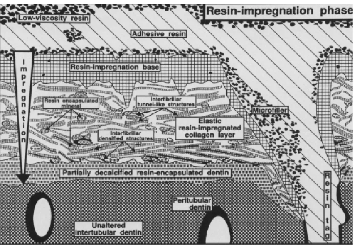
Within the interdiffusion zone, three sublayers were identified (Figs. 1 and 2)
An upper diffuse black layer contained few structural features. Underneath, partially
altered collagen fibrils were closely packed, mostly running parallel with the interface and
perpendicular to the dentinal tubules. At the base of the upper layer, several stained
projections were found to bulge out into the underlying collagen network and appeared to
be confined by obstructive, parallel-running collagen fibrils. Finally, the third dense layer
demarcated the superficially demineralized dentin layer from the deeper unaltered dentin.
Resin diffusion into the decalcified dentin surface layer was evident, but diminished with
depth, presumably reducing deeper resin impregnation into the interfibrillar spaces. The
citric acid applied on dentin probably caused denaturation of the superficial collagen fibrils.
Its decalcifying effect gradually weakened with depth, leaving behind hydroxyapatite
[image:15.612.81.386.449.621.2]crystals at the base of the interdiffusion zone.
particles infiltrated into the IDZ; black arrows= triangular laterally resin impregnated intertubular dentin; and white asterisks= constricted outline of resin tag. The bar represents 5 m. (Taken from Van Meerbeek, 1993)
Figure 2: Schematic representation explaining the ultrastructure of the resin-dentin interdiffusion zone at the resin impregnation phase. (Taken from Van Meerbeek, 1993)
This interdiffusion zone, or hybrid layer, not only provides the retention for the
restoration, but it also seals the dentin surface. This seal is supposed to keep bacteria out
while providing thermal insulation.
For years several authors have been proposing explanations for the postoperative
sensitivity and suggesting methods to avoid or at least diminish it.2, 5, 37-44 Practitioners around the world have been trying them, but the issue of sensitivity is still there. One way
to approach this problem is by using new materials and new adhesives. Self-etch
adhesives have been proposed as a better way to form a hybrid layer without the
undesired effect of sensitivity. Few clinical studies have dealt with sensitivity and the
1.3.2 Total-etch vs. Self-etch
The fifth generation of dental adhesives can be summarized as the etch and bond
technique.26 The fifth generation consists of two different types of adhesive materials: the so-called “one-bottle systems” and the self-etching primer bonding systems.45
To facilitate clinical use, “one-bottle” systems combined the primer and adhesives
into one solution to be applied after etching enamel and dentin simultaneously (the
total-etch wet-bonding technique) with 35 to 37 percent phosphoric acid for 15 to 20 seconds.
These bonding systems create a mechanical interlocking with etched dentin by means of
resin tags, adhesive lateral branches and hybrid layer formation and show high
bond-strength values both to the etched enamel and dentin.46
Watanabe and Nakabayashi developed a self-etching primer that was an aqueous
solution of 20 percent phenyl-P in 30 percent HEMA for bonding to enamel and dentin
simultaneously. The combination of etching and priming steps reduce the working time,
eliminate the washing out of the acidic gel and also eliminate the risk of collagen collapse.
However, the self-etching primer solution also has some disadvantages. For example, the
solution must be refreshed continuously because its liquid formulation cannot be controlled
where it is placed, and often a residual smear layer remained in between adhesive
material and dentin. Also the effectiveness of self-etching primer systems on properly
etching the enamel was less predictable than the result obtained with phosphoric acid
gel.47 Toida advised that removal of the smear layer by a separate etching step before bonding would produce a more reliable and durable bond to dentin. Bond strength tests
made under laboratory conditions often did not demonstrate statistically significant
Leakage tests conducted under laboratory and clinical conditions showed that the seal
achieved at the enamel margins with one-bottle systems is superior to that resulting from
self-etching primer.48
In recent times, several bonding systems were developed and proposed as the sixth
generation of adhesive materials. The sixth generation eliminated the need for the etching
step. These bonding systems achieve a proper bond to enamel and dentin using only one
solution. The first evaluations of these new systems showed a sufficient bond to
conditioned dentin, but not so to enamel. The sixth-generation systems are composed of
an acidic solution that cannot be kept in place and have a pK that is not enough to properly
etch enamel.45
Several studies show that self-etch adhesives achieve similar bond strengths to the
total-etch adhesives. Toledano et al. determined the bond strength of five adhesive
systems to either superficial or deep dentin.49 They used extracted human third molars and had their crowns transversally sectioned either just below the occlusal DEJ or next to the
pulp, to expose flat, superficial or deep dentin surfaces. The surfaces were bonded with: 1)
three 2-step, total-etch, self-priming adhesives (Single Bond, Prime&Bond NT, and Excite),
2) a 2-step, self-etching primer (Clearfil SE Bond), and 3) a single-step, self-etching
all-in-one adhesive (Etch & Prime 3.0) according to manufacturers' directions. Composite
build-ups were constructed incrementally with Z250. Bonded interfaces were examined by TEM.
Nanoleakage was examined using a silver-staining technique. Single Bond, Prime&Bond
NT and Clearfil SE Bond performed equally when bonded to superficial dentin; the lowest
value was obtained with Etch & Prime 3.0. On deep dentin, the highest bond strengths
were attained with Clearfil SE Bond and Prime & Bond NT. Nanoleakage was manifested
Molla et al. compared five 2-step and two 3-step total-etch (TE) bonding systems,
two systems with self-conditioning (SC) primers, and one SC all-in-one adhesive by use of
the microtensile bond test.50 Hybrid resin composites were bonded to the occlusal dentin of 50 extracted human molars. Microtensile bond strength was determined and debonded
surfaces were examined under the SEM for mode of failure. Mean bond strengths of the
simplified (2-step) TE systems (OptiBond Solo, Gluma One Bond, Solobond M,
Prime&Bond NT, One Coat Bond; 19.9 MPa to 39.9 MPa) were not significantly lower than
that of the traditional 3-step TE systems (EBS Multi: 26.0 MPa; OptiBond FL: 32.7 MPa),
and not related to phosphoric acid concentration. Dentin treatment with SC primers
(Clearfil Liner Bond 2: 22.0 MPa; Clearfil Liner Bond 2V: 22.4 MPa) was as effective as
etching with phosphoric acid. The SC all-in-one adhesive (Etch&Prime 3.0: 10.1 MPa)
produced significantly lower bond strength than all other systems evaluated. The authors
concluded that the use of adhesive/composite combinations including simplified bonding
systems does not necessarily result in reduced bond strength to dentin. SC primers (2
bottle self-etch adhesives) offer a promising alternative to phosphoric acid etching as far
as bonding to dentin is concerned. In contrast, the SC all-in-one adhesive evaluated needs
to be improved.
These studies corroborate that the 6th generation adhesives perform in a similar way to 5th generation adhesives. All-in-one adhesive Etch&Prime 3.0 (6th generation) though, has proven to achieve significantly lower bond strengths.
Atash et al. compared the shear and tensile bond strengths of eight adhesive
systems to the enamel and dentin of primary bovine teeth.51 They used two hundred and fifty-six noncarious bovine mandibular primary incisors. The tested adhesives were: Clearfil
(AS); Xeno III (XE); Scotch Bond 1 (SB); Etch & Prime 3.0 (EP); and I Bond (IB). For the
shear bonding test and the tensile bonding test, the labial surfaces of primary incisors were
used. Shear bond strength values ranged from 18.1 to 8.9 MPa on enamel (in decreasing
order, SE, LP, OB, AS, XE, SB, EP and IB) (Table 1), and from 17.8 to 8.2 MPa on dentin
(in decreasing order, SE, SB, OB, AS, XE, LP, IB and EP) (Table 2). Tensile bond strength
values ranged from 13.1 to 6.7 MPa on enamel (in decreasing order, SE, OB, AS, LP, XE,
IB, SB and EP) (Table 3), and from 12.1 to 5.7 MPa on dentin (in deceasing order, SE, SB,
OB, AS, XE, LP, IB and EP) (Table 4). The differences in bond strengths between the
eight systems on enamel and dentin were all statistically significant for both the shear and
tensile bond strengths. The authors found that the highest shear bond strength was
achieved by SE on enamel and dentin, and the lowest by IB on enamel and EP on dentin.
The highest tensile bond strength was obtained by SE on enamel and dentin, and the
lowest by EP. Shear bond strengths were significantly higher on enamel when compared
to dentin for five of the eight adhesives systems, and tensile bond strengths were
significantly higher on enamel when compared to dentin for all but two systems.
Vuu et al. investigated the tensile bond strengths of 37% phosphoric acid/One-Step
Plus (PA, 5th generation) and Tyrian SPE/One-Step Plus (SPE, 6th generation) bonding
systems to human enamel and superficial dentin with 5 composites.52 Buccal and lingual enamel and superficial dentin surfaces of molars were prepared. Five composites were
used for bonding to the teeth with 2 bonding systems in the form of a truncated cone.
Bonding systems were used following manufacturer's instructions. Specimens were
subjected to 1000 thermocycles in 5o and 55oC water with a dwell time of 20 seconds in each temperature bath. Specimens were debonded with a testing machine at 0.5 mm/min.
bonding system in both enamel and dentin with all 5 composites. The SPE bonding system
performed better on enamel than dentin with all 5 composites. Some authors suggest
therefore, the use of acid-etch only on enamel to improve the bond strength of self-etch
adhesives.
Van Landuyt et al. tested the hypothesis that the two-step self-etch adhesive Clearfil
SE Bond (C-SE; Kuraray, Osaka, Japan) bonds equally effectively to enamel/dentin either
with or without prior etching with phosphoric acid. Bur-cut enamel/dentin surfaces prepared
from human molars were partially split in two halves by cutting a shallow groove.53 One half was first etched with 40% phosphoric acid (K-etchant), while protecting the other half
by holding a razor blade in the groove. Next, C-SE was applied strictly following the
manufacturer's instructions, after which the surface was built up using Z100 (3M Espe).
After 24-h water storage, micro-specimens were prepared with the interface circularly
constricted using a Micro-Specimen Former prior to micro-tensile bond strength ( TBS)
(MPa) measurement. In addition, interfaces of C-SE with enamel/dentin prepared with and
without acid etching were examined by Field Emission Gun-Scanning Electron Microscopy
(Feg-SEM) and Transmission Electron Microscopy (TEM). Etching significantly increased
the bonding effectiveness of C-SE to enamel. A clearly more micro-retentive surface was
revealed by TEM and Feg-SEM when enamel was etched. Phosphoric-acid etching prior to
C-SE application on dentin significantly decreased the TBS to dentin. TEM provided
indications of a low-quality hybrid layer with phosphoric-acid etching. Using C-SE,
additional etching with phosphoric acid to improve bonding effectiveness should be limited
to enamel.
Self-etch adhesives do not remove the smear layer completely. The weak acidity of
reach the underlying dentinal structures. Wang and Spencer provided information
regarding morphology, quality and chemistry of the interfaces between three self-etching
primers/adhesives and dentin.54 The occlusal one-third of the crown was removed from 18 human third molars. The prepared dentin surfaces were randomly selected for treatment
with one of three commercial self-etching bonding agents according to manufacturers'
instructions. One 2-step etching adhesive (Clearfil SE Bond) and two 1-step
self-etching adhesives (One-Up Bond F and Prompt L-Pop) were selected. Five-micron-thick
sections of adhesive/dentin interface specimens were cut and stained with Goldner's
trichrome for light microscopy. Companion slabs were analyzed with micro-Raman
spectroscopy and scanning electron microscopy (SEM). It was shown that the difference in
aggressiveness of the three self-etching systems produced a different thickness of hybrid
layer. Staining technique showed a distinct colored line/zone at the adhesive/dentin
interfaces for all three bonding systems. The width of this line varied, and was
approximately 1, 1-2, 2-3 micron for Clearfil SE Bond, One-Up Bond F and Prompt L-Pop,
respectively. The color differences in the stained interface sections, which are reflected by
the extent to which the adhesive encapsulates the demineralized dentin matrix, indicated
that collagen fibrils at the interfaces were not totally encased in all three self-etching
adhesives. Raman results showed that Prompt L-Pop is the most aggressive system in this
study. It almost totally demineralized the 2-micron deep subsurface dentine, while Clearfil
SE is mild, and only partially demineralized the first micron deep dentine. In comparison
with two-step self-etching system, the aggressive one-step system produces more
complex interfaces. It is believed that a part of the smear layer is removed and the
remainder is penetrated through diffusion channels which permits the adhesive to infiltrate
Suppa, in a correlative Field Emission InLens-Scanning Electron microscopy /
Transmission Electron Microscopy (FEISEM/TEM) study found in a TEM of Clearfil SE
Bond, a 1- m-thick, partially demineralized hybrid layer that included loose smear layer
remnants along the surface of the hybrid layer.55
Arrais evaluated the morphology and thickness of the resin-infiltrated dentinal layer
after the application of adhesive systems.57 The dentin-bonding agents were evaluated on flat dentinal preparations confected on the occlusal surfaces of human teeth. The test
specimens were prepared and inspected under scanning electron microscopy at a
magnification of 2,000x. The adhesive systems were responsible for different hybrid layer
thicknesses (p < 0.05), and the mean values were: for Scotchbond MP Plus (SM), 7.41 +/-
1.24 micrometer; for Single Bond (SB), 5.55 +/- 0.82 micrometer; for Etch & Prime 3.0
(EP), 3.86 +/- 1.17 micrometer and for Clearfil SE Bond (CB), 1.22 +/- 0.45 micrometer.
The results suggest that the conventional three-step adhesive system (SM) was
responsible for the thickest hybrid layer, followed by the one-bottle adhesive (SB). The
self-etching adhesives, EP and CB, formed the thinnest hybrid layers. The author
concluded that self-etching adhesives form a much thinner hybrid layer than any of the
total-etch systems.
The studies reviewed showed that the self-etch adhesives, by having less acidity,
penetrate less into the dentin, forming a thinner hybrid layer. This means that the adhesive
has less depth of exposed collagen to penetrate and it is therefore easier to seal than the
acid-etch removed dentinal surface.
1.3.3 Sensitivity
The Total-Etch technique exposes the dentinal tubules after removing the smear
layer. According to the hydrodynamic theory, it is believed that the removal of the smear
plug allows for movement of dentinal fluid within the dentinal tubules, causing sensitivity.59,
60
Alternative explanations were the presence of remaining bacteria7 or the prolonged use of Zinc Oxide Eugenol cements (ZOE-cements)6 as the cause of the post-operative sensitivity. Some authors also believed that the postoperative sensitivity could be related
to cuspal deformation caused by the polymerization shrinkage and composite deformation
under occlusal stress. In 2002, in an opinion article, Christensen stated that the subject of
sensitivity is brought-up in many courses by practitioners. The author calls postoperative
sensitivity an “unpredictable problem” in dentistry that practitioners face despite of
meticulous use of adhesives.
Akpata et al. in 2001 compared objective and subjective assessments of
post-operative sensitivity when class 1 cavities, lined with glass-ionomer or adhesive bonding
system, were restored with resin-based composite (RBC).38 Occlusal cavities on homologous contra-lateral posterior teeth in 44 male patients attending primary health
centers in Riyadh, Saudi Arabia were restored with RBC after a cavity lining of either a
light-cured glass-ionomer cement (Vitrebond) or an adhesive bonding system (One-Step).
Cold response measurements 24 hrs, 7 days and 1 month post-operatively showed that
the threshold of pulpal response was significantly lower (P< 0.05) in the restored teeth
when the adhesive bonding system served as cavity liner. In addition, based on the
patients' subjective assessments, the prevalence of mild or severe post-operative
sensitivity was significantly higher (P< 0.05), 24 hrs and 7 days post-operatively, in the
teeth with the adhesive bonding system as a cavity liner. After a post-operative period of 1
post-operative sensitivity when the restored teeth received a lining of either glass-ionomer
or adhesive bonding system.
Perdigao et al. in 2003 placed 30 restorations with the Clearfil SE (Clearfil SE Bond,
Kuraray America, New York) and 36 restorations with Prime & Bond NT (Dentsply Caulk,
Milford, Del.), which uses 34 percent phosphoric acid to etch enamel and dentin
simultaneously.34 Teeth were restored with the proprietary hybrid resin-based composite indicated for posterior restorations: Clearfil AP-X for Clearfil SE Bond or Esthet-X Micro
Matrix Restorative for Prime & Bond NT. The restored teeth were evaluated preoperatively
and at two weeks, eight weeks and six months postoperatively for sensitivity to cold (ice
stick), compressed air and masticatory forces as the patient’s spoken response to a visual
analog scale from 0 to 10, as well as for marginal discoloration. This study revealed no
statistically significant differences in postoperative sensitivity between the SE and TE
materials at any recall time. Only one tooth displayed sensitivity to occlusal forces at six
months. The authors concluded that the SE adhesive did not differ from the TE adhesive in
regard to sensitivity and marginal discoloration. Perdigao didn’t find a significant difference
between adhesives and concluded that the technique is probably more important than the
adhesive type itself.34
Hayashi et al. in 2003 stated that post-operative sensitivity (POS) may be observed
in recently placed posterior composites. This study examined a retrospective analysis of
the findings of a unique multi-center clinical trial to investigate the five-year risk of failure of
posterior composites with POS and to determine the factors likely to have an important
impact on the prognosis of the restorations. Longitudinal five-year data from the
POS were more likely to have failed at five years than the restorations of Occlusin without
POS, with odds ratios ranging from 1.73 (95% CI: 1.04, 2.87) to 1.97 (95% CI: 1.36, 2.85).
Distributions of patient age and cavity size were significantly different for successful and
failed restorations (chi-square test, p < 0.05). Logistic regression indicated that cavity size
was the only factor likely to have influenced the prognosis of the restorations with POS (p
= 0.041, odds ratio 3.21, 95% CI 1.05: 9.70). Restorations with POS in large cavities were
more likely to have failed by five years than restorations in small cavities. It was concluded
that the restorations with Occlusin included in the Occlusin trial program were more likely
to have failed at five years if POS occurred within one month of placement. Cavity size has
been shown to have been an important factor in the prognosis of Occlusin trial restorations
with POS.
Sarret in 2005, in a review article, stated that the clinical problems related to early
composite materials are no longer serious clinical challenges.62 The author concluded that post-operative sensitivity appears to be more related to the dentin adhesives' ability to seal
open dentinal tubules rather than the effects of polymerization shrinkage on cuspal
deflections and marginal adaptation.
De Souza et al. in 2005, evaluated the clinical performance of two packable and
one microhybrid resin composites in occlusal cavities of posterior permanent teeth after 1
year.63 Sixty occlusal restorations were placed in 18 patients. The restorations were divided into three groups according to the restorative material: G1 (Surefil + Prime&Bond
NT); G2 (Filtek P60 + Singlebond), and G3 (Suprafill + Suprafill). They were placed by two
previously calibrated operators. The restorations were directly evaluated for color
matching, marginal discoloration, secondary caries, wear, marginal adaptation, and
matching; 98.2% for marginal discoloration; 100% for secondary caries; 92.6% for wear;
and 92.6% for marginal adaptation. Postoperative sensitivity was reported in 5% of the
restorations.
Sobral et al. in 2005, evaluated the effects of pre-treatments with a 35%
hydroxyethyl metacrylate/5% glutaraldehyde dentin desensitizer (Gluma Desensitizer) and
a 2% chlorexidine-based cavity disinfectant (Cav-Clean) on postoperative sensitivity.64 Three premolar teeth with no pain symptoms were selected from each one of 17 patients,
totaling 51 teeth, for which Class II restoration using a composite was indicated. Each one
of the three premolar teeth of the same patient was submitted to a different treatment.
After acid etching, only a dental adhesive was applied to the first tooth, which served as
the control. Gluma Desensitizer dentinal desensitizer was applied to the second premolar
tooth prior to applying the dental adhesive. Cav-Clean cavity disinfectant was used on the
third premolar tooth before applying the dental adhesive. All premolar teeth were restored
with a condensable composite. Sensitivity to different stimuli (cold, heat, sweet and dental
floss) was assessed on Day 1, Day 4 and Day 7 by questionnaire following restorative
procedures. The results showed that there was no statistically significant difference in the
three different treatments (P>0.05). Postoperative sensitivity resulting from Class II
restorations using composite resin cannot be completely eliminated with the prior use of a
dentinal desensitizer or a cavity disinfectant. In day-to-day clinical treatment, postoperative
sensitivity may possibly be related to the technique employed.
Sensitivity, being a form of pain, is a subjective experience. The same stimuli that
can elicit a response in one subject can be imperceptible by another. To be able to study
sensitivity, it must be somehow quantified [65]. Several attempts were made in the past to
and vary from a visual analogue scale (VAS), a numerical rating scale from 0 to 10 (NRS),
a verbal rating scale (VRS), the McGill Pain Questionnaire, to the Integrated Pain Score
(IPS) which is an instrument designed at the Pain Therapy and Palliative Care Division of
the National Cancer Institute of Milan to integrate pain intensity and duration in a single
measure.15
1.3.4 Visual Analog Scales
The Visual Analog Scale, originally developed over 70 years ago, is popular for
measuring subjective phenomena. Huskisson used a VAS to measure intensity of pain and
researchers have been using it ever since, to measure pain.69
A Visual Analog Scale is a useful instrument to measure the response to stimuli. It
consists of a 100mm line with a start and end point that are the limits. The start point
means “no pain” and the end point means “severe pain”. The patient being tested is asked
to place a vertical mark on the line indicating the level of the response to the stimulus.
Visual Analog Scales measure the intensity or magnitude of sensations and subjective
feelings, and the relative strength of attitudes and opinions about specific stimuli. The
reliability of the Visual Analog Scale (VAS) has been determined by many authors in
several studies and has been tested many times.16, 70, 71
Holland in a review article published in 1997 reporting the consensus of a
committee of interested persons from academia and industry that convened to discuss the
subject of clinical trials on dentin hypersensitivity stated that sensitivity may be assessed
either in terms of the stimulus intensity required to evoke pain or the subjective evaluation
Price et al. used the Visual Analog Scale to measure sensory and affective
responses to 6 noxious thermal stimuli (43, 45, 47, 48, 49 and 51 degrees C) applied for 5
sec to the forearm by a contact thermode. Sensory VAS and affective VAS responses to
these temperatures yielded power functions with exponents 2.1 and 3.8, respectively;
these functions were similar for pain patients and for volunteers. The power functions were
predictive of estimated ratios of sensation or affect produced by pairs of standard
temperatures (e.g. 47 and 49 degrees C), thereby providing direct evidence for ratio
scaling properties of VAS. VAS sensory intensity responses to experimental pain, VAS
sensory intensity responses to different levels of chronic pain, and direct temperature
(experimental pain) matches to 3 levels of chronic pain were all internally consistent,
thereby demonstrating the valid use of VAS for the measurement of and comparison
between chronic pain and experimental heat pain.71 Internal consistency can be defined as the extent to which tests or procedures assess the same characteristic, skill or quality. It
is a measure of the precision between the observers or of the measuring instruments used
in a study. This type of reliability often helps researchers interpret data and predict the
value of scores and the limits of the relationship among variables.72
In a review of the literature in an article published in 2004 by Coll et al. that included
papers published since 1983, the author found that the definition of pain has been evolving
and so have the methods to measure it.14 This shows the vast array of measurement tools are not consistent and lead to ineffective pain management. VAS was found to be
methodologically sound, conceptually simple, easy to administer and unobtrusive to the
respondent. Hence, it seems to be the most suitable for measuring intensity of pain.
Averbuch in a randomized double-blind naproxen sodium and placebo-controlled
both visual analog and categorical scales simultaneously, found that both appeared as
effective.10 The authors concluded, though, that a combined metric scale for pain measurement that provides the subject with multiple cues may improve communication
and concordance between scales for individual pain determination
The VAS is the most widely used assessment tool in the measurement of pain and
has been widely recommended for the study of pain and sensitivity. It allows the
researcher to quantify a subjective experience and make statistical calculations with the
measurements obtained.
Torabinejad et al. in 2005 used the VAS in a study that compared levels of
postoperative discomfort after cleaning and shaping of root canals using two protocols for
removal of smear layer. Seventy-three consecutive patients requiring root canal treatment
were included.73 At random, canals were cleaned and shaped with one of the following protocols. In group 1, 5.25% sodium hypochlorite was used as the root canal irrigant. The
smear layer was removed by placing 17% EDTA in the canal(s) for 1 min followed by a
5-ml rinse with 5.25% NaOCl. In group 2, canals were irrigated with 1.3% NaOCl; the smear
layer was removed by placing MTAD in the canal(s) for 5 min. Access cavities were closed
with a sterile cotton pellet and Cavit. The patients recorded degree of discomfort at various
time intervals after cleaning and shaping on a visual analogue scale (VAS) for 1 wk. No
significant statistical difference was found in the degree of discomfort between the two
groups (p = 0.58).
Polat et. al. in 2005, in a study to determine the pain sequelae in fixed orthodontic
treatment and to evaluate comparatively the analgesic effects of nonsteroidal
assigned to one of six groups: (1) placebo/placebo, (2) ibuprofen/ibuprofen, (3)
flurbiprofen/flurbiprofen, (4) acetaminophen/acetaminophen, (5) naproxen
sodium/naproxen sodium, and (6) aspirin/aspirin. The pain evaluations were made during
chewing, biting, fitting the front teeth, and fitting the back teeth using a 100-mm visual
analogue scale (VAS) for seven days. All the analgesics succeeded in decreasing the pain
levels compared with the placebo group. However, naproxen sodium and aspirin groups
showed the lowest pain values, and the acetaminophen group showed VAS results similar
to those of the two analgesics.
Burke et al. in 2000, in a study to examine the effectiveness of a dentin bonding
system in the treatment of dentinal hypersensitivity in dental practice conditions used the
VAS.75 Dentists in two dental practices agreed to carry out the project. One practice was in the UK, the other in India. A total of 34 patients who were diagnosed to have dentinal
hypersensitivity were treated using the dentin bonding system. Patients were requested to
record their perception of their pain on a 100mm linear scale, pre-treatment, one day and
one week post-treatment. All patients experienced relief of pain, both 1 day and 1 week
after treatment. Profile plots of the patients' perceived pain scores for the two practices
separately indicated that there was a general trend for these to fall quite sharply one day
after treatment and then generally level out one week post-treatment. There was evidence
indicating a possible difference in pain perception in the two communities from which the
patients were drawn. The author concluded that the dentin bonding system evaluated was
successful in reducing the pain of dentinal hypersensitivity, at least in the short term.
Caselli et al. in 2006, evaluated the postoperative sensitivity of posterior Class I
composite resin restorations, restored with a self-etching or a total-etch one-bottle
patients. Each patient received two restorations. After cavity preparations were completed
under rubber-dam isolation, they were restored using Clearfil SE Bond or Single Bond and
a resin-based restorative material (Filtek Z250). Sensitivity was evaluated at 0 and 7 days
and 6 months using cold stimuli, and recorded using a visual analogue scale. If sensitivity
was experienced on day 7, patients were also contacted on days 14 and 30 to assess the
degree of sensitivity. No statistically significant differences in sensitivity were found
between the two adhesive systems at days 0 and 7 or at 6 months. No spontaneous
postoperative sensitivity was reported. The author reported that the adhesive systems
used in this study showed no differences in postoperative sensitivity, and did not show
spontaneous sensitivity after 6 months.
More specifically, Perdigao in 2003 in a study where teeth were restored with the
proprietary hybrid resin-based composite indicated for posterior restorations and then
tested preoperatively and at two weeks, eight weeks and six months postoperatively for
sensitivity to cold (ice stick), compressed air and masticatory forces, used the Visual
Analog Scale to record the patients response. This study revealed no statistically
significant differences in postoperative sensitivity between the SE and TE materials at any
recall time. Perdigao didn’t find a significant difference between adhesives and concluded
that the technique is probably more important than the adhesive type itself. 34
1.3.5 Ceramic Restorations
The clinical performance of ceramic restorations has been studied by several
authors. The generalized results are that the ceramic inlays and onlays have an excellent
clinical performance.
Coelho Santos in a controlled clinical trial evaluated the clinical performance of
D and pressable (IPS Empress, Ivoclar-Vivadent) after two years. Eighty-six restorations,
44 IPS and 42 D, were cemented into the mouths of 35 patients. Twenty-seven premolars
and 59 molars received Class II preparations totaling 33 onlays and 53 inlays. All
restorations were cemented with dual-cured resin cement (Variolink II, Ivoclar-Vivadent)
and Syntac Classic adhesive under rubber dam. The evaluations were conducted by two
independent investigators at the baseline and after one and two years using the modified
USPHS criteria. After two years, 100% of the restorations were assessed and all the
restorations were considered clinically excellent or acceptable. Among the analyzed
criteria, the following received Bravo ratings: marginal discoloration--IPS (31.82%), D
(23.81%); marginal integrity--IPS (18.18%), D (11.9%), color match-IPS (4.55%), D
(9.52%) and surface texture-IPS (2.27%); D (14.29%). No "Charlie" or "Delta" scores were
attributed to the restorations. The author’s conclusion is that these two types of ceramic
materials demonstrated excellent clinical performance after two years. 77
In 2005, Hayashi, M et al., evaluated the quality of fired feldspathic ceramic inlay
(G-Cera Cosmotech II, GC Co, Tokyo, Japan) after eight years in vivo. 78 Forty-five fired ceramic inlays (for 26 premolars and 19 molars; Class I in 12 teeth, Class II in 31 teeth and
onlay in two teeth) were placed in 25 patients. All restorations were evaluated at the time
of placement and at 6 months, 1, 2, 4, 6 and 8 years after placement using modified
USPHS criteria. Replicas of the restorations were observed with a scanning electron
microscope (SEM) to evaluate the degradation of the marginal area and wear loss of the
restoration. Longevity was observed in 80% of the fired ceramic inlay restorations at eight
years (Kaplan-Meier method), although it was 92% at the six-year observation. Marginal
fracture was detected in 11 restorations (22%), including bulk fracture in five (11%), which
cases and marginal discoloration in 14 (31%). SEM evaluation disclosed marginal
microfractures in 77% of the restorations, wear in 36% and wear of the resin cement along
the margin in 74% at eight years. No significant difference was observed between molars
and premolars. This longitudinal eight-year clinical observation suggested that fired
ceramic inlay restorations made by the G-Cera Cosmotech II system are clinically
acceptable. However, critical failure as bulk fracture may become a future problem since
marginal disintegration was detected in 77% of the restorations from microscopic and
macroscopic perspectives.
El-Mowafy in a review of the literature that only included studies that lasted over 2
years regarding survival of inlays, onlays and crowns made of IPS-Empress cemented with
resin cement, found that the survival for inlays and onlays ranged from 96% at 4.5 years to
91% at 7 years, with most failures being caused by bulk fracture. The survival of crowns
ranged from 92% to 99% at 3-3.5 years, with failure caused by fracture. The author
concluded that dentists should inform their patients about these survival rates when
offering such treatment and that the use of IPS-Empress crowns in the posterior region of
the mouth is not recommended until the results of more long-term clinical trials are
available. 79
Ceramic inlays are a very esthetic alternative to the traditional gold restorations.
Gandjour et al. in a Cochrane review including publications between 1966 and June 2003
that reported annual survival probabilities and annual observations found that
laboratory-fabricated ceramic, chairside CAD/CAM ceramic, and gold inlays had similar failure-free
survival rate, but laboratory-fabricated ceramic inlays had the highest costs and, thus,
laboratory-fabricated ceramic, chairside CAD/CAM ceramic, and gold inlays have “a
strikingly similar” failure-free survival rate.
All-ceramic crowns were always a desirable, yet unreliable, treatment option.
Lehner et al., in a review of articles published between November 1990 and December
1991, stated that despite the good appearance and biocompatibility of dental porcelains,
failures are still of considerable concern because of some limited properties (fracture
toughness) common to all-ceramic crown systems. The author concluded that only
long-term clinical trials will validate achievements compared with other all-ceramic systems and
with well-established metal ceramics. 81
Several materials have been used in search of an esthetic and strong alternative to
metal ceramic restorations. There has been a struggle to satisfy the demand for more
esthetic options for the posterior region and to have long term success. Leucite-reinforced
ceramics, pressable ceramics, etc. have limited success and are recommended for the
esthetic zone only. It was the Alumina (AlO3) and Zirconia (Y-TZP) materials that allowed
the use of all-ceramic crowns with confidence in the posterior region.
Luthard et al. in 2002, in a study to determine if the strength and reliability of yttria
stabilized tetragonal zirconia (Y-TZP) ceramics were affected by the inner surface grinding
of crowns; found that inner surface grinding significantly reduces the strength and reliability
of Y-TZP zirconia compared with the lapped control sample. Co-analysis of flexural
strength, Weibull parameter, and fracture toughness showed counteracting effects of
surface compressive stress and grinding-introduced surface flaws. The authors concluded
that grinding of Y-TZP needs to be optimized to achieve the CAD/CAM manufacture of
Kosmac et al. in 2000, in a study to evaluate the effects of dental grinding and
sandblasting on the biaxial flexural strength and Weibull modulus of various yttria
stabilized tetragonal zirconia (Y-TZP) ceramics containing 3 mol% yttria, found that surface
grinding and sandblasting showed a counteracting effect on the strength of Y-TZP
ceramics. Dental grinding lowered the mean strength and Weibull modulus, whereas
sandblasting provided a powerful method for strengthening. The finest-grained material
exhibited the highest strength after sintering, but it was less damage tolerant than tougher,
coarse-grained materials. Upon extraction with the acetic acid solution and the ammonia
solution, a significant amount of tetragonal zirconia had transformed to monoclinic, but
extensive microcracking and attendant strength degradation had not yet occurred.
Standard grade Y-TZP ceramics are more resistant in an alkaline than in an acidic
environment, and there was a strong grain-size dependence of the diffusion-controlled
transformation. Since a special Y-TZP grade containing a small amount of alumina
exhibited the highest damage tolerance and superior stability in an acidic environment, the
authors concluded that this material shows considerable promise for dental applications. 82,
83
Potiket et al.in 2004, in an in-vitro study using extracted intact human maxillary
incisors, found that there was no significant difference in the fracture strength of the teeth
restored with all-ceramic crowns with 0.4- and 0.6-mm aluminum oxide copings, 0.6-mm
zirconia ceramic copings, and metal ceramic crowns. Forty intact, noncarious human
maxillary central incisors were divided into 4 groups (n=10): Group MCC (control),
metal-ceramic crown (JRVT High Noble Alloy); Group AC4, crown with 0.4-mm aluminum oxide
coping (Procera AllCeram); Group AC6, crown with 0.6-mm aluminum oxide coping
AllZirkon). Teeth were prepared for complete-coverage all-ceramic crowns so that a final
dimension of 5.5 +/- 0.5 mm was achieved incisocervically, mesiodistally, and
faciolingually. A 1.0-mm deep shoulder finish line was used with a rounded internal line
angle. All restorations were treated with bonding agent (Clearfil SE Bond) and luted with
phosphate-monomer-modified adhesive cement (Panavia 21). Fracture strength was
tested with a universal testing machine at a crosshead speed of 2 mm per minute with an
angle of 30 degrees to the long axis of the tooth after restorations were stored in 100%
relative humidity of a normal saline solution for 7 days. The mode of fracture was
examined visually. Means were calculated and analyzed with 1-way ANOVA and Tukey's
HSD (alpha=.05). The means of fracture strength were: Group MCC, 405 +/- 130 N; Group
AC4, 447 +/- 123 N; Group AC6, 476 +/- 174 N; and Group ZC6, 381 +/- 166 N. There was
no significant difference between groups (P =.501). The mode of failure for all specimens
was fracture of the natural tooth. 84 This study shows that all-ceramic crowns are as strong as porcelain-fused-to-metal crowns.
White et al. investigated the strength of a wide variety of layered zirconia and
porcelain beams to determine whether the inclusion of zirconia cores results in improved
strength. Eight types of layered or simple zirconia and porcelain beams (n = 10),
approximately fixed partial denture-size, were made of a tetragonal polycrystalline
zirconium dioxide partially stabilized with yttria core (Lava System Frame) and a
feldspathic dental porcelain (Lava Ceram veneer ceramic). Elastic moduli of the materials
were measured using an acoustic method. Maximum force and modulus of rupture were
determined using 3-point flexural testing and a universal testing machine. Descriptive
statistical methods were used. Beams with porcelain tensile surfaces recorded mean
tensile surfaces recorded moduli of rupture almost an order of magnitude higher, 636 to
786 MPa. The elastic moduli of the porcelain and zirconia materials were 71 and 224 GPa,
respectively. Crack propagation following initial tensile cracking often involved the
porcelain-zirconia interface, as well as bulk porcelain and zirconia. The layered
zirconia-porcelain system tested recorded substantially higher moduli of rupture than have been
previously reported for other layered all-ceramic systems. 81
In 2005, Vult von Steyern performed two simultaneous clinical studies investigating
one alumina-based and one zirconia-based material system. The objective was to
compare the strength of a zirconia system with that of an alumina equivalent with known
long-term clinical performance. 85 The author found that the success rate of the clinical alumina study was 90% after 5 years. After a total of 11 years (+/-1 year), the
success/survival rate was 65%. In the second clinical study, the success rates of the 2-
and 3-year follow-ups were 100%. In the three in-vitro studies, the following results were
found: (1a) the mean flexural strength of the specimens in the group that was exposed to
saliva first after glazing was significantly higher (P < 0.001) than that of the specimens in
the group that was exposed to saliva before glazing, (1b) the FPDs luted on shoulder
preparations resisted higher loads than the FPDs luted on chamfer preparations (P =
0.051), 2) total fractures were more frequent in the alumina than in the zirconia group (P <
0.001), 3) FPDs loaded on implants resisted higher loads (mean = 604 N, SD=184 N ) than
FPDs loaded on abutment teeth (mean= 378 N, SD=152 N, P = 0.003). These studies
justified the use of shorter alumina- (< or = three-unit) and zirconia-based (< or = five-unit)
FPDs as the clinical results are acceptable. The clinical performance of alumina is,
however, not as good as that of comparable high-gold alloy based
fractures) differs from that of zirconia crowns (veneer fractures), suggesting that the
zirconia core is stronger than the alumina core. The consensus is that crowns made with
an aluminous core can be used with confidence to restore teeth in the posterior region.
Zirconia core crowns are even stronger and provide better long term results.
In the proposed study, therefore, crowns with a Zirconia core and pressable ceramic
inlays and onlays were used to test the self-etch, self-cure adhesive.
1.3.6 Clinical evaluation
To evaluate objectively the quality of dental work several methods have been
proposed in the literature. 86-89 To evaluate the clinical performance of the ceramic restorations the most common instrument is the USPHS criteria. The majority of the
studies on clinical performance use it. It was originally developed by Ryge and Snyder in
1973 in an attempt to provide a method for rating the quality of restorations (amalgams
and composites) clinically. Four operational categories are included, 2 satisfactory (Alpha,
Bravo) and 2 not acceptable (Charlie, Delta). 90 The original Ryge criteria have been modified to be used in the evaluation of a variety of restorations. The original categories
Alpha, Bravo, Charlie and Delta, have been further subdivided (i.e. Alpha-1, Alpha-2, etc)
to suit the evaluation needs in many studies.
Perdigao in 2003, in a study where 30 restorations were placed with the Clearfil SE
(Clearfil SE Bond, Kuraray America, New York) and 36 restorations with Prime & Bond NT
(Dentsply Caulk, Milford, Del.), which uses 34 percent phosphoric acid to etch enamel and
dentin simultaneously used a modification of the original USPHS criteria. The restored
teeth were evaluated preoperatively and at two weeks, eight weeks and six months
photographs were taken at baseline and at each recall appointment. The operators
evaluated marginal discoloration at 6 months using this scale: Alpha= no marginal
discoloration; Bravo= slight staining that disappears on polishing; Charlie= discoloration
that penetrates the interface and cannot be polished; Delta= evidence of caries. This study
revealed no clinical signs of marginal degradation at six months. The authors concluded
that the SE adhesive did not differ from the TE adhesive in regards to sensitivity and
marginal discoloration. 34
Kramer et al., in 2005 published a study to clinically evaluate the effect of two
different adhesive/resin composite combinations for luting of IPS Empress inlays. 91 Ninety-four IPS Empress restorations were placed in 31 patients in a controlled
prospective clinical split-mouth study. The restorations were luted with EBS
Multi/Compolute (3M Espe) or with Syntac/Variolink II low (Ivoclar Vivadent) without lining.
The ceramic restorations were examined according to modified USPHS codes and criteria
at baseline and after 0.5, 1, 2, and 4 years. Two patients including four restorations missed
the 4 years recall. After 4 years of clinical service, four restorations in two patients (three
luted with Compolute, one with Variolink II) had to be replaced due to hypersensitivity, 90
inlays and onlays were acceptable. Between the five recalls, a statistically significant
deterioration was found for the criteria marginal adaptation and inlay fracture (Friedman
2-way ANOVA; p<0.05). No statistical difference was found between the adhesives. At
baseline, 95% of the restorations revealed luting composite overhangs. After 4 years, 55%
of cases had overhangs and 38% showed marginal ditching. No differences were found for
surface roughness, color matching, tooth integrity, proximal contact, hypersensitivity, and
between the two luting systems was detectable. The overall failure rate after 4 years was
4%.
In 2004, Santos et al., presented a study to evaluate the clinical performance of
ceramic inlays and onlays made with two systems: sintered (Duceram [D], DeguDent) and
pressable (IPS Empress [IPS], Ivoclar-Vivadent) after 1 year. 92 Seventy-four restorations - 37 IPS and 37 D - were cemented in 34 patients. Twenty-four premolars and 50 molars
received Class II cavity preparations, totaling 28 onlays and 46 inlays. The restorations
were evaluated by two independent investigators at baseline, 6 months and 1 year,
according to modified USPHS criteria. After one year, 100% of the restorations were
assessed and all the restorations were considered clinically excellent or acceptable.
Among the analyzed criteria, only the following received "Bravo" ratings: marginal
discoloration: IPS (24.32%), D (13.51%); marginal integrity: IPS (10.81%), D (8.11%);
color match: IPS (5.41%), D (5.41%); surface texture: IPS (2.70%), D (10.81%). No
"Charlie" or "Delta" scores were given to the restorations. The authors reached to the
following conclusion: only marginal discoloration differed statistically significantly from the
results of the baseline examination for IPS Empress ceramic restorations (p = 0.008). No
significant differences were found between the two ceramics. The two ceramic systems
demonstrated excellent clinical performance after a period of 1 year.
Many other clinical studies have used the modified USPHS criteria for clinical
performance evaluation. 77, 93
In the proposed study, therefore, a modification of the USPHS criteria was used to
1.4 References
1. Buonocuore MG. A simple method of increasing the adhesion of acrylic filling
materials to enamel surfaces. J Dent Res. 1955; 34: 849-53.
2. Christensen GJ. Preventing postoperative tooth sensitivity in class I, II and V
3. Trowbridge HO. Tooth sensitivity associated with the use of luting cements. Penn
Dent J (Phila). 1995; 94(1-2): 4-5, 24-6.
4. Brannstrom M, Johnson G. Movements of the dentine and pulp liquids on
application of thermal stimuli. An in vitro study. Acta Odontol Scand. 1970; 28(1):
59-70.
5. Eick JD, Welch F. Polymerization shrinkage of posterior composite resins and its
possible influence on postoperative sensitivity. Quintessence Int. 1986; 17: 103-11.
6. Kanca J. An alternative hypothesis to the cause of pulpal inflammation in teeth
treated with phosphoric acid on the dentin. Quintessence Int. 1990; 21(2): 83-6.
7. Bergenholtz G. Evidence for bacterial causation of adverse pulpal responses in
resin-based dental restorations. Crit Rev Oral Biol Med. 2000; 11(4): 467-80.
8. Swift EJ Jr. Dentin bonding: what is the state of the art? Compend Contin Educ
Dent. 2001; 22(12 Suppl): 4-7; quiz 18.
9. Holland GR, Narhi M, Addy M, Gangarosa L, Orchardson R. Guidelines for the
design and conduct of clinical trials on dentine hypersensitivity. J Clin Periodontol.
1997; 24(11): 808-13.
10. Averbuch M, Katzper M. Assessment of Visual Analog versus Categorical Scale for
Measurement of Osteoarthritis Pain. J Clin Pharmacol. 2004; 44(4): 368-72.
11. Briggs M, Closs J. A descriptive study of the use of visual analogue scales and
verbal rating scales for the assessment of postoperative pain in orthopedic patients.
J Pain Symptom Manage. 1999; 18(6): 438-46.
12. Choiniere M, Auger F, Latarjet J. Visual analogue thermometer: a valid and useful