Prophylaxis of post-ERCP pancreatitis:
European Society of Gastrointestinal Endoscopy (ESGE)
Guideline
–
Updated June 2014
Authors Jean-Marc Dumonceau1, Angelo Andriulli2, B. Joseph Elmunzer3, Alberto Mariani4, Tobias Meister5, Jacques Deviere6, Tomasz Marek7, Todd H. Baron8, Cesare Hassan9, Pier A. Testoni4, Christine Kapral10
Institutions Institutions are listed at the end of article.
Bibliography
DOIhttp://dx.doi.org/ 10.1055/s-0034-1377875 Published online: 22.8.2014 Endoscopy 2014; 46: 799–815 © Georg Thieme Verlag KG Stuttgart · New York ISSN 0013-726X Corresponding author
Jean-Marc Dumonceau, MD PhD
Gedyt Endoscopy Center Beruti 2347 (C1117AAA) Buenos Aires Argentina Fax: +52886100 jmdumonceau@hotmail.com
Abbreviations
! CT computed tomography DGW double guidewireEPBD endoscopic papillary balloon dilation ESGE European Society of Gastrointestinal
Endoscopy
ERCP endoscopic retrograde
cholangio-pancreatography
NSAID nonsteroidal anti-inflammatory drug
PEP post-ERCP pancreatitis
PGW pancreatic guidewire
RCT randomized controlled trial
SOD sphincter of Oddi dysfunction
SOM sphincter of Oddi manometry
ULN upper limit of normal
1. Introduction
!
The Guideline on prophylaxis of post-ERCP pan-creatitis (PEP) issued by the European Society of Gastrointestinal Endoscopy (ESGE) in 2010 aimed to provide a qualified basis for gastrointestinal endoscopists to take measures to minimize the incidence and severity of PEP [1]. Shortly before the publication of the ESGE Guideline, nonsteroi-dal anti-inflammatory drugs (NSAIDs) were re-portedly rarely used in clinical practice for pre-vention of PEP (16 % of respondents to a survey performed in June 2009), and this was attributed by survey participants to the lack of sufficient data [2]. Similarly, in an Austrian nationwide ERCP survey, PEP prophylaxis was administered This Guideline is an official statement of the European Society of Gastrointestinal Endoscopy (ESGE). It addresses the prophylaxis of post-endoscopic retrograde cholangiopancreatography (post-ERCP) pancreatitis.
Main recommendations
1ESGE recommends routine rectal administra-tion of 100 mg of diclofenac or indomethacin immediately before or after ERCP in all patients without contraindication. In addition to this, in the case of high risk for post-ERCP pancreatitis (PEP), the placement of a 5-Fr prophylactic pan-creatic stent should be strongly considered. Sub-lingually administered glyceryl trinitrate or 250 µg somatostatin given in bolus injection might be considered as an option in high risk cases if nonsteroidal anti-inflammatory drugs (NSAIDs) are contraindicated and if prophylactic pancre-atic stenting is not possible or successful.
2 ESGE recommends keeping the number of
cannulation attempts as low as possible. 3ESGE suggests restricting the use of a pancre-atic guidewire as a backup technique for biliary cannulation to cases with repeated inadvertent cannulation of the pancreatic duct; if this meth-od is used, deep biliary cannulation should be
attempted using a guidewire rather than the contrast-assisted method and a prophylactic pancreatic stent should be placed.
4ESGE suggests that needle-knife fistulotomy should be the preferred precut technique in pa-tients with a bile duct dilated down to the papil-la. Conventional precut and transpancreatic sphincterotomy present similar success and complication rates; if conventional precut is se-lected and pancreatic cannulation is easily ob-tained, ESGE suggests attempting to place a small-diameter (3-Fr or 5-Fr) pancreatic stent to guide the cut and leaving the pancreatic stent in place at the end of ERCP for a minimum of 12–24 hours.
4ESGE does not recommend endoscopic papil-lary balloon dilation as an alternative to sphinc-terotomy in routine ERCP, but it may be advan-tageous in selected patients; if this technique is used, the duration of dilation should be longer than 1 minute.
in only 4.0 % of patients in 2010 and in 7.0 % of patients in 2011 [3]. A more recent survey from the UK found that the proportion of endoscopists using NSAIDs had increased to 34.6 % in 2012 [4]. Obviously, prophylactic measures against PEP are still greatly un-derused in daily clinical practice. At the same time, PEP is still the most frequent and severe complication encountered following ERCP.
New evidence that has become available since the publication of the ESGE Guideline in 2010 is discussed in the present update and new recommendations are issued.
2. Methods
!
ESGE commissioned this update of the Guideline on prevention of PEP. Methods similar to those used in the previous Guideline were applied [1]. A literature search of PubMed/MEDLINE, a search using the Cochrane Library, Embase, and the internet was performed to identify publications since 2009 on this topic. The search focused on fully published prospective studies, particular-ly randomized controlled trials (RCTs) and meta-anaparticular-lyses. Retro-spective analyses and pilot studies were also included if they ad-dressed topics not covered in the prospective studies.
Thereafter, the commissioned authors met once and subsequent-ly developed the updated Guideline. The Guideline committee chairs (C.K., J.M.D.) worked with the subgroup leaders (C.K., T.M., A.A., T.M., P.T., T.B., J.M.D.) who developed draft proposals that were distributed and reviewed electronically. In May 2014, a draft prepared by C.K. and J.M.D. was sent to all group members. After agreement on a final version, the manuscript was sent to all individual ESGE members and individual ESGE member societies, and was reviewed by two experts selected by the ESGE Govern-ing Board. After incorporation of comments, the manuscript was then sent to the journal Endoscopy for publication. The final wording of the Guideline document was agreed by all commis-sioned authors.
3. Definitions
! ▶Statement 2010: None. ▶Statement 2014:Two definitions of PEP may currently be used, neither of these being ideal in the setting of PEP: the consensus definition and grading of severity of PEP according to Cotton et al. and the more recent revised Atlanta international consensus definition and classification of acute pancreatitis.
Background:
The consensus definition and grading of severity of PEP devel-oped by Cotton et al. has been used for > 20 years [5]. It has al-lowed standardized reporting of the incidence and severity of PEP. Post-ERCP pancreatitis was originally defined as“clinical pancreatitis with amylase at least three times the upper limit of normal at more than 24 hours after the procedure, requiring hos-pital admission or a prolongation of planned admission.”Various modifications were introduced by Freeman et al. who proposed using lipase as a possible alternative to amylase and defining clin-ical pancreatitis as“new or worsened abdominal pain,” hence taking into account patients who undergo ERCP in the setting of acute pancreatitis or of a flare of chronic pancreatitis [6]. The grading system for the severity of PEP by the consensus
defini-tion is not ideal as it is mainly based on the length of hospitaliza-tion.
New information since 2009:
The Atlanta classification of acute pancreatitis was updated in 2012 [7]. Although this classification provides clear definitions to classify acute pancreatitis, its limitations include the fact that it was not primarily developed to define PEP. Also, the benefit of a contrast-enhanced computed tomography (CT) scan has not been demonstrated in the setting of PEP (contrast-enhanced CT scan is required if abdominal pain suggests strongly that acute pancreatitis is present, but the serum amylase and/or lipase ac-tivity is less than three times the upper limit of normal [ULN]). According to this classification, the diagnosis of PEP requires two of the three following criteria: (i) abdominal pain consistent with acute pancreatitis (acute onset of a persistent, severe, epi-gastric pain often radiating to the back); (ii) serum lipase or amy-lase activity at least three times greater than the ULN; and (iii) characteristic findings of acute pancreatitis on contrast-en-hanced CT and, less commonly, magnetic resonance imaging or transabdominal ultrasonography. This classification defines three degrees of severity based on the presence or absence of or-gan failure (plus its duration) and of local or systemic complica-tions.
A prospective study has shown the two definitions presented above to be poorly correlated [8].
The Pancreatitis Across Nations Clinical Research and Education Alliance (PANCREA) has defined four degrees of severity for pan-creatitis, based on the presence or absence of complications, both local (necrosis of the pancreas and/or peripancreatic tissue) and systemic (cardiovascular, renal, or respiratory organ failure) [9].
4. Incidence, risk factors, and severity of PEP
!
4.1. Incidence
▶Statement 2010:
Pancreatitis is the most frequent complication after ERCP with an incidence of 3.5 % in unselected patients; it is of mild or moderate severity in approximately 90 % of cases.
▶Statement 2014: No changes. Background:
Data on incidence rate and severity of PEP were mainly based on a systematic review of 21 prospective studies involving more than 16 000 patients [10]. Post-ERCP pancreatitis was found to be the most frequent complication following ERCP, with an inci-dence of 3.47 % (95 % confiinci-dence interval [95 %CI] 3.19 %–3.75 %). Based upon data from studies that have included unselected pa-tients, PEP is mild, moderate, and severe in 45 %, 44 %, and 11 % of cases, respectively. Death occurs in 3 % of cases of PEP (95 %CI 1.65 %–4.51 %) [10].
New information since 2009:
Few new data have become available and these have yielded sim-ilar results. In an ERCP benchmarking program, PEP was reported to occur in 4.2 % of 13 513 unselected procedures [3]. In a retro-spective study of 886 procedures, 39 patients (4.4 %) were diag-nosed with pancreatitis, of mild moderate, and severe type in 69 %, 23 %, and 8 %, respectively [11].
4.2. Risk stratification
4.2.1 Hospital and endoscopist volume for ERCP
▶Statement 2010:There is no evidence that hospital ERCP volume has an influence on the incidence of PEP; data about a potential relationship be-tween PEP incidence and endoscopist case volume are conflict-ing. Low annual case volumes, of endoscopists and centers, are associated with higher ERCP failure rates (Evidence level 2 + ).
▶Statement 2014: No changes. Background:
Factors that may affect the outcome of ERCP that are specifically related to hospital procedure volume include availability of equipment and adequacy of anesthesia, endoscopic and radiolo-gic support, and nursing assistance. The number of ERCPs per-formed in many centers is not as high as commonly believed: in three large (regional or national) studies, the median annual number of ERCPs was between 49 and 235 [12–14]. In one large study, the median annual number of ERCPs per endoscopist was 111 and 40 % of endoscopists performed fewer than 50 sphinc-terotomies/year [15].
Multivariate analyses from two prospective audits performed in England and Italy (66 and 9 centers, respectively) found no signif-icant association between annual hospital volume of ERCPs and incidence of PEP [16, 17].
New information since 2009:
A prospective Swedish study of 12 718 procedures showed no significant difference among PEP rates in centers with low (< 100 ERCPs/year), medium, and high ( > 500 ERCPs/year) volumes [18]. In a prospective multicenter study that included 3635 ERCPs in 11 high volume ( > 200 ERCPs/year) and 10 low volume centers (median of 275 and 45 ERCPs/year, respectively), there was no significant difference in the incidence of PEP (3.9 % vs. 3.1 %) [19]. However, these results were confounded because a higher proportion of patients at high risk of PEP was treated in high vol-ume centers. In this study, the PEP rates did not differ significant-ly between expert and nonexpert operators (3.8 % vs. 5.5 %, respectively;P= 0.34).
4.2.2. Patient- and procedure-related risk factors for PEP
▶Statement 2010:Independent patient-related and procedure-related risk factors for PEP are listed in
●
" Table 1. Risk factors synergistically increase the risk of PEP (Evidence level 1 + ).▶Statement 2014:
Risk factors for PEP, in particular those related to the procedure (cannulation attempts > 10 minutes and pancreatic guidewire passages > 1) have been updated in
●
" Table 1. Risk factors syner-gistically increase the risk of PEP.Background:
Independent risk factors for PEP were presented in a table based on data from a meta-analysis [20] plus those from five prospec-Table 1 Independent risk factors for post-endoscopic retrograde cholangiopancreatography (post-ERCP) pancreatitis (PEP).1 Adjusted odds ratios (95 %
confi-dence intervals in parentheses except where indicated other-wise)
Pooled incidence of PEP in patients with vs. those without risk factor
Patient-related risk factors
Definite risk factors Suspected sphincter of Oddi dysfunction (SOD)
1.91 (1.37–2.65) 8.6 % vs. 2.5 %
Female gender 3.5 (1.1–10.6) 4.0 % vs. 2.1 %*
Previous pancreatitis 2.46 (1.93–3.12) 6.7 % vs. 3.8 %
Likely risk factors
Previous PEP 8.7 (3.2–23.86) 30 % vs. 3.5 %
Younger age Range of odds ratios: 1.09–2.87 6.2 % vs. 2.6 %
Nondilated extrahepatic bile ducts 3.8 % vs. 2.3 %
Absence of chronic pancreatitis 1.87 (1.00–3.48) 4.0 % vs. 3.1 %
Normal serum bilirubin 1.89 (1.22–2.93) 4.15 % vs. 1.43 %
Procedure-related risk factors
Definite risk factors
Cannulation attempts duration > 10 minutes2
1.76 (1.13–2.74) 3.8 % vs. 10.8 %
Pancreatic guidewire passages > 1 2.77 (1.79–4.30) 2.9 % vs. 9.5 %
Pancreatic injection 2.2 (1.60–3.01) 3.3 % vs. 1.7 %
Likely risk factors
Precut sphincterotomy3 2.3 (1.4–3.7) 5.3 % vs. 3.1 %
Pancreatic sphincterotomy 3.07 (1.64–5.75) 2.6 % vs. 2.3 %
Biliary balloon sphincter dilation 4.51 (1.51–13.46) 9.3 % vs. 2.6 %
Failure to clear bile duct stones 3.35 (1.33–9.10) 1.7 % vs. 1.6 %
Intraductal ultrasound (IDUS)4 2.41 (1.33–4.39) 8.37 % vs. 2.76 %
1For definite or likely risk factors, adjusted odds ratios are reproduced either from Masci et al. [20] or from included studies that identified the characteristic as an independent risk factor. Pooled incidences were calculated using figures available in all of the included studies that provided sufficient data for calculation [6, 16, 17, 19, 21, 22, 25–27, 115, 116, 177]. (See text for details about included studies) 2Cannulation attempts of duration > 5 minutes may already increase the incidence of PEP as shown by Halttunen et al. (11.7 % vs. 2.7 %
for cannulation attempts≥5 minutes vs. < 5 minutes, respectively) [115].
3Evidence is growing that precut sphincterotomy is not a definite risk factor for PEP by itself, the increased risk of PEP being related to cannulation efforts that preceded precut [28].
tive, multicenter studies that analyzed potential risk factors for PEP using multivariate analysis [6, 16, 17, 21, 22]. The list was not exhaustive because not all potential risk factors had been ana-lyzed. For example, ampullectomy is generally considered to be a definitive risk factor for PEP on the basis of several small pro-spective studies [23, 24].
As risk factors for PEP were shown to be independent by multi-variate analysis, they might have a cumulative effect. Freeman et al. calculated the adjusted odds ratio (OR) for various combina-tions of risk factors by using data prospectively collected from about 2000 ERCPs: the highest risk of PEP (42 %) was found for fe-male patients with a normal serum bilirubin level, suspected sphincter of Oddi dysfunction (SOD), and difficult biliary cannu-lation [21].
New information since 2009:
Suspected or known dysfunction of the sphincter of Oddi, female gender, younger age, and previous history of pancreatitis are well-known independent risk factors for PEP. A recent prospec-tive study confirmed these findings [25]. In a large ERCP series that included 11 497 procedures, SOD was confirmed as an inde-pendent risk factor [26]. Recently, in a Swedish case–control study of 12 718 ERCP procedures, independent risk factors were young age, female gender, prolonged procedure time, and elec-tive ERCP; whereas rendezvous procedures reduced the risk of PEP [18]. Another prospective multicenter study confirmed inde-pendent procedure- and patient-related risk factors for PEP (> 10 attempts to cannulate the papilla of Vater, OR 14.9; previous PEP, OR 8.7; precut, OR 3.1; pancreatic duct cannulation, OR 2.1) [19]. Some of these predictors were confirmed in two other studies [25, 27]. However, needle-knife sphincterotomy was found not to be an independent risk factor for PEP [3, 25, 28].
The role of intraductal ultrasound as a risk factor for PEP remains unclear but this factor was identified by multivariate analysis (OR 2.41,P= 0.004) in a retrospective analysis of 2364 ERCP proce-dures [29].
No new data have become available regarding the synergistic ef-fect of risk factors for PEP.
4.3. Prediction of PEP
▶Statement 2010:
Serum amylase values less than 1.5 times the ULN, obtained at 2– 4 hours post-ERCP, almost exclude PEP; values more than 3 or 5 times the ULN at 4–6 hours post-ERCP have increasing positive predictive values for PEP (Evidence level 2 + ). It is recommended that serum amylase be determined in patients to be discharged on the day of ERCP; patients with amylase values less than 1.5 times the ULN can be discharged without concern about risk of PEP (Recommendation grade B).
▶Statement 2014:
Serum amylase or lipase values less than 1.5 and 4 times the ULN, respectively, obtained at 2–4 hours post-ERCP have a very high negative predictive value for PEP (Evidence level 2 +). ESGE sug-gests testing serum amylase or lipase 2–6 hours after ERCP in pa-tients presenting with pain and who are to be discharged on the day of ERCP; patients with amylase or lipase values less than 1.5 and 4 times the ULN, respectively, can be discharged without concern about risk of PEP (Recommendation grade B).
Background:
The recommendations were based on five studies that reported similar predictive values based on serum amylase levels obtained 2 to 6 hours following ERCP. In one study of PEP [30], lipase values
at a cutoff of 4 times the ULN had a negative and positive predic-tive value for PEP of 99 % and 15 %, respecpredic-tively.
New information since 2009:
Two studies confirmed previous findings. A prospective study from Brazil that included 300 patients showed that serum hyper-amylasemia < 1.5 times the ULN at 4 hours and < 2 times the ULN at 12 hours had a negative predictive value of 94 % for the devel-opment of PEP [8]. Serum hyperamylasemia following ERCP had a poor positive predictive value for PEP. A retrospective study in-vestigated, in addition to the 4-hour post-ERCP serum amylase level, the impact of having a pancreatogram in predicting PEP among 886 ERCPs [11]; the negative predictive value of serum amylase < 2.5 times ULN for moderate or severe PEP was 99.2 % and 100 %, in patients who, respectively, did and did not have a pancreatogram.
5. Pharmacologic agents available for PEP prophylaxis
!
5.1. Introduction
Post-ERCP pancreatitis appears unavoidable even in the hands of expert endoscopists. Consequently, attempts to reduce the rate of this complication by pharmacological intervention should be pursued. While a few medications have proven effective for pre-venting PEP, we acknowledge that multiple factors in addition to efficacy influence the decision to issue a clinical recommenda-tion. In particular, the magnitude of benefit, as expressed by the number needed to treat (NNT), the robustness and consistency of supporting RCTs, the safety profile of the medication, its ease of administration, availability, and cost were considered in issuing these recommendations.
5.2. Drugs with proven efficacy
5.2.1. Nonsteroidal anti-inflammatory drugs (NSAIDs)
▶Statement 2010:NSAIDs reduce the incidence of PEP; effective PEP prophylaxis has only been demonstrated using 100 mg of diclofenac or indo-methacin administered rectally (Evidence level 1 ++). Routine rec-tal administration of 100 mg of diclofenac or indomethacin im-mediately before or after ERCP is recommended (Recommenda-tion grade A).
▶Statement 2014:
NSAIDs reduce the incidence of PEP in patients at high as well as low risk for PEP; effective PEP prophylaxis has only been demon-strated using diclofenac or indomethacin administered rectally (Evidence level 1 ++ ). ESGE recommends routine rectal adminis-tration of 100 mg of diclofenac or indomethacin immediately be-fore or after ERCP in all patients without contraindication (Re-commendation grade A).
Background:
Three different meta-analyses pooled data from four RCTs that compared rectally administered diclofenac or indomethacin at a dose of 100 mg vs. placebo [31–33]. Two RCTs evaluated the ef-fect of rectal administration of 100 mg diclofenac immediately after the procedure, while the other two evaluated rectal admin-istration of 100 mg indomethacin immediately before the proce-dure. Both schedules showed similar results. Patients who were considered to be at high risk for PEP were included in two stud-ies. Overall, PEP occurred in 4.4 % patients in the treatment groups vs. 12.5 % patients in the placebo groups with an estima-ted pooled relative risk (RR) of 0.36 (95 %CI 0.22–0.60), and an NNT to prevent one episode of PEP of 15. The administration of
NSAIDs was associated with a similar decrease in the incidence of PEP regardless of risk [33]. No adverse events attributable to NSAIDs were reported.
New information since 2009:
Despite previous meta-analytical results, NSAIDs were not com-monly used by endoscopists in clinical practice: only 16 % of re-spondents to a survey performed in June 2009, shortly before publication of the ESGE Guideline, used NSAIDs for PEP prophy-laxis and this low figure was attributed to the lack of sufficient data [2]. A more recent survey (June 2012) found that the propor-tion of endoscopists using NSAIDs has increased to 35 % [4]. In a multicenter RCT [34], patients at high risk for PEP received either a single dose of rectal indomethacin or of placebo immedi-ately after ERCP. A total of 602 patients were enrolled, including 82 % with a clinical suspicion of SOD and 82 % who received pro-phylactic pancreatic stenting based on the high risk of PEP. Post-ERCP pancreatitis developed in 9.2 % vs. 16.9 % of patients in the indomethacin vs. placebo group, respectively (P= 0.005). Of note, moderate-to-severe PEP was less frequent in the indomethacin vs. placebo group (4.4 % vs. 8.8 %, respectively;P= 0.03). The ben-efit of 100 mg rectal indomethacin was also confirmed in an RCT
that included 228 patients but the results were not significantly different from placebo, likely owing to the small sample size [35]. A lower dosage of diclofenac, either 50 mg or 25 mg in patients weighing≥50 kg or < 50 kg, respectively, was compared with pla-cebo in a small RCT from Japan: the PEP incidence was 3.9 % vs. 18.9 % in the diclofenac vs. the control group, respectively (P= 0.017) [36]. A small RCT of 80 patients demonstrated a nonsigni-ficant trend toward benefit associated with the combination of intramuscular diclofenac and intensive intravenous fluid admin-istration [37]. On the other hand, it seems that selective cyclo-oxygenase-2 inhibitor (coxib) administration is ineffective, as shown by an RCT that included 371 patients where intravenous valdecoxib or glyceryl trinitrate (GTN) patch were compared with placebo: the incidence of PEP was similar in all treatment groups (10 %,P= 0.986) [38].
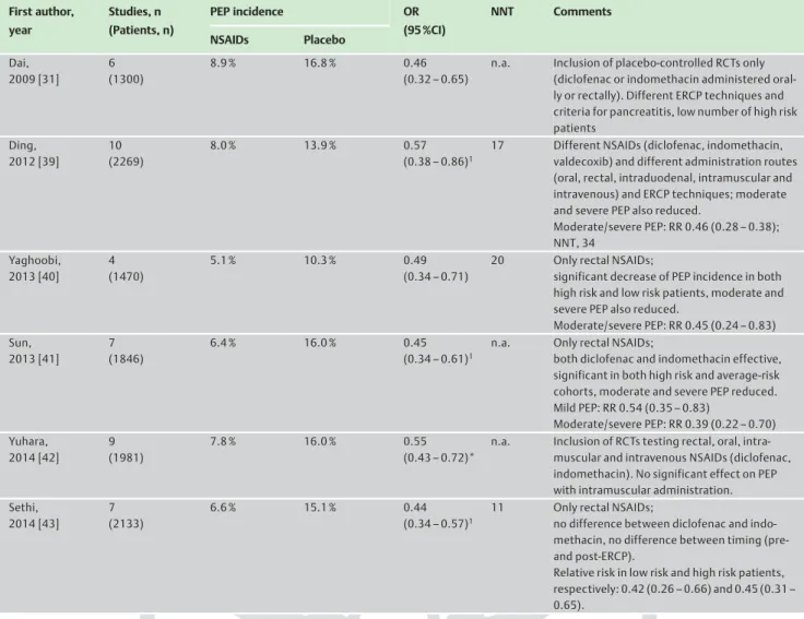
Six meta-analyses published between 2009 and 2014 (
●
" Table 2) compared NSAIDs vs. placebo administration for prevention of PEP, and all of them concordantly showed the benefit of NSAIDs in preventing either mild or moderate/severe PEP. The NNT fig-ures, reported in the majority of these studies, have varied from 11 to 34 [31, 39–43].Table 2 Meta-analyses published in 2009 or later that assessed the effect of NSAIDs on post-endoscopic retrograde cholangiopancreatography (post-ERCP) pancreatitis (PEP).1 First author, year Studies, n (Patients, n) PEP incidence OR (95 %CI) NNT Comments NSAIDs Placebo Dai, 2009 [31] 6 (1300) 8.9 % 16.8 % 0.46 (0.32–0.65)
n.a. Inclusion of placebo-controlled RCTs only (diclofenac or indomethacin administered oral-ly or rectaloral-ly). Different ERCP techniques and criteria for pancreatitis, low number of high risk patients Ding, 2012 [39] 10 (2269) 8.0 % 13.9 % 0.57 (0.38–0.86)1
17 Different NSAIDs (diclofenac, indomethacin, valdecoxib) and different administration routes (oral, rectal, intraduodenal, intramuscular and intravenous) and ERCP techniques; moderate and severe PEP also reduced.
Moderate/severe PEP: RR 0.46 (0.28–0.38); NNT, 34 Yaghoobi, 2013 [40] 4 (1470) 5.1 % 10.3 % 0.49 (0.34–0.71)
20 Only rectal NSAIDs;
significant decrease of PEP incidence in both high risk and low risk patients, moderate and severe PEP also reduced.
Moderate/severe PEP: RR 0.45 (0.24–0.83) Sun, 2013 [41] 7 (1846) 6.4 % 16.0 % 0.45 (0.34–0.61)1
n.a. Only rectal NSAIDs;
both diclofenac and indomethacin effective, significant in both high risk and average-risk cohorts, moderate and severe PEP reduced. Mild PEP: RR 0.54 (0.35–0.83) Moderate/severe PEP: RR 0.39 (0.22–0.70) Yuhara, 2014 [42] 9 (1981) 7.8 % 16.0 % 0.55 (0.43–0.72)*
n.a. Inclusion of RCTs testing rectal, oral, intra-muscular and intravenous NSAIDs (diclofenac, indomethacin). No significant effect on PEP with intramuscular administration. Sethi, 2014 [43] 7 (2133) 6.6 % 15.1 % 0.44 (0.34–0.57)1
11 Only rectal NSAIDs;
no difference between diclofenac and indo-methacin, no difference between timing (pre-and post-ERCP).
Relative risk in low risk and high risk patients, respectively: 0.42 (0.26–0.66) and 0.45 (0.31– 0.65).
NSAIDs, nonsteroidal anti-inflammatory drugs; OR, odds ratio; CI, confidence interval; NNT, number needed to treat; n.a., not available; RCT, randomized controlled trial; RR, relative risk.
In a post hoc analysis of an RCT of NSAIDs vs. placebo for PEP pro-phylaxis, administration of rectal NSAIDs alone was more effec-tive and less costly than prophylactic pancreatic stent placement alone or combined with rectal NSAIDs [44]. However, the authors cautioned that these findings should not change current clinical practice because their post hoc observational study did not pro-duce the same quality of evidence as their RCT, which supported the use of indomethacin in addition to prophylactic pancreatic stenting in high risk cases [34].
5.3. Possibly effective drugs
5.3.1. Somatostatin and octreotide
▶Statement 2010:Based on an ad hoc meta-analysis of results from 10 high quality RCTs, somatostatin proved to be ineffective in preventing PEP (Evidence level 1 ++). We do not recommend universal adminis-tration of prophylactic somatostatin in average-risk patients un-dergoing ERCP (Recommendation grade A). Administration of so-matostatin might be more efficacious using specific dose sche-dules, but caution is needed when interpreting the results of sub-group analyses as they often exaggerate differences between treatments in RCTs.
Octreotide administration did not affect the overall incidence of PEP when data from eight high quality trials were pooled (Evi-dence level 1 ++). Prophylaxis with octreotide is not recommen-ded (Recommendation grade A). In future studies the efficacy of prophylactic administration of octreotide should be evaluated using a dose greater than or equal to 0.5 mg.
▶Statement 2014:
Some meta-analytical results seem to support the benefit of so-matostatin and octreotide for averting PEP but their clinical use cannot be recommended except in well selected cases, owing to discordant data from different routes or dosages and the exces-sively high NNT values (Recommendation grade A).
Background:
Three meta-analyses assessed the effect of somatostatin/octreo-tide for PEP prophylaxis before the 2010 ESGE Guideline publica-tion [1]; an updated meta-analysis of high quality RCTs (Jadad score > 3) was performed for writing the ESGE Guideline. Respec-tively, 10 and 8 RCTs were included for assessing somatostatin and octreotide. Overall, the updated meta-analysis found no sig-nificant reduction of PEP incidence with somatostatin vs. placebo (OR 0.57; 95 %CI 0.32–1.03) or with octreotide vs. placebo (OR 0.73; 95 % CI, 0.41–1.30). However, subgroup analyses suggested that some dosages or administration schedules might be asso-ciated with a protective effect against PEP.
New information since 2009:
A meta-analysis that compared octreotide vs. placebo (18 RCTs, 3171 patients) found no significant difference in PEP incidence (OR 0.77; 95 %CI 0.56–1.05) [45]. However, at post hoc subgroup analysis, dosage seemed to have an impact: when the compound was given at a dose≥0.5 mg (6 RCTs; 1470 patients) the OR for de-veloping PEP dropped to 0.45 (95 %CI 0.28–0.73; NNT = 25), whereas the agent proved ineffective at a dosage < 0.5 mg. Other subgroup analyses of octreotide administration that assessed the route (intravenous vs. subcutaneous) and the schedule (be-fore or after ERCP) were inconclusive.
A second meta-analysis assessed the effect of somatostatin (10 RCTs) and of octreotide (7 RCTs) in a total of 3818 patients [46]. Overall, somatostatin reduced the risk of PEP (RR 0.52; 95 %CI 0.30–0.90), while octreotide was not effective (RR 0.86; 95 %CI 0.45–1.63). Subgroup analyses suggested that higher doses of
so-matostatin (3 mg given as an infusion over 12 hours) or lower do-ses (250 µg) given as bolus injection may be most efficacious, especially for the subgroup of patients at relatively higher risk for PEP, i. e., those undergoing pancreatic duct injection (OR 0.35; 95 %CI 0.15–0.82) and biliary sphincterotomy (OR 0.33; 95 %CI 0.16–0.70). With regard to octreotide, subgroup analysis showed a protective effect when administered at high dose (OR 0.42; 95 %CI 0.20–0.90).
More recently, two RCTs compared the effect of somatostatin vs. placebo for PEP prophylaxis. In one RCT, the efficacy of somatos-tatin alone could not be assessed because the treatment group re-ceived somatostatin plus diclofenac [47]; the other RCT found that high dose somatostatin was associated with a reduction of post-ERCP hyperamylasemia, but not incidence of PEP [48].
5.3.2. Protease inhibitors
▶Statement 2010:Prophylaxis with gabexate or ulinastatin does not reduce the in-cidence of PEP (Evidence 1 ++). Neither drug is recommended for prophylaxis of PEP (Recommendation grade A).
▶Statement 2014:
Some meta-analytical results seem to support the benefit of ga-bexate and ulinastatin at high doses for averting PEP but their clinical use cannot be recommended owing to the discordance of the data. A novel protease inhibitor, nafamostat, is likely effec-tive for preventing PEP in patients at low risk of PEP but not in high risk patients.
Background:
Gabexate for PEP prophylaxis has been evaluated in six high qual-ity RCTs [49–54]; when the results were pooled, no significant difference was found between the control and treatment groups. Ulinastatin for PEP prophylaxis has been compared with placebo (two RCTs) and with gabexate (two RCTs), with contradictory re-sults [55–58]. Nafamostat is a novel protease inhibitor that inhi-bits trypsin, a proteolytic enzyme considered to play an initial role in the pathogenesis of pancreatitis; compared with gabexate its half-life is 20 times longer and its potency 10 to 100 times greater.
New information since 2009:
Four meta-analyses assessed the efficacy of protease inhibitors for PEP prophylaxis [42, 59–61]. Subgroup analysis of 8 high quality (Jadad score > 3) RCTs on gabexate administration showed that the agent was not associated with a decreased risk of PEP (RR 0.64; 95 %CI 0.36–1.13) and there was great heteroge-neity among the studies. Subgroup analysis of 6 ulinastatin RCTs showed the agent was not associated with a decreased risk of PEP in either high quality studies (RR 0.65; 95 %CI 0.33–1.30) or low quality studies (RR 0.75; 95 %CI 0.49–1.16).
The question of drug dosage was addressed in one meta-analysis: when ulinastatin was administered at sufficient doses (≥150 000 units), it averted PEP (OR 0.39; 95 %CI 0.19–0.81; NNT 6); in a similar fashion, gabexate was effective at either slow infusion of high dose (≥150 000 units) (OR 0.44; 95 %CI 0.25–0.79; NNT 7) or rapid infusion of low dose (OR 0.37; 95 %CI 0.20–0.69; NNT 6) [61].
The benefit of nafamostat has been assessed in a meta-analysis that pooled data from 5 RCTs and 2678 patients [42]. This agent lowered the incidence of PEP (RR 0.47; 95 %CI 0.33–0.67). All three RCTs available as full text found nafamostat to be effective in low risk patients but not in high risk patients [62–64]. When the results observed for high risk patients in the three RCTs were pooled, PEP incidence was not statistically different in the
treat-ment vs. the placebo group (8.9 % [32 /358] vs. 13.1 % [37 /283], respectively;P= 0.12).
5.3.3. Drugs influencing sphincter of Oddi pressure
▶Statement 2010:Nitroglycerin reduces the incidence of PEP; however, when ad-ministered transdermally, it is ineffective (Evidence grade 1 ++). Side effects such as transient hypotension and headache may oc-cur. We do not recommend the routine use of nitroglycerin for prophylaxis of PEP (Recommendation grade A).
There is no evidence that…drugs reducing sphincter of Oddi pressure (other than nitroglycerin) [namely, botulinum toxin, epinephrine, lidocaine, and nifedipine]…reduce the incidence of PEP…[cf. section 5.4 below].
▶Statement 2014:
Glyceryl trinitrate (GTN) may be effective in preventing PEP when administered sublingually. Topical epinephrine may be ef-fective to prevent PEP in purely diagnostic ERCP. ESGE does not recommend the routine use of GTN or of epinephrine for PEP pro-phylaxis. No changes concerning botulinum toxin, lidocaine, and nifedipine.
Background:
The influence of GTN on the incidence of PEP was evaluated in two meta-analyses that pooled data from 5 RCTs involving 1662 patients [65, 66]. Both meta-analyses showed an overall signifi-cant reduction of PEP with an RR of 0.61 (95 %CI 0.44–0.86) and an NNT of 26. In the majority of the patients, GTN was adminis-tered transdermally. In a subgroup analysis, transdermal GTN failed to show a significant reduction of PEP (RR 0.66; 95 %CI 0.43–1.01).
Botulinum toxin [67], epinephrine [68], lidocaine [69], and nife-dipine [70, 71], were not found to prevent PEP in the correspond-ing RCTs.
New information since 2009:
A meta-analysis that pooled data from 8 RCTs (1920 patients) found that GTN decreases PEP incidence compared with placebo (5.9 % vs. 9.8 %, respectively;P= 0.002) [72]. A more recent meta-analysis extended the observation to 12 RCTs (2649 patients) and found again that GTN reduces the overall incidence of PEP (RR 0.67; 95 %CI 0.52–0.87) but was, however, ineffective in lowering the incidence of moderate to severe PEP (RR 0.70; 95 %CI 0.42– 1.15) [73]. The route of GTN administration may influence its ef-fectiveness. Subgroup analyses revealed that sublingual adminis-tration of GTN was more effective than transdermal and topical administration (RR 0.47; 95 %CI 0.28–0.78).
The prophylactic merit of topical epinephrine (0.02 %, 20 mL sprayed on the papilla) was compared with placebo in diagnos-tic-only ERCP in two RCTs [68, 74]. Matsushita et al. originally evaluated spraying of epinephrine onto the papilla in 370 pa-tients undergoing diagnostic ERCP [68]; the incidence of PEP was 0 % in the epinephrine group vs. 2.2 % in the control group (not significant). In the second study, Xu et al. randomized 941 patients to topical epinephrine vs. placebo; the rates of PEP were 1.95 % vs. 6.45 %, respectively (P= 0.0086) [74]. When the re-sults of the two studies were pooled in a meta-analysis, topical epinephrine proved efficacious in reducing PEP (OR 0.25; 95 %CI 0.06–0.65; NNT 15) [75]. However, in the two RCTs of epine-phrine, only patients with purely diagnostic ERCP were included, no guidewire was initially used, cannulation times were very long, and the definition of PEP was not standard. Based on these shortcomings, routine use of topical epinephrine cannot be re-commended for PEP prophylaxis.
5.3.4. Antibiotics
▶Statement 2010:Ceftazidime reduced the incidence of PEP in a single study (Evi-dence grade 1–). Further data are needed before recommending ceftazidime for the prophylaxis of PEP (Recommendation grade C).
▶Statement 2014:
Antibiotics have not been proven effective in PEP prophylaxis; further data are needed (Recommendation grade C).
Background:
In an RCT that tested ceftazidime for prophylaxis of PEP, the inci-dence of PEP was lower in the treatment than in the control group (2.6 % vs. 9.4 %, respectively;P= 0.009) [76]. This study was of low methodological quality owing to unclear allocation con-cealment.
New information since 2009:
A network meta-analysis ranked antibiotics in fourth position among 16 drugs for the efficacy of PEP prophylaxis (OR 0.46; 95 %CI 0.15–1.07; NNT 21) [75]. However, the difference ob-served between antibiotics and placebo was not statistically sig-nificant, only 254 patients were included in the treatment arms of four RCTs [76–79] included in the meta-analysis (not 1082 as stated in the meta-analysis), and two of these RCTs did not have PEP prophylaxis as their primary study endpoint [76, 78].
5.3.5. Intensive hydration
▶Statement 2010:No statement.
▶Statement 2014:
In a pilot study, intensive hydration seemed to effectively prevent PEP. Large-scale RCTs to establish an evidence-based approach to intensive hydration are needed.
New information since 2009:
Based on a pilot study in 62 patients, intensive hydration in the periprocedural period with intravenous lactated Ringer’s solu-tion appears to reduce PEP incidence [80]. None of the patients who received aggressive hydration developed PEP, compared with 17 % of patients who received standard hydration (P= 0.016). No patients had evidence of volume overload. Two obser-vational studies support this strategy of hydration for attenuating the severity of PEP [81, 82].
5.4. Drugs proven ineffective
▶Statement 2010:
There is no evidence that glucocorticoids, drugs reducing sphinc-ter of Oddi pressure (other than nitroglycerin), antioxidants, he-parin, interleukin-10, or some anti-inflammatory drugs (other than diclofenac and indomethacin), such as pentoxifylline, sema-pimod, and the recombinant platelet-activating factor acetylhy-drolase, reduce the incidence of PEP (Evidence levels from 1–to 1 ++). None of these drugs is recommended for PEP prophylaxis (Recommendation grade A).
▶Statement 2014:
No change except for GTN and epinephrine (cf. section 5.3.3 above).
Background:
The efficacy of glucocorticoids for PEP prophylaxis was evaluated in two meta-analyses that included 6 RCTs [83, 84]; the incidence of PEP was not significantly different in the glucocorticoids vs. the control group (11.8 % vs. 10.6 %, respectively). Three RCTs that evaluated interleukin-10 have yielded contradictory results [85–87]. Subcutaneous heparin was not found to reduce PEP
in-cidence in 2 RCTs [88, 89]. Drugs potentially reducing the sphinc-ter of Oddi pressure (other than GTN and topical epinephrine), including botulinum toxin, lidocaine, and nifedipine, have not been found to reduce PEP incidence in RCTs [67, 68,90–92]. Three antioxidant agents, namely allopurinol, N-acetylcysteine, and natural beta-carotene were not found to be effective in pre-venting PEP in 5 RCTs and one meta-analysis [93–98].
New information since 2009:
Regarding heparin, a meta-analysis that analyzed four clinical trials (including 3 RCTs) with a total of 1438 patients showed no beneficial effect of prophylactic heparin for the prevention of PEP [99]. Regarding antioxidant supplementation, a meta-analysis of 11 RCTs (total, 3010 patients) showed no beneficial effect on the incidence and the severity of PEP [100]. Regarding allopurinol, an RCT found no effect for the prevention of PEP [101]. No new data are available for glucocorticoids, interleukin-10, pentoxifylline, semapimod, and the recombinant platelet-activating factor acet-ylhydrolase.
6. Pancreatic stent placement for PEP prophylaxis
!
▶Statement 2010:
Prophylactic pancreatic stent placement is recommended to pre-vent PEP in patients who are at high risk for development of PEP. Short, 5-Fr diameter, plastic pancreatic stents are currently re-commended. Passage of the stent from the pancreatic duct should be evaluated within 5 to 10 days of placement and re-tained stents should be promptly removed endoscopically (Evi-dence level 1 + ; Recommendation grade A).
▶Statement 2014:
Prophylactic pancreatic stenting decreases the risk of PEP in high risk and mixed-case groups; it nearly eliminates the risk of severe PEP. 5-Fr pancreatic stents are more efficacious than 3-Fr stents in preventing PEP. ESGE recommends the placement of 5-Fr pan-creatic stents in cases at high risk of PEP. Passage of the stent from the pancreatic duct should be evaluated within 5 to 10 days of placement and retained stents should be promptly removed endoscopically (Evidence level 1 + ; Recommendation grade A). Background:
Two meta-analyses have demonstrated that, in patients at high risk of PEP, prophylactic pancreatic stent placement significantly reduces the incidence of PEP [102, 103]. The OR was 0.44 (95 %CI 0.24–0.81), with an absolute risk reduction of 12.0 % (95 %CI 3.0– 21.0). A multicenter RCT (201 patients) showed a decreased inci-dence of PEP when prophylactic pancreatic stent placement was performed, regardless of the concomitant occurrence of other known risk factors for PEP (PEP incidence in the stent vs. no-stent group, 3.2 % vs. 13.6 %, respectively; P= 0.019) [104]. In these studies, the risk of severe PEP was nearly eliminated following successful placement of a prophylactic pancreatic stent.
Pancreatic stent placement was shown to be cost-effective only in patients at high risk for PEP [105].
New information since 2009:
A meta-analysis of 14 RCTs with a total of 1541 patients showed a significant reduction in the incidence and the severity of PEP when prophylactic pancreatic stenting was used [106]. In addi-tion, subgroup analysis showed that pancreatic stenting reduced the risk of PEP in high risk and mixed-case groups. In a network meta-analysis, prophylactic pancreatic stenting alone was shown to be less effective than NSAIDs alone, and the combination of
NSAIDs with prophylactic pancreatic stenting did not further re-duce the risk of PEP [107].
The ideal stent characteristics for PEP prophylaxis and the opti-mal duration of stent placement are not definitively known. However, a network meta-analysis showed that the probabilities of 5-Fr and 3-Fr stents being ranked as the most efficacious for the prevention of PEP were 96.8 % vs. 3.1 %, respectively, with 5-Fr single-pigtail, unflanged stents and 5-5-Fr straight, flanged stents producing similar results [108]. Furthermore, placement of 5-Fr stents requires fewer guidewires and is easier than that of 3-Fr stents [109, 110]. It is believed that stents need to remain in place for a minimum of 12–24 hours to provide benefit, since removal at the end of ERCP negates the protection from PEP [111] and early outward migration may also result in PEP [112]. Ad-verse events related to attempted prophylactic pancreatic stent-ing include PEP, stent-induced pancreatic ductal damage, and in-ward migration [113]. Removal of proximally migrated small-di-ameter stents can be technically challenging, if not impossible [114].
7. ERCP techniques
!7.1. General considerations
7.1.1. Patient position
▶Statement 2010:There is no evidence that the incidence of PEP is influenced by patient position during ERCP (Evidence level 2 + + ). Therefore, no recommendation is made regarding patient position.
▶Statement 2014: No changes. Background:
Comparisons in RCTs of different patient positions during ERCP showed no significant difference in PEP incidence.
New information since 2009: None.
7.1.2. Cannulation attempts
▶Statement 2010:Trauma resulting from repeated attempts at biliary cannulation has been proven to be a risk factor for the development of PEP (Evidence level 2 + + ). The number of cannulation attempts should be minimized (Recommendation grade B).
▶Statement 2014:
ESGE recommends keeping the number of cannulation attempts as low as possible (Recommendation grade B).
Background:
The risk of PEP is higher after multiple attempts at duct tion [6, 21,115,116]. The rendezvous technique allows cannula-tion trauma to be minimized, and has been used in most studies that evaluated intraoperative endoscopic sphincterotomy. New information since 2009:
A meta-analysis assessed 5 RCTs that compared preoperative vs. intraoperative endoscopic sphincterotomy [117]; PEP was less frequent with the latter approach while the incidence of other ERCP-related complications was similar with both techniques. A nationwide case–control study from Sweden confirmed that the rendezvous technique was associated with a reduction in the risk of PEP compared with standard cannulation techniques, from 3.6 % to 2.2 % (OR 0.5; 95 %CI 0.2–0.9;P= 0.02) [18].
7.1.3. Contrast medium
▶Statement 2010:Injection of contrast medium into the pancreatic duct is an inde-pendent predictor of PEP (Evidence level 1 + ). If pancreatic duct injection occurs incidentally or is required, the number of injec-tions and volume of contrast medium injected into the pancreatic duct should be kept as low as possible (Recommendation grade B). Compared with traditional, high-osmolality contrast agents, low-osmolality contrast agents are costlier but are not associated with reduction in the rates of PEP (Evidence level 1–). The rou-tine use of these agents for ERCP is not recommended (Recom-mendation grade B).
▶Statement 2014: No changes. Background:
In a large meta-analysis, pancreatic duct injection was found to be an independent predictor of PEP (RR 2.2; 95 %CI 1.60–3.01) [20]. In a retrospective study that included more than 14 000 ERCPs, the extent of pancreatic duct injection (head-only vs. head and body vs. injection to the tail) was independently asso-ciated with PEP [118]. The hypothesis that low-osmolality con-trast agents would be less harmful than high-osmolality concon-trast agents because of less important fluid shifts in the pancreas was invalidated in a meta-analysis of 13 RCTs that involved 3381 pa-tients [119].
New information since 2009:
One article examined the role of low-osmolality contrast agents but it does not warrant any change in the Guideline [120].
7.1.4. Carbon dioxide
▶Statement 2010:Use of carbon dioxide (CO2) as a replacement for air for luminal insufflation during ERCP does not influence the incidence of PEP but decreases the incidence and severity of post-procedural ab-dominal pain (Evidence level 1 + ). Carbon dioxide is recommen-ded for insufflation, and might be particularly useful for outpati-ent ERCPs, to reduce post-procedural abdominal pain and to avoid confusion with PEP (Recommendation grade B).
▶Statement 2014: No changes. Background:
Because of its higher solubility in water compared with nitrogen and oxygen, the main components of air, carbon dioxide is cleared from the bowel following endoscopy much faster than air (through the bloodstream and respiration).
New information since 2009:
Three meta-analyses, which included between 5 and 7 RCTs (be-tween 446 and 818 patients), compared carbon dioxide vs. air for gut distension during ERCP exclusively [121–123]. All these meta-analyses found that the use of carbon dioxide reduces post-ERCP abdominal pain without change in other complication rates or in procedure duration. A survey showed that the use of carbon dioxide during endoscopy is uncommon; this may be related to implementation costs and unawareness by endosco-pists of the advantages of carbon dioxide [124].
7.1.5. Cannulation techniques
▶Statement 2010:For deep biliary cannulation, the wire-guided technique reduces the risk of PEP and increases the success rate of primary cannula-tion when compared with the standard contrast-assisted method
(Evidence level 1 + + ). The wire-guided technique is recommen-ded for deep biliary cannulation (Recommendation grade A).
▶Statement 2014: No changes. Background:
The wire-guided biliary cannulation technique entails passage of a guidewire inserted through a catheter (most often a hydrophilic guidewire inserted into a sphincterotome) for deeply cannulating the bile duct. Two meta-analyses published in 2009 showed that, in RCTs, the incidence of PEP was significantly lower with the wire-guided as compared with the standard contrast-assisted cannulation technique [125, 126].
New information since 2009:
Five comparative studies and a meta-analysis comparing the wire-guided vs. the standard contrast-assisted method for selec-tive biliary cannulation were published between 2009 and 2013 [127–132]. Four studies [128–131], two of which were RCTs [128, 131], did not confirm the results of previous meta-analyses that showed a lower risk of PEP with the wire-guided method. In most studies, the wire-guided method shortened cannulation and fluoroscopy times. However, in a recent meta-analysis that extended the analysis to 12 RCTs (3450 patients), the wire-guid-ed method significantly lowerwire-guid-ed the incidence of PEP comparwire-guid-ed with the contrast-assisted method (RR 0.51; 95 %CI, 0.32–0.82) [132]. In addition, the wire-guided cannulation technique was associated with greater primary cannulation success (RR 1.07; 95 %CI 1.00–1.15), fewer precut sphincterotomies (RR 0.75; 95 % CI 0.60–0.95), and no increase in other ERCP-related complica-tions.
7.1.6. Electrosurgical current
▶Statement 2010:The incidence of post-sphincterotomy pancreatitis is not influ-enced by the type of electrosurgical current used (whether pucut or blended) (Evidence level 1 + ). Blended current is re-commended for biliary sphincterotomy, particularly in patients at high risk of bleeding (Recommendation grade A).
▶Statement 2014: No changes. Background:
As pure-cut current produces less edema than blended current [133], it was hypothesized that it might reduce the incidence of PEP after biliary sphincterotomy. A meta-analysis of four RCTs that included 804 patients found no significant difference in the incidence of PEP following the use of pure vs. blended current [134]. However, the incidence of bleeding was significantly high-er when pure-cut current was used.
New information since 2009:
No new evidence has become available.
7.2. Effect of difficult biliary cannulation
7.2.1 Definition
▶Statement 2010: None.▶Statement 2014:
ESGE recommends that future studies define difficult biliary can-nulation in an intact papilla as any of the following: cancan-nulation attempts of duration > 5 minutes, > 5 attempts, or 2 pancreatic guidewire passages.
Background:
Many different definitions of“difficult”biliary cannulation have been used, which make comparisons between studies impracti-cal.
New information since 2009:
In an effort to standardize this definition, Halttunen et al. pro-spectively collected data on 907 biliary cannulations attempted by experienced endoscopists at 10 centers [115]. The authors found the incidence of PEP progressively increased with various factors perceived as causing difficulty in cannulation. Any of the following factors was associated with a PEP incidence of > 10 % during wire-guided cannulation of a native papilla: cannulation attempts of duration > 5 minutes, > 5 attempts, or 2 pancreatic guidewire passages. The latter was also noted in another prospec-tive study [116].
For difficult cannulation, commonly used options include persist-ent attempts at cannulation using standard methods, pancreatic guidewire placement (with biliary cannulation attempted either using a guidewire, the so-called“double guidewire”(DGW) tech-nique, or using contrast medium injection), precut of various types, repeat attempts at ERCP 24–48 hours later, and patient re-ferral to another endoscopist.
7.2.2. Pancreatic guidewire-assisted technique
▶Statement 2010:Data about the usefulness and safety of pancreatic guide wire placement to facilitate biliary cannulation in difficult cases are conflicting. Prophylactic pancreatic stent placement decreases the incidence of PEP with this technique (Evidence level 2 +). Pan-creatic guide wire assistance may facilitate biliary cannulation mostly in the case of inadvertent but repeated cannulation of the pancreatic duct; if this method is used, a pancreatic stent should be placed for PEP prophylaxis (Recommendation grade B).
▶Statement 2014:
In cases of difficult biliary cannulation, pancreatic guidewire (PGW) placement allows biliary cannulation in a proportion of cases similar to persistence in attempting cannulation with standard cannulation techniques (or precut if it is used as a back-up technique), but the risk of PEP is likely higher. In such circum-stances, PEP is effectively prevented by prophylactic pancreatic stenting (Evidence level 1–). ESGE suggests restricting the use of a PGW as a backup technique to cases with repeated inadvertent cannulation of the pancreatic duct; if this method is used, deep biliary cannulation should be attempted using a guidewire rather than the contrast-assisted method and a prophylactic pancreatic stent should be placed (Recommendation grade B).
Background:
In the PGW-assisted technique, a guidewire is inserted in the main pancreatic duct to facilitate deep biliary cannulation by straightening the papillary anatomy and to prevent repeated can-nulation of the pancreatic duct [135, 136]. In two RCTs, compared with persistence in applying the standard cannulation technique, the PGW technique yielded overall similarly low success rates for biliary cannulation (means, 57 % vs. 56 %, respectively) and a non-significantly higher incidence of PEP (means, 14 % vs. 6 %, respec-tively) [137, 138]. Discordances between these RCTs in terms of cannulation success and of PEP incidence may be related to differ-ences in the inclusion criteria (i. e., difficult biliary cannulation alone or combined with repeated unintended pancreatic cannu-lation) and in the use of prophylactic pancreatic stenting.
New information since 2009:
Seven new studies are summarized in
●
" Table 3[139–145]. In two RCTs that compared the PGW vs. the precut techniques [142, 143], success rates of biliary cannulation were similar but, in one of the RCTs [143], the PGW technique was plagued by a higher incidence of PEP (38 % vs. 11 %, respectively;P= 0.01). Pro-phylactic pancreatic stenting was not used in any of the RCTs mentioned above. Another RCT showed that prophylactic pancre-atic stenting significantly decreased the incidence of PEP after the PGW technique had been used [146]. In a retrospective study that included 146 patients, prophylactic pancreatic stenting was always attempted after the PGW technique had been used, and failed prophylactic pancreatic stenting was the only independent predictor of PEP [144]. In another retrospective study that in-volved 142 patients, the incidence of PEP decreased after the au-thors changed their cannulation technique following PGW place-ment, from contrast-assisted to guidewire-assisted biliary can-nulation [147].7.2.3. Precut biliary sphincterotomy
▶Statement 2010:Various techniques of precut biliary sphincterotomy have been described; the fistulotomy technique may present a lower inci-dence of PEP than standard needle-knife sphincterotomy but fur-ther RCTs are required to determine which technique is safer and more effective, based upon the papillary anatomy. There is no evidence that the success and complication rates of biliary precut are affected by the level of endoscopist experience in this tech-nique but published data only report on the experience of one endoscopist (Evidence level 2–). Prolonged cannulation attempts using standard techniques may impart a risk for PEP greater than the precut sphincterotomy itself (Evidence level 2 +). Precut sphincterotomy should be performed by endoscopists with ex-pertise in standard cannulation techniques (Recommendation grade D). The decision to perform precut biliary sphincterotomy, the timing, and the technique, are based on anatomic findings, endoscopist preference, and procedural indication (Recommen-dation grade C).
▶Statement 2014:
In cases of difficult biliary cannulation, early precut is associated with a lower PEP incidence than persistent attempts using the standard approach but the overall success and complication rates are similar with both approaches. Needle-knife fistulotomy seems to be associated with fewer complications, including PEP, compared with other precut techniques.
ESGE suggests that needle-knife fistulotomy should be the pre-ferred precut technique in patients with a bile duct dilated down to the papilla. Conventional precut and transpancreatic sphincterotomy present similar success and complication rates; if conventional precut is elected and pancreatic cannulation is ea-sily obtained, ESGE suggests attempting to place a small-diame-ter (3-Fr or 5-Fr) pancreatic stent to guide the cut and leaving the pancreatic stent in place at the end of ERCP for a minimum of 12–24 hours (Recommendation grade B).
Background:
Precut biliary sphincterotomy can be done by different tech-niques: needle-knife precut starting at the orifice of the papilla (conventional precut), needle-knife above the orifice (fistulot-omy), transpancreatic sphincterotomy (septotomy) and needle-knife over a pancreatic stent. Compared with prolonged attempts at biliary cannulation using standard techniques, the use of
pre-cut sphincterotomy has long been thought to increase the success of biliary cannulation and the incidence of PEP [6, 20].
New information since 2009:
Two meta-analyses investigated the issue of timing of the precut procedure in 6 RCTs [148, 149]. Aside from the definition of early precut, differences between studies included the technique of precut and the randomization ratio (from 1:1 to 1:3). Endosco-pists experienced in precut techniques performed all procedures. The overall incidence of PEP was lower in patients allocated to early precut than to persistence in the use of standard techniques (2.5 % vs. 5.3 %; OR 0.47; 95 %CI 0.24–0.91) but the overall cannu-lation and complication rates were similar in patients allocated to early precut or to persistence in the use of standard cannulation techniques.
In a retrospective study comparing different precut techniques, the risk of PEP was significantly lower after fistulotomy (2.6 %) compared with conventional precut (20.9 %) and pancreatic sep-totomy (22.4 %); the overall complication rate was also signifi-cantly lower after fistulotomy vs. other precut techniques [150]. In another retrospective study of needle-knife fistulotomy per-formed in 204 patients, complications (mostly PEP) progressively increased with decreasing common bile duct (CBD) diameter, up to 14 % in patients with a CBD diameter < 4 mm [151]. Two retro-spective studies compared transpancreatic septotomy and nee-dle-knife precut (with pancreatic stenting or not) for biliary
ac-cess [152, 153]; they found no significant differences in terms of PEP, overall complication and overall success rates.
Needle-knife precut assisted by pancreatic stenting has been pro-posed for reducing PEP. One RCT found that, compared with stent removal at the end of the procedure, leaving the stent for 7–10 days reduces the incidence and severity of PEP (P< 0.05 for both comparisons) [111].
7.3. Specific therapeutic techniques
7.3.1. Balloon dilation as a substitute for endoscopic
sphincterotomy
▶Statement 2010:
Compared with endoscopic sphincterotomy, endoscopic papil-lary balloon dilation (EPBD) using small-caliber balloons (≤10 mm) is associated with a significantly higher incidence of PEP and significantly less bleeding (Evidence level 1 ++). EPBD is not recommended as an alternative to sphincterotomy in routine ERCP but may be useful in patients with coagulopathy and al-tered anatomy (e. g. Billroth II) (Recommendation grade A). If bal-loon dilation is performed in young patients, the placement of a prophylactic pancreatic stent should be strongly considered (Evi-dence level 4; Recommendation grade D).
▶Statement 2014:
Compared with endoscopic sphincterotomy, endoscopic papil-lary balloon dilation (EPBD) using small-caliber balloons (≤10 mm) is associated with more PEP, less bleeding, and fewer late Table 3 New studies evaluating pancreatic guidewire (PGW) placement for biliary cannulation in the case of difficult biliary cannulation.
First author, year Design Patients, n Additional inclusion criteria Pancreatic stenting Successful biliary cannulation rate
PEP rate Note
Xinopoulos, 2011 [139] Retrospective 112 Repeated uninten-tional pancreatic duct cannulation No 44 % (vs. 81 % with
precut in patients who had no repeated un-intentional pancreatic duct cannulation)
6.1 % (similar to group with easy biliary cannula-tion [5.3 %] and to group with precut in place of PGW [7.5 %]) Comparative study Grönroos, 2011 [140] Prospective 50 Repeated uninten-tional pancreatic duct cannulation No 66 % 2 % Authors advocate to shift to other technique if DGW is cumbersome Belverde, 2012 [141] Retrospective 121 Selective pancreat-ic duct cannulation achieved No 97 % 2.6 % Angsuwatcharakon, 2012 [142] RCT 44 No No PGW vs. precut: 74 % vs. 81 % (n.s.) PGW vs. precut: 17.4 % vs. 9.5 % Shorter cannula-tion time with the PGW vs. precut technique (172 s vs. 394 s (P< 0.001) Yoo, 2013 [143] RCT 71 Selective pancrea-tic duct cannula-tion achieved No DGW vs. precut: 91.2 % vs. 91.9 % (n.s.) DGW vs. precut: 38 % vs. 11 % (P= 0.01) Ito, 2013 [144]* Retrospective 146
n.a. Attempted in all PGW: 70 %;
if failure with PGW, DGW was successful in 72 % PGW, 8 %; DGW, 4 % Independent predictor of PEP: failed pancreatic stenting (OR 8.3; 95 %CI 2.3–30) Tanaka, 2013 [145]1 Retrospective 79 Opacification of the pancreatic duct
40 /79 82 % (PGW) vs. 83 %
(DGW)
11 % (PGW) vs. 7 % (DGW)
PEP, post-endoscopic retrograde cholangiopancreatography (post-ERCP) pancreatitis; PGW, pancreatic guidewire; DGW, double guidewire; RCT, randomized controlled trial; n.s., not significant; n.a., not available; OR, odds ratio; CI, confidence interval
1Biliary cannulation attempted by searching the bile duct through contrast medium injection with a guidewire in the main pancreatic duct (PGW technique), as opposed to searching through manipulation of a guidewire inserted in a catheter (DGW technique).
stone recurrences; the risk of PEP is inversely associated with the duration of EPBD (Evidence level 1 ++). ESGE does not recom-mend endoscopic papillary balloon dilation as an alternative to sphincterotomy in routine ERCP, but it may be advantageous in selected patients; if this technique is used, the duration of dila-tion should be longer than 1 minute (Recommendadila-tion grade A). Background:
The use of EPBD may be advantageous compared with endo-scopic sphincterotomy, by decreasing clinically significant bleed-ing in patients with coagulopathy, for preservbleed-ing sphincter of Oddi function in younger patients [154], and in patients with al-tered anatomy (Billroth II) where sphincterotomy is technically difficult. In two meta-analyses, the use of EPBD resulted in a low-er success rate than endoscopic sphinctlow-erotomy for the initial re-moval of biliary stones, with a significantly higher incidence of PEP and significantly lower incidence of bleeding [155, 156]. New information since 2009:
A meta-analysis of 10 high quality RCTs (1451 patients) that com-pared EPBD vs. endoscopic sphincterotomy for biliary stone ex-traction found similar success rates of biliary stone exex-traction with both methods but did not bring new findings with regard to complications [157]; indeed it included fewer RCTs than the previously published Cochrane meta-analysis [156].
A meta-analysis that included 12 RCTs (1649 patients) indica-ted that duration of EPBD is inversely associaindica-ted with the inci-dence of PEP: short (≤1 minute) EPBD was associated with a higher risk of PEP (OR 3.87; 95 %CI 1.08–13.84) compared with endoscopic sphincterotomy (4 RCTs), but long EPBD infla-tion time (> 1 minute) was not (OR 1.14; 95 %CI 0.56–2.35) (6 RCTs) [158]. A lower risk of PEP after long EPBD was also re-ported in the single RCT that compared long (5-minute) vs. short (1-minute) EPBD (RR 0.32; 95 %CI 0.11–0.93) [159]. The authors suggested that the increased risk of PEP after short EPBD might be related to inadequate balloon expansion result-ing in a worsened compression of the pancreatic duct. A meta-analysis that included 3 RCTs (496 patients) with a fol-low-up longer than 1 year showed that stone recurrence was less frequent after EPBD vs. endoscopic sphincterotomy (OR 0.48; 95 %CI 0.26–0.90) [160]. Another RCT that was not included in this meta-analysis and included 474 patients also reported that, in patients with biliary stones≤8 mm, overall late complica-tions and stone recurrence were less frequent after EPBD than after endoscopic sphincterotomy (5.3 % vs. 17.3 %,P= 0.009; 4.4 % vs. 12.7 %;P= 0.048, respectively) (mean follow-up, 55 months); the difference was not significant for patients with stones > 8 mm [161]. A retrospective cohort study with a median follow-up of 92 months also showed a lower incidence of common bile duct stone recurrence after EPBD vs. endoscopic sphincterotomy [162].
7.3.2. Large-balloon dilation for extraction of
difficult biliary stones
▶Statement 2010:
Potential advantages of performing large-balloon dilation in ad-dition to endoscopic sphincterotomy for extraction of difficult biliary stones remain unclear (Evidence level 3). Endoscopic sphincterotomy plus large-balloon dilation does not seem to in-crease the risk of PEP and can avoid the need for mechanical li-thotripsy in selected patients, but not enough data are available to recommend routine use over biliary sphincterotomy alone in conjunction to lithotripsy techniques (Recommendation grade D).
▶Statement 2014:
For the extraction of difficult biliary stones, endoscopic sphinc-terotomy plus large-balloon dilation presents a risk of PEP similar to that of endoscopic sphincterotomy alone; it presents a lower bleeding risk and, possibly, lower overall morbidity and requires less use of mechanical lithotripsy. ESGE suggests performing endoscopic sphincterotomy plus large-balloon dilation in place of endoscopic sphincterotomy alone for the extraction of selected difficult biliary stones (Recommendation grade B).
Background:
In patients with a tapered distal bile duct or large biliary stones, endoscopic sphincterotomy followed by biliary dilation using a large-diameter (12–20 mm) balloon has been proposed to facili-tate stone extraction [163]. This technique has been associated with high success rates for stone extraction without the need for mechanical lithotripsy and with acceptable complication rates in case series [164–167]; the single RCT that had compared this method with endoscopic sphincterotomy at the time of publica-tion of the first Guideline had found no differences in rates of suc-cessful stone clearance, need for mechanical lithotripsy, and complications [168].
New information since 2009:
A meta-analysis of 7 RCTs that included 790 patients found that, compared with endoscopic sphincterotomy alone, endoscopic sphincterotomy followed by large balloon dilation was associated with similar PEP rates but significantly fewer overall complica-tions and significantly less bleeding [169]. However in this meta-analysis, studies which, according to their authors, were retrospective, were considered to be RCTs [170]. Two other meta-analyses that included 3 RCTs and 4–6 retrospective stud-ies (including between 90 and 1295 patients) found similar inci-dences of PEP but significantly lower bleeding risk with endo-scopic sphincterotomy plus large balloon dilation vs. endoendo-scopic sphincterotomy [171, 172]. Additional differences in favor of endoscopic sphincterotomy plus large balloon dilation were mostly attributable to the results of retrospective studies; they included a lower use of mechanical lithotripsy and a lower over-all complication rate [171, 172].
7.3.3. Sphincter of Oddi manometry
▶Statement 2010:In patients undergoing pancreatic sphincter of Oddi manometry, use of the standard perfusion catheter without an aspiration port has been shown to increase the risk of PEP compared with mod-ified water perfusion catheters (Evidence level 2 ++). Pancreatic sphincter of Oddi manometry should be done using a modified triple-lumen perfusion catheter with simultaneous aspiration or a microtransducer catheter (non-water-perfused) (Recommen-dation grade B).
▶Statement 2014:
ESGE recommends that all patients undergoing ERCP for known or suspected sphincter of Oddi dysfunction (SOD) receive rectal NSAIDs combined with pancreatic stenting. Pancreatic sphincter of Oddi manometry should be done using a modified triple-lu-men perfusion catheter with simultaneous aspiration or a micro-transducer catheter (non-water-perfused) (Recommendation grade B).
Background:
It is well documented that patients undergoing ERCP for known or suspected SOD are at high risk of PEP, regardless of whether sphincter of Oddi manometry (SOM) is performed [173]. Biliary SOM does not appear to increase the risk of PEP [173]; pancreatic