Autologous Fat Grafts Harvested and Refined by
the Coleman Technique: A Comparative Study
Lee L. Q. Pu, M.D., Ph.D. Sydney R. Coleman, M.D. Xiangdong Cui, M.D. Robert E. H. Ferguson, Jr., M.D. Henry C. Vasconez, M.D. Sacramento, Calif.; New York, N.Y.; and Lexington, Ky.
Background: The viability of fat grafts obtained by even a well-established tech-nique remains poorly studied and unknown. This study was designed to deter-mine the viability of fat grafts harvested and refined by the Coleman technique. Methods: Sixteen adult white women were enrolled in this study. In group 1 (n⫽8), fat grafts were harvested and processed with the Coleman technique by a single surgeon from the abdomen of each patient according to his standardized method. In group 2 (n ⫽ 8), fat grafts were harvested with the conventional liposuction by another surgeon. After centrifugation, the resulting middle layer of tissue was collected. All fat graft samples were analyzed for the following studies: trypan blue vital staining for viable adipocyte counts, glycerol-3-phophatase dehydrogenase assay, and routine histologic examination. Results: The higher viable adipocyte counts were found in group 1 compared with group 2 (4.11⫾1.11 versus 2.57⫾0.56⫻106cells/ml;p⬍0.004). The level of glycerol-3-phophatase dehydrogenase activity was significantly higher in group 1 compared with group 2 (0.66⫾ 0.09 versus 0.34 ⫾ 0.13 U/ml; p⬍
0.0001). Histologic examination showed normal structure of fragmented fatty tissues in both groups.
Conclusions: Although fat grafts obtained by both methods maintain normal histologic structure, the Coleman technique yields a greater number of viable adipocytes and sustains a more optimal level of cellular function within fat grafts and should be considered superior to conventional liposuction as a preferred method of choice for fat graft harvesting. (Plast. Reconstr. Surg.122: 932, 2008.)
A
utologous fat grafts have been used success-fully for structural fat grafting in facial, lip, and hand rejuvenation and body contour improvement.1–5Most investigators believe that fat,as autologous tissue, can be considered the ideal soft-tissue filler because it is abundant, readily avail-able, inexpensive, host compatible, and can be harvested easily and repeatedly. If fat grafts can truly survive after transplantation, structural fat grafting may present a safe, long-lasting, natural-appearing method for soft-tissue augmentation in
patients.6 –10 However, one of the main concerns
after fat grafting can be the potential high rate of absorption over time in the grafted site, which may reach up to 70 percent of the filled volume.6,7The
most acceptable explanation for absorption has been based on the Peer’s cell survival theory, which states that the number of viable adipocytes at the time of transplantation may correlate with ultimate fat graft survival volume.11
To obtain long-term survival of transplanted autologous fatty tissue, the harvested and pro-cessed fat grafts must remain viable before
im-From the Division of Plastic Surgery, University of Califor-nia, Davis, the Institute of Reconstructive Plastic Surgery, New York University School of Medicine, and the Division of Plastic Surgery, University of Kentucky.
Received for publication December 11, 2007; accepted Feb-ruary 26, 2008.
Presented at the 14th International Congress of the Inter-national Confederation for Plastic, Reconstructive and Aes-thetic Surgery, in Berlin, Germany, June 26 through 30, 2007; and at the 2007 Annual Scientific Meeting of the American Society of Plastic Surgeons, in Baltimore, Mary-land, October 26 through 31, 2007.
Copyright ©2008 by the American Society of Plastic Surgeons
DOI: 10.1097/PRS.0b013e3181811ff0
Disclosure: Dr. Sydney R. Coleman is an unpaid consultant and has stock options for Cytori Thera-peutics and Juniper Medical. He is also an unpaid consultant for Beacon Medical and receives royalties from instruments sold by Mentor and from books sold by Quality Medical Publishing. None of the other authors has any commercial associations or financial interests in any of the drugs, products, or instruments used in this study.
plantation. In 1994, Coleman first described his technique, which uses a syringe, cannula, and cen-trifuge, for structural fat grafting.1He later refined
and popularized his technique for fat graft har-vesting and processing with the Coleman instru-ments and centrifuge and a centrifugation proto-col, often referred to as the Coleman technique.2,3
By using his established technique for fat graft harvesting and processing along with his refined placement technique, many surgeons are able to achieve good long-term results with structural fat grafing.1–5,12
Although several studies were performed in the past searching for improvement of fat transfer,13–15
the viability of fat grafts harvested and processed by even a well-established technique remains poorly studied and unknown. The present study was there-fore designed to evaluate the viability of fat grafts immediately after harvesting and refining by an ex-perienced, reputable surgeon with the Coleman technique using a syringe, cannula, and centrifuge.
MATERIALS AND METHODS
Fat Graft Harvesting and Refining
Sixteen adult white women, aged 23 to 57 years, who had no major systemic metabolic dis-eases or lipid disorders were enrolled in this study, and the study was approved by the university’s institutional review board. In group 1 (n⫽8), fat grafts were harvested with the Coleman technique by a single surgeon (S.R.C.) from the abdomen of each patient according to his well-described and standardized method.1,2,8Briefly, through a small
incision, a mixed solution (0.5% lidocaine with 1:200,000 of epinephrine in lactated Ringer’s so-lution) was infiltrated into the lower abdominal donor site using a blunt Lamis infiltrator (Byron Medical, Inc., Tucson, Ariz.). The solutions were infiltrated at a ratio of 1 cc of solution per cubic centimeter of fat grafts to be harvested. The fat grafts were harvested through the same incisions made previously. The harvesting canula was 3 mm in di-ameter and 15 or 23 cm in length, with a blunt tip (Byron Medical). It was connected to a 10-cc Luer-Lok syringe. Gently pulling back on the plunger of a 10-cc syringe provided a light negative pressure while the cannula was advanced and retracted through the harvested site. After filling the syringe with harvested tissue, the cannula was removed from the syringe. A Luer-Lok plug was twisted onto the syringe to seal the Luer-Lok aperture, and the plunger was removed from the barrel of the sy-ringe and the body of the filled sysy-ringe was placed into a centrifuge (Byron Medical) and spun at
3000 rpm for 3 minutes. After centrifugation, the oil layer (upper level) was decanted and the aqueous layer (lower level) was also drained out of the syringe. The middle layer, composed of predominantly fat grafts (Fig. 1), was studied subsequently.
In group 2 (n⫽8), fat grafts were harvested from the abdomen of each patient according to a standard liposuction method with conventional liposuction (suction-assisted lipectomy) by an-other experienced surgeon (L.L.Q.P.). Through a small incision, a mixed solution (0.5% lidocaine with 1:200,000 of epinephrine in lactated Ringer’s solution) was infiltrated into the lower abdomen. A Byron aspiration cannula (3 or 4 mm in diam-eter) was connected to a liposuction machine (By-ron Medical) and used to harvest adipose aspirates by the surgeon as if one were performing liposuc-tion. The negative pressure of the machine during liposuction was set up at a pressure setting of no greater than 20 cmH2O. The adipose aspirates
(approximately 100 cc) were collected in a bottle at the time of liposuction and transferred imme-diately to the laboratory. The specimens were then spun at 500 rpm for 10 minutes on a large-capacity centrifuge (Mistral 3000i; Curtin Matheson Scien-tific, Inc., Houston, Tex.) to separate adipose tissue from oil and soluble liquid. The middle layer of adipose aspirates after centrifugation, which con-tained more viable adipocytes,16 was taken as fat
grafts for the subsequent comparative studies (Fig. 2). All fat graft samples in this study were analyzed within 30 minutes after they were harvested and processed.
Fig. 1. Fat grafts were harvested and processed with the Coleman technique and spun at 3000 rpm for 3 minutes accord-ing to his protocol. After centrifugation, both upper and lower levels of components were removed and the remaining fat grafts within syringes were studied subsequently.
Evaluation of Fat Grafts Viable Adipocyte Count
One gram of fat graft specimen from each patient was collected in both groups 1 and 2. Each specimen was washed three times with phosphate-buffered saline. It was then mixed with 1 mg/cc of type I collagenase (Sigma, St. Louis, Mo.) in phos-phate-buffered saline containing 5% bovine se-rum albumin (Sigma) for digestion and incubated at 37°C in a carbon dioxide incubator. After 1 hour of incubation, the digestion was terminated with 10% (volume/volume) fetal calf serum (Sigma), and any remaining tissue fragments were removed by straining the digested fatty tissues through a piece of large-weave gauze. The digested fat grafts were fractionated into mature adipocytes (top layer) and stromal pellet (bottom) after a centrif-ugation at 200 g for 10 minutes. The viable adi-pocytes were determined after 0.4% trypan blue vital stain (Sigma) from a 100-l sample with 1:1 dilution with trypan blue. The number of viable adipocytes was then counted with a hemocytom-eter under a microscope with 400⫻magnification.
Glycerol-3-Phosphate Dehydrogenase Assay
Glycerol-3-phophatase dehydrogenase assay was chosen in this study to assess cellular function of fat grafts because it is relatively simple but is adipocyte specific. According to the instructions from the manufacturer (Kamiya Biomedical Co., Seattle, Wash.), glycerol-3-phophatase dehydroge-nase activity within fat grafts was evaluated using a spectrophotometric assay. Briefly, 1 g of fat graft specimen was mixed with 4 ml of 0.25M cane sugar solution and homogenized. The mixture was then
spun at 700 g at 4°C for 10 minutes and the su-pernatant was taken to the special centrifuge tube, which was again spun but at 54,000gfor 60 min-utes. The supernatant obtained after the second centrifugation was diluted approximately 20 to 100 times with an enzyme-extracting reagent. Fi-nal procedures of the assay were as follows: the substrate reagent (400 l) was dispensed into an assay well and heated to 25°C; the diluted super-natant was also heated to 25°C and 200l of it was added to the well and mixed with the substrate reagent; the optical absorption at 340 nm was mea-sured for 3 to 10 minutes and plotted on a graph; the change in optical density per minute from the linear position of the curve was obtained; and glycerol-3-phophatase dehydrogenase activity was calculated based on the formula glycerol-3-phophatase dehydrogenase activity (units per mil-liliter) ⫽ change in optical density at 340 nm/ minute⫻0.482) and the value expressed as units per milliliter.
Histology
Each fat graft specimen (approximately 3 g) was fixed immediately in 10% buffered formalin, concentrated by gravity filtration through a porus paper, processed through graded alcohols and xylene, embedded in paraffin, sectioned at 5m in thickness, and stained with hematoxylin and eosin. All histologic slides were examined by an experienced pathologist in a single-blinded fash-ion for architectural disruptfash-ion, adipocyte degen-eration, or necrosis.
Statistical Analysis
All data in this study are expressed as mean⫾ SD. A two-tailed unpaired t testwas used to assess the difference between the two groups. A value of
p ⬍0.05 was considered statistically significant.
RESULTS
In this study, the total number of viable adi-pocytes was 4.11 ⫾1.11 ⫻106 cells/ml in group
1 and 2.57⫾0.56⫻106cells/ml in group 2. The
higher viable adipocyte count was found in group 1 compared with group 2. The difference of viable adipocyte counts between the two groups was found to be statistically significant (p ⬍0.004).
Glycerol-3-phophatase dehydrogenase assay was used in this study to assess cellular function of fat grafts in each group. The higher the enzyme activity level, the better the cellular function of adipocytes within fat grafts. The glycerol-3-phophatase dehydrogenase activity was 0.66 ⫾ 0.09 U/ml in group 1 and 0.34 ⫾ 0.13 U/ml in
Fig. 2. Fat grafts were harvested with conventional liposuction and spun at 500 rpm for 10 minutes. After centrifugation, the resulting middle layer of adipose aspirates was then studied for comparison.
group 2. The higher level of the enzyme activity was found in group 1 compared with group 2. The difference of glycerol-3-phophatase dehydroge-nase assay between the two groups was found to be significantly significant (p⬍0.0001).
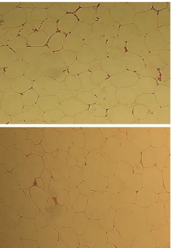
There was no evidence of fatty tissue degen-eration or necrosis in either group. Normal struc-ture of fragment fatty tissues was found primarily, and the basic structure of fragmental fatty tissues appeared to be maintained in both groups. No distinguishable differences were seen histologi-cally in group 1 compared with group 2 (Fig. 3).
DISCUSSION
The results from the present study demon-strated for the first time that there were more viable adipocytes and better cellular function of fat grafts harvested and processed by a well-established tech-nique, the Coleman techtech-nique, than those of fat grafts harvested by conventional liposuction
per-formed by an experienced surgeon and then pro-cessed with centrifugation, a technique used by some surgeons with various modifications.4,12,16 –18
However, fat grafts harvested and processed with both techniques were able to maintain normal histologic structure as fragmental fatty tissues, with no distinguishable differences. The above findings indicate that the Coleman technique may be superior to conventional liposuction for har-vesting and processing fat grafts because the Coleman fat grafts have a greater number of viable adipocytes and sustain more optimal cellular func-tion than fat grafts harvested with convenfunc-tional liposuction performed by an experienced sur-geon. One of the explanations for our findings can be attributed to the less traumatic method of the Coleman technique when it is used to harvest fat grafts because the syringe aspiration can only gen-erate very low but consistent negative pressure. Such a low pressure may minimize the force ex-erted on the adipocytes; therefore, the integrity of these cells within the fat grafts can be preserved.19
In addition, higher negative pressure (20 cmH2O)
generated by conventional liposuction may lead to suboptimal cellular function of adipocytes within fat grafts, even if their structures remain intact.20
Despite a growing interest in autologous fat grafting, there is no consensus and agreement on what is the best technique with which to obtain fat grafts. Based on a recent survey of the members of the American Society for Aesthetic Plastic Surgery, the Coleman technique was the most common method of autologous fat transfer (54 percent), followed by the standard liposuction method (25 percent), other syringe techniques (16 percent), and direct excision (5 percent).12 However, no
previous scientific studies have been conducted to compare the viability of fat grafts obtained by the Coleman technique and conventional liposuc-tion, two of the most common techniques used by plastic surgeons to harvest fat grafts in the United States. The findings from the present study pro-vide scientific epro-vidence that the Coleman tech-nique is a better techtech-nique with which to obtain fat grafts and should be the preferred method in autologous fat grafting.
Other methods have been reported in the liter-ature and used clinically to obtain fat grafts. Har-Shai et al. advocated an integrated approach for increas-ing the survival of autologous fat grafts. In their study, fat grafts were harvested with conventional liposuction and processed by slow centrifugation (200g). The fat grafts after centrifugation were sus-pended with cell culture medium. In 15 patients who were enrolled in the study, the amount of graft taken
Fig. 3. Histologic examination (hematoxylin and eosin stain; original magnification,⫻100) showed primarily normal structure of fragmented fatty tissues, with no obvious differences between the Coleman fat grafts (above) and fat grafts harvested with con-ventional liposuction (below).
in the recipient sites ranged between 50 and 90 percent clinically at 6 to 24 months’ follow-up.19
Ramon et al. reported their preferred technique for obtaining fat grafts. In their study, fat grafts were harvested with conventional liposuction. The fat grafts were then placed on a sterile cotton towel, called the “towel” technique. Significantly less fibro-sis was noted with this technique compared with the centrifugation technique.14Hu et al. reported their
modified technique, including syringe technique for fat graft harvest, separation of adipose tissues with gravity, and additional purification with cotton sticks. They observed good volume maintained after fat injection in a total of 17 patients who were fol-lowed for 13 to 37 months.21However, no study was
conducted comparing their preferred method and the Coleman technique.
The findings from the present study support a previously accepted hypothesis that conventional liposuction may have a more detrimental effect on fat cells than syringe aspiration for fat graft harvest and that fat grafts harvested with conventional liposuction may have fewer viable fat cells.22,23
However, contrary to our findings, Smith et al., in their recent study, concluded that no significant difference in adipocyte viability was observed with regard to harvesting techniques using syringe as-piration and standard liposuction.15Their finding
was supported by a previous study that found that conventional liposuction fat harvest might not re-sult in increased fat cell damage compared with fat harvested by direct excision.24A recent study from
our laboratory found that although fat grafts har-vested by conventional liposuction were able to maintain normal structure with near the same number of viable fat cells compared with fresh fatty tissues harvested by direct excision, they had a less-than-optimal level of cellular function.20
Therefore, controversy remains regarding whether higher suction pressure used in conventional li-posuction can truly cause any significant degree of cellular damage within fat grafts. Further studies may be warranted to determine the effect of higher pressure used during harvest on the via-bility of fat cells within fat grafts.
Because the Coleman technique can be time-consuming and operator dependent for fat graft harvest if it is performed by less experi-enced surgeons, the method may be limited to obtain only a small amount of fat graft. The technique may not be the preferred technique by many surgeons for obtaining a large amount of fat graft for fat grafting to the breasts or buttocks because at least several hundreds cubic centime-ters of fat graft is needed for these procedures.5,27
Some surgeons still routinely harvest fat grafts with liposuction for fat grafting.4,17,18However, those fat
grafts may need to be optimized by further treat-ment with culture medium, insulin, or growth fac-tors to improve their viability before or during implantation.26 –29Nevertheless, the findings from
the present study clearly support the use of the Coleman technique for obtaining fat grafts be-cause the Coleman fat grafts have better viability of adipocytes within harvested fat grafts.
CONCLUSIONS
Our results for the first time demonstrate that although fat grafts obtained by both methods main-tain normal histologic structure, the Coleman tech-nique yields a greater number of viable adipocytes and sustains a more optimal level of cellular func-tion within harvested fat grafts than convenfunc-tional liposuction performed by an experienced sur-geon. Therefore, the Coleman technique should be considered as a standard and preferred method of choice for fat graft harvesting and processing. Future studies may be warranted to possibly de-velop a more speedy technique for obtaining fat grafts or to see whether fat grafts harvested by conventional liposuction can be optimized in vitro before being used as fat grafts for in vivo injection.
Lee L. Q. Pu, M.D., Ph.D.
Division of Plastic Surgery University of California, Davis 2221 Stockton Boulevard, Suite 2123 Sacramento, Calif. 95817 lee.pu@ucdmc.ucdavis.edu
ACKNOWLEDGMENTS
The authors would like to express their appreciation to Michael L. Cibull, M.D., for assistance in histologic study and Christina Chupka for secretarial support in preparation of the article.
REFERENCES
1. Coleman, S. R. Facial recontouring with lipostructure.Clin. Plast. Surg.24: 347, 1997.
2. Coleman, S. R. Hand rejuvenation with structural fat graft-ing.Plast. Reconstr. Surg.11: 1731, 2002.
3. Gatt, J. E. Permanent lip augmentation with serial fat graft-ing.Ann. Plast. Surg.42: 376, 1999.
4. Guerrerosantos, J. Autologous fat grafting for body contour-ing.Clin. Plast. Surg.23: 619, 1996.
5. Roberts, T. L., III, Weinfeld, A. B., Bruner, T. W., and Nguyen, K. “Universal” and ethnic ideals of beautiful but-tocks are best obtained by autologous micro fat grafting and liposuction.Clin. Plast. Surg.33: 371, 2006.
6. Billings, E., Jr., and May, J., Jr. Historical review and present status of free fat graft autotransplantation in plastic and reconstructive surgery.Plast. Reconstr. Surg.83: 368, 1989.
7. Sommer, B., and Sattler, G. Current concepts of fat graft survival: Histology of aspirated adipose tissue and review of the literature.Dermatol. Surg.26: 1159, 2000.
8. Coleman, S. R. Structural fat grafts.Clin. Plast. Surg.28: 111, 2001.
9. Guyuron, B., and Majzoub, R. K. Facial augmentation with core fat graft: A preliminary report.Plast. Reconstr. Surg.120: 295, 2007.
10. Coleman, S. R. Structural fat grafting: More than a perma-nent filler.Plast. Reconstr. Surg.118: 108S, 2006.
11. Peer, L. A. Cell survival theory versus replacement theory. Plast. Reconstr. Surg.16: 161, 1955.
12. Kaufman, M. R., Bradley, J. P., Dickinson, B., et al. Autolo-gous fat transfer national consensus survey: Trends in tech-niques for harvest, preparation, and application, and per-ception of short- and long-term results.Plast. Reconstr. Surg. 119: 323, 2007.
13. Rohrich, R. J., Sorokin, E. S., and Brown, S. A. In search of improved fat transfer viability: A quantitative analysis of the role of centrifugation and harvest site.Plast. Reconstr. Surg. 113: 391, 2004.
14. Ramon, Y., Shoshani, O., Peled, I. J., et al. Enhancing the take of injected adipose tissue by a simple method for concen-trating fat cells.Plast. Reconstr. Surg.115: 197, 2005. 15. Smith, P., Adams, W. P., Jr., Lipschitz, A. H., et al. Autologous
human fat grafting: Effect of harvesting and preparation techniques on adipocyte graft survival.Plast. Reconstr. Surg. 117: 1836, 2006.
16. Boschert, M. T., Beckert, B. W., Puckett, C. L., and Con-cannon, M. J. Analysis of lipocyte viability after liposuction. Plast. Reconstr. Surg.109: 761, 2002.
17. Toledo, L. S. Syringe liposculpture.Clin. Plast. Surg.23: 683, 1996.
18. Murillo, W. L. Buttock augmentation: Case studies of fat injection monitored by magnetic resonance imaging.Plast. Reconstr. Surg.114: 1606, 2004.
19. Har-Shai, Y., Lindenbaum, E. S., Gamliel-Lazarovich, A., Beach, D., and Hirchowitz, B. An integrated approach for
increasing the survival of autologous fat grafts in the treat-ment of contour defects.Plast. Reconstr. Surg.104: 945, 1999. 20. Pu, L. L. Q., Cui, X., Fink, B. F., Cibull, M. L., and Gao, D. The viability of fatty tissues within adipose aspirates after conventional liposuction.Ann. Plast. Surg.54: 288, 2005. 21. Hu, S., Zhang, H., Feng, Y., et al. Introduction of an easy
technique for purification and injection of autogenous free fat parcels in correcting of facial contour deformities.Ann. Plast. Surg.58: 602, 2007.
22. Shiffman, M. A., and Mirrafati, S. Fat transfer techniques: The effect of harvest and transfer methods on adipocyte viability and review of the literature.Dermatol. Surg.27: 819, 2001. 23. Nguyen, A., Pasyk, K. A., Bouvier, T. N., Hassett, C. A., and
Argenta, L. C. Comparative study of survival of autologous adipose tissue taken and transplanted by different tech-niques.Plast. Reconstr. Surg.85: 378, 1990.
24. Lalikos, J. F., Li, Y. Q., Roth, T. P., Doyle, J. W., Matory, W. E., and Lawrence, W. T. Biochemical assessment of cellular dam-age after adipocyte harvest.J. Surg. Res.70: 95, 1997. 25. Coleman, S. R., and Saboeiro, A. P. Fat grafting to the breast
revisited: Safety and efficacy.Plast. Reconstr. Surg.119: 775, 2007.
26. Ullmann, Y., Hyams, M., Ramon, Y., Beach, D., Peled, I. J., and Lindenbaum, E. S. Enhancing the survival of aspirated human fat injected into nude mice.Plast. Reconstr. Surg.101: 1940, 1998.
27. Yuksel, E., Weinfeld, A. B., Cleek, R., et al. Increased free fat-graft survival with the long-term, local delivery of insulin, insu-lin-like growth factor-I, and basic fibroblast growth factor by PLGA/PEG microspheres.Plast. Reconstr. Surg.105: 1712, 2000. 28. Eppley, B. L., Sidner, R. A., Platis, J. M., and Sadove, A. M. Bioactivation of free-fat transfers: A potential new approach to improving graft survival. Plast. Reconstr. Surg.90: 1022, 1992.
29. Shoshani, O., Livne, E., Armoni, M., et al. The effect of interleukin-8 on the viability of injected adipose tissue in nude mice.Plast. Reconstr. Surg.115: 853, 2005.
Membership in the American Society of Plastic Surgeons
For information regarding membership in the American Society of Plastic Surgeons, contact: American Society of Plastic Surgeons
444 E. Algonquin Road Arlington Heights, Ill. 60005 Tel: 847-228-9900