2015-08-04
A Gene-Centered Method For Mapping 3’UTR-RBP Interactions: A
A Gene-Centered Method For Mapping 3’UTR-RBP Interactions: A
Dissertation
Dissertation
Alex M. TamburinoUniversity of Massachusetts Medical School
Let us know how access to this document benefits you.
Follow this and additional works at: https://escholarship.umassmed.edu/gsbs_dissPart of the Computational Biology Commons, Genomics Commons, Molecular Biology Commons, and the Systems Biology Commons
Repository Citation Repository Citation
Tamburino AM. (2015). A Gene-Centered Method For Mapping 3’UTR-RBP Interactions: A Dissertation. GSBS Dissertations and Theses. https://doi.org/10.13028/M2DK5M. Retrieved from
https://escholarship.umassmed.edu/gsbs_diss/793
This material is brought to you by eScholarship@UMMS. It has been accepted for inclusion in GSBS Dissertations and Theses by an authorized administrator of eScholarship@UMMS. For more information, please contact
3’UTR-RBP INTERACTIONS
A Dissertation Presented
By
Alex M. Tamburino
Submitted to the Faculty of the University of Massachusetts Graduate School of Biomedical Sciences, Worcester in partial fulfillment of the requirements for the degree of
DOCTOR OF PHILOSOPHY
August 4, 2015
3’UTR-RBP INTERACTIONS A Dissertation Presented by
Alex M. Tamburino
The signatures of the Dissertation Defense Committee signify completion and approval as to style and content of the Dissertation
Albertha J.M. Walhout, Thesis Advisor Sean P. Ryder, Member of Committee Allan Jacobson, Member of Committee Scot A. Wolfe, Member of Committee Timothy R. Hughes, Member of Committee
The signature of the Chair of the Committee signifies that the written dissertation meets the requirements of the Dissertation Committee
Job Dekker, Chair of Committee
The signature of the Dean of the Graduate School of Biomedical Sciences signifies that the student has met all graduation requirements of the school.
Anthony Carruthers, Ph.D.,
Dean of the Graduate School of Biomedical Sciences Interdisciplinary Graduate Program
I wish to dedicate this dissertation to my mentor, Marian. I rotated in the lab back in 2009 working on a project studying DNA-TF interactions using the yeast one-hybrid system. I didn’t love the project (sorry!) but I loved the lab and I knew that I wanted to join.
I pitched a project to Marian...or more accurately several projects. The first few were duds, but she steered me through the process. Finally I pitched an idea to her that had some merit...or at least lacked glaring flaws. From there she guided me through the early stages of the process and connected me with a committee of advisors who provided the additional scientific knowledge and guidance.
All of these efforts helped get me through the many ups and downs I encountered over the years. She always gave me what I needed to suceed whether it be support, guidance, rigor, confidence or a swift kick in the ass.
I would like to thank everyone who helped me throughout this dissertation. My mentor and thesis research advisory committee were instrumental in providing scientific guidance and knowledge from the earliest days of the project. In particular, Allan Jacobson provided critical suggestions that enabled PRIMA to get off the ground and Sean Ryder provided critical insights that helped me to overcome key obstacles and achieve success in the late stages of the project. Additionally Job Dekker helped inspire me and shape my thought process throughout all stages of the technology development.
I would also like to thank my lab mates and collaborators who contributed to and helped shape this project. In particular, Shaleen Shrestha and Ebru Kaymak contributed to the final stage experiments which helped complete this project. I also appreciated the many insightful discussions with my lab mates. Most notably, I recall many hours of deliberations John Reece-Hoyes, Lesley MacNeil and Juan Fuxman-Bass to whom I am grateful.
Interactions between 3´ untranslated regions (UTRs) and RNA-binding proteins (RBPs) play critical roles in post-transcriptional gene regulation. Metazoan genomes encode hundreds of RBPs and thousands of 3’ UTRs have been experimentally identified, yet the spectrum of interactions between 3´UTRs and RBPs remains largely unknown. Several methods are available to map these interactions, including protein-centered methods such as RBP immunoprecipitation (RIP) and cross-link immunoprecipitation (CLIP), yeast three-hybrid assays and RNAcompete. However, there is a paucity of RNA-centered approaches for assaying an RNA element of interest against multiple RBPs in a parallel, scalable manner.
Here, I present a strategy for delineating protein-RNA interaction networks using a gene centered approach. This approach includes annotating RBPs and identifying physical interactions between an RNA of interest and these RBPs using the Protein-RNA Interaction Mapping Assay (PRIMA). Few RBPs have been experimentally determined in most eukaryotic organisms. Therefore I show that existing RBP annotations can be supplemented using computational predictions of RNA binding domains (RBD) from protein sequences. A single RNA of interest can be tested using PRIMA against a library of RBPs constructed from these annotations. PRIMA utilizes the green fluorescent protein (GFP) in yeast as a reporter.
ends of an mRNA, which increases mRNA stability and enhances translation. PRIMA recapitulates known and uncovers new interactions involving RBPs from human, Caenorhabditis elegans and bacteriophage
with short RNA fragments and full-length 3´UTRs. The development of RBP prey libraries will enable the testing of 3´UTRs against the hundreds of RBPs, which is essential to gain broad insights into post-transcriptional gene regulation at a systems level.
Chapter 1 Introduction………...1 Can we assay a single 3'UTR against a library of RBPs?...7 Protein-centered techniques for identifying 3'UTR -RBP……. interactions………...9 Gene-centered techniques for identifying 3'UTR -RBP…….... interactions………...11
C. elegans as a model to study 3'UTR - RBP interactions….18
How to assay a single 3'UTR against a library of RBPs…….20
Chapter 2 A compendium of C. elegans RNA binding proteins predicts
extensive regulation at multiple levels.………26 wRBP1.0……….28 RNA binding domains (RBDs)...31 RBP-encoding genes are bound by more TFs, more RBPs, and have more splice variants………....35 RBP 3'UTRs are extensively regulated………...39 RBPs are more extensively phosphorylated………...46 Comparison of gene-specific RBPs (Group 1) with TFs…….48 Conclusions………....52 Materials and Methods……….55
Chapter 3 Protein-RNA Interaction Mapping Assay (PRIMA)...101
Cloning of RNA elements and RBPs……….…..122
Yeast manipulations and assay conditions………...125
Data processing and quantitative scoring………126
RNAi and imaging of worm strains………...127
Chapter 4 Technical variations in the development of PRIMA………129
Reporter choice………...129
Detection method………135
Yeast mating type and strains………...142
Conclusion………....145
Chapter 5 Discussion………147
PRIMA’s impact on identifying RNA-RBP interactions……..148
Additional improvements to PRIMA………..149
Potential PRIMA experiments………151
Conclusion………....155
Figure 1.1: Three general states of an mRNA………...3
Figure 1.2: Combinatorial regulation of a 3’UTR by RBPs………...6
Figure 1.3: Comparison of gene- and protein-centered approaches……….8
Figure 1.4: Diagram of Y3H………..13-14 Figure 1.5: Outline of a gene-centered approach to RNA-RBP……….……. interaction mapping………...22-23 Figure 2.1: Venn diagrams……….…30
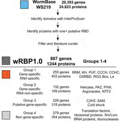
Figure 2.2: wRBP1.0………...32
Figure 2.3: RBPs are extensively regulated by TFs and RBPs…………....37
Figure 2.4: RBPs are extensively regulated through 3'UTRs…………..40-41 Figure 2.5: Quartile binned boxplots of miRNAs targeting RBP 3'UTRs vs. 3’UTRome……….43
Figure 2.6: miRNA targeting……….…..45
Figure 2.7: Normalization of proteomic data………...47
Figure 2.8: RBPs are extensively regulated post-translationally………...49
Figure 2.9: Comparison of gene-specific RBPs (Group 1) to TFs……..50-51 Table S2.1 RBDs, groups and examples………...59-60 Table S2.2 wRBP1.0……….61-99 Figure 3.1 PRIMA design and experimental workflow………...104-106 Figure 3.2 Data filtering example……….…...108
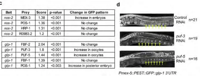
Figure 3.4 Known RNA-RBP interactions can be detected by PRIMA……... ……….……….... 113-115 Figure 3.5 Specific interactions were detected for two full-length 3´UTRs… ………..118-119 Figure 3.6 The glp-1 and nos-2 3´UTRs are regulated by RBPs in the……..
embryo………....121 Figure 3.7 DNA sequences and plasmid configurations used in this……… manuscript………..123 Figure 4.1 PRIMA development flow chart ………...130 Figure 4.2 The LacZ and HIS3 reporters were tested in PRIMA……...134
Figure 4.3 Detection of GFP using fluorescence microscopy…...137-139 Figure 4.4 Detection of GFP using flow cytometry………...140-141 Figure 4.5 The use of diploid and haploid yeast………..….144 Figure 4.6 The use of different strains………...146
3-AT 3-aminotriazole (3-AT) AD Activation domain
APA Alternative polyadenylation
C2H2 Cysteine (2) Histidine (2) zinc finger DNA binding domain CCCH Cysteine (3) Histidine (1) zinc finger DNA binding domain cDNA Complementary DNA
ChIP Chromatin Immunoprecipitation CLIP Cross-link Immunoprecipitation DB DNA binding domain
dsRBD Double stranded RNA binding domain eIF4E Eukaryotic initiation factor 4E
eIF4G Eukaryotic initiation factor 4G EMSA Electrophoretic mobility shift assay FBE FBF binding element
FL1 Fluorescence channel 1 FSC Forward scatter
GFP Green fluorescent protein gs gene specific
H2B Histone 2B
IPTG isopropyl 1-thio-β-D-galactopyranoside KH K homology domain
MCS Multiple cloning site miRNA micro RNA
mRNA messenger RNA
MS2 Bacteriophage MS2 coat protein MS2BS MS2 binding site
NRE NOVA1 recognition element
nt nucleotide
ONPG ortho-Nitrophenyl-β-galactoside ORF Open reading frame
Pab1p Poly(A) binding protein
PAGE Polyacrylamide gel electrophoresis PBS Phosphate buffered saline
PCR Polymerase chain reaction PNA Peptide nucleic acid
PRIMA Protein-RNA interaction mapping assay PUF Pumilio/FBF domain
RBD RNA binding domain RBP RNA binding protein RNAi RNA interference
RPM Rotations per minute RRM RNA recognition motif SAM Sterile alpha motif SCR Spatial control region
SELEX Systematic evolution of ligands by exponential enrichment SLBP Stem-loop binding protein
SSC Side scatter SXL Sex lethal
TCR Temporal control region TF Transcription factor UTR Untranslated region
wRBP1.0 worm RNA binding protein list 1.0
X-gal 5-bromo-4-chloro-3-indolyl-β-d-galactoside Y3H Yeast three-hybrid
CHAPTER I Introduction
Proper gene expression is vital to organismal development and responses to environmental cues. Regulation of gene expression occurs at multiple levels including transcriptionally, post-transcriptionally, and post-translationally. Transcriptional and post-translational regulation of gene expression have long been a focus of genomics and systems biology, however post-transcriptional gene regulation has not received the same attention. Regulation by micro RNAs (miRNAs) is being increasingly studied, but few large scale analyses of messenger RNA (mRNA) regulation by RNA binding proteins (RBP) have been performed to date.
Many aspects of an mRNA’s life cycle are regulated by RBPs including splicing, 3’ end formation, nuclear-cytoplasmic export, localization, translation and stability (Moore, 2005 ; Glisovic et al 2008; Martin and Ephrussi, 2009 ; Mitchell and Parker, 2014). The current understanding is that mRNAs do not exist alone, but rather as ribonucleoprotein complexes (RNPs) (Keene 2007, Mitchell and Parker 2014). At the onset of transcription an mRNA receives a 7-methyl-guanosine cap at their 5’ end and is subsequently bound by
eIF4E, the cap binding protein. The elongating transcript then interacts with additional RBP and RNP complexes including splicing proteins, small nuclear RNPs, exon junction proteins, and polyadenylation factors (Moore 2005). In most cases, the mRNA is polyadenylated at its 3’ end and subsequently bound by the polyA binding protein, Pab1p (Mangus et al. 2003).
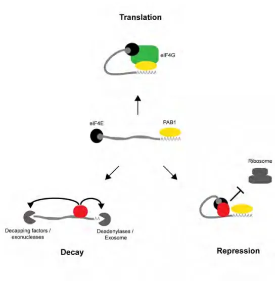
In the cytoplasm, mRNAs primarily exist in three general states: active translation, repression or degradation (Buchan and Parker 2009). The circularization of the 5’ and 3’ ends of the mRNA serves to promote active translation and prevent degradation of the transcript (Wells et al.
1998, Coller et al. 1998, Gray et al. 2000). The 5’ and 3’ ends are bound by eIF4E and Pab1p, respectively. Both bind the scaffold protein, eIF4G, thereby circularizing the transcript and promoting translation through enhanced ribosome binding (Wells et al. 1998). Further, the closed-loop scaffold provides a framework for the regulation of translation and decay by trans-acting RBPs (Szostak and Gebauer, 2013) (Figure 1.1).
RBPs can regulate the mRNA state by affecting ribosome recruitment, initiation and elongation (repression). During Drosophila
melanogaster embryogenesis translation of many mRNAs is repressed spatially to create asymmetric protein distributions (Nakamura et al. 2004,
Figure 1.1: Three general states of an mRNA. In the cytoplasm, mRNAs primarily exist in three general states: active translation, repression or decay. The mRNA is bound at the 5’ and 3’ ends by eIF4E and Pab1p, respectively (center). Formation of the closed-loop by eIF4E results in efficient translation (top). RBPs which block this interaction result in repression of ribosomal recruitment (right). RBPs which remove the polyA tail and/or cap result in RNA decay (left).
Cho et al. 2005, Beckmann et al. 2005). For instance, oskar mRNA is regulated through its 3’ untranslated region (UTR) by the protein Bruno. Bruno interacts directly with eIF4E thereby disrupting the translation competent form of the closed-loop (Nakamura et al. 2004). The caudal mRNA is repressed in a related manner by Bicoid which binds the 3'UTR and recruits d4EHP. This paralog of eIF4E binds the 5’ cap but does not associate with the eIF4G scaffold protein (Cho et al. 2005). Lastly, the msl-2 mRNA is inhibited by Sex-lethal (SXL) through dual mechanisms. SXL binds to the 3'UTR of msl-2 and prevents binding of the 43S ribosomal preinitiation complex to the mRNA. Complexes which escape this first level of repression are then blocked from scanning to the downstream translation initiation codon (Beckmann et al. 2005).
RBPs can regulate the mRNA state by recruiting deadenylation and decapping factors that disrupt the closed-loop and destabilize the transcript (decay). Entry into mRNA decay typically begins with shortening of the poly(A) tail and removal of the 5’ cap followed by exonuclease degradation at the 5’ and 3’ ends (Garneau et al. 2007). In Saccharomyces cerevisiae , HO mRNA is regulated by two different RBPs, Puf4p and Puf5p (Hook et
al. 2007, Chritton et al. 2010). The RBPs bind to separate elements in the HO 3'UTR and decrease HO mRNA stability by promoting deadenylation and disruption of the closed-loop (Hook et al. 2007). In particular, Puf5p
recruits the deadenylase Ccr4p (via Pop2p) and the decapping factor Dhh1p (via Eap1p) (Goldstrohm et al. 2007, Blewett and Goldstrohm 2012). Additionally, Puf5p represses translation in vitro independent of its effects on HO stability (Chritton et al. 2010).
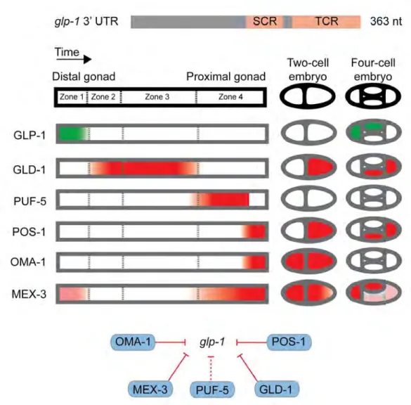
C. elegans glp-1 illustrates the regulation of a single 3'UTR by many RBPs. GLP-1 is a conserved protein that is essential for germline development. glp-1 mRNA is present throughout development, however GLP-1 protein is only expressed at distinct points (Crittenden et al. 1994) (Figure 1.2). The glp-1 3’UTR was determined sufficient for patterning of GLP-1 expression and subsequent mutation of the 3'UTR determined there were two regions which exhibited control over the temporal (TCR) and spatial (SCR) expression patterns (Evans et al.1994).
The regulation of glp-1 3'UTR elements by several RBPs was discovered through a combination of approaches. GLD-1 and POS-1 were postulated to be regulators based on correlative expression patterns and phenotypes (Marin and Evans 2003, Ogura et al. 2003). SPN-4 was suspected based on its physical association with POS-1 (Ogura et al.
2003). MEX-3, MEX-5 and OMA-1 were associated with glp-1 through ‘protein-centered’ studies where the RBP was in focus (Pagano et al. 2007, 2009, Kaymak et al. 2013). In contrast, a large-scale ‘gene-centered’ screen focusing on the glp-1 3’UTR associated PUF-5
Figure 1.2: Combinatorial regulation of a 3’UTR by RBPs. The glp-1 3’UTR contains two cis-regulatory regions, SCR and TCR. Embryogenesis proceeds in an orderly fashion from the distal to proximal gonad then the two- and four-cell stages. GLP-1 protein is expressed in Zone 1 of the distal gonad and the anterior blastomeres of the four-cell stage (green). Five different RBPs known to repress glp-1 mRNA have mostly opposite expression patterns (red).
with glp-1. In total, two decades of research has identified numerous regulators but few studies have taken an unbiased, comprehensive approach to identifying physical or regulatory interactions between mRNAs and RBPs. A new approach towards identifying physical interactions between a 3’UTR and many RBPs would facilitate such studies of glp-1 and additional UTRs.
Can we assay a single 3'UTR against a library of RBPs?
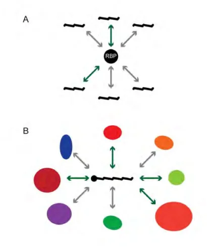
Conceptually, there are two approaches for dissecting interactions with, or regulation of 3'UTRs by RBPs: protein-centered and gene-centered (Figure 1.3). Protein-centered approaches begin with an RBP of interest and seek to determine the 3'UTRs it binds ; whereas, a gene-centered approach begins with the 3'UTR and seeks to determine the RBPs which bind it. Protein-centered approaches include in vivo interaction detection methods such as RBP immunoprecipitation (RIP) and cross-link immunoprecipitation (CLIP) or RBP specificity determination through electrophoretic mobility shift assays (EMSA), systematic evolution of ligands by exponential enrichment (SELEX), and RNAcompete (Keene et al. 2006, Ule et al. 2003, Pagano et al. 2011, Tuerk and Gold 1990, Ray et
Figure 1.3: Comparison of gene- and protein-centered approaches. There are two approaches to identifying mRNA-RBP interactions: protein-centered and gene-centered. Protein-centered methods begin with a protein of interest and test for interactions amongst a set of RNAs or 3'UTRs. Gene-centered methods begin with an RNA of interest and test for interactions amongst a set of RBPs.
al. 2009). A major gene-centered approach is RNAi where the expression of a target mRNA or protein is assessed following the knockdown of individual RBPs (Kamath et al. 2003). A final method, yeast three-hybrid (Y3H) has been used in both manners (SenGupta et al. 1996).
Protein-centered techniques for identifying 3'UTR -RBP interactions
The primary in vivo protein-centered techniques are RIP and CLIP (Keene et al. 2006, Ule et al. 2003). In both methods a given RBP is immunoprecipitated (IP) from cell lines and the bound RNAs are identified using microarrays or RNA sequencing (Keene et al. 2006, Ule et al. 2005). The main difference is that with CLIP a cross-linking step is added before IP. The advantage of cross-linking is that more stringent washes can be utilized without losing lower affinity interactions. Both RIP and CLIP have been used to study the C. elegans RBPs FBF-1/2, RNP-8, and GLD-1 (Kershner et al. 2010, Kim et al. 2010, Wright et al. 2011, Jungkamp et al.
2011).
The primary in vitro techniques include EMSA, SELEX and RNAcompete. In all three methods a purified RBP is tested against a series of RNAs individually (EMSA) or as a library (SELEX, RNAcompete). In
EMSAs, an RNA is labeled radioactively ( e.g. 32P) or fluorescently ( e.g. fluorescein) (Pagano et al. 2011). The labeled probe is incubated with purified protein and native polyacrylamide gel electrophoresis (PAGE) is performed. Binding by an RBP decreases the mobility of the probe. Quantitative measurement of the RBP-RNA interactions can be assessed by comparing binding of multiple protein concentrations against a fixed concentration of RNA (Pagano et al. 2011). Additionally, the RBP specificity can be quantitatively assessed by making single nucleotide mutations in the RNA and measuring the binding affinities. The specificity of multiple C. elegans RBPs has been determined using this approach (Pagano et al. 2007, 2009, Farley et al. 2008, 2012, Kaymak et al. 2013).
SELEX and RNAcompete both test a purified RBP against a library of short RNA sequences. In SELEX, the RBP is incubated with a randomized pool of purified RNAs (Tuerk and Gold 1990, Ellington and Szostak 1990). The RBP is captured then the bound RNAs are eluted and reverse transcribed into complementary strands of DNA (cDNA). The DNA is amplified via polymerase chain reaction (PCR) and transcribed back into RNA for subsequent rounds of selection. After multiple rounds, the DNA is sequenced and the enriched sequences are compiled into a representative binding motif. In RNAcompete, the library of RNAs is synthesized on an oligonucleotide microarray (Ray et al. 2009). The design of the microarray
helps ensure that individual RNA sequences are well represented in the library and it enables detection of bound RNAs on the same microarrays following a single round of selection. SELEX and RNAcompete have both been used to determine RNA binding specificities of several C. elegans RBPs (Pagano et al. 2009, Kaymak et al. 2013, Ray et al. 2013).
Protein-centered approaches are not ideal for research directed at understanding the regulation of a single 3'UTR. The primary reason is that there are hundreds of potential RBPs in eukaryotic organisms. To test all RBPs each technique would have to be performed hundreds of times. Additionally RIP and CLIP require suitable antibodies or epitope-tagged RBPs thus limiting broad use of these techniques. EMSA, SELEX and RNAcompete require protein purifications increasing the workload for each individual experiment. RNAcompete has been performed for hundreds of purified RBPs, however the resulting RNA binding motifs are often short and degenerate (Ray et al. 2013). These motifs are found in thousands of 3'UTRs, therefore additional assays are needed to test physical and regulatory interactions that are predicted to occur in vivo (Pagano et al. 2007, 2009, Kaymak et al. 2013).
In comparison, gene-centered approaches such as RNAi enable direct testing of a single mRNA against many RBPs in vivo . In particular, transgenic cell lines and animal strains can be constructed wherein green fluorescent protein (GFP) is constitutively expressed from an mRNA containing a 3'UTR of interest. The expression patterns can be visualized via fluorescence microscopy. Regulatory interactions by RBPs can be determined by evaluating GFP expression following RNAi of RBPs (Pagano et al. 2009, Kaymak et al. 2013). This is a valuable approach and has the necessary throughput for testing hundreds of RBPs. Missed interactions can result due to subtle regulation, redundancy, and context dependent interactions. Also, interactions determined by RNAi are regulatory and therefore cannot be determined to occur through direct physical interaction without secondary assays.
When this project began, yeast three-hybrid (Y3H) was the best suited assay for the gene-centered detection of physical interactions between 3'UTRs and RBPs (Figure 1.4A). In Y3H, an RNA element of
Figure 1.4: Diagram of Y3H. A) Y3H tests RNA-RBP interactions using two fusion proteins and one fusion RNA. A DNA-binding domain (DB) - MS2 fusion protein binds to an RNA through two fused MS2 binding sites localizing the RNA upstream of a reporter gene. An RBP - activation domain (AD) fusion protein is coexpressed. If the RNA-RBP interaction occurs the AD is recruited upstream of the reporter thereby activating reporter expression. B) In protein-centered Y3H, a library of randomized RNA encoding plasmids are transformed into yeast containing the RBP-AD fusion and plated on selective media. If interactions occur with any transformed RNA the resulting yeast cell grows to form a visible colony. The selected colonies can be sequenced to identify the interacting RNA and a composite RBP binding site can be determined. C) In gene-centered Y3H, a library of RBP-AD encoding plasmids is transformed into yeast containing the MS2 binding site - RNA encoding plasmids. If interactions occur with any transformed RBP the resulting yeast cell grows to form a visible colony. The selected colonies can be sequenced to identify the interacting RBP.
interest is transcribed with two covalently attached high affinity sites for the bacteriophage MS2 coat protein (MS2). The RNA is constitutively expressed from an RNA polymerase III promoter and contains a RNAseP 5’ leader sequence that promotes nuclear retention of the transcript. Additionally a DNA binding domain (DB) - MS2 fusion protein is co-expressed. The fusion protein serves to tether the RNA upstream of a reporter gene ( e.g. HIS3 ) by binding the RNA (via MS2) and DNA binding sites (via DB) (SenGupta et al. 1996, Zhang et al. 1999). To test for an RNA-RBP interaction, the RNA ‘bait’ yeast is transformed with a RBP ‘prey’ that is fused to a TF transcription activation domain (AD). If the RBP interacts with the RNA element then the AD is recruited to the reporter gene thereby promoting reporter expression.
HIS3 and LacZ are frequently used as reporter genes. HIS3 is essential for the biosynthesis of histidine and therefore yeast does not grow without the addition of exogenous histidine to the media. Additionally, low levels of 3-aminotriazole (3-AT), a competitive inhibitor of His3p, can be added to the media to increase selective pressure against low level reporter expression. Reporter activation is detected by assessing yeast growth on media lacking histidine and including 3-AT. LacZ encodes the beta-galactosidase protein. This enzyme converts the colorless compounds 5-bromo-4-chloro-3-indolyl-β-d-galactoside (X-gal) and
ortho-Nitrophenyl-β-galactoside (ONPG) into blue and yellow compounds, respectively. Reporter activation is detected by measuring the amount of color produced in the presence of either compound.
Y3H has been successfully utilized in both protein- and gene-centered configurations. The largest efforts using Y3H have centered on dissecting the nucleotide binding specificity of Pumilio/FBF (PUF) family members (Wickens et al. 2002). In a collection of works, PUF RBPs were tested in a protein-centered manner vs. a library of randomized Y3H RNA baits (SenGupta et al. 1999, Bernstein et al. 2005 ; Opperman et al. 2005, Stumpf et al. 2008, Koh et al. 2009) (Figure 1.4B). Bait RNAs were transformed into a yeast strain that expressed a PUF-AD prey and utilized the HIS3 reporter. Thousands of individual colonies were screened following growth on selective media. Individual baits were sequenced to determine the interacting RNA elements and composite representations of each PUF RBP specificity was determined. As with other protein-centered assays, these specificities were used to predict target 3'UTRs which were bound by the given RBP. Physical interactions were assessed using Y3H and the LacZ reporter, or with orthologous assays such as EMSAs.
Most work used Y3H in the protein-centered configuration, however, it was initially utilized in a gene-centered manner (Wang et al. 1996, Zhang et al. 1997, Martin et al. 1997). A forward genetic screen studying C.
elegans sex determination identified 17 different gain-of-function mutants within a five nucleotide stretch of the fem-3 3'UTR (Ahringer and Kimble 1991). Additional evidence suggested that a post-transcriptional regulator was present in the worm germline, however the identity remained unknown (Ahringer et al. 1992). Y3H was used with the HIS3 reporter to identify the regulatory RBPs. The wild-type fem-3 3’UTR element was the bait and a cDNA library of RBP-AD fusion preys was transformed into the bait strain (Figure 1.4C). Screening selected colonies identified two closely related fem-3 binding factors, FBF-1 and FBF-2 (Zhang et al. 1997). Subsequent Y3H and RNAi experiments confirmed these proteins as regulatory factors that directly bound the fem-3 3'UTR.
Despite successful application of Y3H as a gene-centered method it has not been widely used to the study individual 3'UTRs. There are several potential, non-trivial explanations for this. First, there is a large drop in signal strength for RNA baits longer than 150 nucleotides (nt) (Zhang et al.
1999). The majority of metazoan 3'UTRs are longer than 150 nt (Mangone et al. 2010, Ulitsky et al. 2012, Derti et al. 2012) and would therefore be ineffective Y3H baits. Second, Y3H RNAs are transcribed by RNA polymerase III which is terminated by a tetrauridine signal (Zhang et al.
1999). 3'UTRs are particularly uridine rich and many contain tetrauridine sequences (Mangone et al. 2010, Jan et al. 2011, Ulitsky et al. 2012, Derti
et al. 2012). Thus testing many full-length 3'UTRs would be challenging using Y3H. Theoretically, an RNA polymerase II system without the same restrictions could be established, but this too has not been widely adapted (Zhang et al. 1999).
C. elegans as a model to study 3'UTR - RBP interactions
Briefly, C. elegans is a hermaphroditic nematode with 959 somatic cells. It is transparent, easy to culture and many techniques for genetic manipulations have been developed. In particular, transgenic strains can be generated including those which constitutively express GFP mRNAs fused to a 3’UTR of interest (Merritt et al. 1998). Additionally, mutant strains and RNAi clone libraries are available for the removal / downregulation of RBPs (Caenorhabditis Genome Center, Fraser et al.
2000, Kamath et al. 2003)
Development in C. elegans is an ordered process. There are two
gonad arms which produce both sperm and oocytes (Hubbard and Greenstein, 2005). Within each gonad gametes move from the distal region proximally towards the vulva in an orderly fashion. In the distal region, a population of nuclei mitotically divide in syncytium replenishing the progenitor population (Kimble and Crittenden, 2005). As the nuclei
progress proximally they form cell walls and enter meiosis. Development continues as the oocyte moves through the loop and proximal regions. As the oocyte passes the spermatheca it is fertilized and the egg is laid at the two-cell stage of the embryo (Hubbard and Greenstein, 2005).
Transcription is absent from early regions of the germline until the 4-cell stage embryo (Seydoux et al. 1994, 1996, 1997). This suggested that the primary means of gene regulation in the C. elegans germline is post-transcriptional. Transgenic strains for 30 genes with reported germline expression patterns were generated to test this hypothesis (Merritt et al.
2008). The 3'UTR of each gene was cloned downstream of a GFP:Histone 2B (H2B) reporter driven by the germline ubiquitous pie-1 promoter.
Transgenic strains were generated via microparticle bombardment and randomly integrated into the genome. 17 distinct expression patterns were observed following examination of the resulting strains. Comparisons of the resulting expression patterns to those reported for the endogenous proteins showed concordance for 24/30 strains. The strains which did not match reported expression patterns were sperm-specific genes. In summary, the study demonstrated that 3'UTRs are sufficient for many germline expression patterns. Additional studies have identified many RBPs which regulate those, and other 3'UTRs. Studying C. elegans germline expressed
3'UTR-RBP interactions is an ideal system, however large scale analyses of C. elegans RBPs have not been performed.
How to assay a single 3'UTR against a library of RBPs?
The goal of this project was to develop an approach for testing a single 3'UTR against a library of C. elegans RBPs. The gene-centered approach had the following objectives:
1) Curate a compendium of C. elegans RBPs
The spectrum of C. elegans RBPs was not known. Cursory examinations of public databases and computational predictions identified 92 and 319 candidate RBPs indicating the total number of RBP was in the hundreds (Lublin and Evans 2007, Wang et al. 2009). There was a clear need for a dedicated analysis of the C. elegans proteome and literature similar to that performed for C. elegans transcription factors (TFs) (Reece-Hoyes et al. 2005). This database of C. elegans RBPs could then facilitate the construction of physical resources for gene-centered studies of 3'UTR interactions with many RBPs.
Curation of the C. elegans RBPs involved computational predictions of protein sequences coupled with manual annotation of RBPs with direct experimental evidence (Figure 1.5A). Known RNA binding domains were identified from literature and domain signatures were used to scan the C.
elegans proteome for high scoring sequences. Additionally, homologs of known RBPs were identified. Lastly, RBPs were identified through a directed literature search of C. elegans and RNA binding proteins.
2) Develop an assay for detecting 3'UTR-RBP interactions in a gene-centered manner
The goal for this assay was to enable detection of physical interactions in a gene-centered manner. Additionally, interactions were to be detected in pair-wise and condition independent manners to remove complicating factors such as developmental or environmental states. Ideally, the method was designed in a scalable manner to enable future large scale screens. Lastly, emphases were placed on developing a cell-based system wherein the bait RNA was in a functional environment.
The assay was designed such that reporter mRNA activation by a heterologous RBP would result in high levels of signal ( i.e. GFP fluorescence) in yeast (Figure 1.5B). The yeast based system enables the
Figure 1.5: Outline of a gene-centered approach to RNA-RBP interactaction mapping. A) The proteome of C. elegans was computationally analyzed to determine proteins sequences with one or more RNA binding domains. The computationally determined list was filtered manually to remove spurious predictions and to add literature curated RBPs that were missed. B) An assay was developed which enables the specific activation of the GFP reporter gene in yeast when an interaction occurs between an RNA and RBP. This assay is gene-centered, meaning that an individual RNA bait strain can be tested against a library of RBP preys. C) RNA-RBP interactions were tested in a pair-wise manner and those with increased signals were considered preliminary physical interactions. D) Assay defined interactions were tested in vivo using RNAi of the indicated RBP in a transgenic GFP : 3'UTR strain.
use of standard genetic techniques including integration of the bait-encoding DNA and subsequent transformation of the RBP prey library. This enables direct pair-wise testing of interactions in a condition independent manner. Additionally, the intact cellular environment places the interaction in a functional context. Lastly, yeast based assays can be scaled-up for genome level interaction screens (Simonis et al. 2009, Reece-Hoyes et al. 2013)
3) Test a single 3'UTR against a library of C. elegans RBPs
To demonstrate the effectiveness of the gene-centered approach a single 3'UTR was tested against a library of RBPs (Figure 1.5C). The ideal candidate for this is the C. elegans glp-1 3'UTR because there are several known RBPs which bind to and regulate it. Also, there may be additional unknown RBPs. To perform the test a subset of the curated, C. elegans
germline expressed RBPs were tested in the assay. Comparisons were then made to known interactions and newly discovered interactions were tested using follow-up assays (Figure 1.5D).
CHAPTER II
Preface
This chapter comprises published work from the following reference:
Tamburino, A. M., Ryder, S. P., & Walhout, A. J. M. (2013). A Compendium of Caenorhabditis elegans RNA Binding Proteins Predicts Extensive Regulation at Multiple Levels. G3, 3, 297-304.
It is the product of my own work performed under the guidance of Marian Walhout and Sean Ryder.
A compendium of C. elegans RNA binding proteins predicts extensive regulation at multiple levels
Generating the right protein at the right place, the right time, and the right levels is critical during all aspects of life. Multiple levels of gene regulation coordinate the precise expression of genes throughout development and in response to environmental cues and insults. In genomics and systems biology, much attention has focused on the elucidation of regulatory networks involving transcription factors (TFs) or microRNAs (miRNAs) (Martinez and Walhout 2009 ; Arda and Walhout 2010). These networks include interactions in which these factors both regulate and are regulated by other molecules (Reece-Hoyes et al. 2011; Bartel 2009 ; Deplancke et al. 2006; Martinez et al. 2008; Harbison et al. 2004; Arda et al. 2010). RNA binding proteins (RBPs) are another important class of gene regulators ; however, the regulatory networks in which they function remain largely uncharacterized.
Although TFs bind DNA and miRNAs interact with mRNAs, RBPs can interact with the entire spectrum of RNAs. These RNAs occur throughout the cell and can take on a vast array of functions, including serving as templates for protein synthesis (mRNA), participating as structural components of the splicing and translation machinery (rRNA, tRNA,
snRNA), and providing regulatory activity to modulate transcription, translation and chromatin structure (miRNA, siRNA, piRNA, lncRNA) (Lee and Schedl 2005, Steitz 2008 ; Moore and Proudfoot 2009 ; Carthew and Sontheimer 2009 ; Wahl et al. 2009). Physical interactions between RNA and RBPs are crucial to RNA regulation, for instance, to mediate precise mRNA 3’ end formation, splicing, localization, stability, and translation. As a result of these physical interactions, RBPs can control transcript localization, levels, and translation (Shepard et al. 2003; Glisovic et al. 2008).
In contrast to RBPs, TFs are rapidly being characterized at a systems level using genome-scale methods such as chromatin immunoprecipitation (ChIP) and yeast one-hybrid assays (Walhout 2011). Among other findings these studies have demonstrated degenerate DNA binding of TFs, extensive combinatorial complexity of interactions between TFs and gene promoters, as well as both specific and promiscuous protein interactions between divergent members of the same TF family (Deplancke et al. 2006; Badis et al. 2009; Grove et al. 2009; Zinzen et al. 2009). The systems-level characterization of TFs has been greatly facilitated by high-confidence predictions of which genes in a genome encode such proteins (Reece-Hoyes et al. 2005; Kummerfeld and Teichmann 2006 ; Vaquerizas
et al. 2009). However, such compendia are not yet available for RBPs in multicellular model organisms.
Here, we present a compendium of predicted RBPs for the nematode Caenorhabditis elegans (wRBP1.0). We have used wRBP1.0 to begin the analysis of RBPs at a genome-wide level, using publicly available datasets. We found that RBP-encoding mRNAs have more alternative isoforms, longer 3’ untranslated regions (UTRs), and more alternative polyadenylation (APA) sites than other mRNAs. In addition, RBP gene promoters interact with more TFs, RBP mRNAs are bound by more RBPs, and the 3'UTRs of RBP-encoding mRNAs are targeted by more miRNAs. Finally, RBPs are phosphorylated more frequently than other proteins. Together, our compendium and analyses provide a first step toward the characterization of RBP regulatory networks in C. elegans and serve as a model for the continued study of RBPs in other organisms, including humans.
RESULTS AND DISCUSSION
wRBP1.0
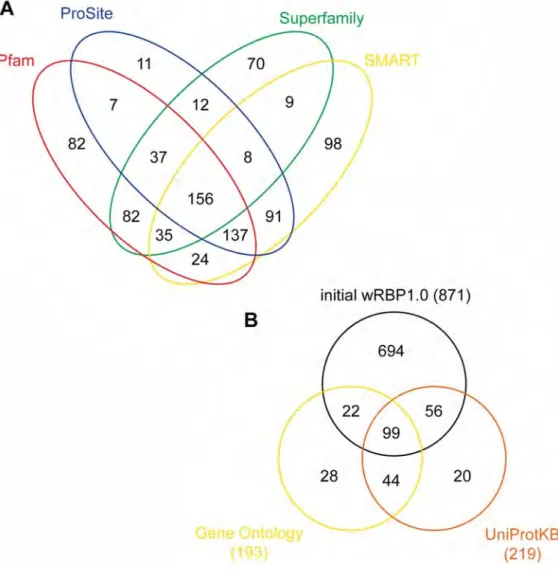
To curate the compendium of putative RBPs in C. elegans , we searched the proteome (version WS219) for each of 17 RBDs [see the section RNA binding domains (RBDs)] based on domain sequence signatures from the unified InterPro database (Quevillon et al. 2005; Hunter et al. 2009). Proteins were annotated for the presence of each domain using four separate databases (see Materials and Methods) and each protein possessing one or more RBD was included in the compendium. Low-confidence calls were removed (see Materials and Methods), and the curations were supplemented with RBPs that we identified from the literature but that were missed in the computational search. Of the total RBP set, 67% were identified by more than one method, which illustrates the robustness of our predictions (Figure 2.1A). Furthermore, the initial list contains greater than 93% of proteins that were previously curated as RNA binding (Wang et al. 2009), which illustrates the sensitivity of our method. It is important to note that we increased the number of putative C. elegans RBPs by almost threefold relative to this study (from 319 to 887). Two major reasons for this include the inclusion of additional RBDs and protein classes ( i.e., dsRBDs, ribosomal proteins, C2H2 zinc fingers, SAM domains) and the inclusion of additional RBPs possessing each domain (i.e., 10–60% increase in KH, RRM, helicase, and CCCH zinc finger domain containing proteins). Further, 66% of the
Figure 2.1: Venn diagrams of: A) Cross-validation of programs used by
InteroProScan, and B) initial wRBP1.0 list together with Gene Ontology and
UniProtKB listed RBPs
RBPs (177 of 269) annotated in Gene Ontology and UniProtKB databases as ‘RNA binding’ were included, again demonstrating high sensitivity (Figure 2.1B) (Gene Ontology Consortium 2000 ; Uniprot Consortium 2009). Next, we manually evaluated 96 RBPs that were not included in our initial list but that were annotated as RNA binding by Gene Ontology, UniProtKB or Wang et al . 2009. After careful consideration, we judged 16 of these to be candidate RBPs, whereas we did not have sufficient confidence to include the other 80 (data not shown). Finally, we determined that wRBP1.0 includes 220 of 230 protein listed in RBPDB (Cook et al. 2011) including 22 of 23 proteins with experimental evidence of RNA binding [AIN-1 is associated with the miRNA silencing complex but does not require RNA for binding (Wormbase.org)]. Altogether, this generated a final wRBP1.0 compendium of 887 genes. RBPs were then classified into Groups 1-4 based on the domains they possess (Figure 1, see below).
RNA binding domains (RBDs)
We identified a set of 17 RBDs by literature searches for proteins that bind to RNA (Figure 2.2, Table S2.1). Altogether, we identified 887 putative RBP-encoding genes (Table S2.2; see below). We divided these
Figure 2.2: wRBP1.0. Pipeline for C. elegans RBP predictions. RBDs were
predicted from WormBase protein annotations then filtered and literature
curated. RBPs were separated into four groups according to their RBDs as
genes into four groups based on whether they are more likely to bind and regulate RNA in a gene-specific or nonspecific manner. Many RBPs contain multiple RBDs ; however, only 10 of 887 genes contain domains from two or more different groups (Table S2.2). The classification of these 10 genes was first based on the presence of a sequence or structure-specific RBD.
Group 1: Gene-specific RBDs that bind RNA in a sequence-specific manner: This group contains eight RBDs that mediate binding to specific mRNAs in a sequence and/or structure-specific manner (Figure 2.2). RNA binding by these domains has been demonstrated for several individual proteins in vitro , and gene-specific binding has been detected for several proteins in vivo (Table S2.1) (Ryder et al. 2004; Bernstein et al. 2005; Opperman et al. 2005 ; Pagano et al. 2007; Farley et al. 2008; Pagano et al. 2009; Kershner and Kimble 2010 ; Wright et al. 2010). Direct, sequence-specific RNA binding has been shown for some C. elegans
RBPs, but the vast majority remains untested. For instance, GLD-1 (KH domain) and FBF-1 both bind specific sequences in vitro (Ryder et al.
2004; Bernstein et al. 2005) and associate with specific mRNAs in vivo (Kershner and Kimble 2010 ; Wright et al. 2010; Jungkamp et al. 2011). Altogether, 250 of the 887 RBP-encoding genes are included in Group 1.
Group 2: Gene-specific RBDs that do not bind RNA in a sequence specific manner: RBDs within Group 2 bind RNA in a gene-specific manner in vivo. However, contrary to Group 1 RBDs, the means for this RNA binding specificity are unknown or occur in a manner that is not inherent to the RBD itself (i.e., the domain contributes to RNA binding affinity rather than specificity). For instance, the argonautes ALG-1 and ALG-2 bind miRNAs through their PAZ/PIWI domains. Complementary base pairing by these miRNAs directs targeting of these proteins to specific mRNAs. Out of the 17 RBDs considered, four are placed in this group: helicase, PAZ, PIWI, and NTF2, altogether encoding 169 proteins.
Group 3: Putative gene-specific RBDs: Group 3 proteins are predicted to bind RNA in a gene- and sequence-specific manner. However, we have separated Group 3 proteins from those in Group 1 because their RBDs could be involved not only in RNA binding but also in DNA binding, or protein-protein interactions, thus making the prediction of their function ambiguous (see Table S1 for references). For instance, Xenopus laevis TFIIIA can bind both DNA and RNA through various combinations of its C2H2 zinc fingers (Theunissen et al. 1992 ; Luet al. 2003). All proteins with the domains of group 3 are included although we expect that not all of them
will mediate RNA binding ( e.g. , many C2H2 zinc fingers occur in TFs that bind DNA). Group 3 contains three of the 17 RBDs and 226 genes.
Group 4: Non-gene-specific RBPs, with some exceptions: The fourth group contains RBDs that typically do not bind RNA in a gene-specific manner. Many essential factors involved in general gene expression are in this group, including ribosomal proteins, transfer RNA-binding proteins, translation initiation factors, core splicing proteins and RNA degradation proteins such as ribonucleases and exosome components. Two of the 17 domains are included in this category and because many general RBPs lack clear domains, additional proteins are included based upon conservation to RBPs in other organisms. Altogether, this group contains 279 genes.
RBP-encoding genes are bound by more TFs, more RBPs, and have more splice variants
RBPs have been proposed to both fine tune gene expression as well as drive tissue and stage-specific gene expression (Blencowe 2006 ; Glisovic et al. 2008). Therefore, we hypothesized that RBPs may, as a group, be extensively regulated to mediate these functions. Here, we
tested this hypothesis using the wRBP1.0 compendium and several publicly available datasets.
Transcriptional regulation mediated by the binding of TFs to gene promoters provides a first and important level of regulation. There are 937 predicted TFs encoded by the C. elegans genome (Reece-Hoyes et al. 2005; Reece-Hoyes et al. 2011), and binding of 22 of these TFs (~2%) has been examined by ChIP-seq (Gerstein et al. 2010). Based on these data, we found that promoters of RBP genes are bound by more TFs than promoters of other genes (Figure 2.3A). Both gene-specific and general RBP promoters are bound by significantly more TFs (p<1e-9), indicating that transcriptional regulation is an important first step toward RBP expression. Importantly, these data were obtained using transgenic TF fusion strains. Because transgenes are often silenced in the germline (Cui and Han 2007) where many RBPs are expressed, it is possible that our analyses underestimate the enrichment. Further, this analysis was based on only 22 TFs; future studies will reveal the generality of our observation.
We next analyzed publicly available RBP-mRNA interactions. We obtained three RIP-Chip datasets for the C. elegans RBPs FBF-1, GLD-1 and RNP-8 (Kershner and Kimble 2010; Kim et al. 2010; Wright et al.
Figure 2.3: RBPs are extensively regulated by TFs and RBPs. A) More TFs
bind to RBP promoters than the promoters of other genes B) RBPs bind to
a higher proportion of RBP-encoding mRNAs. C) RBP genes are more
frequently spliced than other genes. *p<0.05,**p<0.005, relative to
proteome, hypergeometric test (frequency data), Komologorov-Smirnov
2010) and found that 73% of RBP mRNAs are bound by at least one RBP, compared with only 35% of the total transcriptome (Figure 2.3B). The number of RBP mRNAs from Group 4 bound is even greater (86%). Our result is consistent with Gene Ontology enrichment analysis performed in the original studies that retrieved enrichment for ‘RNA binding’ and ‘Nucleic acid binding’ terms, respectively (Kim et al. 2010 and Kershner and Kimble 2010).
The binding of RBPs to mRNAs affects numerous steps of an mRNA’s lifecycle, including alternative splicing (Blencowe 2006 ; Glisovic et
al. 2008). To test whether C. elegans RBP-encoding mRNAs are more extensively spliced than other genes, we evaluated the number of protein isoforms per RBP-encoding gene by using comprehensive WormBase annotations. Approximately one-quarter of the 887 RBP-encoding genes (212; 23.9%) encode multiple isoforms, which is significantly more than the 14.4% of genes that undergo alternative splicing in the entire genome (Figure 2.3C). An even greater percentage of mRNAs encoding gene-specific RBPs in Group 1 are alternatively spliced (30.4% ; Figure 2C). Through alternative splicing, the total number of RBPs increased by more than 40% (from 887 genes to 1242 proteins) and, interestingly, the number of distinct gene-specific RBPs increased by ~60% (250 genes
encoding 401 proteins). Thus, alternative splicing increases the effective number of RBPs in the C. elegans proteome.
RBP 3'UTRs are extensively regulated
3'UTRs affect gene expression via interactions with RBPs and miRNAs (Bartel 2009 ; Kuersten and Goodwin 2003). Concordantly, C.
elegans 3'UTRs contain numerous conserved sequence elements that may interact with miRNAs or RBPs (Mangone et al. 2010; Jan et al. 2011). Using comprehensive 3'UTR annotations (www.UTRome.org), we found that RBP-encoding mRNAs have significantly longer 3'UTRs, with a median length of 156 nucleotides (nt), compared with 129 nt for the whole transcriptome (Figure 2.4A). The 3'UTRs of gene-specific RBP mRNAs (Group 1) are even longer (215 nt), whereas general RBPs have shorter 3'UTRs (Group 4; 100 nt).
Longer 3'UTRs can contain more regulatory sites, which implies that gene-specific RBPs may be more heavily regulated via their 3'UTRs, whereas general RBPs may be less extensively regulated. To test this, we first assessed the degree to which miRNAs target RBP 3'UTRs relative to all genes. In the absence of comprehensive experimental miRNA targeting data, predictions for bound target mRNAs can be made using
Figure 2.4: RBPs are extensively regulated through 3'UTRs A) RBP transcripts have longer 3'UTRs. B) RBP 3'UTRs are more heavily targeted by miRNAs. C) More miRNA families target RBP 3'UTRs. D) 3'UTR annotations show that more RBPs utilize alternative 3'UTRs, and E) that RBP genes have more alternative 3'UTRs. F) Combined miRNA target predictions and 3'UTR annotations reveal that APA affects predicted miRNA targeting. *p<0.05,**p<0.005, relative to proteome, hypergeometric test (frequency data), Komologorov-Smirnov test (cumulative frequency data).
the miRNA seed sequences (Bartel 2009). We used target predictions from TargetScan for all C. elegans 3'UTR sequences experimentally determined by 3P-Sequencing (3Pseq) (Jan et al. 2011). TargetScan predicts miRNA targets based upon stringent seed pairing as well as site number, type, context, and conservation (Bartel 2009). Comparison of RBP-encoding mRNA 3'UTRs to the 3'UTRs of all C. elegans mRNAs revealed that significantly more RBP 3'UTRs are predicted targets of miRNAs (Figure 2.4B). Furthermore, significantly more miRNA families target each gene-specific RBP 3'UTR compared with all 3'UTRs, indicating a potential for increased combinatorial complexity (Figure 2.4C). In contrast, general RBPs showed no significant difference in miRNA targeting compared to the total transcriptome.
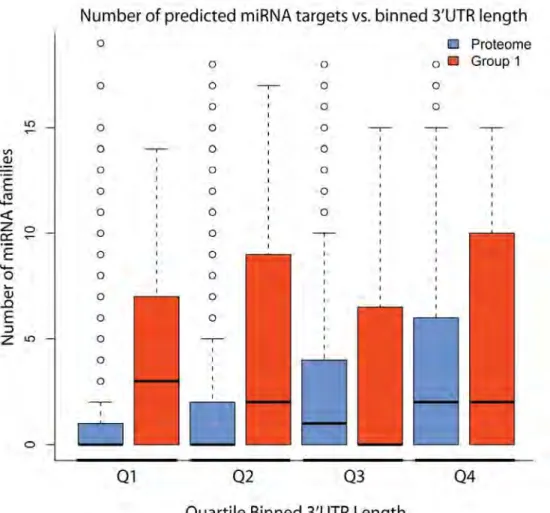
It is important to note that these predictions are based on conservation of the site in multiple species and availability of the site in folded RNA. This implies that the increased number of miRNA families targeting 3'UTRs is not solely a consequence of 3'UTR length. To confirm this, we compared RBP 3'UTRs with similar length 3'UTRs from the total transcriptome by binning 3'UTRs by length (Figure 2.5). This analysis confirmed that, among the shortest 3'UTRs ( i.e., the first two quartiles), more miRNAs are indeed predicted to target RBP 3'UTRs, while we did not observe a difference for the longest 3'UTRs.
Figure 2.5: Quartile binned boxplots of miRNAs targeting RBP 3'UTRs vs. 3’UTRome. Quartiles were determined using the total 3’UTRome. The
We further evaluated miRNA targeting to RBP 3'UTRs using predictions made by mirWIP (Hammell et al. 2008) and argonaute ALG-1 bound 3'UTRs determined using cross-link immunoprecipitation (Figure 2.6) (Zisoulis et al. 2010). Both of these analyses showed that RBP 3'UTRs are indeed more frequently targeted by miRNAs, which further supports the observations made with TargetScan predictions.
Alternative 3'UTR usage provides additional unique sites of regulation for miRNAs and RBPs or, conversely, can eliminate regulatory sites for these same factors. Recently, it has been shown that shortening of 3'UTRs by alternative polyadenylation (APA) alters protein expression in proliferating cells, an effect partly attributed to the loss of miRNA binding sites (Sandberg et al. 2008; Mayr and Bartel 2009). Using 3'UTR annotations determined by 3P-Seq (Jan et al. 2011), we found that more RBPs use APA and that RBPs possess more distinct 3'UTRs than the total transcriptome (Figures 2.4D,E ; results with 3'UTRome annotations were consistent, data not shown). Once again, the effect was especially pronounced for gene-specific RBPs (Group 1). We calculated the number of genes in which APA eliminates all predicted targeting sites for one or more miRNA family, thereby preventing miRNA repression and increasing gene expression. Using 3P-seq-derived 3'UTRs and TargetScan miRNA target predictions, we found that more than 15% of the gene-specific
Figure 2.6: miRNA targeting. Frequency of 3'UTRs targeted by miRNAs
RBPs could evade potential repression by at least one miRNA family using APA, a fraction that is more than twice that of the total transcriptome (Figure 2.4F). The predicted effects of APA may also affect gene expression through the distinct binding of RBPs to alternate 3'UTRs.
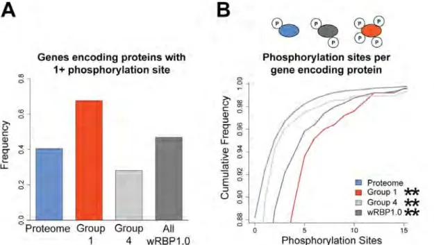
RBPs are more extensively phosphorylated
Posttranslational modifications provide another mechanism to create protein diversity. In particular, phosphorylation can affect the ability of proteins to function and/or interact with binding partners (Deribe et al. 2010). To evaluate the degree to which RBPs are phosphorylated, we interrogated phosphoproteome data that were obtained by tandem mass spectrometry of synchronized adult worms and that identified 6,780 phosphorylation sites on 2,373 proteins (Zielinska et al. 2009). Because many factors can affect the ability for certain proteins to be detected in mass spectrometry, we corrected for potential biases by normalizing the frequency of detected RBPs in each group by a separate mass spectrometry study that analyzed the proteome of mixed stage worms and did not enrich for phophopeptides (Figure 2.7) (Merrihew et al. 2008). We found that more gene-specific RBPs are phosphorylated relative to the