1
2012 (to be reviewed and updated 2017)
International Framework for Examination of the Cervical Region for potential of Cervical Arterial Dysfunction prior to Orthopaedic Manual Therapy Intervention
Authors: Rushton A, Rivett D, Carlesso L, Flynn T, Hing W, Kerry R. Background
This consensus document presents a clinical reasoning framework for best practice developed by an international collaboration of the Standards Committee of the International Federation of
Orthopaedic Manipulative Physical Therapists (IFOMPT) and international subject matter experts. This framework was informed by the consensus forum at the IFOMPT International Conference in Rotterdam (June 2008) where nominated experts from each Member Organisation of IFOMPT were invited to participate. Prior to this forum, the issues central to this document were explored through oral presentations and discussion at the World Confederation for Physical Therapy (WCPT)
International Congress (June 2007, Vancouver) and in Rotterdam. Drafts of the document have been developed since 2008 through an iterative consultative process with experts and Member
Organisations. This document has been developed as a resource to educate members of the Member Organisations of IFOMPT, physical therapists internationally, and through physical therapists, the public. The framework has subsequently been agreed on by all Member
Organisations of IFOMPT. It has been a challenging process to produce an international agreed document, emphasising the need for Member Organisations of IFOMPT to operationalise the framework in line with their national legislation / practice
IFOMPT
Please see www.ifompt.org for all information. The vision statement of IFOMPT is:
"World-wide promotion of excellence and unity in clinical and academic standards for manual/musculoskeletal physiotherapists."
The vision statement summarises the mission of IFOMPT that as an organisation it aims to: 1. Promote and maintain the high standards of specialist education and clinical practice in
manual/musculoskeletal physiotherapists.
2. Promote and facilitate evidence based practice and research amongst its members.
3. Communicate widely the purpose and level of the specialisation of manual/musculoskeletal physiotherapists amongst physiotherapists, other healthcare disciplines and the general public.
4. Work towards international unity/conformity of educational standards of practice amongst manual/musculoskeletal physiotherapists.
2
5. Communicate and collaborate effectively with individuals within the organisation and with other organisations.
The Standards Committee of IFOMPT is a sub-committee of the Executive Committee and responsible for advising the Executive on educational issues and maintaining standards. The Standards Document is the guideline document which IFOMPT provides for groups of Manual Therapists who wish to seek membership of IFOMPT through the creation of postgraduate
educational programmes in Orthopaedic Manual Therapy (OMT). Part A of the Standards Document (IFOMPT, 2008) details the educational standards.
3 Executive summary
Key principles of the international framework
The framework provides guidance for the assessment of the cervical spine region for potential of Cervical Artery Dysfunction (CAD) in advance of planned OMT interventions Although events and presentations of CAD are rare, they are an important consideration as
part of an OMT assessment
The framework is based on best available evidence and is intended to be informative and not prescriptive
The framework enhances the physical therapist’s clinical reasoning as part of the process of patient assessment and treatment
An important underlying principle of the framework is that physical therapists cannot rely on the results of only one test to draw conclusions, and therefore development of an
understanding of the patient’s presentation following an informed, planned and individualised assessment is essential
The framework is designed to be an aid to patient-centred clinical reasoning
The framework requires effective clinical reasoning to enable effective, efficient and safe assessment and management of the cervical region
The physical therapist’s aim during the patient history is to make the best judgment on the probability of serious pathology and contraindications to treatment based on available information
A process of planning the physical examination to interpret the data from the patient history and define the main hypotheses is essential to an effective physical examination
It is important that the tests within the physical examination provide reliable and valid data to enable evaluation of the main hypotheses
A risk versus benefit model is advocated to provide a simple framework for decision-making through consideration of risk factors, predicted benefit of OMT intervention, and analysis of possible action
A flowchart of clinical reasoning is provided
Informed consent must be obtained prior to treatment interventions, following adequate disclosure of information
Key considerations are provided for the physical therapist during the selection and application of cervical manipulation as a treatment intervention
Guidance is provided on alternative approaches to direct cervical treatment, frequency of treatment, minimising end-range cervical techniques, force minimisation, and monitoring for adverse effects
4 Flowchart of clinical reasoning
Data obtained from patient history
Planning the physical examination
Possible cervical vasculogenic contribution?
Any gaps in data from patient history?
Quality of data obtained?
Any precautions or contraindications?
Which physical tests to use?
Priority for physical testing?
Interpretation of data from patient history using evidence informed knowledge, and cognitive and metacognitive processes.
Interpretation includes analysis of patient’s preferences.
Interpretation of data from physical examination using
evidence informed knowledge, and cognitive and metacognitive processes. Interpretation includes analysis of patient’s preferences. Data obtained from physical examination
Evaluation of patient’s presentation Any gaps in data from assessment?
Quality of data obtained?
Risk versus benefit analysis?
Decision regarding action?
Best decision regarding management In collaboration with the patient
5 Aim of the framework
The framework is designed to provide guidance for the assessment of the cervical spine region for potential of Cervical Artery Dysfunction (CAD) in advance of planned OMT interventions. The IFOMPT definition of Orthopaedic Manual Therapy (OMT), voted in at the General Meeting in Cape Town, March 2004 is:
OMT is a specialised area of physiotherapy / physical therapy for the management of neuro-musculo-skeletal conditions, based on clinical reasoning, using highly specific treatment approaches including manual techniques and therapeutic exercises.
OMT also encompasses, and is driven by, the available scientific and clinical evidence and the biopsychosocial framework of each individual patient.
OMT interventions for the cervical spine addressed through this framework include: manipulation, mobilisation and exercise. Attention is focused to techniques occurring in end range positions of the cervical spine, during mobilisation, manipulation and exercise interventions.
The framework is based on best available evidence at the time of writing. It is designed to be used in conjunction with the IFOMPT Standards (IFOMPT, 2008) available at www.ifompt.org that define postgraduate best practice in OMT internationally. Central to this framework are sound clinical reasoning and evidence based practice.
Within the cervical spine, events and presentations of CAD are rare, but are an important
consideration as part of an OMT assessment. Arterial dissection (and other vascular) presentations are fairly recognisable if the appropriate questions are asked during the patient history, if
interpretation of elicited data enables recognition of this potential, and if the physical examination can be adapted to explore any potential vasculogenic hypothesis further. The framework is therefore reflective of best practice and aims to place risk in an appropriate context that is informed by the evidence. In this context, the framework considers ischaemic and non-ischaemic presentations to identify risk, prior to overt symptoms in a patient presenting for cervical management.
An important underlying principle of the framework is that physical therapists cannot rely on the results of only one test to draw conclusions, and therefore development of an understanding of the patient’s presentation following an informed, planned and individualised assessment is essential. There are multiple sources of information available from the process of patient assessment to improve the confidence of estimating the probability of CAD. Data available to inform clinical reasoning will improve and change with ongoing research. This framework therefore encourages physical therapists to critically read the current literature to enable support for their clinical
decisions, rather than provide specific data prescriptive guidance, as the evidence base for this is not available.
The framework is intended to be informative and not prescriptive, and aims to enhance the physical therapist’s clinical reasoning as part of the process of patient assessment and treatment. The framework is intended as simple and flexible. The physical therapist should be able to apply it to their individual patients thereby facilitating patient centred-practice.
6
The framework is divided into the following sections, and is designed to be used in conjunction with key literature sources identified in the context section:
1. Context to assessment of the cervical region 2. Clinical reasoning as a framework
3. Patient history
4. Planning the physical examination 5. Physical examination
6. Risk versus benefit analysis 7. Flowchart of clinical reasoning
8. Informed consent and medico-legal framework
9. Safe OMT practice, including emergency management of an adverse situation 10. Teaching OMT for the cervical region
11. Proposed response to the media: key messages to communicate 12. References.
7
Section 1: Context to assessment of the cervical region 1.1 IFOMPT
The vision of IFOMPT is for worldwide promotion of excellence and unity in clinical and academic standards for manual/musculoskeletal physiotherapists, with its mission statement including to work towards international unity/conformity of educational standards of practice amongst manual/musculoskeletal physiotherapists. The process of development of this framework has been guided by this vision and mission, commencing with an exploration of the key issues in 2007.
1.2 Process of development
At the World Confederation for Physical Therapy Congress in Vancouver (2007) IFOMPT coordinated a session of speakers and discussion titled ‘VBI [vertebro-basilar insufficiency] session’ to address a topic that generates frequent questions from Member Organisations of IFOMPT and individual physical therapists. The session involved much discussion about pre-manipulative screening in the cervical spine, and as a result of the session, the IFOMPT Standards Committee was asked to take the key issues forward. At the request of the Standards Committee, a survey using a questionnaire regarding pre-manipulative screening was carried out (DR/LC). The questionnaire was sent to all Member Organisations and Registered Interest Groups (RIGs) of IFOMPT in late 2007. Results of the survey were presented at the IFOMPT Conference in Rotterdam in 2008. In addition, a discussion forum was facilitated in Rotterdam (AR) and contributed to by the development team
(DR/LC/TF/WH/RK), involving a nominated expert from each MO. The forum concluded that the development of an agreed international framework was required to inform OMT practice in this area.
1.3 Key findings from the 2007 survey (Carlesso and Rivett, 2011)
Twenty Member Organisations (100%) and 2 RIGs responded. MO membership varied between countries with 7 small (≤100), 8 moderate (101-399), and 5 large (≥400). Seven Member
Organisations (35%) had their own guidelines or protocol, and 10 Member Organisations (50%) and 1 RIG essentially used that of another country (9 Member Organisations reported using those from Australia, and 1 reported using those from the UK). Thus, the majority of Member Organisations (85%) used pre-manipulative guidelines, with the Australian guidelines commonly adopted
internationally. Only 5 (25%) Member Organisations had a patient information sheet about cervical manipulation and its risks. Eight Member Organisations (40%) and 1 RIG recommended warning patients about the small risk of stroke and death, while 3 Member Organisations recommended informing re stroke only. Therefore provision of information re serious adverse responses was not standard practice in all countries. Only 3 Member Organisations were aware of cases of stroke attributed to a manipulative physical therapist in their country.
For the physical examination of patients, 17 Member Organisations (85%) and 2 RIGs taught
screening positional tests involving extension and rotation (2 using rotation only), and all 20 Member Organisations (100%) and 2 RIGs recommended the use of the sustained pre-manipulative position as a screening test. Fifteen Member Organisations and 1 RIG taught other pre-manipulative screening tests, including: craniovertebral ligament tests (8), dizziness differentiation tests (2), and Hautant’s test (2).
In exploring the use of manipulation in the cervical spine, 8 Member Organisations (40%) and 1 RIG reported that members had decreased the use of manipulation in the upper cervical spine in the last 10 years. Nineteen Member Organisations (95%) and 1 RIG continued to teach upper cervical
8
manipulations, with 3 Member Organisations teaching upper cervical spine manipulations involving end-range rotation. Thirteen Member Organisations (65%) and 1 RIG indicated that the manipulation techniques taught had been changed to limit the amount of rotation used for upper cervical
techniques.
It is acknowledged that practice may have changed in some countries since the survey was conducted, but these data provide a useful overview to inform the content of this document. 1.4 Key points to emerge from the discussion forum in Rotterdam 2008
The forum in Rotterdam agreed that an international framework was required, and agreed the following points and guiding principles to inform a first draft of a consensus document:
1.4.1 Existing documents need to inform development of an international framework. Particularly; Clinical Guidelines for Assessing Vertebrobasilar Insufficiency in the Management of
Cervical Spine Disorders (Rivett et al, 2006)
Manipulation Association of Chartered Physiotherapists, Cervical Arterial
Dysfunction and Manipulative Physiotherapy: information document (Kerry et al, 2007).
1.4.2 Inclusion of key aspects of the framework as detailed on page 6.
1.4.3 Consideration be given to including the pre-manipulative positional test.
1.4.4 Consideration be given to including information on craniovertebral ligament testing. 1.4.5 Recommendations on informed consent need to be sufficiently flexible for different
jurisdictions (to be inclusive of all Member Organisations). 1.4.6 Preferred options to be included on manipulative practices. 1.4.7 An IFOMPT endorsed document must be:
reflective of best practice and research flexible and simple in application legally suitable to individual countries an aid to patient-centred clinical reasoning
9
Section 2: Clinical reasoning to underpin this framework
Clinical reasoning is employed to underpin the framework detailed in this document. The cognitive and metacognitive processes of reasoning, using evidence-informed knowledge within OMT are the central components to expertise of practice in OMT (Rushton and Lindsay, 2010).
2.1 IFOMPT Standards
The IFOMPT Standards Document (IFOMPT, 2008) states that:
“Advanced clinical reasoning skills are central to the practice of OMT Physical Therapists, ultimately leading to decisions formulated to provide the best patient care. Clinical decisions are established following consideration of the patient’s clinical and physical circumstances to establish a clinical physical diagnosis and treatment options. The decisions are informed by research evidence concerning the efficacy, risks, effectiveness, and efficiency of the options (Haynes, 2002). Given the likely consequences associated with each option, decisions are made using a model that views the patient’s role within decision making as central to practice (Higgs and Jones, 2000), thus describing a patient centred model of practice”. “The application of OMT is based on a comprehensive assessment of the patient’s neuromusculoskeletal system and of the patient’s functional abilities. This examination serves to define the presenting dysfunction(s) in the articular, muscular, nervous and other relevant systems; and how these relate to any disability or functional limitation as described by the World Health Organisation’s International Classification of Functioning, Disability and Health (World Health Organisation, 2001). Equally, the examination aims to distinguish those conditions that are indications or contraindications to OMT Physical Therapy and / or demand special precautions, as well as those where anatomical anomalies or pathological processes limit or direct the use of OMT procedures”.
2.2 IFOMPT competencies relating to clinical reasoning
Dimension 6 of the detailed competencies relates to clinical reasoning in postgraduate physical therapy practice in OMT, as follows:
Table 2.1 Dimension 6 of the IFOMPT Standards
Dimension 6 Demonstration of critical and an advanced level of clinical reasoning skills enabling effective assessment and management of patients with
neuromusculoskeletal dysfunctions Competencies Relating to Knowledge
Competency D6.K1
Demonstrate critical understanding of the process of hypothetico-deductive clinical reasoning, including hypothesis generation and testing
Competency D6.K2
Demonstrate effective use of the process of pattern recognition, including the importance of organising clinical knowledge in patterns
10
D6.K3 OMT, including those related to diagnosis, treatment and prognosis
Competency D6.K4
Demonstrate effective recognition of dysfunction requiring further investigation and / or referral to another healthcare professional
Competency D6.K5
Demonstrate critical evaluation of common clinical reasoning errors
Competencies Relating to Skills Competency
D6.S1
Demonstrate accurate and efficient selection of inquiry strategies based on early recognition and correct interpretation of relevant clinical cues
Competency D6.S2
Demonstrate critical and evaluative collection of clinical data to ensure reliability and validity of data
Competency D6.S3
Demonstrate advanced use of clinical reasoning to integrate scientific evidence, clinical data, the patient’s perceptions and goals, and factors related to the clinical context and the patient’s individual circumstances
Competency D6.S4
Demonstrate integration of evidence based practice and experiential reflective practice in clinical decision making
Competency D6.S5
Demonstrate application of collaborative clinical reasoning with the patient, carers / care-givers and other health professionals in determining management goals, interventions and measurable outcomes
Competency D6.S6
Demonstrate effective prioritisation in the examination and management of patients with neuromusculoskeletal dysfunction
Competency D6.S7
Demonstrate effective use of metacognition in the monitoring and development of clinical reasoning skills
Competencies Relating to Attributes Competency
D6.A1
Demonstrate patient-centred clinical reasoning in all aspects of clinical practice
Competency D6.A2
Demonstrate critical understanding of the key role of clinical reasoning skills in the development of clinical expertise
Competency D6.A3
Demonstrate effective collaborative and communication skills in requesting further investigation or referral to another healthcare professional
Competency D6.A4
Demonstrate learning through critical reflection during and after the clinical encounter
Competency D6.A5
11 2.3 Implications for practice
The framework requires effectiveness in the above clinical reasoning competencies to enable
effective assessment and management of a patient, and thus effective, efficient and safe assessment and management of the cervical region. It is clear that many documented adverse events following the application of cervical manipulation could have been avoided if more thorough clinical reasoning had been exercised by the clinician (Rivett 2004). The framework is therefore designed to be an aid to patient-centred clinical reasoning.
12 Section 3: Patient history
3.1 Clinical reasoning processes
In line with the emphasis on clinical reasoning, it is essential that the patient history is used to establish and test hypotheses related to potential adverse events of OMT. It is important to understand that there are very limited diagnostic utility data related to many factors considered here. Therefore, the physical therapist’s aim during the patient history is to make the best judgment on the probability of serious pathology and contraindications to treatment based on available information.
Many red flags which contraindicate or limit OMT treatment manifest in an obvious way in the patient presentation (Moore et al 2005), such as:
Contraindications to OMT interventions: Multi-level nerve root pathology Worsening neurological function
Unremitting, severe, non-mechanical pain
Unremitting night pain (preventing patient from falling asleep) Relevant recent trauma
Upper motor neuron lesions Spinal cord damage
And the features detailed in section 3.4 Precautions to OMT interventions:
Local infection Inflammatory disease Active cancer
History of cancer Long-term steroid use Osteoporosis
Systemically unwell Hypermobility syndromes Connective tissue disease
A first sudden episode before age 18 or after age 55 Cervical anomalies
Throat infections in children
Recent manipulation by another health professional
However, there are serious conditions which may mimic musculoskeletal dysfunction in the early stages of their pathological progression:
CAD (e.g. vertebrobasilar insufficiency due to dissection) (Kerry et al, 2008)
Upper cervical instability (Niere and Torney, 2004), that could compromise the vascular and neurological structures.
A patient experiencing, for example pain from one of these presentations may seek OMT for the relief of the pain (Murphy, 2010; Taylor and Kerry, 2010). It is therefore important that the subtle
13
symptoms of these pathologies are recognised in the patient history. It is also important to recognise risk factors indicating a potential for neuro-vascular pathology. Information is given below to
highlight the key components of the patient history in this context. 3.2 Risk factors
Cervical arterial dysfunction
The following risk factors are associated with an increased risk of either internal carotid or vertebrobasilar arterial pathology and should be thoroughly assessed during the patient history (Arnold and Bousser, 2005; Kerry et al, 2008):
Past history of trauma to cervical spine / cervical vessels History of migraine-type headache
Hypertension
Hypercholesterolemia / hyperlipidemia
Cardiac disease, vascular disease, previous cerebrovascular accident or transient ischaemic attack
Diabetes mellitus
Blood clotting disorders / alterations in blood properties (e.g. hyperhomocysteinemia) Anticoagulant therapy
Long-term use of steroids History of smoking Recent infection
Immediately post partum
Trivial head or neck trauma (Haneline and Lewkovich, 2005; Thomas et al, 2011) Absence of a plausible mechanical explanation for the patient’s symptoms.
Upper cervical instability
The following risk factors are associated with the potential for bony or ligamentous compromise of the upper cervical spine (Cook et al 2005):
History of trauma (e.g. whiplash, rugby neck injury) Throat infection
Congenital collagenous compromise (e.g. syndromes: Down’s, Ehlers-Danlos, Grisel, Morquio)
Inflammatory arthritides (e.g. rheumatoid arthritis, ankylosing spondylitis) Recent neck/head/dental surgery.
3.3 Importance of observation throughout history
Signs and symptoms of serious pathology and contraindications / precautions to treatment may manifest during the patient history stage of assessment. This is an opportunity to observe and recognise possible red flag indicators such as gait disturbances, subtle signs of disequilibrium, upper motor neuron signs, cranial nerve dysfunction, and behaviour suggestive of upper cervical instability (e.g. anxiety, supporting head/neck) early in the clinical encounter.
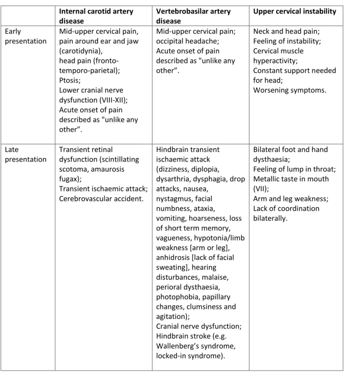
14 3.4 Differentiation
The following information is provided to assist in the differential diagnosis of musculoskeletal dysfunction from serious pathologies which commonly manifest as musculoskeletal dysfunction (Arnold and Bousser, 2005; Arnold et al, 2006; Kerry et al, 2008; Kerry, 2011):
Table 3.1: Differential diagnosis
Internal carotid artery disease
Vertebrobasilar artery disease
Upper cervical instability Early
presentation
Mid-upper cervical pain, pain around ear and jaw (carotidynia),
head pain (fronto-temporo-parietal); Ptosis;
Lower cranial nerve dysfunction (VIII-XII); Acute onset of pain described as "unlike any other”.
Mid-upper cervical pain; occipital headache; Acute onset of pain described as "unlike any other”.
Neck and head pain; Feeling of instability; Cervical muscle hyperactivity;
Constant support needed for head; Worsening symptoms. Late presentation Transient retinal dysfunction (scintillating scotoma, amaurosis fugax);
Transient ischaemic attack; Cerebrovascular accident.
Hindbrain transient ischaemic attack (dizziness, diplopia,
dysarthria, dysphagia, drop attacks, nausea,
nystagmus, facial numbness, ataxia,
vomiting, hoarseness, loss of short term memory, vagueness, hypotonia/limb weakness [arm or leg], anhidrosis [lack of facial sweating], hearing disturbances, malaise, perioral dysthaesia, photophobia, papillary changes, clumsiness and agitation);
Cranial nerve dysfunction; Hindbrain stroke (e.g. Wallenberg’s syndrome, locked-in syndrome).
Bilateral foot and hand dysthaesia;
Feeling of lump in throat; Metallic taste in mouth (VII);
Arm and leg weakness; Lack of coordination bilaterally.
15 3.5 Typical case histories of vascular dysfunction
3.5.1 Common vertebral artery dissection Case:
A 46 year-old female supermarket worker presented to physical therapy with left-sided head (occipital) and neck pain described as “unusual”. She reported a 6 day history of the symptoms following a road traffic accident. The symptoms were progressively worsening. The pain was eased by rest. She reported a history of previous road traffic accidents. Past medical history included hypertension, high cholesterol, and a maternal family history of heart disease and stroke. Cranial nerve tests for VIII, IX, and X were positive and resting blood pressure was 170/110. Two days after assessment, the patient reported an onset of new symptoms including “feels like might be sick”, “throaty” and “feels faint” – especially after performing prescribed neck exercises. Two days after this, she reported a stronger feeling of nausea, loss of balance, swallowing difficulties, speech difficulties and acute loss of memory. Magnetic resonance arteriography revealed an acute hindbrain stroke related to a left vertebral (extra-cranial) artery dissection.
Synopsis:
A typical background of vascular risk factors and trauma, together with a classic pain distribution for vertebral arterial somatic pain which was worsening. Positive signs (blood pressure and cranial nerve dysfunction) were suggestive of cervical vascular pathology. Signs of hindbrain ischaemia developed in a typical time period post-trauma.
3.5.2 Vertebral artery with pain as the only clinical feature Case:
A friend presents to a physical therapist with a sore neck and unremitting headache. The individual complains that they “think” their “neck is out”. They ask if they can have their neck manipulated to “put it back in”. The headache has been present for 3-4 days and is getting worse. They note that the pain has been unrelieved by medication (paracetamol) and it appears to be of a mechanical
presentation. Without taking a full history and carrying out a physical examination, the physical therapist goes ahead and manipulates the neck. The result was the individual experiencing numbness and paralysis to their left arm and hand.
Synopsis:
Investigation post incident identified an intimal tear of the vertebral artery. The key issue in this case is that the presentation was not fully assessed through a detailed history and physical examination. The warning feature from the history of worsening pain, unrelieved by medication, combined with an inadequate physical examination and limited clinical reasoning, all contributed to an unfortunate and probably avoidable outcome.
16 3.5.3 Internal carotid artery dissection
Case:
A 42 year-old accountant presents to physical therapy with a 5 day history of unilateral neck and jaw pain, as well as temporal headache, following a rear-end motor vehicle collision. There is a
movement restriction of the neck and the physical therapist begins to treat with gentle passive joint mobilisations, and advises range of movement exercises. The following day, the patient’s pain is worse, and he has developed an ipsilateral ptosis. The patient’s blood pressure is unusually high. Synopsis:
On medical investigation, an extra-cranial dissection of the internal carotid artery was found. The patient had underlying risk factors for arterial disease, and the presentation was typical of internal carotid artery dissection, with a key differentiator being the ptosis. A dramatic systemic blood pressure response was a result of this vascular insult.
3.5.4 Further examples of similar cases can be found in the literature
(Biousse et al, 1994; Lemesle et al, 1998; Crum et al, 2000; Zetterling et al, 2000; Chan et al, 2001; Caplan and Biousse, 2004; Arnold and Bousser, 2005; Asavasopon et al, 2005;
Rogalewski and Evers, 2005; Taylor and Kerry, 2005; Thanvi et al, 2005; Arnold et al, 2006; Debette and Leys, 2009; Kerry and Taylor, 2009).
17 Section 4: Planning the physical examination 4.1 Necessity for planning
A process of interpreting the data from the patient history and defining the main hypotheses is essential to an effective physical examination (Maitland et al, 2005; Petty, 2011). Hypothesis generation from the history and refining, re-ranking and rejecting of these hypotheses in the physical examination is necessary to facilitate optimal clinical reasoning in OMT (Jones and Rivett, 2004). Therefore careful planning of the physical examination is required. In particular for this framework, the possible vasculogenic (cervical arterial) contribution to the patient’s presentation needs to be clearly evaluated from the patient history data.
4.2 Are any further patient history data required?
An important component of planning is the identification of any further patient history data that may be required. That is, are there any gaps in the information obtained? Is the quality of the information obtained sufficient?
4.3 Decision-making regarding the physical examination
Based upon the evaluation and interpretation of the data from the patient history, the physical therapist needs to decide:
Are there any precautions to OMT? Are there any contraindications to OMT?
What physical tests need to be included in the physical examination?
What is the priority for these physical tests for this specific patient? This is to inform decisions regarding the order of testing and to determine which tests should be completed at the first visit.
18 Section 5: Physical examination
5.1 Blood pressure
Hypertension is considered a risk factor for carotid and vertebral artery disease. More acutely, an increase in blood pressure may be related to acute arterial trauma, including of the internal carotid and vertebral arteries (Arnold and Bousser, 2006). Evaluation of blood pressure as part of the physical examination may therefore be a valuable test to inform clinical reasoning.
Resting blood pressure should be taken in either sitting or lying, with the arm (brachial pulse site) being at the same level (in relation to gravity) as the heart / 4th intercostal space. A validated monitoring unit should be used ensuring the correct cuff-size. The cuff should be fitted so that two adult fingers can be inserted at the top and bottom when deflated. The patient should remain static in a calm environment for at least five minutes prior to testing. Repeat measurements can be taken leaving two minutes between each measurement.
Normotensive range (non-diabetic adult) is systolic 120-140mmHg / 70-90mmHg diastolic (Mancia et al, 2007).
Although hypertension is an undoubted strong predictor of cardiovascular disease, interpretation of readings must be in the context of other findings, and sound clinical reasoning. Vascular disease is an interplay between various factors, of which high blood pressure is just one (albeit a consistently important one). Blood pressure is a graduated, continuous measure and as such cannot have a threshold. The physical therapist should keep these points in mind during clinical decision-making. Hypertension and neck pain are only two of the many factors which influence the decision on probability of vascular pathology. Data regarding scaled risk is equally as clinically useful. There is a positive correlation between increased systolic and diastolic pressure and risk of stroke, which is the higher the pressure, the greater the risk. This would mean that a patient with say 190mmHg / 100mmHg is at greater risk than a patient with 160mmHg / 95mmHg. Thus, the risk is different even though they are both hypertensive. However, to reiterate, the actual utility of these data in isolation is limited as the true clinical risk is dependent on additional co-existing factors (Nash, 2007).
Patients with hypertension that has not been previously identified should be advised to discuss its implications with their primary care provider.
5.2 Craniovertebral ligament testing
Instability of the craniovertebral ligaments could compromise the vascular and neurological
structures in the upper cervical region. Mechanisms for causing symptoms and signs include: C1-C2 instability causing abnormal pressure on cervical nerves, vertebral artery compromise (Savitz and Caplan, 2005; Thanvi et al, 2005), and cord compression (Bernhardt et al, 1993; Rao, 2002). Whether to test for instability is therefore an important decision when suspecting CAD. The presence of instability is a clear contraindication to the use of OMT techniques (Gibbons and Tehan, 2006). There are a variety of ligaments that act together to maintain stability, and yet allow flexibility of the cervical region. These include the anterior and posterior longitudinal, interspinous, intertransverse, tectorial membrane, alar, transverse and ligamentum flavum ligaments (Panjabi and White, 1990).
19
Symptoms and signs of instability include (Gibbons and Tehan, 2005):
1. Facial paraesthesia secondary to dysfunction of the connections of the hypoglossal nerve, as well as the ventral ramus (neck-tongue paraesthesia) and the dorsal ramus (facial
numbness) of C2 2. Drop attacks
3. Bilateral or quadrilateral paraesthesia or motor deficits including weakness / incoordination 4. Nystagmus
5. Nausea.
Traditional instability testing techniques of the cervical region included the Sharp-Purser test, which is a comparatively safe procedure to perform to test the excursion of movement when relocating the dens to the atlas, in order to assess the transverse ligament. Other assessment procedures for instability included the tectorial membrane distraction and the alar ligament side flexion/bending and rotation tests (Cattrysse et al, 1997; Gibbons and Tehan, 2005). However in recent times, assessment of ligament stability has moved to systematically working through a series of active / patient generated, passive / therapist generated (with overpressure), and passive accessory movement tests, in order to feel the degree of movement or restriction at each joint and therefore ligament integrity, as well as to reproduce the patient’s symptoms.
Examples of active / patient generated tests for assessing cervical ligament integrity include: Atlanto-occipital joint isolation (nod)
C1-C2 rotation with the neck flexed
C2-C3 rotation with protraction and retraction
Upper cervical extension, and rotation and lateral flexion to same side (C0-C3)
Examples of passive / therapist generated (with overpressure) tests for assessing cervical ligament integrity include:
Fixation of C1 via the transverse processes of C1 and passive flexion/extension of the occiput (C0-C1)
Fixation of the C2 spinous process with passive side bending or rotation of the occiput (C0-C2)
Examples of accessory movement tests for assessing cervical ligament integrity include (Gibbons and Tehan, 2005):
Transverse atlantal ligament stress test (modified Sharp-Purser test) Alar ligament test.
(A useful resource for description of these tests is Mintken et al [2008a], which includes reference to videos that are available online).
Signs of instability from the aforementioned tests may include: 1. Increase in motion or empty end-feel
2. Reproduction of symptoms of instability 3. Production of lateral nystagmus and nausea.
For each individual patient, a decision needs to be made regarding the value of performing any craniovertebral ligament tests, evaluating the risks and benefits of any specific test procedure using
20
current evidence from research investigating validity of testing (e.g. Kaale et al, 2008). However, the evidence of the predictive ability of these tests to identify instability is lacking and the physical therapist should carefully consider whether physical testing is prudent or safe in the presence of subjective symptoms of instability. In some situations, for example a post acute trauma presentation following a road traffic accident, the best decision would be to support them with a cervical collar pending radiological investigation.
Patients who have age-related loss of spinal movement, or have experienced cervical region trauma (e.g. whiplash), or who have pathological conditions (congenital e.g. Downs syndrome, inflammatory e.g. rheumatoid arthritis, or marked degeneration e.g. osteoarthritis) that may affect cervical spine ligament integrity require further craniovertebral ligament screening e.g. flexion-extension
radiographic views and / or MRI. 5.3 Neurological examination
Examination of the peripheral nerves, cranial nerves, and for an Upper Motor Neurone lesion will assist in evaluating the potential for neuro-vascular conditions (see Fuller [2008] for a detailed description of how to perform testing or www.neuroexam.com).
5.4 Positional testing
Provocative positional testing is frequently used in practice. It is intended to provide a challenge to the vascular supply to the brain, and the presence of signs or symptoms of cerebrovascular
ischaemia during or immediately post testing is interpreted as a positive test. Sustained end-range rotation has been advocated, and has been described as the most provocative and reliable test (Mitchell et al, 2004). The sustained pre-manipulative test position has also been advocated (Rivett et al, 2006). However, the predictive ability of either of these tests to identify at risk individuals is lacking.
5.5 Palpation of the carotid artery
Palpation of the common and internal carotid arteries is possible due to the size of these vessels and their relatively superficial anatomy. Although no meaningful diagnostic utility statistics exist in relation to its precise role in predicting potentially adverse outcomes, carotid palpation is conventionally used as part of a clinical work-up for carotid artery dysfunction (e.g. Cournot et al 2007; Cury et al 2009; Atallah et al 2010). Asymmetry between left and right vessels is considered. A pulsatile, expandable mass is typical of arterial aneurysm. Such a finding should be considered in the context of other clinical findings. It is possible for dissections and disease of the carotid arteries to exist in the absence of aneurysm formation, therefore a negative finding should not be used to refute the hypothesis of arterial dysfunction.
Palpation of the vertebral arteries is much less likely to provide meaningful information due to the small diameter of the vessel and its relatively inaccessible anatomy.
As pulse palpation is a relatively simple psycho-motor skill, training in this area should be focussed on anatomical landmarks and vessel palpation. Ideally, the physical therapist would aim to
understand and experience both normal and pathological pulse quality. 5.6 Differentiation
Differentiation of a patient’s symptoms originating from a vasculogenic cause with complete certainty is not currently possible from the physical examination. Thus, it is important for the
21
physical therapist to understand that headache / neck pain may be the early presentation of an underlying vascular pathology (Rivett, 2004; Taylor & Kerry, 2010). The task for the therapist is to differentiate the symptoms by:
1. Having a high index of suspicion 2. Testing the vascular hypothesis.
This process of differentiation should take place from an early point in the assessment process i.e. early in the patient history. The symptomology and history of a patient experiencing vascular pathology is what may alert the physical therapist to such an underlying problem (Rivett, 2004; Taylor & Kerry, 2010). A high index of suspicion of cervical vascular involvement is required in cases of acute onset neck/head pain described as “unlike any other” (Taylor & Kerry, 2010). Physical therapists may be exposed to patients presenting with the early signs of stroke (for example, neck pain / headache) and as such need both knowledge and awareness of the mechanisms involved. A basic understanding of vascular anatomy, haemodynamics and the pathogenesis of arterial dysfunction may help the physical therapist differentiate vascular head and neck pain from a musculoskeletal cause (Rivett, 2004; Taylor & Kerry, 2010) through interpretation of the patient history data and tests in the physical examination. Kerry and Taylor (2006) provide a summary of key physical examination tests and their value for differentiating vasculogenic head and neck pain, including: cervical rotation positional test, cervical extension positional test, blood pressure examination, cranial nerve examination, eye examination, use of hand held Doppler ultrasound, holding head and turning body test, and the Dix-Hallpike manoeuvre.
5.7 Refer on for further investigation
There are no standardised clinical guidelines for medical diagnostic work-up in respect of vertebral and carotid arterial dysfunction. It is recommended that the physical therapist follows local policy in referring for further investigation. Conventionally, duplex ultrasound, magnetic resonance
imaging/arteriography, and computed tomography are used in the work-up (Cury et al 2009; Jones et al 2010). Being non-invasive and cheaper, duplex ultrasound is often considered first. The primary aim is to differentiate between haemorrhagic sources for the signs and symptoms and any other cause, as this will dictate the management pathway. It is recommended that physical therapists refer for immediate medical investigation when their clinical suspicion is supported by the reasoned historical details and clinical examination findings as suggested in this document.
5.8 Additional training
It is acknowledged that some physical tests included in this section may not be in the domain of current OMT practice in some countries. It is recommended that in those countries where these tests are not within the domain of current practice that their use is considered. Any additional training required in physical examination techniques could be achieved within a physical therapist’s local environment.
22 Section 6: Risk versus benefit analysis
6.1 Framework for evaluating risk
The risk associated with OMT intervention for musculoskeletal cervical spine disorders should be considered within a clinical reasoning framework. That is, the risk, albeit likely extremely low in general and in comparison to some other conservative treatments (Rivett 2004), may vary somewhat depending on the patient’s individual clinical presentation, and in particular in the presence of risk factors previously discussed (see Sections 3.1 and 3.2). It is therefore the
responsibility of the physical therapist to recognise and consider whether the risk for a particular patient is increased, and to do whatever is reasonable to minimise any risk associated with OMT intervention.
Risk versus benefit analysis:
Data and evidence surrounding the clinical concern of this framework are incomplete and often contradictory. It is important to appreciate that an absolute diagnosis cannot be made by the physical therapist. The physical therapist must accept that the clinical decision is made in the absence of certainty and a decision based on a balance of probabilities is the aim of assessment. Although some presentations absolutely contraindicate OMT intervention, others suggest risk factors for potential adverse events and may co-exist with treatable musculoskeletal dysfunction. It is the responsibility of the physical therapist to make the best decision regarding treatment in these situations using their clinical reasoning skills (Jones and Rivett, 2004; Kerry and Taylor, 2009). The following model provides a simple framework for decision-making regarding risk versus benefit but should not be considered didactic:
Table 6.1: Decision-making framework for analysing risk versus benefit
Risk
Benefit
Action
High number/ severe nature of risk factors
Low predicted benefit of manual therapy
Avoid treatment
Moderate number / moderate nature of risk factors
Moderate predicted benefit of manual therapy
Avoid or delay treatment / monitor and reassess
Low number / low nature of risk factors
Low / moderate / high predicted benefit of manual therapy
Treat with care / continual monitoring for change/new symptoms
23 Section 7: Flowchart of clinical reasoning
Data obtained from patient history
Planning the physical examination
Possible cervical vasculogenic contribution?
Any gaps in data from patient history?
Quality of data obtained?
Any precautions or contraindications?
Which physical tests to use?
Priority for physical testing?
Interpretation of data from patient history using evidence informed knowledge, and cognitive and metacognitive processes.
Interpretation includes analysis of patient’s preferences.
Interpretation of data from physical examination using
evidence informed knowledge, and cognitive and metacognitive processes. Interpretation includes analysis of patient’s preferences. Data obtained from physical examination
Evaluation of patient’s presentation Any gaps in data from assessment?
Quality of data obtained?
Risk versus benefit analysis?
Decision re action?
Best decision regarding management In collaboration with the patient
24
Section 8: Informed consent and medico-legal framework 8.1 Informed consent
Informed consent is comprised of both ethical and legal components. Patient consent to treatment is a standard of physical therapy practice. The specific requirements of informed consent will vary from country to country according to local laws, customs and norms. This section provides physical therapists with information on this process based on the literature and current generally accepted ethical and legal standards.
Application to individual Member Organisations:
Given the international audience of this document, Member Organisations are advised to check local laws and health regulations affecting the informed consent process.
Please consult your MO for any local information or requirements. Member Organisations are encouraged to add any local requirements as an addendum to this document to facilitate use of the document in their local context.
In seeking informed consent, the physical therapist should be confident that the patient will benefit from treatment and that the risk is minimal. Informed consent can be defined as “the voluntary and revocable agreement of a competent individual to participate in a therapeutic or research
procedure, based on an adequate understanding of its nature, purpose and implications” (Sim, 1986). The process of informed consent includes the following components: the types of consent, the requirements of disclosure of information by the therapist, how it is obtained, and the requirements of record keeping of the informed consent process. It is important to note that informed consent is part of the process of clinical reasoning. This acknowledges the importance of dialogue between the physical therapist and patient about treatment alternatives, in combination with the patient’s preferences, so that mutually agreed choices of care can be made (Charles et al, 1997; Jones and Rivett, 2004). Further, it infers the importance of the patient’s autonomy and that their right to make decisions throughout the treatment process is ongoing and not a one-off event (Delany, 2005).
8.2 Types of consent
Express consent is given explicitly either in writing or verbally (Sim, 1997) (e.g. the patient expressly states that they agree, or signs a form indicating agreement). This is recommended when initially seeking informed consent for a treatment intervention e.g. cervical manipulation, as it provides the clearest form of consent and often fulfills legal obligations.
Implied consent is not specifically indicated as in express consent, but is implied by some action which suggests consent (Sim, 1997) (e.g. after having a discussion with the physical therapist regarding treatment, the patient lays down on the treatment bed signaling that they are a willing participant). This form of consent is open to interpretation and is therefore less reliable upon legal scrutiny.
Tacit consent is failure of the patient to disagree or dissent (Sim, 1997). This form of consent is open to interpretation and is therefore less reliable upon legal scrutiny.
Embodied consent is assessment of the patient’s body language for consent to treatment, prior to and during treatment (Fenety et al, 2009). Since express consent is initially recommended for treatment interventions e.g. cervical manipulation, embodied consent becomes important during the treatment. The body language of the patient should be observed specifically during the pre-manipulative hold and assessed for indications that they may be reconsidering the initial express
25
consent that was given. If the therapist observes body language that may indicate the patient is uncomfortable with proceeding, the therapist should stop the procedure and ask the patient if it is acceptable to continue.
Whatever the form of the consent, it should be given voluntarily and without undue influence from the therapist, and once the patient has given consent they can withdraw their consent at any time during treatment.
8.3 Disclosure of Information
It is recommended that physical therapists provide patients with information about the proposed assessment and treatment procedures. The information provided can be communicated verbally or by written material, such as an information brochure. The most prudent approach is to use both verbal and written communication (Purtillo, 1984).
Once again, Member Organisations are advised to check local laws and health regulations affecting the informed consent process as the legal requirements may vary from country to country.
Provision of a brochure is optional, but allows patients time to consider the recommendations, ask questions, and make an informed choice overall. It can be given to the patient to read prior to treatment while they are in the waiting room or in the clinic. If the patient requires further time before making a decision, a brochure can be taken home for consideration. Provision of a brochure ensures that the information is standardised and allows for easy record keeping of the informed consent process by indicating that the brochure was given.
It is recommended that the information provided to the patient cover the following points (Appelbaum et al, 1987; Wear, 1998). It is important to note that the points apply to any physical therapy intervention:
It must be specific to the proposed treatment. It must cover alternative treatment options.
It must cover benefits and risks of the proposed treatment and alternatives.
Note: It is important to remember that an analysis of risk is based upon a physical therapist’s analysis of the situation i.e. the interaction of the patient, therapist and planned
intervention to inform the level of risk.
Omission of any of the above information may invalidate the consent of the patient. It is the responsibility of the physical therapist to ensure that the patient fully understands all of the information that has been provided. It is also the responsibility of the physical therapist to provide further information requested by the patient and to answer all questions asked by the patient in a manner that the patient considers satisfactory (Wear, 1998).
Once again, Member Organisations are advised to check local laws and health regulations affecting the informed consent process as the legal requirements may vary from country to country.
8.4 Obtaining informed consent
Informed consent is obtained when a patient explicitly indicates either verbally or in writing, following adequate disclosure of information about the proposed procedure, their consent to proceed with the treatment. Consent must be obtained before treatment begins. Asking the patient for consent while treatment is in progress may adversely influence the patient’s decision-making and is not recommended (Jensen, 1990).
26
For changes in treatment (introducing a different type of technique), the full process of informed consent must be undertaken and consent explicitly obtained verbally or in writing.
e.g. You have been treating a patient using intervention A. The patient has not responded as you had hoped and you would like to now try intervention B. Intervention B is considered to be a new or different treatment to intervention A. Therefore, if the initial process of obtaining informed consent did not include information pertaining specifically to intervention B, the physical therapist must specifically gain informed consent for the use of intervention B prior to its application.
For continuation of the same treatment (e.g. intervention A), it is recommended that consent be obtained each time it is used. This does not necessarily entail the full disclosure of information that was required the first time. Agreement by the patient verbally to the ongoing use of intervention A in most cases would be sufficient. If however, in follow up discussion with the patient, you perceive that there is a lack of understanding of the previously disclosed information, it is recommended that the full process of disclosure of information be revisited.
8.5 Recording of informed consent
It is recommended that the disclosure of information and the obtaining of informed consent be recorded in a standardised manner in the patient’s clinical record.For each treatment, it is recommended that the obtaining of informed consent be recorded each time.The use of stickers (one for the initial informed consent process and one for follow up visits) is suggested to standardise and facilitate ease of recording. Stickers can be designed with a series of bullet points that can be ticked. Similar strategies can be used in an electronic medical record system.
27 Section 9: Safe OMT practice
9.1 Range of techniques recommended as good practice
OMT practice encompasses a wide range of therapeutic manoeuvres from patient activated forces to therapist activated forces. OMT is integrated into the overall management strategy of patient care. Reports of patient harm from OMT in the cervical region have typically been in the practice of cervical manipulation.
The following are necessary considerations for the physical therapist during the selection and application of cervical manipulation (Rivett, 2004; Childs et al, 2005):
The principle of all techniques is that minimal force should be applied to any structure within the cervical spine i.e. low amplitude, short lever thrusts.
Patient safety and comfort form the basis of appropriate technique selection. Cervical manipulation techniques should be comfortable to the patient.
Cervical manipulation techniques should not be performed at the end of range of cervical movement, particularly extension and rotation.
There is flexibility in the choice of the patient’s position using the principles that the patient needs to be comfortable, and that the physical therapist needs to be able to receive
feedback. The use of the supine lying position with the patient’s head supported on a pillow is encouraged. This position allows the physical therapist to monitor facial expressions, eye features, etc.
Positioning the patient in the pre-manipulative test position prior to a manipulation is good practice to evaluate patient comfort and to enable evaluation of their response.
The patient response to all cervical spine movements, including cervical manipulation interventions is continuously monitored.
The skills of the physical therapist may be a limitation for the selection of manipulation as a treatment technique, even though clinical reasoning may suggest manipulation is the best choice. In this situation, a risk may be introduced owing to limited clinical skills and it would therefore be a responsible decision to not use manipulation. The self-evaluative skills of the physical therapist in evaluating their ability to perform the desired technique safely and efficiently are therefore important. Referral to a colleague suitably qualified/trained in the desired manipulative technique may be appropriate.
9.2 Alternative approaches to direct cervical treatment.
Emerging pain sciences suggests that the effects of manual techniques (such as mobilisation and manipulation) on pain may be largely neurological in nature and not limited to the direct influence of a particular spinal motion segment. Furthermore, clinical trials have reported that thoracic spine manipulation results in improvements in perceived levels of cervical pain, ranges of motion, and disability in patients with mechanical neck pain (Cleland et al, 2005; 2007a and b; Krauss et al, 2008; Gonzalez-Inglesias et al, 2009), although the mechanism by which this occurs is not known. Given the concern regarding the risks associated with cervical spine manipulation, thoracic spine
manipulation provides an alternative, or supplement to, cervical manipulation and mobilisation to maximise the patient’s outcome with an extremely low level of risk. The current evidence suggests that during the initial treatment sessions there is a large likelihood of improved patient outcomes when thoracic manipulation is coupled with cervical active range of movement exercises (Cleland et al, 2005; 2007a and b; Krauss et al, 2008; Gonzalez-Inglesias et al, 2009). Subsequent sessions can then introduce more direct manual cervical treatments if warranted. This approach allows the therapist to observe the patient’s response to treatment over a longer time period and theoretically
28
minimises the risks associated with cervical manipulation in the presence of an emerging cervical vascular disorder, such as arterial dissection.
9.3 Frequency of treatment
Frequency of treatment will vary depending on the individual and injury in question. Current evidence suggests that manual interventions should be coupled with therapeutic exercise when managing a patient’s neck pain and headache (Jull et al, 2002; Kay et al, 2005; Walker et al, 2008). Caution should be applied in situations where the patient’s preference is for repeated manipulation, owing to potential dangers of frequent repeated manipulation and a lack of longer term benefit. 9.4 Minimising end-range cervical techniques
End of range movements are known to stress the cervical arteries and potentially neural structures. Thus avoidance of these positions is recommended during cervical manipulation (Hing et al, 2003; Rivett, 2004). Although evidence is limited, this principle also logically applies to techniques performed in end range neck positions during cervical mobilisation and exercise interventions. 9.5 Force minimisation
OMT techniques used to treat the cervical region should be applied in a controlled, comfortable manner in mid ranges of cervical movement in order to reduce the potential stress on vascular and neurological structures. The influence of the head and cervical spine segments not included in the manipulation can be used to direct loads to the targeted segment. Therefore by doing this, there is little stress on the rest of the neck and the elimination of cervical spine locking positions (Hing et al, 2003).
9.6 Monitor for any adverse effects
Monitoring the patient for response to treatment and any adverse effects is a continual process throughout and after the treatment session. Verbal and physical examination can be carried out while performing a treatment technique through monitoring physical body behaviour, facial expression, muscle tone, and verbal communication / responsiveness. Grading scales designed by Maitland et al (2005) and Kaltenborn (2003) can be used to guide the physical therapist, providing an objective measure of the patient’s progress during treatment. Similarly, in the osteopathic model, there is considerable emphasis placed on the physical examination of the joint ‘barrier’ (Greenman 1996; Hartman 1997) and end-feel. Movement diagrams (Maitland et al, 2005) and other
components of the physical examination can be reviewed post treatment to assess for changes in the physical behaviour of the cervical region. However, the ultimate standard of response should be based on the change in a patient reported outcome measure (e.g. Neck Disability Index, Global Rating of Change, etc).
9.7 Emergency management of an adverse situation
As a health professional, the physical therapist is expected to act swiftly and judiciously when confronted with an emergency situation. A plan of action should be devised, available, and operational for effective management of an adverse situation. If a patient becomes unresponsive during any aspect of physical therapy care, the physical therapist should immediately implement an emergency action plan for cardiopulmonary resuscitation. Emergency help should be sought immediately, such as calling for an ambulance. Training in cardiopulmonary resuscitation should be completed on a regular basis.
29 Section 10: Teaching OMT for the cervical region
10.1 Framework for those teaching cervical assessment and management
A variety of manual assessment and intervention techniques are being used in the assessment and management of the cervical spine. The reports of patient harm from OMT in the cervical region have typically been in the practice of cervical manipulation. The teaching of OMT for the cervical region requires instructors to have a thorough understanding and proficiency in:
assessment for pathology that is outside the usual physical therapist’s scope of practice understanding of the implications of findings from musculoskeletal diagnostic imaging the use of tools to determine baseline status, treatment outcomes, and prognostic
indicators
neuromusculoskeletal examination procedures including sensory-motor function, vascular status and ligamentous integrity
palpatory skills of the cervical region differential diagnosis and clinical reasoning
Practical skills teaching and examination of competency are necessary components of manipulation instruction at all levels of physical therapy education programmes. Based on the available literature, instruction should particularly emphasise the continuum of the amplitude, velocity, patient comfort, and sensitivity and specificity of handling during manipulation tutoring (Flynn et al, 2006; Mintken et al, 2008b). This continuum reflects the excellence in manual skills to enable physical therapists to perform manipulation efficiently and effectively.
Practical skills teaching and examination of competency involves students practising cervical techniques on their peers. Instruction should therefore include a process of evaluation of peers to act as models for OMT technique practice.
10.2 Recommended qualifications for instructors
Educational qualifications for first professional (entry-level) and post-professional training
instructors vary across the world. However, recommended attributes of instructors responsible for teaching the cognitive and psychomotor skills used in cervical manipulation are described below (these are provided to guide educational programmes when planning instructor development processes and resources). Importantly, instructors should:
1. Be actively engaged in clinical practice within the area of their expertise and instruction, and have an appropriate amount of relevant clinical experience.
2. Possess teaching experience that preferably includes mentoring or formal training in adult educational processes and methods.
3. Apply evidence-based concepts within both their clinical practice and teaching.
4. Have been trained and examined in didactic and psychomotor aspects of manual therapy, including manipulation, or the equivalent.
5. Have completed a formally accredited (by an IFOMPT recognised national body) post-professional programme in manual therapy.
30
6. Regularly undertake ongoing professional education and training relevant to cervical manipulation.
The instructor should be appropriately qualified to ensure that the student can:
1. Demonstrate competency in both performing and interpreting examination procedures appropriate for physical therapy management and prevention of musculoskeletal disorders of the cervical spine.
2. Demonstrate competency in both the technical application and interpretation of response to manipulative interventions utilised in the management of musculoskeletal disorders of the cervical spine.
Furthermore, specific safety precautions associated with manipulation in general, and particularly manipulation in the cervical spine are a necessary component of instruction. Students should be competent in making decisions regarding when to utilise manipulation, and when to refer to a physician or other practitioner based on safety or other medical concerns.
10.3 Educational resources
When teaching manipulation techniques in the cervical region it is essential to present techniques which are easy to understand and implement in the clinical setting. There is a vast array of physical therapy and medical resources that describe the management of cervical spine disorders, including those related to manual and manipulative therapy. Physical therapists should be well versed in current best evidence for managing cervical disorders. This document does not endorse any specific philosophy or approach to manipulation, however the physical therapist is responsible for choice, application, and monitoring of responses to manipulative techniques following the principles outlined in this document.