Chapter 13: Viral Diseases
13.1: BK POLYOMA VIRUS13.1.1: We suggest screening all KTRs for BKV with quantitative plasma NAT (2C) at least:
• monthly for the first 3–6 months after transplantation(2D);
• then every 3 months until the end of the first post-transplant year(2D);
• whenever there is an unexplained rise in serum creatinine(2D); and
• after treatment for acute rejection. (2D)
13.1.2: We suggest reducing immunosuppressive medications when BKV plasma NAT is per-sistently greater than 10 000 copies/mL (107copies/L).(2D)
BKV, BK polyoma virus; KTRs, kidney transplant reci-pients; NAT, nucleic acid testing.
Background
BK polyoma virus (BKV) is a member of the polyoma fam-ily of viruses. BKV can cause nephropathy, which is diag-nosed by kidney biopsy. Reduction of immunosuppression is defined as a decrease in the amount and intensity of im-munosuppressive medication. Nucleic acid testing (NAT) is defined as one or more molecular methods used to iden-tify the presence of DNA or RNA (e.g. polymerase chain reaction).
Rationale
• The use of NAT to detect BKV in plasma provides a sensitive method for identifying BKV infection and determining KTRs who are at increased risk for BKV nephropathy.
• Early identification of BKV infection may allow mea-sures to be taken that may prevent BKV nephropathy.
• When NAT is not available, microscopic evaluation of urine for the presence of decoy cells is an accept-able, albeit nonspecific, alternative screening method for BKV disease and the risk for BKV nephropathy.
• Fifty percent of patients who develop BK viremia do so by 3 months after kidney transplantation.
• Ninety-five percent of BKV nephropathy occurs in the first 2 years after kidney transplantation.
• BKV plasma NAT >10 000 copies/mL (107 copies/L)
has a high positive predictive value for BKV nephropathy.
• Reduction of immunosuppressive medication may re-sult in reduced BKV load and decreased risk of BKV nephropathy.
• Histologic evidence of BKV nephropathy may be present in the absence of elevated serum creatinine.
• Reduction in maintenance immunosuppressive medi-cation is the best treatment for BKV nephropathy.
Whether to screen KTRs with NAT of plasma or urine has been controversial. A negative urine NAT for BKV has al-most a 100% negative predictive value (298). By testing urine, one can avoid performing BKV testing of blood on those patients with negative urine studies. Based on this, some experts recommend screening of urine as the defini-tive site for BKV surveillance (298). However, the presence of a positive NAT for BKV in urine, in the absence of an elevated BKV load in the plasma, is not associated with an increased risk for BKV disease (298). Hence, the use of urine screening requires performance of NAT on the blood of those patients whose level of BK viruria exceeds estab-lished thresholds. This requires patients to return to the clinic for the additional test. Accordingly, it is suggested that NAT be performed on plasma, and not the urine of KTRs.
When NAT is not available, microscopic evaluation of the urine for the presence of decoy cells is an acceptable, albeit nonspecific, alternative screening method for BKV disease and the risk for BKV nephropathy. A negative screening test rules out BKV nephropathy in most cases (high negative predictive value). However, a positive screening test has a very low positive predictive value for BKV nephropathy (298,299). Thus, many patients with urine decoy cells will not develop BKV nephropathy. It may be inappropriate to change therapy in such patients based on the presence of urine decoy cells alone.
Emerging data suggest that BKV nephropathy can be pre-vented if immunosuppressive medications are reduced in patients with BKV detected by a high viral load in plasma (determined by NAT) (300).
Timing of BKV NAT
The presence of BKV can be identified prior to the onset of clinical symptoms at a time when only subclinical infection is present, or in association with clinically apparent BKV nephropathy. Evidence to date suggests that the presence
of BK viremia precedes BKV nephropathy by a median of 8 weeks. Approximately, 50% of patients who will develop BK viremia will do so by 3 months after transplant (298). Most BKV nephropathy occurs in the first 2 years after transplant with only 5% of cases occurring between 2 and 5 years after transplant (298). Accordingly, the timing and frequency of testing in recommended screening algo-rithms should reflect these data and balance the cost of screening with the potential to prevent BKV nephropathy. The proposed screening algorithm is most intense early after kidney transplantation, with decreasing frequency as patients are out longer from the transplant. Although we have not recommended screening beyond the first year af-ter transplant, an inaf-ternational consensus conference sug-gested continued annual screening for patients between 2 and 5 years after kidney transplantation (298). Centers with higher frequency of BKV might follow this approach. Screening for the presence of BKV should also be per-formed for patients with unexplained rises in serum cre-atinine, as this may be attributable to BKV nephropathy. Finally, screening should be considered for those patients who have undergone a major increase in immunosuppres-sive medication, as they may be at risk of developing BKV nephropathy.
Rising BKV load
There is increased risk of BKV nephropathy associated with a rising BKV load in plasma (298,299). Although plasma NAT assays for BKV lack standardization, a thresh-old plasma BKV level of>10 000 copies/mL (107copies/L)
is associated with a 93% specificity for the presence of BKV nephropathy. In the absence of evidence of clinical disease, KTRs with BKV levels in excess of this thresh-old are considered to be at risk of progression to BKV nephropathy (298,299). Histologic evidence of early BKV nephropathy may be present prior to detection of elevated serum creatinine (298).
The risk of BKV nephropathy appears to be correlated with the intensity of immunosuppression, and reduction of
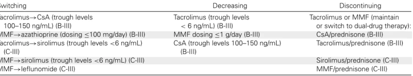
im-Table 14: Treatment of BKV nephropathy by modification of maintenance immunosuppression
Switching Decreasing Discontinuing
Tacrolimus→CsA (trough levels Tacrolimus (trough levels Tacrolimus or MMF (maintain
100–150 ng/mL) (B-III) <6 ng/mL) (B-III) or switch to dual-drug therapy):
MMF→azathioprine (dosing≤100 mg/day) (B-III) MMF dosing≤1 g/day (B-III) CsA/prednisone (B-III)
Tacrolimus→sirolimus (trough levels<6 ng/mL) CsA (trough levels 100–150 ng/mL) Tacrolimus/prednisone (B-III)
(C-III) (B-III)
MMF→sirolimus (trough levels<6 ng/mL) (C-III) Sirolimus/prednisone (C-III)
MMF→leflunomide (C-III) MMF/prednisone (C-III)
BKV, BK polyoma virus; CsA, cyclosporine A; MMF, mycophenolate mofetil.
B-III, ‘moderate evidence to support a recommendation for use’ based on ‘evidence from opinions of respected authorities, based on clinical experience, descriptive studies or reports of expert committees.’ Likely equivalent to a 2D recommendation.
C-III, ‘poor evidence to support a recommendation’ based on ‘evidence from opinions of respected authorities, based on clinical experi-ence, descriptive studies or reports of expert committees.’ Likely equivalent to a 2D recommendation.
Reprinted with permission (298).
munosuppression can result in a decrease in BKV load and a concomitant reduction of risk of development of BKV nephropathy (301). A RCT reported that withdrawal of the antimetabolite resulted in clearance of viremia without pro-gression to BKV nephropathy (300). Although some would use antiviral therapy (including cidofovir, leflunomide and/or ciprofloxacin) as treatment, to date there are no definitive data confirming the effectiveness of these agents for either treatment or prevention of BKV nephropathy (298,299). Some centers may choose different treatment strategies for patients with elevated BKV loads in the absence of any histologic changes, compared to patients with find-ings of BKV nephropathy in the absence of serum creati-nine elevation. The international consensus group recom-mended performance of kidney biopsy for these patients (298). When a kidney biopsy is obtained, it should be eval-uated for the presence of BKV using the cross-reacting antibody for simian virus 40. However, other experts have not recommended the performance of a kidney biopsy for asymptomatic patients with an elevated BKV load (300). Treating biopsy-proven BKV nephropathy
The treatment of BKV nephropathy is unsatisfactory. Although there are some centers that would use an-tiviral therapy (including cidofovir, leflunomide and/or ciprofloxacin) as treatment, to date there are no defini-tive data confirming their effecdefini-tiveness. However, reduc-tion of immunosuppression does appear to have some im-pact on BKV nephropathy, though variable rates of graft loss attributable to BKV nephropathy have been reported even when reduction of immunosuppression has been employed (Table 14). A common practice of immunosup-pressive dose reduction is withdrawal of antimetabolite (azathioprine or MMF) and reduction in CNI dosage by 50%. An algorithm for the treatment of BKV nephropathy through modification of baseline immunosuppression has been proposed (298). Switching from the antimetabolite MMF or EC-MPS to leflunomide (an immunosuppressive agent with antiviral activity) has been associated with de-clining BKV load in blood and improving histology (302),
although convincing evidence of the efficacy of this, or other antiviral agents, is lacking.
Research Recommendations
Studies are needed to determine:• the most cost-effective strategies for screening for BKV in different populations;
• the efficacy of altering immunosuppressive medication regimens and of antiviral agents in the prevention and treatment of BKV nephropathy.
13.2: CYTOMEGALOVIRUS
13.2.1: CMV prophylaxis: We recommend that KTRs (except when donor and recipient both have negative CMV serologies) re-ceive chemoprophylaxis for CMV infection with oral ganciclovir or valganciclovir for at least 3 months after transplantation, (1B) and for 6 weeks after treatment with a T-cell–depleting antibody.(1C)
13.2.2: In patients with CMV disease, we sug-gest weekly monitoring of CMV by NAT or pp65 antigenemia.(2D)
13.2.3: CMV treatment:
13.2.3.1: We recommend that all patients with serious (including most pa-tients with tissue invasive) CMV disease be treated with intra-venous ganciclovir.(1D)
13.2.3.2: We recommend that CMV dis-ease in adult KTRs that is not serious (e.g. episodes that are associated with mild clinical symptoms) be treated with ei-ther intravenous ganciclovir or oral valganciclovir.(1D)
13.2.3.3: We recommend that all CMV disease in pediatric KTRs be treated with intravenous ganci-clovir. (1D)
13.2.3.4: We suggest continuing therapy until CMV is no longer detectable by plasma NAT or pp65 antigen-emia.(2D)
13.2.4: We suggest reducing immunosuppressive medication in life-threatening CMV dis-ease, and CMV disease that persists in the face of treatment, until CMV disease has resolved.(2D)
13.2.4.1: We suggest monitoring graft function closely during CMV dis-ease.(2D)
CMV, cytomegalovirus; KTRs, kidney transplant recipi-ents; NAT, nucleic acid testing.
Background
Cytomegalovirus disease is defined by the presence of clinical signs and symptoms attributable to CMV infec-tion, and the presence of CMV in plasma by NAT or pp65 antigenemia. CMV disease may manifest as a nonspe-cific febrile syndrome (e.g. fever, leukopenia and atypical lymphocytosis) or tissue-invasive infections (e.g. hepati-tis, pneumonitis and enteritis). Tissue-invasive CMV dis-ease is defined as CMV disdis-ease and CMV detected in tissue with histology, NAT or culture. Serologically, nega-tive CMV is defined by the absence of CMV immunoglob-ulin G (IgG) and immunoglobimmunoglob-ulin M. Serologically pos-itive for CMV is defined as being CMV IgG-pospos-itive. Interpretation of CMV serologies may be confounded by the presence of passive antibody that may have been ac-quired from a blood or body-fluid contamination. Chemo-prophylaxis is defined as the use of an antimicrobial agent in the absence of evidence of active infection, to pre-vent the acquisition of infection and the development of disease.
Rationale
• CMV disease is an important cause of morbidity and mortality.
• There are strategies for preventing CMV infection and disease that result in marked improvements in out-comes.
• Risk for CMV after transplantation is strongly depen-dent on donor (D) and recipient (R) serology, with pa-tients who are D+/R−, D+/R+ or D−/R+ at risk for developing CMV infection and disease, and D+/R−at highest risk for severe CMV disease.
• The incidence of CMV disease in D−/R−is<5%.
• Chemoprophylaxis with ganciclovir or valganciclovir for at least 3 months after transplantation reduces CMV infection and disease in high-risk patients.
• Chemoprophylaxis is associated with improved graft survival compared to preemptive antiviral therapy initi-ated in response to increased CMV load.
• The use of a T-cell–depleting antibody is a risk factor for CMV disease.
• Chemoprophylaxis with ganciclovir for patients receiv-ing a T-cell–depletreceiv-ing antibody protects against the de-velopment of CMV disease.
• A detectable CMV load at the end of antiviral therapy is associated with an increased risk of disease recur-rence.
Preventing CMV
Cytomegalovirus is a frequent and important cause of clini-cal disease in KTRs. In the absence of antiviral prophylaxis, symptomatic CMV disease can be seen in approximately 8% of KTRs (303), although older estimates placed it at 10–60% of KTRs (304). In addition to directly attributable morbidity, CMV may also have an immunomodulatory ef-fect, and active CMV disease has been associated with infectious complications as well as acute rejection and CAI (305). Accordingly, strategies that can prevent CMV infec-tion and disease should lead to improved outcomes follow-ing kidney transplantation.
Randomized controlled trials have demonstrated that the incidence of CMV disease can be reduced by pro-phylaxis and preemptive therapies in solid-organ trans-plant recipients (306–308). In trials of KTRs alone, there is low-quality evidence, largely due to sparse data, that prophylaxis results in less acute rejection and CMV infection, with no clear evidence of increased ad-verse events (see Evidence Profile and accompanying evidence in Supporting Tables 48–49 at http://www3. interscience.wiley.com/journal/118499698/toc). However, there is high-quality evidence from a large systematic re-view that CMV prophylaxis in solid-organ transplant recip-ients (307) significantly reduces all-cause mortality, CMV disease mortality, CMV disease, but not acute rejection or graft loss. In most of these trials, the majority of or-gan recipients received kidneys. Thus, the Work Group concluded that overall there is moderate-quality evidence to support this recommendation. Observational data sug-gest that D+/R− KTRs are at the highest risk of devel-oping severe CMV disease compared to all other KTRs (306). Studies in this high-risk population have shown that antiviral chemoprophylaxis reduces the incidence of CMV disease by about 60% (306). The use of antiviral chemo-prophylaxis has also been shown to reduce the incidence of CMV-associated mortality, all-cause mortality, as well as clinically important disease due to opportunistic infections (306). Chemoprophylaxis has also been shown to be effec-tive in KTRs at moderate risk for CMV disease (e.g. CMV D+/R+, or D−/R+).
In contrast to the situation for antiviral chemoprophylaxis, the number of studies evaluating the efficacy of viral load monitoring to inform preemptive therapy in high-risk pa-tients is limited (308). While results of these studies are encouraging, they have only demonstrated a reduction in CMV disease, and this strategy has not yet been shown to reduce CMV-related mortality (306). At the present time, the use of viral load monitoring to prompt preemptive ther-apy is not recommended for these high-risk KTRs (307). The basis for this concern is both a lack of data in CMV D+/R−KTRs, the implications of a failure to comply with the preemptive monitoring approach (an important poten-tial limitation of this strategy) and the relative safety and efficacy of universal chemoprophylaxis in high-risk organ transplant recipients.
The use of CMV viral load monitoring to inform preemp-tive antiviral treatment with ganciclovir in patients at mod-erate risk for developing CMV disease has been shown to be effective (308) and has several potential advantages compared to the use of universal chemoprophylaxis. Pri-mary among these is limiting exposure to antiviral agents only to those KTRs who have demonstrated evidence of subclinical CMV infection. Based upon this, a consensus has existed to limit this approach to patients at moder-ate (but not high) risk for CMV disease (305,307). How-ever, a recently published RCT comparing oral ganciclovir prophylaxis to CMV surveillance monitoring to inform pre-emptive ganciclovir therapy demonstrated an advantage in long-term graft survival in those KTRs randomized to received ganciclovir chemoprophylaxis (309). Accordingly, while many experts have previously felt that both strate-gies (universal chemoprophylaxis or viral load monitoring to inform pre-emptive antiviral therapy) were acceptable for the prevention of CMV disease in this population (305,308), if confirmed, the newer data may provide evidence that all KTRs at risk for the development of CMV should re-ceive chemoprophylaxis and not a preemptive therapy ap-proach. Some experts recommend the use of viral load monitoring to inform preemptive antiviral treatment in this cohort of KTRs at moderate risk for developing CMV disease.
A number of observational studies have shown that the incidence of CMV disease is very low (<5%) in CMV seronegative recipients of CMV seronegative donors (D−/R−) (307). Although there are no cost–benefit studies in this low-risk population, the very low incidence of CMV disease makes it very unlikely that the benefits of pre-ventive strategies outweigh their harm. The latter include adverse effects of medication and costs.
There is strong evidence linking the use of antibody treat-ment of rejection with increased risk of CMV infection and disease. The use of these agents results in activation of CMV from latency to active infection.
Chemoprophylaxis
A variety of potential antiviral agents have been evaluated. RCTs demonstrated that ganciclovir, valganciclovir, acy-clovir and valacyacy-clovir were each effective in the preventing CMV infection and disease (307). However, head-to-head comparisons demonstrated that ganciclovir was more ef-fective than acyclovir in preventing both CMV infection and CMV disease. Oral valganciclovir was as effective as intra-venous ganciclovir in the prevention of both CMV infection and disease. Oral and intravenous ganciclovir yielded sim-ilar results. The use of acyclovir and valacyclovir should be restricted to situations where ganciclovir/valganciclovir cannot be used.
Most recent RCTs evaluating oral antiviral agents for the prevention of CMV disease have treated patients
for 3 months after transplantation (307). A recent meta-analysis did not find a difference in treatment efficacy for patients receiving less or more than 6 weeks of therapy. The impetus behind prolonged treatment is an increasing recognition of late CMV disease. A RCT evaluating 3 vs. 6 months is currently being conducted.
Three studies have evaluated prophylaxis or CMV disease in KTRs treated for acute rejection. Two studies evaluating ganciclovir in patients receiving antilymphocyte antibody therapy demonstrated a reduction in CMV disease (310). A third study evaluated the use of intravenous immunoglob-ulin followed by acyclovir prophylaxis in patients receiv-ing OKT3 (311). This latter study failed to demonstrate a protective effect against CMV compared with no therapy. Accordingly, the use of intravenous ganciclovir or oral val-ganciclovir has been recommended for CMV prophylaxis during antilymphocyte antibody therapy (305). The use of oral ganciclovir should be avoided for patients with high-level CMV viremia (305). The use of acyclovir or famciclovir is not recommended, given the absence of data supporting the efficacy of these agents. It is also suggested that CMV serologies be repeated for patients CMV-seronegative prior to transplant, who require antibody therapy as treatment for rejection to decide their current risk status.
CMV treatment
The presence of CMV in plasma, detected by NAT or pp65 antigenemia, at the end of treatment is a major predictor of recurrent CMV disease (305). Recent evidence suggests that the use of oral valganciclovir was effective in the treat-ment of CMV disease (312). Although the results of this study are encouraging, the determination of what level of disease is appropriate for oral therapy in the ambulatory setting vs. treatment with intravenous ganciclovir (at least initially) remains unclear. At this point, most experts would be willing to use oral therapy to treat adult KTRs with mild CMV disease. A consensus does not exist as to which patients with tissue-invasive disease might be candidates for oral therapy. Clearly, patients with more severe dis-ease, including those with life-threatening disease should be hospitalized and treated with intravenous ganciclovir. It is worth noting that similar data are not available for pe-diatric KTRs or other children undergoing solid-organ trans-plantation. Accordingly, while the use of oral valganciclovir may be appropriate for some adult KTRs experiencing mild to moderate CMV disease, all pediatric KTRs should re-ceive intravenous ganciclovir for the treatment of CMV dis-ease. Further, concern also exists with regards to the use of oral valganciclovir in patients in whom there are ques-tions regarding adequate absorption of this medication.
CMV viral load testing
While resolution of clinical signs and symptoms are criti-cal in the management of CMV disease, measurement of the CMV viral load provides additional useful information.
The use of viral load monitoring identifies both virologic response (guiding duration of therapy) as well as the pos-sible presence of antiviral resistance. The presence of de-tectable CMV load at the end of therapy is associated with an increased rate of recurrent disease (313). The time to clearance of CMV in plasma as measured by NAT may be prolonged compared to pp65, and may be associated with an increase risk of recurrent CMV disease (314).
Immunosuppression and graft function monitoring during CMV disease
The reduction of immunosuppression used as part of the treatment of CMV disease places patients at some risk for the development of rejection. The presence of CMV in-fection and disease has been associated with the develop-ment of rejection independent of reduction of immunosup-pression. Accordingly, careful monitoring of kidney allograft function is warranted during treatment of CMV disease to guide the use of immunosuppression.
Research Recommendations
Randomized controlled trials are needed to determine:
• the benefits and harm of CMV chemoprophylaxis vs. preemptive antiviral therapy informed by CMV viral load monitoring;
• the optimal duration of antiviral chemoprophylaxis. 13.3: EPSTEIN-BARR VIRUS AND POST-TRANSPLANT
LYMPHOPROLIFERATIVE DISEASE
13.3.1: We suggest monitoring high-risk (donor EBV seropositive/recipient seronegative) KTRs for EBV by NAT (2C):
• once in the first week after transplan-tation(2D);
• then at least monthly for the first 3– 6 months after transplantation(2D);
• then every 3 months until the end of the first post-transplant year(2D); and
• additionally after treatment for acute rejection.(2D)
13.3.2: We suggest that EBV-seronegative pa-tients with an increasing EBV load have immunosuppressive medication reduced. (2D)
13.3.3: We recommend that patients with EBV disease, including PTLD, have a reduction or cessation of immunosuppressive med-ication.(1C)
EBV, Epstein-Barr virus; KTRs, kidney transplant recip-ients; NAT, nucleic acid testing; PTLD, post-transplant lymphoproliferative disease.
Background
Epstein-Barr virus (EBV) disease is defined by signs and symptoms of active viral infection and increased EBV load. The EBV viral load is defined as the amount of viral genome that is detectable in the peripheral blood by NAT. PTLD are clinical syndromes associated with EBV and lymphoprolif-eration, which range from self-limited, polyclonal prolifera-tion to malignancies containing clonal chromosomal abnor-malities (315). The World Health Organization (WHO) has developed a histological classification for PTLD (323).
Rationale
• There is a 10- to 50-fold increased risk for EBV dis-ease (including PTLD) in EBV-seronegative compared to EBV-seropositive KTRs.
• The EBV viral load measurement is sensitive, but not specific, for EBV disease and PTLD, particularly in pre-viously seronegative KTRs.
• The EBV viral load becomes positive before the devel-opment of EBV disease.
• Early identification of primary infection and viral load monitoring allows therapeutic interventions to prevent progression to EBV disease.
• Reducing immunosuppressive medication may pre-vent EBV disease and PTLD.
• Reducing immunosuppressive medication is an effec-tive treatment for many patients with EBV disease and PTLD.
• EBV viral load is detectable and elevated in many pa-tients experiencing EBV disease, including PTLD, but can also be elevated in asymptomatic patients.
• The presence of EBV-negative PTLD has been re-ported, and these lesions may behave differently than EBV-positive PTLD lesions.
Primary EBV (human herpes virus 4) infection is associated with an increased incidence of PTLD in KTRs. An EBV-negative KTR from an EBV-positive donor is at increased risk for developing PTLD (316,317). A newly detectable or rising EBV load often precedes EBV disease and PTLD (318). Identification of seronegative patients with a rising EBV load offers the opportunity to preemptively intervene and potentially prevent progression to EBV disease includ-ing PTLD (319). While this has been observed most fre-quently in pediatric KTRs, there is no reason to assume that EBV-seronegative adult KTRs who receive a kidney from a EBV seropositive recipient are not also at increased risk of developing EBV disease, and likely to benefit from EBV load monitoring.
Primary EBV infection in EBV-seronegative organ transplant recipients occurs most frequently in the first 3–6 months following organ transplantation (320). This is most likely
due to the fact that the source of the EBV infection is attributable to either the donor organ or blood products re-ceived by the patient at or near the time of transplant. Serial measurement of EBV loads in previously seronegative pa-tients allows the identification of onset of infection (318). Continued observation of EBV loads in newly infected pa-tients identifies those papa-tients with rapidly rising viral loads who are likely to be at greatest risk of progressing to EBV disease. Because the most likely sources of EBV infection in KTRs are either passenger leukocytes from the donor allograft or blood products exposure (which are more likely at or near the time of transplantation), the likelihood that they will develop primary EBV infection is reduced with time after transplantation. Accordingly, EBV load monitor-ing should be performed most frequently durmonitor-ing the first 3–6 months after transplant. Because the risk of devel-oping EBV infection after this time period is diminished, but not eliminated, continued surveillance of EBV load is recommended, albeit at less frequent intervals.
EBV-seronegative patients with an increasing EBV viral load
The development of primary EBV infection after kidney transplantation is associated with a marked increased risk for the development of EBV disease and PTLD (316,317). High EBV loads have been found at the time of diagno-sis of PTLD. Because the EBV load becomes positive 4– 16 weeks prior to development of PTLD (318), the pres-ence of a rising EBV load identifies patients in whom inter-vention may prevent PTLD.
The potential role of antiviral therapy as a preemptive re-sponse to a rising viral load is controversial. Children un-dergoing liver transplantation had a reduction in the risk for EBV PTLD with reduced immunosuppressive medication (tacrolimus) without concomitant use of antiviral therapy (321). In contrast, evidence is lacking for the efficacy of preemptive antiviral therapy (e.g. acyclovir, ganciclovir) in response to an elevated or rising EBV load in the absence of reduction of immunosuppression.
EBV disease diagnosis
Epstein-Barr virus disease can present with varied mani-festations, including nonspecific febrile illness, gastroen-teritis, hepatitis and other manifestations that may be at-tributable to CMV or other pathogens. Although biopsy to detect the presence of EBV infection within affected tis-sue is the most definitive way to confirm the diagnosis of EBV disease, histological confirmation may not be feasible for patients with some nonspecific clinical syndromes that may not localize to specific tissue (e.g. febrile syndromes). Because the EBV viral load is detectable and elevated in the vast majority of KTRs with EBV disease, including PTLD, the combination of the presence of a compatible clinical syndrome in association with a high EBV load provides a sensitive and specific approach to the diagnosis of EBV disease 322). However, it is still necessary to be cautious
in considering this diagnosis, as many patients may have asymptomatic elevations of EBV load. Accordingly, such patients may be misdiagnosed as having EBV disease, if they develop intercurrent infections due to an alternative pathogen at a time that they are having an asymptomatic elevation in their EBV load. In such patients, a tissue diag-nosis may be the only method of confirming the presence or absence of EBV disease.
EBV-associated PTLD
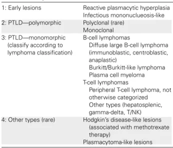
The term PTLD describes a broad category of EBV-related diseases that have distinct histological appearances (Table 15) (323). The approach to the management of PTLD can vary according to the PTLD disease classification. Fur-thermore, EBV-negative PTLD lesions have been reported. These lesions may behave differently then EBV-positive le-sions and may warrant alternative therapeutic options. In addition, lesions with a characteristic clinical appearance on physical examination or imaging studies may be due to alternative pathogens (e.g. pulmonary nodules attributable to fungal pathogens). Because of all these concerns, it is imperative that suspected PTLD lesions be biopsied and undergo histolopathologic evaluation by a pathologist ex-perienced with the diagnosis of PTLD (315).
Observational studies have suggested KTRs with EBV dis-ease are at high risk of developing PTLD (324). Observa-tional studies have also shown that mortality from EBV-associated PTLD is over 50% (325,326). The presence of immunosuppression is major risk factor for the develop-ment of EBV disease, including PTLD, in KTRs (317,327). In most cases, the progression of clinical symptoms is a consequence of the inability to mount an adequate
EBV-Table 15: Categories of PTLD
1: Early lesions Reactive plasmacytic hyperplasia
Infectious mononuclueosis-like
2: PTLD—polymorphic Polyclonal (rare)
Monoclonal 3: PTLD—monomorphic
(classify according to lymphoma classification)
B-cell lymphomas
Diffuse large B-cell lymphoma (immunoblastic, centroblastic, anaplastic)
Burkitt/Burkitt-like lymphoma Plasma cell myeloma T-cell lymphomas
Peripheral T-cell lymphoma, not otherwise categorized Other types (hepatosplenic, gamma-delta, T/NK)
4: Other types (rare) Hodgkin’s disease-like lesions
(associated with methotrexate therapy)
Plasmacytoma-like lesions PTLD, posttransplant lymphoproliferative disease; T/NK, T-cell/ natural killer cell.
Modified with permission (323).
specific cytotoxic T-cell response, because of the immuno-suppressive medications. It is therefore logical to assume that reduction of immunosuppression may result in reso-lution of EBV disease. As many as two thirds of patients presenting with EBV-associated PTLD will respond to re-duction or withdrawal of immunosuppressive medication (315,328). This is less likely to be the case for patients presenting more than 1 year after transplantation, or with EBV-associated lymphoma. In these cases, there is an in-creased tendency for the lesions to behave in a truly malig-nant fashion. However, because some patients presenting late after transplant with biopsy evidence of lymphoma have responded to reduction of immunosuppression, this strategy may still be considered even in these patients, though expectations of efficacy will be reduced.
Epstein-Barr virus disease and PTLD are important causes of morbidity and mortality following kidney transplantation. Rates of PTLD are higher in pediatric KTRs and those pa-tients who are EBV-seronegative prior to transplant who experience primary infection after transplant. While EBV disease and PTLD may be more common among pediatric KTRs, adult EBV-seronegative recipients of kidneys from an EBV-seropositive donor are also felt to be at increased risk for the development of these complications. Because of the complexity of this disease and its management, in-volvement of infectious diseases specialists, oncologists and transplant physicians in a team approach will likely maximize therapeutic outcomes.
13.4: HERPES SIMPLEX VIRUS 1, 2 AND VARICELLA ZOSTER VIRUS
13.4.1: We recommend that KTRs who develop a superficial HSV 1, 2 infection be treated (1B) with an appropriate oral antiviral agent (e.g. acyclovir, valacyclovir, or famci-clovir) until all lesions have resolved.(1D) 13.4.2: We recommend that KTRs with systemic HSV 1, 2 infection be treated(1B) with in-travenous acyclovir and a reduction in im-munosuppressive medication.(1D) 13.4.2.1: We recommend that intravenous
acyclovir continue until the pa-tient has a clinical response,(1B) then switch to an appropriate oral antiviral agent (e.g. acyclovir, valacyclovir, or famciclovir) to complete a total treatment dura-tion of 14–21 days.(2D)
13.4.3: We suggest using a prophylactic antiviral agent for KTRs experiencing frequent re-currences of HSV 1,2 infection.(2D) 13.4.4: We recommend that primary VZV
infec-tion (chicken pox) in KTRs be treated(1C) with either intravenous or oral acyclovir or valacyclovir; and a temporary reduction in amount of immunosuppressive medica-tion.(2D)
13.4.4.1: We recommend that treatment be continued at least until all le-sions have scabbed.(1D)
13.4.5: We recommend that uncomplicated her-pes zoster (shingles) be treated(1B) with oral acyclovir or valacyclovir(1B), at least until all lesions have scabbed.(1D) 13.4.6: We recommend that disseminated or
in-vasive herpes zoster be treated(1B) with intravenous acyclovir and a temporary re-duction in the amount of immunosuppres-sive medication(1C), at least until all le-sions have scabbed.(1D)
13.4.7: We recommend that prevention of pri-mary varicella zoster be instituted in varicella-susceptible patients after expo-sure to individuals with active varicella zoster infection(1D):
• varicella zoster immunoglobulin (or in-travenous immunoglobulin) within 96 hours of exposure(1D);
• if immunoglobulin is not available or more than 96 h have passed, a 7-day course of oral acyclovir begun 7–10 days after varicella exposure.(2D)
HSV, herpes simplex virus; KTRs, kidney transplant recipients; VZV, varicella zoster virus.
Background
Superficial herpes simplex virus (HSV) infection is defined as disease limited to the skin or mucosal surfaces without evidence of dissemination to visceral organs.
Systemic HSV infection is defined by disease involving vis-ceral organs.
Primary varicella zoster virus (VZV) infection is infection in a patient who is immunologically naive to VZV. In gen-eral, primary VZV presents as ‘chickenpox,’ which most fre-quently manifests as multiple crops of cutaneous lesions that evolve from macular, papular, vesicular and pustular stages. The lesions tend to erupt over the entire body and will be in different stages. Disseminated VZV can develop in immunocompromised individuals with involvement of the lungs, liver, central nervous system and other visceral organs.
Uncomplicated herpes zoster (shingles) is defined as the presence of cutaneous zoster limited to no more than three dermatomes.
Disseminated or invasive herpes zoster is defined as the presence of cutaneous zoster in more than three der-matomes, and/or evidence of organ system involvement.
The definition of a clinically significant exposure to an in-dividual with active VZV infection varies by whether the infected individual presents with varicella (chickenpox) or zoster (shingles). Varicella may be spread to a susceptible individual by either airborne exposure or direct contact with a lesion. In contrast, an infectious exposure to someone with zoster requires direct contact with a lesion. Accord-ingly, a significant exposure to varicella is defined by face-to-face contact with someone with chickenpox, while a sig-nificant exposure to someone with zoster requires direct contact with a lesion. The minimum duration of airborne exposure necessary to allow transmission is not known. In general, most experts consider the minimum to be some-where in the range of 5–60 min.
Rationale
• Superficial HSV infections are typically self-limited in immunocompetent patients, but immunosuppressive medication in KTRs increases the risk for invasive and disseminated HSV infection; treatment of superficial HSV infections with oral acyclovir or valacyclovir is safe and effective.
• Systemic HSV infections represent a potentially life-threatening complication to immunosuppressed KTRs. Intensive treatment of systemic HSV infection with in-travenous acyclovir and a reduction in the amount of immunosuppressive medication is warranted to pre-vent progression and further dissemination of HSV.
• Primary VZV infection is potentially life-threatening to KTRs. Treatment with intravenous acyclovir is safe and effective.
• Herpes zoster infection is potentially life-threatening to KTRs. Treatment with oral acyclovir or valacyclovir is safe and effective.
• Disseminated or invasive herpes zoster is life-threatening to KTRs. Treatment with intravenous acy-clovir and a temporary reduction in the amount of im-munosuppressive medication is safe and effective.
• The use of varicella zoster immunoglobulin or commer-cial intravenous immunoglobulin products within 96 h of exposure to VZV prevents or modifies varicella in susceptible individuals.
• Oral acyclovir begun within 7–10 days after varicella exposure and continued for 7 days appears to be a reasonable alternative to immunoglobulin to prevent or modify primary varicella in susceptible individuals (329,330).
Superficial HSV infection
Serologic evidence of HSV1 and HSV2 is common in the general population. Although periodic reactivation of HSV1 and HSV2 infection occurs, these episodes tend to be self-limited in immunocompetent individuals. How-ever, episodes of invasive or disseminated HSV may occur in KTRs receiving immunosuppressive medications, and
indeed the incidence of invasive HSV is higher in KTRs than in the general population (331,332).
The highest incidence of HSV reactivation occurs early after transplantation, with the greatest risk occurring during the first month following transplantation (333). While presen-tation later after transplant is associated with a lower risk of dissemination, treatment of superficial infection with oral acyclovir, valacyclovir or famciclovir is still recommended, given the safety and efficacy of these medications (333). To prevent dissemination, it seems prudent to continue treatment until there are no new, active lesions.
Systemic HSV infection
In contrast to superficial HSV infection, systemic HSV infection involving the lungs, liver, central nervous sys-tem or other visceral organs represents a potentially life-threatening complication. Because systemic HSV is life-threatening, hospitalization and treatment with intra-venous acyclovir is warranted (333). If possible, immuno-suppressive medications should be reduced or withdrawn until the infection has resolved.
Intravenous acyclovir should be continued until there is demonstrative evidence of clinical improvement as mea-sured by resolution of fever, hypoxia and signs or symp-toms of hepatitis. Once the patient has reached this level of improvement, completion of therapy may be carried out using oral acyclovir or valacyclovir.
Primary varicella zoster infection
Varicella zoster infection can be life-threatening in KTRs (334,335). Although some centers have begun to institute the use of oral acyclovir in the outpatient setting for KTRs, there is little evidence to confirm the safety and efficacy of this approach. Careful selection of patients with assurance of close clinical follow-up is necessary if oral acyclovir is to be used in these patients.
Uncomplicated herpes zoster
Although herpes zoster can be seen in immunocompetent patients, the presence of immunosuppression is associ-ated with an increased risk for the development of both uncomplicated and complicated herpes zoster infection. Patients with only skin disease, but who have lesions in-volving more than three dermatomes, are considered to have disseminated cutaneous zoster. Similarly, patients with visceral involvement in addition to skin disease are considered to have disseminated zoster.
Uncomplicated zoster is a clinical syndrome character-ized by cutaneous clustering of vesicular lesions in a der-matomal distribution of one or more adjacent sensory nerves. An important complication of herpes zoster in im-munocompetent adults is the potential development of postherpetic neuralgia. RCTs in healthy adults have demon-strated that the use of acyclovir, valacyclovir or famciclovir have been associated with more rapid healing of the skin,
as well as a decreased incidence of both acute neuritis and postherpetic neuralgia (336,337). In immunocompromised hosts, patients are at risk not only of postherpetic neuralgia but also of severe local dermatomal infection (334). Simi-larly, immunosuppressed patients are at increased risk for the development of disseminated cutaneous zoster and visceral dissemination. The more severe the level of im-munosuppression, the greater the risk of dissemination. Accordingly, prompt initiation of antiviral therapy with close follow-up is warranted for these patients, even if they have only superficial skin infection (333).
Disseminated or invasive herpes zoster
Treatment with intravenous acyclovir and temporary re-duction in the amount of immunosuppressive medication is efficacious (333,338). Although specific evidence is not available to guide which immunosuppressive agent should be reduced, it would seem logical, whenever possible, to reduce the dosage of CNIs as well as steroids. In the ab-sence of any evidence of intercurrent rejection, an effort should be made to maintain the reduced level of immuno-suppression for a minimum of 3–5 days and until there is evidence of clinical improvement.
Prevention of primary varicella zoster infection The use of varicella zoster immunoglobulin has been demonstrated to prevent or modify varicella in immuno-suppressed individuals exposed to varicella (330,333,339). If varicella zoster immunoglobulin is not available, or if>96 h have passed since the exposure, some experts recom-mend prophylaxis with a 7-day course of oral acyclovir (80 mg/kg/day administered in four divided doses with a max-imum of 800 mg per dose) beginning on day 7–10 after varicella exposure (330,339). The use of varicella vaccine is not recommended as a postexposure prophylactic strat-egy in KTRs.
13.5: HEPATITIS C VIRUS
13.5.1: We suggest that HCV-infected KTRs be treated only when the benefits of treat-ment clearly outweigh the risk of allo-graft rejection due to interferon-based therapy (e.g. fibrosing cholestatic hepati-tis, life-threatening vasculitis).(2D) [Based on KDIGO Hepatitis C Recommendation 2.1.5.]
13.5.2: We suggest monotherapy with standard interferon for HCV-infected KTRs in whom the benefits of antiviral treatment clearly outweigh the risks.(2D) [Based on KDIGO Hepatitis C Recommendations 2.2.4 and 4.4.2.]
13.5.3: We suggest that all conventional current induction and maintenance immunosup-pressive regimens can be used in HCV-infected patients. (2D) [Based on KDIGO Hepatitis C Recommendation 4.3.]
13.5.4: Measure ALT in HCV-infected pa-tients monthly for the first 6 months and every 3–6 months, thereafter. Perform imaging annually to look for cirrhosis and hepatocellular carcinoma. (Not Graded) [Based on KDIGO Hepatitis C Recom-mendation 4.4.1.] (See RecomRecom-mendation 19.3.)
13.5.5: Test HCV-infected patients at least every 3–6 months for proteinuria.(Not Graded) [Based on KDIGO Hepatitis C Recommen-dation 4.4.4.]
13.5.5.1: For patients who develop new-onset proteinuria (either urine protein/creatinine ratio >1 or 24-hour urine protein >1 g on two or more occasions), per-form an allograft biopsy with im-munofluorescence and electron microscopy.(Not Graded) [Based on KDIGO Hepatitis C Recom-mendation 4.4.4.]
13.5.6: We suggest that patients with HCV-associated glomerulopathy not receive in-terferon.(2D) [Based on KDIGO Hepatitis C Recommendation 4.4.5.]
ALT, alanine aminotransferase; HCV, hepatitis C virus; KDIGO, Kidney Disease: Improving Global Outcomes; KTRs, kidney transplant recipients.
Background
The Work Group reviewed the KDIGO Hepatitis C Guide-lines (340) that were applicable to KTRs, and ultimately agreed with the pertinent guideline statements. Only mi-nor modifications (to guideline statement 4.4.1 in the KDIGO Hepatitis C Guidelines) were made, resulting in recommendation statement 13.5.4. The Transplant Work Group did not conduct a systematic review, but relied on the evidence reviewed by the Hepatitis C Work Group. A brief synopsis of the rationale for the KDIGO Hepatitis C Guidelines that are pertinent to KTRs is presented, with further discussion to the modification of recommendation 13.5.4. Details may be found in the Hepatitis C guidelines.
Rationale
Kidney transplant recipients infected with hepatitis C virus (HCV) have worse patient- and allograft-survival rates than KTRs without HCV infection. In addition, HCV-infected KTRs are at increased risk for several complications, includ-ing worseninclud-ing liver disease, NODAT and glomerulonephri-tis. Thus, close follow-up of the HCV-infected KTR is pru-dent.
There are few data to suggest when and how to screen HCV-infected KTRs for posttransplant complications. How-ever, given the higher level of immunosuppression early after transplantation, the Transplant Guideline Work Group determined that liver enzymes should be checked ev-ery month for the first 6 months of the posttransplant period, and every 3 months thereafter. The detection of clinically worsening liver enzymes should prompt refer-ral for hepatologic evaluation. Annual liver ultrasound and alpha-fetoprotein level to screen for hepatocellular carci-noma should be considered in patients with cirrhosis on liver biopsy.
Available evidence indicates that all currently available in-duction and maintenance immunosuppressive agents can be used in KTRs infected with HCV. Although immunosup-pression may cause or contribute to complications of HCV in KTRs, there is scant evidence that one type of immuno-suppressive agent is more or less likely to be harmful. The exception is tacrolimus, which increases the risk for NO-DAT, and might be expected to impart at least an additive risk for NODAT to HCV-infected KTRs.
Interferon is effective for viral eradication in HCV-infected patients, especially when combined with ribavirin. How-ever, the administration of interferon after kidney trans-plantation can be deleterious to the allograft and should generally be avoided in KTRs, unless there is indication of worsening hepatic injury.
Hepatitis C virus infection has also been implicated in the pathogenesis of glomerular disease in both native and transplanted kidneys. Therefore, the Hepatitis C and Trans-plant Guideline Work Groups concluded that HCV-infected KTRs should be tested for proteinuria every 3–6 months. As recommended for all KTRs, patients who develop new-onset proteinuria (either urine protein/creatinine ratio>1 or 24-hour urine protein greater than 1 g on two or more occasions) should have an allograft biopsy with immunoflu-orescence and electron microscopy.
Interferon-based therapies may be effective in treating HCV-related glomerulopathy in native kidney disease. How-ever, interferon use in KTRs is associated with an increased risk of rejection. The risk of kidney allograft loss from pro-gressive HCV-associated glomerulopathy compared to that from interferon-induced rejection is unknown. Ribavirin can reduce proteinuria in HCV-associated glomerulopathy, al-though its impact on kidney function is unknown and it does not lead to viral clearance.
13.6: HEPATITIS B VIRUS
13.6.1: We suggest that any currently available induction and maintenance immunosup-pressive medication can be used in HBV-infected KTRs.(2D)
13.6.2: We suggest that interferon treatment should generally be avoided in HBV-infected KTRs.(2C)
13.6.3: We suggest that all HBsAg-positive KTRs receive prophylaxis with tenofovir, ente-cavir, or lamivudine.(2B)
13.6.3.1: Tenofovir or entecavir are prefer-able to lamivudine, to mini-mize development of potential drug resistance, unless medica-tion cost requires that lamivu-dine be used.(Not Graded) 13.6.3.2: During therapy with antivirals,
measure HBV DNA and ALT lev-els every 3 months to monitor efficacy and to detect drug resis-tance.(Not Graded)
13.6.4: We suggest treatment with adefovir or tenofovir for KTRs with lamivudine re-sistance (>5 log10 copies/mL rebound of HBV-DNA).(2D)
13.6.5: Screen HBsAg-positive patients with cir-rhosis for hepatocellular carcinoma every 12 months with liver ultrasound and al-pha feto-protein. (Not Graded) (See Rec-ommendation 19.3.)
13.6.6: We suggest that patients who are neg-ative for HBsAg and have HBsAb titer
<10 mIU/mL receive booster vaccination to raise the titer to≥100 mIU/mL.(2D)
ALT, alanine aminotransferase; HBsAb, antibody to hepatitis B surface antigen; HBsAg, hepatitis B surface antigen; HBV, hepatitis B virus; KTRs, kidney transplant recipients.
Background
Patients with CKD stage 5 are at increased risk of acquiring HBV infection. Infection can be acquired through infected blood products, or transmission from another infected pa-tient in a dialysis unit. The risk has come down considerably in Western countries following the introduction of univer-sal immunization and strict isolation practices, but remains substantial in developing countries. Screening for HBV in-fection is done by serologic testing for hepatitis B surface antigen (HBsAg). NAT for the presence of HBV DNA gives a more accurate idea of the infection load. Viral replication is accelerated following introduction of immunosuppression in KTRs. A number of studies have shown that HBV infec-tion increases the risk of mortality, most often due to liver disease and graft failure. Effective antiviral therapy permits inhibition of viral replication and retards development of progressive liver disease, and may lower the risk of liver cancer.
Rationale
• HBV-infected patients exhibit increased viral replication and are at risk for progressive liver disease after kidney transplantation.
• HBsAg positivity is an independent risk factor for mor-tality and graft failure.
• HBsAg-negative patients are at low risk of increased viral replication and progressive liver disease.
• Prospective studies have shown that antiviral agents normalize alanine aminotransferase (ALT), and induce clearance of HBV-DNA and hepatitis B E antigen (HBeAg). Antiviral agents are best used as prophylaxis, since KTRs not initiated on antiviral agents at the time of transplantation often develop enhanced viral replica-tion and hepatic dysfuncreplica-tion.
• ALT activity is lower in KTRs than in the general pop-ulation, and is unreliable as a marker of liver disease activity by itself. Serial monitoring of HBV DNA is re-quired to assess treatment efficacy. A rise in DNA copy number suggests development of resistance.
• The newer nucleoside analogues, adefovir and teno-fovir are effective for treatment of lamivudine-resistant HBV infection.
Hepatitis B virus infected patients are at risk of exacerba-tion of the infecexacerba-tion, progressive liver disease and devel-opment of hepatocellular carcinoma after kidney transplan-tation. The rate of HBV infection in CKD stage 5 patients as determined by seropositivity for HBsAg varies between 0% and 8% in developed countries (341). The US Centers for Disease Control and Prevention (CDC) estimates that the prevalence of HBsAg-positive patients in the US dial-ysis population has declined from 7.8% to 0.9%, with an estimated incidence of disease in 2000 of 0.05% (342). This has largely been due to widespread use of univer-sal precautions, screening of the blood supply, the use of erythropoiesis-stimulating agents (ESAs), HBV vaccina-tion and strict implementavaccina-tion of segregavaccina-tion of HBsAg-positive from HBsAg-negative patients during hemodialy-sis with dedicated machines and staff for each group. The prevalence, however, is much higher (10–20%) in develop-ing countries.
Hepatitis B virus infection in CKD stage 5 patients is usually asymptomatic even in the acute phase, with about 80% of patients progressing to a chronic carrier state (343). Im-munosuppression following kidney transplantation leads to increased replication of HBV and results in progressive liver disease. Assessing the natural history of hepatitis B among KTRs is difficult for several reasons (344). Amino-transferase activity is lower in this population, which ham-pers recognition of HBV-related liver disease (345). In a meta-analysis (346) of six observational studies (6050 patients), HBsAg positivity was found to be an
independent and significant risk factor for mortality (RR 2.49, 95% CI 1.64–3.78) and graft failure (RR 1.44, 95% CI 1.02–2.04). This finding was confirmed in later observa-tional studies. In a study of 286 kidney transplant patients, liver-related death was the most common cause of death in HBV-positive patients (347). A survey from the South East-ern Organ Procurement Foundation demonstrated a detri-mental effect of HBV infection on patient survival (p=0.02) and graft survival (p=0.05) in 13287 patients who under-went kidney transplantation between 1977 and 1987 in the United States (348). Patient survival was 62% and 66% at 10 years for HBsAg-positive and -negative KTRs (p=0.02). The 10-year survival rate of HBsAg-positive KTRs (45%) compares poorly with HCV-infected patients (65%). In pa-tients with biopsy diagnosis of cirrhosis, 10-year survival was 26% (349).
Many studies provided only limited details of virology and did not incorporate liver histology before kidney trans-plantation, leading to underestimation of the severity of liver disease at the time of transplantation. The only study that carried out serial biopsies found histological deterio-ration in 85% of HBsAg-positive patients at a mean inter-val of 66 months. Approximately, 28% showed cirrhosis, whereas no patients had been cirrhotic on baseline biopsy (350). Among those with cirrhosis, hepatocellular carci-noma was found in 23%, suggesting an annual incidence of between 2.5% and 5%. Based on these data, an expert group recommended hepatic imaging every 3 months to detect hepatocellular carcinoma in patients with cirrhosis (351).
The standard practice of screening for HBV infection is testing for HBsAg. The place of routine NAT in these pa-tients is unclear. Some recent studies have shown that a proportion of dialysis patients may exhibit occult HBV in-fection as detected by NAT in the face of a negative HBsAg (352–360) but not all (361–363). These patients have gen-erally low viral loads and may have mutations that prevent appearance of HBsAg. A large proportion of those with occult infection have antibody to hepatitis B core antigen (HBcAb) and it has been suggested that testing these pa-tients by NAT may be a cost-effective strategy for confirm-ing occult infection. The risk of reactivation of HBV among patients who are HBsAg-negative and HBcAb-positive is low, however (364). Berger et al. (365) found recurrence in 2 of 229 (0.9%) such patients. Savas et al. (366) reported two cases of reactivation and provided a review of 25 pre-viously reported cases. They noted a wide age range of patients experiencing recurrence (22–75 years), a male pre-ponderance, and a posttransplant time of onset between 8 weeks and 15 years. All but one patient had HBsAb titers of less than 100 mIU/mL, leading the authors to suggest that vaccination of such patients may be an effective pre-ventative measure. An expert group recommended routine use of vaccination in such patients to boost the titers above 100 mIU/mL and lamivudine prophylaxis (see section
‘Phar-macotherapy,’ below) during periods of intensified im-munosuppression (351).
The primary goals of management are maximal suppres-sion of viral replication, while minimizing development of resistance and prevention of hepatic fibrosis. In view of the poor likelihood of seroconversion to HBsAb, low rates of conversion from HBeAg to anti-HBeAg antibody positiv-ity, and poor reliability of following ALT as a measure of activity, HBV DNA levels need to be followed to assess re-sponse to therapy. Serological markers of fibrosis, such as the commercially available Fibrotest panel, have not been evaluated in KTRs with HBV infection. Since the replication is dependent on the overall extent of immunosuppression rather than an individual drug, efforts should be made to minimize the doses of all immunosuppressive drugs with-out compromising graft with-outcomes. These include use of the lowest possible dose of steroids. Currently, there is no evidence for the differential effect of any specific immuno-suppressive agent on HBV replication.
Pharmacotherapy
There are currently seven medications available for the treatment of hepatitis B: interferon alfa-2b, pegylated in-terferon alfa 2a, lamivudine, adefovir, tenofovir, telbivudine and entecavir. Interferon therapy for HBV infection in KTRs is associated with high rates of graft loss due to rejec-tion. In a series (367) of 31 HBsAg-positive KTRs treated with recombinant interferon-alpha (three million interna-tional units) three times a week for 6 months, long-term ALT normalization was noted in 47% of patients and 13% cleared HBeAg. However, graft loss occurred in five out of 17 patients during therapy and an additional four patients after the completion of therapy. The use of interferon in this setting, therefore, is not recommended (351). Lamivudine, a cytosine analog that inhibits HBV reverse transcriptase, has been used extensively in KTRs with HBV infection (Table 16). The utility of lamivudine in stabiliza-tion of liver funcstabiliza-tion was shown in several observastabiliza-tional studies. A meta-analysis (368) that included 14 prospec-tive cohort studies (184 patients) determined the mean overall estimate for ALT normalization, and HBV-DNA and HBeAg clearance at 81% (95% CI 70–92%), 91% (95% CI 86–96%) and 27% (95% CI 16–39%), respectively. The duration of lamivudine therapy was 6–12 months in the ma-jority (11 of 14) of the studies. Later clinical trials (369–375) have shown similar results with lamivudine monotherapy given for 24–69 months. HBeAg and HBV-DNA clearance occurred in 0–25% and 43–78%, respectively. Changes in ALT paralleled those in viremia, and 33–77% of patients maintained normal ALT levels.
Timing of initiation
Data on optimal timing of initiation of antiviral therapy are scarce. However, the available data support starting
Table 16: Outcomes of clinical trials of lamivudine therapy
ALT HBsAg HBeAg HBeAg HBV DNA
normalization clearance clearance seroconversion clearance
Author (year) (ref no) (%) (%) (%) (%) (%)
Rostaing (1997) (376) 4/5 (80) 0 0 NA 6/6 (100) Goffin (1998) (377) 4/4 (100) 0 0 0/1 (0) 4/4 (100) Jung (1998) (378) 6/6 (100) 0 1/3 (33) NA 6/6 (100) Kletzmayr (2000) (379) 3/3 (100) 0 2/12 (17) 2/12 (17) 15/16 (93) Tsai (2000) (380) NA 0 0 NA 7/8 (87.5) Lewandowska (2000) (381) 17/28 (61) 0 2/26 (8) NA 10/10 (100) Antoine (2000) (382) NA 0 8/12 (67) NA 9/12 (75) Mouquet (2000) (383) 8/15 (53) 0 NA NA 13/15 (87) Fontaine (2000) (384) NA 0 6/13 (46) 6/13 (46) 26/26 (100) Lee (2001) (385) NA 1/13 (8) 3/8 (37.5) 3/8 (37.5) 10/13 (77) Han (2001) (386) 6/6 (100) 0 2/3 (67) NA 6/6 (100) Chan (2002) (369) 14/14 (100) 0 3/14 (21) NA 26/26 (100) Park (2001) (387) 8/10 (80) 0 1/5 (20) NA 7/10 (70) Mosconi (2001) (388) NA 0 NA NA 4/4 (100)
ALT, alanine aminotransferase; HBV, hepatitis B virus; HBeAg, hepatitis B E antigen; HBsAg, hepatitis B surface antigen; NA, not available. Reproduced with permission (368).
treatment at the time of transplantation in HBsAg-positive patients, irrespective of HBV DNA levels. In a study of 15 patients with normal preoperative ALT (389), seven were started on lamivudine at the time of kidney transplantation. Half of those not treated showed transaminase elevations and HBV viremia in the first year of follow-up, requiring initiation of lamivudine therapy. In contrast, all seven indi-viduals who received lamivudine at the time of transplan-tation continued to have normal ALT and were negative for HBV DNA throughout the follow-up. In another study of HBsAg-positive KTRs (386), where lamivudine was given prophylactically (HBV DNA negative) or preemptively (HBV DNA positive) to 10 patients or reserved for hepatic dys-function in 10 patients, 42% in the latter group developed viremia during follow-up, compared to 10% in the former. Six in the reactive group developed hepatic dysfunction compared to none in the prophylactic/preemptive group. In another study (369) where the decision to start lamivu-dine was based on HBV DNA levels or liver function status, all patients had to be started on lamivudine at a mean time period of 8 months after transplant. More than half the patients were started on treatment because of abnormal ALT.
Duration of therapy
The optimal duration of therapy that ensures long-term remission of viremia and maintenance of normal liver func-tion and minimizes the development of resistance is not known. In a meta-analysis, increased duration of lamivu-dine therapy was positively associated with frequency of HBeAg loss (r=0.51, p=0.04) (Figure 1) (368). Lamivu-dine discontinuation was attempted by Chan et al. (369) in 12 low-risk patients after stabilization, and was successful in only five (42%).
At least 24 months of prophylactic treatment has been rec-ommended (390). The optimal treatment and the choice of drugs require further study. Withdrawal of antiviral ther-apy may be associated with a relapse and increased viral replication, even resulting in liver failure.
Development of resistance is a major clinical problem with long-term lamivudine use. This is usually reflected by a sec-ondary increase in the HBV DNA titers. A commonly used definition is demonstration of>5 log10copies/mL rebound
of HBV DNA. In most, but not all, instances, it is caused by a mutation in the tyrosine–methionine–aspartate–aspartate (YMDD) locus of the HBV DNA polymerase (384). The clin-ical presentation varies. While some patients show no sig-nificant biochemical changes or clinical symptoms, others develop deterioration in liver function (391).
In a study of 29 KTRs (392), resistance was noted in 48% of patients during a mean follow-up period of 69 months; all due to YMDD mutations. Resistance was not related to
Figure 1: HBeAg clearance vs. lamivudine duration.HBeAg,
hepatitis B E antigen; Lam, lamivudine. Reproduced with permis-sion (368).
patient demographics, HBeAg status, seroconversion rates or genotype. About 80% with the YMDD mutation had a hepatitis flare. In the meta-analysis (368), the mean overall estimate for lamivudine resistance was 18% (95% CI 10– 37%). An increased duration of lamivudine therapy was positively associated with lamivudine resistance (r=0.62, p=0.02). The cumulative probability of developing resis-tance was approximately 60% in the later studies. Patients with lamivudine resistance should be treated with adefovir or tenofovir. Limited data are available regarding use of these agents in KTRs. Fontaine et al. (393) gave adefovir to 11 KTRs with lamivudine-resistant HBV infec-tion and found it to be effective in bringing about a re-duction in serum HBV DNA, without any significant ad-verse effects. Entecavir, a guanosine analog, is 30 times more potent than lamivudine in suppressing viral replica-tion. In a multicenter, double-blind RCT comparing ente-cavir to lamivudine in the general population, enteente-cavir was shown to result in larger reductions in HBV DNA than lamivudine. At a dose of 0.5 mg daily, 83% of patients treated with entecavir had undetectable HBV DNA com-pared to 58% of those treated with lamivudine (394). In a study (395) that treated eight adefovir- and lamivudine-resistant KTRs with entecavir for 16.5 months, there was a significant decrease in HBV DNA viral load without any significant adverse effects. Data in the non-CKD popula-tion shows that, while the risk of resistance to entecavir is low in treatment-na¨ıve patients, it may be as high as 51% at 5 years (396) in lamivudine-resistant cases. In a recent study, tenofovir was shown to be superior to ade-fovir in achieving remission of HBV viremia and hepatic histologic scores in non-CKD patients. Tenofovir was ef-fective in lamivudine-resistant cases, and did not produce resistance up to 48 months of treatment (397). Of the two agents, tenofovir has a much lower renal toxicity than ade-fovir, and hence would be the preferred agent in KTRs. It is not known whether substitution of lamivudine with ente-cavir or tenofovir for prophylaxis will prevent development of resistance.
Research Recommendations
• The frequency of occult HBV infection in patients with CKD stage 5 should be evaluated in different parts of the world, and its impact on posttransplant outcomes determined.
• Studies are required to determine whether substitution of lamivudine with entecavir or tenofovir for prophy-laxis will prevent development of resistance in KTRs.
13.7: HUMAN IMMUNODEFICIENCY VIRUS
13.7.1: If not already done, screen for HIV infec-tion.(Not Graded)
13.7.2: To determine antiretroviral therapy, refer HIV-infected KTRs to an HIV specialist, who should pay special attention to drug– drug interactions and appropriate dosing of medications.(Not Graded)
HIV, human immunodeficiency virus; KTRs, kidney transplant recipients.
Background
Screening for human immunodeficiency virus (HIV) infec-tion is defined as the performance of serologic testing for HIV. A two-step screening is usually performed. In the first step, patients are screened for the presence of antibodies against HIV, usually with an enzyme-linked immunosorbent assay (ELISA). This is an extremely sensitive test. However, it is not specific. Accordingly, those patients who are posi-tive on ELISA are then screened using a Western Blot as-say. The presence of a positive Western Blot assay for HIV confirms the diagnosis of HIV infectionexcept in children <18 months of age, where a positive serologic test may be attributable to the presence of passive antibody acquired from the child’s mother during the pregnancy. NAT for the presence of HIV DNA or HIV RNA viral load should be performed on children<18 months of age with a positive HIV antibody. Antiretroviral medications are used specifi-cally for the treatment of HIV infection. Drug–drug interac-tions are pharmacokinetic interacinterac-tions between separate medications that may result in accumulation or more rapid metabolism of one or both compounds.
Rationale
• Patients with HIV require specialized care in centers with appropriate expertise.
• Screening for HIV infection should be carried out on all KTRs (ideally before transplantation) in order to identify those KTRs that will require specialized care.
• Antiretroviral therapy is necessary to maintain virologic suppression and normal immunologic function in HIV patients undergoing kidney transplantation.
• The concomitant use of antiretroviral agents and im-munosuppressive medications creates the potential for drug–drug interactions that may substantially alter blood levels of drugs and require appropriate monitor-ing and adjustments in dosmonitor-ing.
Case series have documented successful outcomes of KTRs with HIV (398–400). However, these HIV patients had been carefully selected and adequately treated for HIV at the time of transplantation (400). Although HIV is not an absolute contraindication to kidney transplantation, the presence of HIV has major implications in the manage-ment of patients following transplantation. A major issue of
concern in the management of HIV patients is the need to be aware of potential drug–drug interactions among an-tiretroviral therapy and other medications, including im-munosuppressants. Care must be taken to identify and select those HIV-infected patients who are most likely to benefit from kidney transplantation without an unaccept-ably high risk of opportunistic infections.
Evidence from a National Institutes of Health (NIH)— sponsored study of organ transplantation in HIV patients has demonstrated both the effectiveness of transplanta-tion as well as the complexity of management of KTRs with HIV (400). Data accrued from the NIH-sponsored study of organ transplantation in HIV-infected patients has identi-fied specific drug combinations that are associated with drug–drug interactions in these patients (401).Accordingly, attention must be paid and caution must be used in these patients to account for the potential impact of these
inter-actions. Although the data from the NIH study demonstrate the feasibility of transplantation for HIV-infected KTRs, the limited number of HIV patients with CKD stage 5 under-going kidney transplantation to date suggests the need to continue performing this procedure under research proto-cols and in selected centers with appropriate expertise. Finally, it is worth noting that review of experience to date suggests that there may be an increased risk for the de-velopment of acute cellular rejection in patients with HIV undergoing organ transplantation.
Research Recommendations
• There is a need to determine the optimal immuno-suppression medication regimen, as well as the best antiretroviral regimens, for HIV-infected KTRs.