LECTURE NOTES
For Medical Laboratory Technology Students
Parasitology
Girma Mekete
Mohamed Awole Adem
Jimma University
In collaboration with the Ethiopia Public Health Training Initiative, The Carter Center, the Ethiopia Ministry of Health, and the Ethiopia Ministry of Education
Funded under USAID Cooperative Agreement No. 663-A-00-00-0358-00.
Produced in collaboration with the Ethiopia Public Health Training Initiative, The Carter Center, the Ethiopia Ministry of Health, and the Ethiopia Ministry of Education.
Important Guidelines for Printing and Photocopying
Limited permission is granted free of charge to print or photocopy all pages of this publication for educational, not-for-profit use by health care workers, students or faculty. All copies must retain all author credits and copyright notices included in the original document. Under no circumstances is it permissible to sell or distribute on a commercial basis, or to claim authorship of, copies of material reproduced from this publication.
©2003 by Girma Mekete and Mohamed Awole Adem
All rights reserved. Except as expressly provided above, no part of this publication may be reproduced or transmitted in any form or by any means, electronic or mechanical, including photocopying, recording, or by any information storage and retrieval system, without written permission of the author or authors.
This material is intended for educational use only by practicing health care workers or
students and faculty in a health care field.
Preface
The problem faced today in the learning and teaching of Parasitology for laboratory technicians in universities, colleges, health institutions, training health centers and hospitals emanates primarily from the unavailability of textbooks that focus on the needs of Ethiopian students. This lecture note has been prepared with the primary aim of alleviating the problems encountered in the teaching of Medical Parasitology course and in minimizing discrepancies prevailing among the different teaching and training health institutions. It can also be used in teaching any introductory course on medical parasitology and as a reference material. This lecture note is devoted to providing general aspects of parasitology in addition to covering human parasites in two major groups -the protozoa and helminths- including their distribution, habitat, morphology, life cycle, pathogenicity, prevention and control, laboratory diagnosis and their relevance to Ethiopia. It has also appendices, which discuss the collection of laboratory specimens, preservatives of stool sample, frequently used parasitological diagnostic methods and reagent preparation. Finally, it contains a glossary, which summarizes important terminologies used in the text. Each chapter begins by specific learning objectives and after each objective and after each class of parasites review questions are also included.
No systemic study has been conducted on the prevalence of human parasites in different ecological zones of Ethiopia but past surveys indicate the presence of all parasites except some that are found in the Far East, South East Asian and Latin American countries and which require specific intermediate hosts. This lecture note tries as far as possible to summarize local literatures that deal with parasite prevalence
in Ethiopia so that it may address itself particularly to the needs of Ethiopian students.
We welcoming the reviewers and users input regarding this edition so that future editions will be better.
Acknowledgments
We would like to acknowledge The Carter Center for its initiative, financial, material and logistic supports for the preparation of this teaching material. We are indebted to The Jimma University and other institutions that support directly or indirectly for the visibility of this lecture note preparation.
Our deepest gratitude to Professor Dennis Carlson with out whom these lectures note preparation is not visible. We greatly thank him for his attitude, knowledge, and practice and above all his commitment, concern and dedication toward solving the health problem of Ethiopia.
We extend our appreciation to the reviewers and teaching staffs in the different institution for their unreserved contribution for the materialization of this lecture note preparation.
Contents
I. Preface i
II. Acknowledgement ii
III. Overview iii
III. General objectives v
CHAPTER ONE - INTRODUCTION 1
1.1 Definition of terms used in parasitology 1
1.2 Sources of exposure to
parasitic infections 5
1.3 Mode of transmission 6
1.3.1 Direct mode of transmission 6
1.3.2 Indirect mode of transmission 7
1.4 Route of transmission 7
1.5 Host parasite relationship 8
1.5.1 Effects of parasites on
there hosts 8
1.5.2 Host susceptibility factors 8
1.5.3 Escape mechanism of the parasite
from the immune system
1.6 General life cycle of parasites 9 1.6.1
Direct life cycle 9
1.6.2 Indirect life cycle 9
1.7 Types of specimen used for
parasitological examination 9
1.8 Classification of Parasites 10
protozoa and metazoa 11
CHAPTER TWO - MEDICAL PROTOZOOLOGY13
2.1 Class- Rhizopoda ( Amoebae)
2.1.1Free living amoeba 11 2.1.2
Free living pathogenic amoebae 28
2.2 Class - Zoomastigophora ( Flagellates) 32
2.2.1 The Oro-intestinal and Urogenital
flagellates 32
2.2.2 The Haemo-Somatic Flagellates 45
2.3. Class -Telosporidia 63
2.3.1 Intestinal and tissue
Coccidian Parasite 64
2.3.2 Haemosporidia 72
2.4 Class -Ciliatea ( Ciliates) 87
CHAPTER THREE -MEDICAL
HELMINTHOLOGY 91
3.1 Platyhelminths 92
3.1.1 Class cestoda (tapeworm) 92
3.1.2 Class trematodes(flukes) 3.1.2.1 Blood Flukes (Schistosmes) 115 3.1.2.2 Liver Flukes 131 3.2.2.3 Intestinal Flukes 139 3.2.2.4Lung Flukes 142 3.2. Nemathelminths 145 3.3.1. Class Nematoda 145 3.3.1.1Intestinal nematodes 145
3.3.1.2. Tissue nematodes 168
Appendix I - Laboratory Examination of Specimens 191
A. Stool 193
B. Urine 206
C. Vaginal and urethral discharge 208
D. Blood 209
E. Skin 216
F. The Gram scotch 217
Appendix II - Preservatives of specimens 219
Glossary 228
Bibliography 241
Overview
The earliest book devoted to Parasitology was published in 1684 by Redi, who provided descriptions of reproductive organs and eggs of
Ascaris. In 1817 Lancisi recorded studies of mosquitoes and Vague Surmises about their role in the cause of intermittent fevers. Goldfuss, in 1817, first used the word ‘’protozoa,’’ which was given modern meaning in 1845 by Siebold, Leeuwenhoek (1632-1723) had devised and used simple microscopes, but achromatic objectives in a compound microscope were not used in England until 1824. Gross, in 1849, was the
first to describe an amebic parasite in man Entamoeba gingival and
Losch identified E. histolytica in 1875. Then came the discovery of
mosquito hosts for filariae by Manson (1877 -1878) and Plasmodia by
Laveran in 1880, transmission of babesiosis by ticks by Smith and Kilburne in 1894, trypanosomes and their transmission by tsetse flies by Bruce (1895 - 1896), and mosquito transmission of plasmodia by Ross (1897- 1898).
After these discoveries, the science of parasitology expanded rapidly. With the aid of microscopes, morphological characters of various parasites were first studied and species and group characteristics were determined. Then several stages of the organisms were related to one another in life-cycle sequence. This provided important information on extrinsic as well as intrinsic development and paved the way for epidemiologic studies. Moreover, the relationship between parasite to host provided a background for studying the pathogenesis of infection in man and reservoir hosts and, indirectly, for understanding clinical aspects.
Recent investigations have been largely concerned with the ecology of parasitic infections, anatomy and physiology as revealed by electron microscopy, metabolism of the parasite in its host, immunologic phenomena, and the rationale of chemotherapy. Meanwhile, practical methods are being developed to control these infections and to reduce human exposure to them.
A landmark of great significance was the publication in 1978 of a two-volume work by B.H. Kean, K.E. Mott and A.J. Russell, which brought together in English translation as the articles on the major discoveries and earliest descriptions by workers in tropical medicine and parasitology. Having set the historical background; now we will see the importance and the scope of Parasitology.
Parasitology is a science that deals with an organism that lives in or on another organism in order to have shelter and /or nutrition. Medical parasitology study parasites that is capable of causing disease in humans. In the context of this ecture note, the term parasite refers to organisms, which belong to protozoa (i.e., Rhizopoda, flagellets ciliates, sporozoans, coccidians and Microsporidian) Helminths (Nematodes, Cestodes and Trematodes).
Human welfare has suffered greatly because of parasites. A lot of economic loss occurs as a result of infection of domestic animals by parasite, which causes diseases such as fascioliasis and trypanosomiasis.
The study of parasitology has more importance to developing countries where the social and economic conditions require great deal of improvement in terms of better clothing, shelter, food, provisions of wells
and latrines and sewages and other waste disposal facilities together with the means of controlling vectors.
Most of the developing countries lie within the tropics. The tropical or the semi-tropical nature of places where most of the people of the developing nations live not only provides better environmental conditions for larval development of parasites than that of temprate regions where most of the developed countries are found, but also provides better conditions for the multiplication of vectors.
This lecture note will discuss specific parasite of medical importance, including the information necessary to assist in the diagnosis.
General Objectives
- Understand the concepts of parasitism, the relationships between parasites and hosts,
between parasites and environment and the cultural and socioeconomic factors affecting the transmission of parasites
- Know the general epidemiological aspects of parasites that affect
human
- Apply simple preventive measures for specific parasites
- Know the life cycle of specific parasites and identify the important
parasitic agents affecting human health
- Be able to prepare reagents necessary for parasitology lab.
- Use effectively the basic laboratory equipment
- Apply the necessary procedures for the diagnosis of parasites in the
medical laboratory and reporting of results properly
- Apply the basic methods of specimens collection, preservation and
processing
CHAPTER ONE
Introduction
Specific Learning Objectives
At the end successful student will be able to:
1. Identify organisms which parasitizes man
2. Define common terms used in Medical Parasitology
3. List the various environmental, cultural and socioeconomic
factors that affect the distribution of parasites
4. Explain effect caused by parasites
5. Describe the classification and characteristics of parasite groups
6. Explain mode of transmission, source of infection, and portal of entry of parasites
1.1
Definition of Terms Used in Parasitology
Parasitology:- is a science that deals with parasites.
Medical Parasitology:- Is the study of parasites that causes disease in man.
Parasite:- is an organism living temporarily or permanently in or on another organism (host) from which is physically or physiologically dependant upon other.
Nature of Parasites- A parasite could be unicellular, worm or an arthropode.
Features of Parasites
1. Smaller than their host, 2. Outnumber the host,
3. Short life span than their host, and
4. Have greater reproductive potential than their host.
Association of Organisms
When there is an association between two organisms their relation will be one of the following type:
1. Mutualism;- Mutual benefit is derived from the association.
2. Symbiosis:- Permanent association between two different
organisms, so dependant on each other, that their life part is impossible.
3. Commensalism:- When the parasite benefited from the host while the host neither benefited nor harmed.
4. Parasitism:-One organism live at the expense of the other, The later usually suffers from the association.
Parasites can be Classified:- I. According to their habitat:
1. Ectoparasites: parasites living on or affecting the skin
surface of the host. E.g. lice, tick, etc.
2. Endoparasites: Parasites living within the body of the host.
E.g. Leishmania species, Ascaris lumbricoides, etc.
II. According to their dependence on the host:
1. Permanent (obligate) parasites: The parasite depends completely upon its host for metabolites, shelter, and transportation. This
parasite can not live outside its host. E.g. Plasmodium species,
2. Temporary (facultative) parasite: The parasite is capable of
independent existence in addition to parasitic life. E.g. Strongyloids
stercolaris,Naegleria fowleri, etc.
III. According to their Pathogenicity:
1. Pathogenic parasites:- It causes disease in the host.
E.g., E. histolytica
2. Non-Pathogenic (commensal) parasite:-The parasite derives food and protection from the host without causing harm to the host. E.g.
Entamoeba coli
3. Opportunistic parasites:- Parasites which cause mild disease in immunologically healthy individuals, but they cause severe disease in immuno-deficient hosts.
E.g. Pneumocystis carnii, Toxcoplasma gondii, Isospora belli
Host :- Hosts are organism which harbors the parasite.
Types of
Hosts:-1. Definitive host:- Depending on the parasitic species, it is either a host which harbors the adult stage of a parasite or most highly developed form of the parasite occurs; or sexually mature stages of a parasite and fertilization takes place in it, e.g., man is the definitive
host of Taenia saginata. When the mature or most highly developed
form is not obvious the definitive host is the mammalian host, e.g., human is the definitive host for trypanosomes that cause African trypanosomiasis.
2. Intermediate host:- Is a host harboring sexually immature or larval
stage of a parasiteand in which no fertilization takes place in it.
Amplifier host- Intermediate hosts in which parasites under go multiplication.
3. Reservoir host:- A wild or domestic animal which harbors a parasite and acts as sources of infection to humans.
4. Carrier host:- A host harboring and disseminating a parasite but
exhibiting no clinical sign.
5. Accidental (Incidental) host:- Infection of a host other than the normal host species. A parasite may or may not continue full development in this host.
Vector:
-
Any arthropod or other living carrier which transports a pathogenicmicroorganisms from an infected to non-infected host.A. Biological vectors:-Those vectors that complete the life cycle a parasite
E.g. Anopheles (Vector of Plasmodium), Phlebotomus
(Vector of Leishmania), Glossina (vector of Trypanosoma), Simulium
(Vector of Onchocerca), etc.
B. Mechanical (Parathenic or transport) Vectors: They are passive carriers of parasites, not essential in the life cycle. E.g. House fly and
Chocroach as a mechanical vector for Amoebae, Giardia
, etc.
Diagnostic Stage:- A developmental stage of a pathogenic organism that can be detected in stool, blood, urine, sputum, CSF or other human body secretions.
Infective Stage:- The stage of parasite at which it is capable of entering the host and continue development within the host.
Infection:- Invasion of the body by any pathogenic organism
(except )arthropods and the reaction of the hosts tissue to the presence of the parasite or related toxins.
Infestation:- The establishment of arthropods upon or within a host.
Zoonosis:- Diseases of animals. Today this term is applied for those diseases that are transmittable to man.
Biological Incubation (Prepatent) Period:- It is time elapsing between initial infection with the parasite and demonstration of the parasites or their stages in excreta, blood, aspirate and other diagnostic material.
Clinical Incubation Period:- It is the interval between exposure and the earliest manifestation or infestation.
Autoinfection:- An infected individual acts as a source for
hyperinfection to himself.
Superinfection (Hyperinfection):- When an individual harboring the parasite is reinfected by the same parasite.
Retroinfection:- A retrograde infection caused by the newly hatched
larva of E. vermicularis from the perianal region to reach the colon,
where the adolescent form of the parasite develop.
1.2 Sources of Exposure to Parasitic Infections
A. Contaminated soil:- Soils polluted with human excreta is commonly
responsible for exposure to infection with Ascaris lumbricoides,
S.stercolaris, Trichuris trichuria and hook worms.
B. Contaminated water:- Water may contain
(a) Viable cysts of Amoeba, flagellates and T. solium eggs,
(b) Cercarial stages of human blood fluke,
(c) Cyclops containing larva of Dracunculus medinesis,
(d) Fresh water fishes which are sources for fish tape worm, and intestinal flukes infection
(e) Crab or cray fishes that are sources for lung fluke and
C. Insufficiently cooked meat of pork and beef which contains infective stage of the parasite.
E.g.,Trichenilla spiralis, Taenia species.
D. Blood sucking arthropods:-These are responsible for transmission of: e.g.,
1. Malaria parasites by female anopheles mosquito
2. Leishmania by phlebotomus 3. Trypanosoma by tsetse fly 4. Wuchreria by Culicine mosquito
E. Animals (a domestic or wild animals harboring the parasite), e.g.
1. Dogs are direct sources for human infection with the hydatid
cyst caused by E. granulosus and cutaneous larva migrans
caused by Toxocara canis,
2. Herbivores animals commonly constitute the source for
human infection with Trychostrongylus species.
F. Human beings:-Another person his clothing, bedding or the immediate environment that he contaminated are directly responsible for all or a considerable amount of infection with a
pathogenic amoeba E. histolytica, E. vermicularis,
H. nana .
G. Sexual intercourse :- e.g., Trichomonas vaginalis
H. Autoinfection :- e.g., S. stercoralis, E. vermicularis, and T. solium
1.3
Mode of Transmission
1.3.1 Direct mode of Transmission:-
The parasite dose not require biological vectors and/or intermediate hosts and require only a single host to complete its life cycle. It may use mechanical vectors for transmission.
Direct Mode of Transmission can be classified as:
I. Horizontal Direct Mode of Transmission: Transmission is mainly effected through:- Feco-oral route:
Most intestinal parasites transmitted in this way.
- Sexual intercourse
- Blood transfusion
- Direct skin penetration (soil transmitted helminthes)
II. Vertical Direct Mode of Transmission:
Transmission of the parasite is from the mother to child through: - Congenital / transplacental
- Transmammary (breast milk)
1.3.2 Indirect Mode of Transmission
The parasite has complex life cycle and requires biological vectors and/or one or more intermediate hosts for transmission.
1.4
Route of Transmission
The infective stage of the parasite may be transmitted in the following ways:
I. By ingesting infective stage of parasites:
1. In food, water or from hands that have been contaminated with faeces,
E.g. E. histolytica, E. vermicularis
2. In raw or undercooked meat, e.g. T. saginata, T. solium, T.
spiralis
3. In raw or undercooked fish, crab, or water vegetation e.g. intestinal flukes
II. Penetration of Skin When in Contact with
1. Faecally polluted soil, e.g., S.stercoralis, Hook worms
2. Water containing infective stages of the parasite E.g., Cercaria of Schistosome species.
III. Through Insect Bite
e.g, filarial worms, Trypanosoma species, Plasmodium
species , Leishmania species
IV. Sexual Contact, e.g., Trichomonas vaginalis
V. Transmammary, e.g., S. stercoralis
VI. Inhalation of contaminated air, e.g., E. vermicularis, P. carnii
VII. Transplacental, e.g., T. gondii
VIII. Kissing, e.g., Trichomonas gingivalis, Trichomonas tenax
1.5
Host Parasite Relationship
1.5.1 Effects of Parasites on their Hosts
A Parasite can affect the host in a number of ways such as:-
1. Consumption of the nutritive elements of the host
E.g. Hookworm –sucks blood, D. latum selectively remove V B12.
2. Obstruction of passages
E.g., heavy infection with adult Ascaris may cause intestinal
obstruction
3. Bleeding e.g. Schistosomes eggs
4. Destruction of tissues: e.g. Trophozoites of E. histolytica causes
necrosis of liver, Leishmania donovani results marked destruction
of marrow elements.
5. Compression of organs, e.g. Hydatid cysts in liver, brain cause
6. Release of toxic substances, e.g., Rupture of E. granulosus cyst result anaphylactic shock
7. Opening path way to secondary infections e.g. Ulcer formed
as a result of D. medinensis infection exposes to Bacterial, Viral
infection
8. Allergy development, e.g., Bite of arthropode
9. Transmission of pathogens to man, e.g., lice transmitting Rickettsia
10. Predisposition to malignancy-e.g., Infection with bilharziasis
predisposes to maliganacy
11. Chronic immune stimulation leading to unresponsiveness to
infections.
1.5.2. Host Susceptibility Factors
Not all parasitic infection causes disease of clinical significance. Both host and parasitic factors are involved.
1.5.2.1 Host Factors
1. Genetic constitution
2. Age
3. Sex
4. Level of immunity: natural and acquired immunity.
5. Nutrition (malnutrition or under nutrition)
6. Intensity and frequency of infections
7.
Presence of co-existing disease or conditions which reduces
immune response. e.g. Pregnancy, HIV
8. Life style and occupation
1.5.2.2 Parasite factors
1. Strain of the parasite and adaptation to human host 2. Parasite load ( number of parasite )
4. Metabolic process of the parasite, particularly the nature of any waste products or toxins produced by the parasite during its growth and reproduction.
1.5.3. Escape mechanism of parasite from the immune system
That parasitism is wide spread in almost all species of animals would imply that parasites have developed the capacity to escape or render ineffective the host internal defense mechanisms. Parasites can evade the host immune responses by variety mechanisms:
1. Site
Intracellular parasites as T. cruzi, Leishmania and the intracellular stage
of Plasmodia are to some extent protected from the action of antibodies
as are those forming cysts as T. gondii and larva of T. solium,
Echinococcus and Trichinella spiralis.
Parasite living in macrophages as Toxoplasma, T. cruzi and Leishmania
are able to avoid or inactivate the lysosomal enzymes, which are the cells weapons of offences against microbial organisms.
2. Avoidance of recognition :
This can be accomplished by:
2.1. Production of successive waves of progeny with different surface antigens
(i.e., variation of antigens) as in African trypanosomes.
2.2. Molecular mimicry: Certain parasites are recognized as self and
consequently do not stimulate immunologic reactions in their
host. Thus Schistosome worms are capable of masking their
foreigners by acquiring a surface layer of host antigens which possibly protect them from antibody damage. These are called “eclipsed” antigens, since these antigens by resembling those of the host are not recognized as foreign and therefore are hidden
from the immune recognition. This phenomenon of antigen sharing between a parasite and a host is called Molecular mimicry.
3. Suppression of immune response:
Several parasitic species e.g, Plasmodium, Toxoplasm,
Trypanosoma and Trichinella are able to suppress the ability of the host to respond immunologically. This sometimes, results in an increase in the severity of any viral or bacterial infection also present. Immuno-supression is due to production by the parasite of large quantities of soluble antigens which:
3.1. Combine with the antibody and preventing it from attaching to the parasite
3.2. Induce B or T-cell tolerance either by blocking antibody forming cells or by depleting the stock of mature antigen- specific lymphocites ( clonal exhaustion ).
1.6 General Life Cycles of Parasites
1.6.1. Direct Life Cycle
A parasite that can complete its life cycle in a single host.
E.g., S. stercoralis, Hook worms, G. lamblia, E,. histolytica, etc.
1.6.2. Indirect Life Cycle:
When a parasite requires an intermediate host or vector to complete its development.
E.g., Plasmodium species, Leishmania species , Taenia species. etc.
1.7 Types of Specimen Used For Parasitological
Examination
Stool :-.e. g., intestinal nematodes, cestodes, trematodes and protozoa.
Blood :- e.g., Haemoparasites
Urine :- e.g., S. hematobium,T. vaginalis,
Sputum :- e.g., P. westermani.
Skin :- e.g., L. aethopica, O. volvulus, D. medinensis and E. vermiculari
Cerebro-Spinal fluid:- e.g., Trypanosoma rhodisense and
Naegleria fowleri.
Bone marrow:- e.g., L. donovani and T.gondii
Lymphgland aspirates:- e.g Trypanosoma rhodisense, L..donovani and T. gondii
Liver aspirate :e.g.,E.histolytica, L..donovani and T.gondii
Spleen aspirate:- e.g L..donovani and T.gondii
Muscle biopsy:- e.g., T. spiralis
Rectal scraping:- e.g., Schistosoma species
Duodenal aspirate:- e.g., G. lamblia, F. hepatica and S. stercoralis
Bronchial biopsy :- e.g., P.carnii
1.8
Classification of Parasites
Nomenclature
All animals and plants must have names by which they can be distinguished. Although common names are frequently used for this purpose, these are not universally understood, partly because of language barriers and partly because of a common name not necessarily applied to the same organism in different countries. To overcome this difficulty, a binomial scientific name is used, consisting of a generic and a specific designation based on the International Code of Zoological Nomenclature. The first name in the binomial is that of the genus to which the organism belongs, and the second is that of the species. This combination of in designating an animal or plant species is termed binomial nomenclature.
Taxonomic classification of medically important parasites of man belong to the kingdom of Animalia and most parasites are members of three phyla:
- Phylum Protozoa
- Phylum Platyhelminths and
Basic Classification of Parasites of Medical Importance Kingdom Animalia
I
___________ I_____________ I I Sub-kingdom Protozoa Metazoa I I
I ________________________ I I I I Phylum Protozoa Platyhelminthes I Arthropoda
I I Nemathelminthes I Class I 1) Trematoda I I I 2)Cestoda I 1) Insecta, 1) Rhizopoda 2) Arachnid, 2) Zoomastigophora I etc. 3) Telosporidea Nematoda 4) Ciliata
1.9 Major Differences between Parasitic Protozoa
and Metazoa
Differences Protozoa Metazoa
1. Number of cells Unicellular Multicellular
2. Mode of multiplication Asexual(withexception) Sexual(with exception)
3. Infection caused by Multiplication Accumulation
4. Rate of Multiplication Fast Slow
Review Questions
1. What is the difference between? a. Commenslalism and parasitism b. Obligate and temporary parasite c. Definitive and intermediate host
d. Biological vector and mechanical vector
2. List the sources of exposure to parasite.
3. Mention possible parasites that are found in the following specimens
a. Stool b. Blood c. Urine d. Sputum e. CSF
4. Identify the possible sources of specimen for the following parasites
a. Schistosoma mansoni
b. E. vermicularis
c. Trypanosome species
d. Filaria worms
e. Giardia lamblia
5. Parasites that have indirect life cycle are more difficult to control. Why?
CHAPTER TWO
Medical Protozoology
Specific Learning Objectives
At the end, successful students will be able to
1. To recognize the general epidemiological aspects of protozoa.
2. Identify pathogenic and non-pathogenic protozoa.
3. Discuss the characteristics of each class of protozoan parasite in general and each parasite in particular.
4. Illustrate the life cycle of each parasite.
5. Apply the necessary procedures for the diagnosis of protozoan parasites and be able to identify them in the procedures used.
Introduction
Protozoa consists of a vast assemblage of single cell micro-organisms that are placed in the subkingdom, or phylum protozoa. They are made of a mass of protoplasm differentiated in to cytoplasm and nucleoplasm .The cytoplasm consists of ectoplasm and endoplasm. The ectoplasm function in protection, locomotion, ingestion of food, excretion, respiration. The endoplasm is concerned with metabolism. It contains the nucleus and many organelles. Reproduction and maintenance of life is performed by the nucleus. The protozoa of medical importance to humans include Amoebas, Flagellates, Ciliates, Coccidia, sporozoa and
Microsporidia.
Many protozoan species are not pathogenic. However, they may be difficult to differentiate from pathogenic species. For this reason the laboratory technician must be familiar with characteristic of pathogenic as
well as non-pathogenic species. Protozoa may colonize or infect intestinal tract, pharynx, and the uro-genital tract of humans. The majority of this parasite belongs to the Amoeba or Flagellate; however infection with Ciliate, Coccidian or Microsporidian parasite may also be encountered. These organisms are generally of world wide distribution and almost are acquired by fecal- oral contamination. In review of stool specimens examined for intestinal parasite in United State, non pathogenic protozoa were detected in 10.8% of specimens. There is no such similar review in Ethiopia.
The protozoa of blood and tissues include the sporozoan parasites
Plasmodium, Babesia and Toxoplasma gondii; the hemoflagellates
Leishmania and Trypanosoma; and the free living amoeba Naegleria and
Acathamoeba. Pnumocystis carnii is also included as a sporozoan tissue parasite, although it will likely be classified as fungus based on recent studies of ribosomal RNA sequence.
The major clinical manifestations of the protozoa causing blood stream infection (malaria and babesia) are secondary to the destruction of RBCs or slugging of infected RBCs in the microvasculature of the brain and other organs.
The protozoa causing tissue infections cause significant damage to specific organs such as the eyes (toxoplasmosis, acanthamoeba, keratitis), the brain (toxoplasmosis, amoebic meningoencephalitis, African sleeping sickness), the heart (toxoplasmosis, chagas disease),or
gastrointestinal tract (chagas disease). Pneumocystis carnii primarily
causes pneumonia; however invasion of other sites such as the eye have been reported.
As a parasite protozoa play a double role; they can attack man and cause disease or they can affect him economically by attacking domestic
animals. Malaria is still the world’s most important disease. Trypanosomes have made many a grazing land in Africa inaccessible to livestock.
The general procedures utilized for diagnosis of the protozoa vary according to where the parasite is found in the body. The malarial
parasites and blood or tissue flagellates (Trypanosome or Leishmania)
are usually detected in stained smears of blood or tissue. In the case of the blood or tissue flagellates cultivation procedures and animal inoculations are often important tools. The intestinal and atrial parasites, with few exceptions, may be found in stool as a motile trophozoite stage or a non-motile, resistant cyst stages.
An increasing number of parasites are being associated with human immonodeficiency virus (HIV) / AIDS; where most of them belong to protozoa. They include:
- Cryptosporidium parvum, I.bellie and Cyclospora catayenesis, causing enterities with secretory diarrhea
- Microsporidia species causing a diarrhea with wasting, eye disease, and disseminated disease
- Pneumocytis carnii, causing life threatening pneumonia
- Blastocystis hominis which can cause severe enteritis
Other parasites causing opportunistic infections in those infected with
HIV, include: Leishmania species – emerging as a major pathogens in
HIV persons
- T.gondii, causing cerebral, toxoplasmosis
- Acanthamoeba species, causing ulceration of skin and infections in other tissues
2.1 Class Rhizopoda (Amoebae) (Rhiza = root, pod=
foot) Amoebae
Protozoan parasite belongs to the class Rizopodea characteristically move by pseudopodia which present the organ of locomotion. Seven ameba, are belonging to the order amoebida, are found in man. One of them is found in the oral cavity and the remaining six species are found
in the large intestine, these include: Entamoeba histolytica, E.dispar, E.
Coli, Endolimax nana, Iodamoeba butschlii and Entamoeba polecki; of
these only one, i.e. E.histolytica is pathogenic to man, E. nana and other
amoebae may coexist in the large gut as commensals. E. gingivalis is
commonly found carious teeth disease gum and tonsils.
All human intestinal amoebae have: 1) a trophozoite from which is motile organism, feed, and reproduce, and, 2) a cystic form which is the
non-feeding, non motile, dormant stage of protozoa. Among amoeba, E.
gingivalis has only a trophozoite from. The trophozoite stage consists of a shapeless mass of moving cytoplasm which is divided into granular endoplasm and clear ectoplasm. Digested food substances are stored as glycogen and chromatoid bodies. Amoeba reproduce asexually by simply dividing into two (binnary fission).
Before going into structural details here for each of them, their nuclear character for identification is considered:
E. histolytica, E. coli and the mouth ameba E gingivalis., have conspicuous peripheral chromatin, arranged on the inner surface of nuclear membrane; where as such chromatin material is lacking in the
remaining other three amoebae. The nuclear membrane in E. histolytica
is delicate and the chromatin substance on the inner side of the nuclear membrane appears as fine beads, which are uniform in size and evenly
arranged. The central chromatin substance is called the Karyosome. It is small and has a halo around it.
In E. coli, the nuclear membrane is thicker and the chromatin on the inner surface of the membrane is distributed irregularly in the form of coarse plaques. The karyosome in this case is coarser and eccentrically placed.
This is called ‘’Coli type’’ of nucleus.
The special feature of E. nana is the large karyosome located in the
center or slightly eccentric. In I. butschlii, the karyosome is large but it is
surrounded by a ring of achromatic granules giving a halo effect around the karyosome.
Figure 2.1.
Nuclei of the different species of amoebae (From Dey TK and
Entamoeba histolytica
Geographical Distribution:- Cosmopolitan distribution, mainly in the tropics and subtropics, and is mainly related to inadequate personal hygiene environmental sanitation, lack of safe water supply and poor socioeconomic situation.
Habitat:-Trophozoite:- Large intestine, liver abscesses and other extra- intestinal organs
Cyst:- found in the stools of chronic dysenteric patients and carriers.
Morphology
Trophozoite:
Size:- 12 to 35μm, Usually as long as 3 or 4 red blood cells .
Shape:- elongated form when actively motile and rounded form
when at rest.
Motility:- Active, Progressive , directional amoeboid motility in fresh
warm stool specimen.
Pseudopodia:- Finger like, broadly rounded end.
Cytoplasm:- Well differentiated into ectoplasm and endoplasm. - May contain ingested host’s red blood cells in dysenteric
specimens
Nucleus:- Single nucleus, not visible in the motile form but in iodine
stained smear clearly seen.
Cyst:- Size: 12-15μm (1½-2 red blood cells)
Shape: spherical
Nuclei: 1-4 nuclei
Nuclear membrane: thin, regular and circular lined with fine chromatin granules internally , and small, compact central karyosome.
Cytoplasm: Yellowish-gray and granular in iodine stained smear.
Stored food: Sausage shaped chromatoidal bars with blunt ends and glycogen mass in immature cysts
with one or two nuclei.
Life cycle:
Entamoeba histolytica requires a single host to complete its life cycle. When mature tetra-nucleated cyst from contaminated food or drink or form hands contaminated with feces is ingested it excysts in the small intestine to produce metacystic trophozoite by a process of binary fission. The immature trophozoites migrate to the colon and grow to become mature trophozoite stage, multiply by binnary fission to invade the mucus membrane of the large intestine. Some times it can perforate the intestinal wall causing extra-intestinal amoebiasis. The trophozoite stage may pass with diarrhea or dysentery.
After a period of growth and multiplication, encystment occurs in the large intestine. In the process of cyst formation, the trophozoite discharge undigested food appears spherical in shape and condense to become pre-cyst. The pre-cyst secrets cyst wall to form a mono-nucleated cyst which is followed by a nuclear division to produce a bi-nucleated and then a tetra-nucleated mature cyst. Cyst and precyst will also pass in semi- formed or formed stool, where cyst is infective if it is ingested by any means of transmission.
Figure 2.2.
Life cycle of E.histolytica. (From Jeffrey HC and Leach RM.
Mode of Transmission
Infective stage: Tetra-nucleated mature cyst.
Man acquires infection of E.histolytica from:
- Ingestion of food or drink contaminated by infective cyst.
Clinical Features and Pathology
May be asymptomatic or exhibit amoebic dysentery or extra-intestinal amoebiasis in the liver, brain, spleen, lung, etc. Amoebic dysentery
occurs when E histolytica trophozoites invade the wall of the large
intestine and multiply in the submucosa, forming large flask shaped ulcers. The amoeaba ingest red cells from, damaged capillaries.
Amoebic Liver Abscess:
Occasionally amoeba is carried to the liver in the portal circulation and form abscesses, usually in the right lobe. There is pain & tenderness over the liver, wasting and fever with chills & night sweats. Patients with large or multiple abscess may become jaundiced and anemic.
Formerly a pathogenic invasive strain & a non pathogenic strain, of E.
histolytica were thought to exist. The two ‘strains’ have now been
recognized as separate species. E. histolyrtica is the invasive pathogenic
and E. dispar has been designated the non- invasive non-pathogenic species. The two species are morphologically identical.
Prevention and Control
1. Cooking of food and vegetables
2. Hand washing after defecation and before eating 3. Safe water supply (treatment, boiling, filtration, etc.) 4. Control of mechanical vectors
5. Avoid use of night soil as a fertilizer proper sanitary disposal of faeces.
6. Treatment of infected individuals and health education.
Laboratory Diagnosis
Laboratory diagnosis of intestinal amoebiasis is based on:
1) Examination of a fresh diarrheic or dysenteric faecal specimen or rectal scraping for motile amoebae using saline, or
2) Examination of formed or semi-formed faeces for cyst stages. Such stool can be examined by direct saline and/or iodine smear, and Zinc sulphate floatation or centrifugal floatation method.
- Charcot-Leyden crystals, representing the crystallized contents
of granules from eosinophil leukocytes may also be found in a fecal smear.
- Specimens must be examined without delay
otherwiseidentification of the trophozoites becomes impossible because the amoeba lose their motility, extrude vacoules containing red cells, round up
- With the recognition that E.histolytica is morphologically identical
but genetically distinct form E.dispar, cysts, formerly reported as
E.histolytica should be now reported as E.historlytica / E.dispar.
Differential Diagnosis of Amoebic Dysentery and Bacillary Dysentery
Amoebic dysentery Bacillary dysentery
Odor Offensive Odorless
Color of blood Dark red Bright red
Exudate Few pus cells Many pus cells
E.histolytica- Present Absent
container
Has charcoat -Leyden crystals No crystal
Appearance - Fecal matter with stratum of Hardly any fecal matter
blood and mucus seen over the consists of blood and
surface mucus
Bacteria Numerous, motile Scanty, non motile
Relevance to Ethiopia
E. histolytica has a worldwide distribution but it is more common in developing countries. The parasite is very common amongst children. General parasitological survey indicates that the infection rate is variable. Of the seven species of amoeba which inhabit the human intestinal tract,
Only E.histolyica is pathogenic to man, as the agent of amoebiasis.
Slightly over 80, 000 new cases of amoebiasis were reported by the Ethiopian MOH in 1988-89, based on outpatient reports. The infection was common in all age groups, and even 3,716 children under 1 year were treated for this disease (MOH, Addis Abeba, 1991).
A wider range in prevalence has been reported from community surveys
in other parts of the country. Extremely high E. histolytica infection rates
(55%) were found among the isolated Saysay shifting cultivators in the Blue NileGorge in Welega. In a survey of 1,850 school children in 50 farming communities on the central and northern plateaus covering five administrative regions, the parasite was found in 94% of the communities, with prevalence rates ranging from 3% to 50 %; on the average, 19% of the school population were infected (Lo CT, et al. 1980). Twelve percent of 698 school-aged children in 17 rural communities in the highlands of Showa were infected (Kloos H et al. 1980).
Low to intermediate levels of amoebiasis prevalence have been reported from towns, apparently due to the effect of urbanization on transmission; 0.5% of 468 school children in Addis Ababa and 2.2% of 90 school
children in Debre Zeit were found to be infected. Rates were highest among poor children (2.8%). [Taticheff et al, 1981].
The highest prevalence of amoebiasis in Ethiopia was found the potential for nosocomial infections in Ethiopian health institutions (Editorial, Ethiop Med J, 1972).
In a relatively recent country wide survey of amoebiasis, a totalof 12,457 persons in 97 communities was stool examined by formol ether concentration technique. The cyst passsers, inschoolchildren and non-schoolchildren coimmunities were 15.0% and 3.5%, respectively. Slightly more females (18.4%) than males (14.2%) were infected among school children but the the difference was not significant non-schoolcommunities.There was a tendency but not a statistically significant decline of cyst –excretion with increasing age. So far as our survey goes, the influence of altitude on the prevalence of amoebiasis appeared not to be significant.Health education,improvement of sanitationand personal hygiene are suggested as realistic measures toreducethe transmissionof the parasite (Erko B, et al. Trop Geog Med, 1995).
The above stuidies did not differentiate between E.histolytica and E.
dispar. A single study done in Wongi areas of central Ethiopia, where an increased incidence of amoebic infection has been reported, of the 29 amoebic isolates successfully stabilized, cloned and characterized
bybSargeaunt’s electrophoretic technique, 27 (93.1%)were of E.dispar
zymodemes and two were (6.9%)wereof E.histolytica (Gatti S et al, Ann
Entamoeba Hertmanni
Geographical Distribution: Cosmopolitan
Habitat: both trophozoite and cyst live in the small intestine
Morphology
Known as “small race” of E.histolytica because of similarity in their
morphology. It feeds on bacteria not red blood cells.
Trophozoite
4-12μm, smaller than E.histolytica
Active but less vigorous directional motility by finger-like pseudopodia Single nucleus with fine bead-like coarse chromatine granules on the thick nuclear membrane and has large centeral karyosome.
Cyst:- 5-10μm in size and spherical in shape.
1-4 nuclei; Minute rice grain shaped chromatoidal bars and glycogen mass in the immature cyst stage.
Life cycle
Similar to the life cycle of E.histolytica. It requires a single host and has
five main developmental stages.
Mode of Transmission
Through contaminated food or drink, or from hands contaminated with faeces.
Pathology:- Harmless commensal
Tetranucleated cyst→Metacyst→Metacystic
Laboratory Diagnosis:
- Finding the characteristic trophozoite and cyst stages in stool
specimen. Differential characters:
- Cyst of E.hertmanni is similar to that of E.histolytica / E. dispar but
the former is smaller in size.
- Cyst of E.hertmanni is also similar to that of E.nana but the later has
4-hole like nucleus and don’t have chromatoid body
Entamoeba coli
Geographical Distribution: Cosmopolitan.
Habitat
Both trophozoite and cysts in the large intestine of man
Morphology Trophozoite:-
Size: 15-50μm(average 25μm), usually bigger than E.histolytica
Shape : Oval or elongated
Motility:- Sluggish, non -progressive and non-directional
- Short blunt pseudopodia
Cytoplasm: Ectoplasm and endoplasm not well differentiated. Nucleus: Single nucleus, visible in the fresh state without staining.
Thick nuclear membrane lined with coarse chromatin granules and eccentric karyosome .
Inclusions: Bacteria, Yeasts, but never red blood cells.
Cyst:-
Size: 12-20μm a little larger than the cyst of E.histolytica.
Shape: rounded or slightly oval.
Nucleus: 1-8 nuclei; thick irregular nuclear membrane large, diffuse,often eccentric karyosome
Cytoplasm: bright pale yellow in iodine stained smear.
Life cycle
Similar to the life cycle of E.histolytica.
Mode of transmission
Ingestion of contaminated food or drink by infective cyst.
Pathology:- Harmless commensal, may cause diarrhea in children.
Laboratory diagnosis:-
Finding the characteristic trophozoite and cyst stages in stool specimen.
Can be differentiated from E.histolytica by its larger size. The cyst of
E.coli shows a greater variation in shape and size than those of E.histolytica.
Entamoeba gingivalis
Geographical distribution: world wide distribution
Habitat: Oral cavity
Morphology:-Has trophozoite stage only, no cyst stage
Trophozoite:-
Size:-10-20 nm Motility: sluggish
Cytoplasm: well differentiate into ectoplasm and endoplasm Pseudopodia:- multiple
Nucleus:- single, delicate nuclear membrane lined with fine chromatin granules and Small central karyosome.
Octanucleated cyst Metacyst Metacystic Trophozoite Trophozoite precyst unincleated cyst binucleated cyst Tetranucleated cyst
Life cycle:-It is reproduced by binary fission and transmitted from one person to another through kissing , droplets spray from the mouth,contaminated spoons or cups.
Pathology: non pathogenic commensal amoebae
Laboratory Diagnosis:-Finding the characteristic trophozoite stage from swab of the oral cavity. It is the only species to ingest host’s leukocytes and has numerous food vacuoles.It should be differentiated from
Trichomonas tenax which belong to flagellates and found in oral cavity.
Endolimax nana
Geographical distribution: cosmopolitan.
Habitat: Trophozoite and cyst in the large intestine.
Morphology:- Trophozoite
Size: 6-15μm (average 10μm)
Motility: multiple small rounded blunt pseudopodia moving slowly in all direction.
Cytoplasm: very granular with small vacuoles
Inclusion: Bacteria.
Nucleus: single large irregularly shaped eccentric karyosome and thick nuclear membrane with out lining of chromatin granules.
Cyst:-
Size: 8-10μm
Shape: oval
Nucleus: 1-4 nuclei large, irregular karyosome
Cytoplasm: Deep yellowish color in iodine smear.
Life Cycle:-Trphozoite stage reproduces by binary fission and man acquires infection from contaminated food or drink with
mature cyst stage.
Laboratory Diagnosis:-Finding of the cyst and trophozoite stages in fecal smear.
Iodamoeba butschili
Geographical distribution: Cosmopolitan.
Habitat: both trophozoite and cyst in the large intestine.
Morphology:- Trophozoite:-
Size: 10-25μm
Shape: compact, leaf -like
Motility: sluggish by clear, rounded, finger like pseudopodia. Nucleus: single, no chromatin granules on the nuclear membrane and has large karyosome surrounded by achromatic granules.
Inclusion: bacteria; large food vacuole packed with glycogen mass that stains deep brown with iodine solution.
Cyst:-
Size: 8-10 μm
Shape: rounded, oval or irregular
Nucleus: single, very large oval karyosome surrounded with cluster of granules.
Vacuole: very large glycogen vacuole often taking up half part of the cyst and stains brownish red by iodine solution hence the name was given as Iodamoeba.
Life Cycle:-The trophozoite reproduces in the large intestine by binary fission. The infective stage is the mature cyst stage and man acquires infection from contaminated food or drink.
Pathology: It is non-pathogenic.
Laboratory diagnosis:-Finding the characteristic trophozoite and cyst stages in the direct fecal smear examination or using concentration technique.
Entamoeba polecki
Most commonly infects pigs and monkeys; but may cause mild diarrhea in human hhumaman
Figure 2.3.
Ameba found in stool specimens of humans. (From SmithJW,et al. Diagnostic Medical Parasitology : intestinal protozoa. American Society
of Clinical Pathologist, 1976.) Amoeba
2.1.2 Free living Pathogenic Amoebae
The free living amoeba constitute a large group, inhabiting fresh, brackish and salt water, moist soil and decaying vegetation. Some are
coprozoic.For convenience this large and diverse group is separated into
two groups on the bases of their ability to undergo transformation from an
amoeba to a flagellate stage. Naegleria belongs to the family
Vahkampfiidea, members of which are characterized as amoebaflagellates able to assume a temporary flagellate form while
completely devoid of flagella in their amoeboid stage. Acanthamoeba,
belonging to the family Acanthamoebidea, never produce flagella. Most cases of primary amebic meningoencephalitis can be ascribed to
infection with Naegleria; less commonly Acanthamoeba is involved.
Naegleria fowleri
Geographical Distribution:-It is free living pathogenic amoebae with worldwide distribution.
This genus of amoeboflagellates has an amoeboid phase, which alternates with one possessing two flagella. The forms found in the tissues are ameboid, and in the tissue they are distinguished with difficulty from acanthamoeba. Most case have occurred during the hot summer months in young persons who within the preceding week swam or dived in fresh or brackhish water, lakes , hot springs and swimming pools have been apparent sources of the infection.
That the source of infection is not always aquatic is illustrated by a case report from Nigeria where the organisms were apparently inhaled during a dust storm by an 8-month old infant, from whose nasal mucosa and spinal fluid the organism were recovered prior to death. Another interesting report from Nigeria concerns a Muslim farmer thought to have
become infected during the ritual washing before prayer, which involved sniffing water up his nose.
Habitat: Nasal cavity, Central nervous system,
Morphology
Trophozoites
Size: 10-15μm
Cytoplasm - Well differentiated into ectoplasm and endoplasm Motility - actively motile with broad pseudopodia elongated Nucleus - Single with central karyosome
Inclusion -does not ingest host RBCs
Cyst - One nucleus and thin cystcyst wall which has no pore.
Flagellate
Shape - Oval
Nuclues - similar as above
Flagella - two flagella equal in length and longer than broad
Pathology
Causes primary or acute meningoencephalitis. Major symptoms include fever, stiff neck (meningitis), frontal headache, altered test (food or drink), vomiting, white blood cell count about 24,000 with many neutrophils.
Prevention and Control:-Infection can be prevented by avoiding contact, swimming and snuffing in waters of ponds, lakes, treatment of water and proper sewage disposal, treatment of infected individuals and health education.
Laboratory Diagnosis:-Only amoeboid trophozoite stage is found in man. Finding the amoeboid trophozoite stage in cerebrospinal fluid (CSF). It can be found in unstained or Giemsa stained CSF smear. The
CSF appears to be claudy, purulent and may contain eosinophils and red blood cells.
Acanthamoeba Species
Similar with that of N.fowleri except the following differences: 1. Trophozoiutes are larger 15-25μm
2. Pseudopodea arefilamentous (spiky pseudopodia or acathopodia) 3. It does notforma flagellate stage in water
4. Cysts are thickwalled with many pores 5. It may encysting the tissue
6. Pathology:- Causes secondary or chronic meningoencephalitis.. Symptoms include fever, headache, rhinitis, meningoencephalitis, conjunctivitis, corneal ulceration, keratitis and loss of vision
7. Notkilledbydessication
Review Questions
1. What are the differences among cysts and trophozote of E.hisolytica,
E.hertmani and E.coli?
2 Which amoeba do not have cyst stage in its life cycle?
3 Explain the general prevention and control methods of amoeba.
4 List pathogenic amoeba.
5. Discuss the general mode of transmission and laboratory diagnosis of amoeba.
6. How do you differentiate amoebic dysentery from bacilliary?
7. What is the clinical significance of studying Naegleria and
2.2 Class: Zoomastigophorea
Mastigophora: Flagellates
Flagellates infecting man are divided into two groups. 1. The oro-intestinal and urogenital flagellates and 2. The Hemo-somatic flagellates.
The oro-intestinal and urogenital flagellates are found in the intestine, oral cavity and genital tract. Many of them are not pathogenic. They are classified in to 2 orders, namely; Protomonadida and Diplonadida, The former is characterized by one nucleus and flagella at the anterior end; where as the latter has a pair of nuclei and flagella, which are symmetrically distributed at the interior end.
The hemoflagellates are present in the blood and invade various tissues of the body; remain either in the intercellular fluid, bathing the cells; as in trypanosoma, or are engulfed by the Red cells and leucocytes as in
Leshmania. Of the six genera the parasite athogenic to man belong to
two genera under the family Trypansomatidae, these are Trypanosoma
and Leishmania.
2.2.1 The Oro-intestinal and Urogenital Flagellates General Characteristics
1. Uses flagellum as locomotory organell 2. Reproduce by simple binary fission
3. Complete their life cycles in a single host and a second host whom they infect is necessary for the continuation of the species.
4. Most are commensal forms except G.lamblia, T.vaginalis and
D.fragilis
6. Except the species of Trichomonas and Dienatamoeba fragilis, all have both cyst and trophozoite stages.
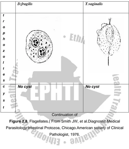
Dientamoeba fragilis
Dientamoeba fragilis originally classified as an amoeba, is now considered an amoeba like flagellate more closely related to the genus
Trichomonas. Electron microscopic studies have revealed that the internal structures are typical of flagellate.
Geographical Distribution: World wide
Habitat: In the large intestine.
Morphology: Has trophozoite stage only, No cyst stage.
Trophozoite:-
Size: 6-15μm
Motility: Either non-motile (most often), or very actively motile in very fresh fluid stools with fan-like multiple pseudopodia. It becomes non-motile under the cover slip or disintegrates immediately.
Cytoplasm: clear ectoplasm.
Nucleus: Usually one or two nuclei but 3or 4 nuclei may be found rarely. Karyosomes split into 4-6 granules.
Inclusion bodies: Bacteria
Life cycle:- The mode of transmission is uncertain but most likely is feco-oral nature. It is postulated that the delicate trophozoite is transported from person to person inside the protective shell of helminth
ova such as Entrobius vermicularis. It reproduces asexually by binary
fission. It is considered to be harmless commensal
Laboratory Diagnosis:-The trophozoite stage is highly fragile and
immediate examination of fresh stool specimen to find the trophozoite stage.
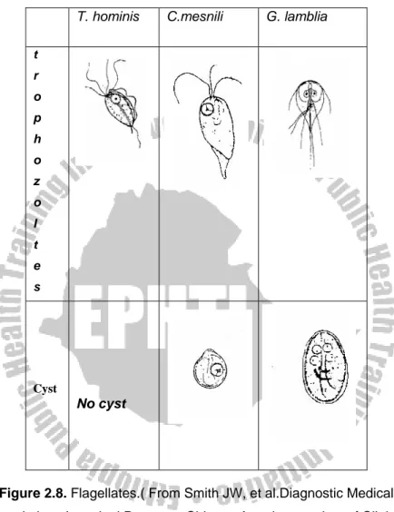
Chilomastix mesnili
Geographical Distribution: cosmopolitan but mostly prevalent in warm climates.
Habitat: Trophozoite and cyst live in the colon and caecum of the large intestine.
Morphology
Trophozoite:- Size: 6-20 by 3-10μm
Shape: Triangular and tapered at one end Motility: spiral in one definite direction. Cytoplasm:
- Spiral groove that makes asymmetrical flagellate
- cytostome (mouth-like cleft) at the rounded end.
Nucleus: one nucleus, easily visible in unstained preparation Flagella: Six flagella. Three anterior free flagella, one delicate flagellum lying in the cytostome and two flagella on the lateral margin of the cytostome
Cyst:- Size: 6-8 by 4-6μm
Shape: pear or lemon shaped
Cystostome and remains of locomotory organelles can be seen.
Nucleus: single; Thick nuclear membrane with small central karyosome.
Life Cycle
Cyst→Excystation→Trophozoite→Binary fission→encystation→Cyst in the faeces
Trophozoite stage reproduces by binary fission. The infective stage is the cyst from contaminated food or drink. Excystation occurs in the large intestine and trophozoite multiplies by binary fission.
Pathology: It is commensal
Laboratory Diagnosis:-Finding the trophozoite and cyst stages in stool
specimen. The trophozoite stage is very similar to Giardia lamblia and
Trichomonas hominis; and needs careful identification.
Giardia lamblia
Also called Giardia intestinalis and G.duodenale
Geographical Distribution:- Cosmopolitan distribution in warm climate and is more prevalent in children than in adults. It is the most commonly diagnosed flagellate of the human intestinal tract. High prevalence occurs in young, malnourished children in large families, orphan asylums, and elementary schools.
Habitat: Upper parts of the small intestine mainly in the duodenum and jejunum.
Morphology:
Trophozoite:-Size: 10-21 by 5-15μm
Shape: pyriform (pear-shaped), i.e. rounded anteriorly and
pointed posteriorly.
Motility: Progressive, rapid, tumbling and spinning often linked to a “falling leaf” type of motility in fresh liquid stools.
Bilaterally symmetrical
Covex dorsal surface and a flattened ventral side Contents:
- Anteriorly there are two sucking discs each contains a nucleus, 4 pairs (8) flagella, Parabasal body and axonemes
Figure 2.5. Trophozoite of G.lamblia. (From Jeffrey HC and Leach RM. Atlas of Medical Helminthology and Protozoology, 1975.)
Cyst :- Size: 8-12μm, oval shape with thick cyst wall.
Finely granular cytoplasm clearly separated from cyst wall. 2-4 oval nuclei at one pole, each with small, central
karyosome.
Cytoplasm: clear when unstained; yellowish green or bluish in iodine solution.
Fibril: thread-like remains of flagella; axonemes and parabasal bodies folded as S-shaped placed length wise in the center of the cyst.
Life Cycle
Requires a single host to complete its cycle and reproduces by a simple longitudinal binary fission
Infection occurs by ingestion of mature tetranucleated cyst with contaminated food, drink, finger, etc. Following ingestion, the cyst excyst in the upper part of the small intestin to form flagellates. They become attached to the intestinal wall by a sucking disc and absorb nourishment through their body surface. They multiply by longitudinal binar fission and some of them are carried down the intestinal tract to undergo encystation. The trophozoites and infective cysts are excreted in the faeces.
Clinical Feature and Pathology:-Major symptoms includes duodenitis, excess secretion of mucus or malabsorption of fat (steatorrhoea), sugar and vitamins, dehydration, diarrhoea, weight loss, poor appetite, vomiting, lethargy bile passage obstruction
Figure 2.6. Life cycle of Giardia lamblia. (From Nasir NT.
Prevention and Control:
1. Improving personal, family and group sanitation and hygiene. 2. Avoid contamination of food, drink and hands with the faeces. 3. Safe water supply and latrine construction.
5. Treatment of infected individuals and health education.
Laboratory Diagnosis:-Finding the trophozoite and cyst stages in stool specimen. The stool is usually offensive, bulky, pale, mucoid (fatty), diarrheic (watery) but there is no blood in the stool. Several specimens collected at different time need to be examined because trophozoites and cysts are excreted irregularly.
Intestinal and non-pathogenic flagellate that require differentiation from
G.lamblia include: C.mesnili and Pentatrichomonas hominis (formerly
T.hominis).
Trophozoites of the above mentioned flagellates can be easily
differentiated from G.lamblia by their shape and movement (in fresh
sample) and bcause they have only one nucleus (and fewer flagella). The
only other trophozote that has two nuclei is D.fragilis but this organism
has no flagella or median bodies and look likes a small amoeba.
Cyst of intestinal flagellates can be easily differentiated from those of G.lamblia because they are smaller and do not have the same characterstic appearance of G. lamblia (do not contain remains of
flagella). C.mensili cysts are lemon shape and D. fagilis does not has
cyst stage.
Relevance to Ethiopia
Infection by Giardia lamblia has a cosmopolitan distribution both in
developed and developing nations. Infection rates ranging from 1% to 50% or so have been reported from various parts of the world. In African,
Asian and Latin American countries, about 200 million cases of Giardia lamblia infections have been estimated to occur annually. The infection may be endemic as in the tropics where it is a familial infection passed around by faecal-oral route, sporadic as in travelers, or epidemic as waterborne or institutional outbreaks (Helmut K and Zein AZ, 1993) Giardiasis is wide spread in Ethiopia. A countrywide survey of giardiasis, using formal-ether concentration method, among school children and residents showed overall prevalence rates of 8.9% and 3.1% respectively. The corresponding rate for non-school children (5-19 years of age), however, was 4.4% showing that the School children are more
significantly i