Karolinska Institutet, Stockholm, Sweden
WHERE IMAGINATION MEETS
SENSATION
MENTAL IMAGERY, PERCEPTION, AND MULTISENSORY
INTEGRATION
Christopher C. Berger
All previously published papers were reproduced with permission from the publisher. Published by Karolinska Institutet.
Printed by AJ E-print AB © Christopher C. Berger, 2016 ISBN 978-91-7676-317-9
Mental Imagery, Perception, and Multisensory
Integration
Thesis for Doctoral Degree (Ph.D.)
By
Christopher C. Berger
M.A.
Principal Supervisor: Prof. Henrik Ehrsson Department of Neuroscience Karolinska Institutet
Co-supervisor: Dr. Predrag Petrovic
Department of Clinical Neuroscience Karolinska Institutet
Opponent:
Prof. Shinsuke Shimojo
Biology Division; Computation & Neural Systems California Institute of Technology
Examination Board: Dr. India Morrison
Department of Clinical & Experimental Medicine Linköping University
Prof. Abdel El Manira Department of Neuroscience Karolinska Institutet Prof. Gustaf Gredebäck Department of Psychology Uppsala University
“Isn't it pretty to think so.” Ernest Hemingway The Sun Also Rises
ABSTRACT
What happens if we imagine seeing something while we are listening to something? Will it change what we hear? What happens if we imagine hearing something while we are looking at something? Will it change what we see? In everyday perception, our brains integrate the information provided to us by our different senses in order to form a coherent perception of the world around us in a process referred to as multisensory integration. However, sometimes the information provided to our senses arises from within, as is the case when we imagine a sensory stimulus; for example, when you picture in your mind the face of a loved one, or imagine how they sound when they say your name. The term mental imagery is used to refer to these willed simulations of sensory stimuli in our minds. Empirical research on mental imagery has demonstrated that there is a great deal of similarity in how we consciously experience, and in how our brains process the sensory stimuli we imagine and the sensory stimuli we perceive from the external world. However, whether our brains integrate stimuli that are imagined in one sense and perceived in the other has never before been explored. The main aim of this thesis was to investigate this possibility.
There were two main goals of the work comprising this thesis. First, to examine whether mental imagery is integrated with incoming sensory stimuli from a different sensory modality to change perception, and second, to examine the neural correlates of these mental imagery-induced changes in perception. Multisensory illusions have come to be a hallmark of multisensory integration as they are an easy and demonstrable way of measuring the integration of cross-modal sensory stimuli. Here, we have made use of classic multisensory illusions, and adapted them to investigate whether mental imagery in one sensory modality can integrate with veridical sensation in another sensory modality to produced fused multisensory percepts. We also used functional magnetic resonance imaging (fMRI) to examine whether brain areas related to multisensory integration of real stimuli were involved in multisensory integration of real and imagined stimuli.
By supplanting a real sensory stimulus with an imagined one in three different classic multisensory illusions, we found that imagined sensory stimuli were integrated with real sensory stimuli from a different sensory modality to change perception. Moreover, we found that these imagery-induced multisensory illusions followed the same spatial and temporal rules as classic multisensory illusions (Study I), as well as the unity-assumption rule of multisensory integration (Study II). Furthermore, we found that the neural correlates of a mental imagery-induced multisensory illusion were closely related to those known to be involved in integration of real multisensory stimuli (Study III). Lastly, consistent with what is known about adaptation to real multisensory stimuli, we found that repeated pairings of imagined and real stimuli from different sensory modalities lead to changes in future perception of the latter (Study IV). Together, these findings suggest that, indeed, what we imagine hearing can change what we see, and what we imagine seeing can change what we hear, affording mental imagery a larger role in multisensory perception than has ever before been considered.
This thesis is based on the following publications, which are referred to in the text by their roman numerals (Studies I-IV)1:
I. Berger, C. C. & Ehrsson, H. H. (2013). Mental Imagery Changes
Multisensory Perception. Current Biology 23(14), 1367-72.
II. Berger, C. C. & Ehrsson, H. H. What We Imagine Hearing Changes What
We See. Submitted Manuscript.
III. Berger, C. C. & Ehrsson, H. H. (2014). The Fusion of Mental Imagery and Sensation in the Temporal Association Cortex. The Journal of Neuroscience 34(41), 13684-13692.
IV. Berger, C. C. & Ehrsson, H. H. Rapidly Induced Auditory Plasticity by
Imagined Visual Stimuli. Manuscript.
CONTENTS
1 Introduction 1
1.1 Mental Imagery 1
1.2 Mental Imagery and Perception 3
1.3 Neural Foundations of Mental Imagery 12
1.4 Perception and Multisensory Integration 19
2 Aims 25
2.1 Study I Aim 25
2.2 Study II Aim 25
2.3 Study III Aim 25
2.4 Study IV Aim 25
3 Methods 27
3.1 Participants 27
3.2 Experimental stimuli 27
3.2.1 Stimuli for Study I 27
3.2.2 Stimuli for Study II 29
3.2.3 Stimuli for Study III 29
3.2.4 Stimuli for Study IV 30
3.3 Imagery Instructions 31
3.3.1 Imagery Instructions for Experiments in Study I 31
3.3.2 Imagery Instructions for Study II 32
3.3.3 Imagery Instructions for Study III 33
3.3.4 Imagery Instructions for Study IV 34
3.4 Psychophysical Techniques 34
3.5 fMRI: Methodological Considerations 37
4 Overview of Studies 41
4.1 Study I: Mental Imagery Changes Multisensory Perception 41
4.1.1 Study I Background and Rationale 41
4.1.2 Study I Results and Conclusions 42
4.2 Study II: What We Imagine Hearing Changes What we See 47
4.2.1 Study II Background and Rationale 47
4.2.2 Study II Results and Conclusions 47
4.3 Study III: The Fusion of Sensation and Mental Imagery in the Temporal Association
Cortex 51
4.3.1 Study III Background and Rationale 51
4.3.2 Study III Results and Conclusions 51
4.4 Study IV: Rapidly Induced Auditory Plasticity by Imagined Visual Stimuli 55
4.4.1 Study IV Background and Rationale 55
4.4.2 Study IV Results and Conclusions 55
5 Discussion 59
6 Acknowledgments 71
ANOVA Analysis of Variance
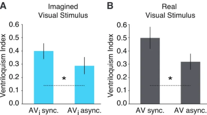
AV async. Audiovisual Asynchronous, Real Visual Stimulus AVsync. Audiovisual Synchronous, Real Visual Stimulus AVi async. Audiovisual Asynchronous, Imagined Visual Stimulus
AVi sync. Audiovisual Synchronous, Imagined Visual Stimulus
BOLD Blood Oxygen Level Dependent
EEG Electroencephalography
ERP Event Related Potential
fMRI Functional Magnetic Resonance Imaging
GLM General Linear Model
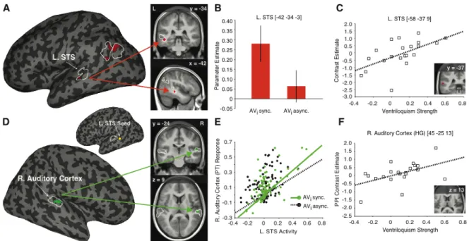
L. STS Left Superior Temporal Sulcus
LCD Liquid-Crystal Display
MEI Multisensory Enhancement Index
MR Magnetic Resonance
MRI Magnetic Resonance Imaging
MVPA Multi-Voxel Pattern Analysis
PET Positron Emission Tomography
PPI Psycho-Physiological Interaction
PSE Point of Subjective Equality
SEM Standard Error of the Mean
1 INTRODUCTION
1.1 MENTAL IMAGERY
imag·i·na·tion (i-ˌma-jə-ˈnā-shən) n. 1. The act or power of forming a mental image of something not present to the senses or never before wholly perceived in reality (“Imagination”, 2016)
As humans, we have an uncanny ability to imagine sensations in our minds at will. If I asked you to imagine seeing your nation’s flag, or to imagine hearing the voice of a loved one, or the tactile sensation of a kiss on your cheek, you would probably have an easy enough time doing that. It is a fascinating, almost oxymoronic ability we have: to see what we do not see, to hear what we do not hear, to move what we do not move, to feel what we do not feel. Of course, despite how peculiar it seems on the surface, for most of us, it is a completely ordinary part of our lives. We do it when we daydream (read: procrastinate), when we imagine descriptions we read in a book (you are welcome E. L. James), and, perhaps most importantly, when we cannot remember what comes after the letter Q in the alphabet, and rehearse the ‘alphabet song’ in our mind to help us along. We imagine all sorts of things that we are not perceiving all the time, yet figuring out what exactly these mental images are, how they are generated by the brain, and their purpose, is—to put it figuratively for maximum imagery within this paragraph—one slippery bar of soap.
This act of imaging—or imagination as one might call it—has come to be called mental imagery by those who study it; mostly in order to avoid confusion with the more fanciful interpretations of the word imagination, and perhaps to create some distance from it in order to sound more scientific. Imagination is just one of those words that means too many things to too many people anyway, and so for our purposes, I will use the phrase mental imagery to refer to our ability to generate sensory experiences in our mind of things we are not actually perceiving: what you ‘saw’ when you imagined the flag, what you ‘heard’ when you imagined the voice of the loved one, and what you ‘felt’ when you imagined the kiss on your cheek in the examples above. Although philosophers and great thinkers have pondered the nature of mental imagery since antiquity, it was not until late into the 19th century, with the development of experimental psychology, that it really moved into the realm of scientific inquiry (Kosslyn, 1994).
As the field of experimental psychology really started to get going at the turn of the 20th century, a lively debate began to develop amongst experimental psychologists about the nature of mental imagery. At the real heart of the debate was a disagreement about how ‘perception-like’ mental imagery is. On one side, there were those who argued that there is no fundamental difference between the things we imagine in our mind an the things we perceive (James, 1890; Kosslyn, 1994; Perky, 1910). On the other side of the debate were those who argue that there is some other underlying mental process at play that is fundamentally different from perception (Pylyshyn, 1973, 2002). This argument would be
taken up in different ways throughout the 20th century, but the gist of the argument remains the same.
In addition to believing that mental imagery and perception did not differ in kind, proponents of the perception-like theories in the early part of the 20th century, believed that mental images were the most fundamental form of thought (Boring, 1942; Danziger, 1979; Titchener, 1902). That is, that all the thoughts we have, however complex, could be reduced to sensory-like impressions in our mind. Researchers on the other side of the debate, however, believed to have found empirical evidence of ‘imageless thought’—thoughts that were completely devoid of sensory information (Angell, 1911; Boring, 1942). As was common practice in the early days of psychology, both sides conducted experiments that relied mostly on ‘trained-observers’ to introspect on the contents of their mind, and therefore both sides of the debate tended to produce results consistent with their theory (of course). This debate would ultimately contribute to a loss of faith in what can be learned by studying the contents of the mind at all, and the field of psychology would move towards studying behavior (i.e., behaviorism) almost exclusively for several decades (Hothersall, 2004). In the later part of the 20th century, researchers would begin to investigate the contents of
the mind once again, this time with new theories and new experimental techniques. And as the field began to revive itself, so to did the debate about mental imagery. On one side of the argument were proponents of the ‘picture-like’ (or analog) theory of mental imagery, arguing once again that the images we conjure up in our minds are very much like pictures; they are copies of sensory impressions from the external world that maintain essential features of the physical world (Halpern, 1988; Kosslyn, 1973, 1994). On the other side, were proponents of propositional-theory (also known as discrete theory), proposing that mental imagery is the product of a language-like more basic form of thought. That is, that mental imagery is the result of accessing and combining discrete symbols, which form an abstract (rather than analog) impression of the thing in mind, and that this is sufficient to account for our perception-like experiences when we imagine stimuli (Pylyshyn, 1973, 2002).
This debate in its original form, however, has died down considerably in recent years, with behavioral and neuroimaging studies having made great strides in our understanding of mental imagery and its relation to perception. Proponents of perception-like theories of imagery have come to modify their theories to accommodate mental processes that are decidedly non-sensory-like, while still maintaining that mental imagery and perception still rely on overlapping mechanisms (Borst, Kosslyn, & Denis, 2006; Kosslyn, 1994). Propositional theory on the other hand, has few hard-lined supporters today; mainly in light of its inability to account for mounting evidence that mental imagery and perception seem to rely on largely overlapping, rather than distinct neural representations (Ganis, Thompson, & Kosslyn, 2004; Kosslyn, Ganis, & Thompson, 2001; Kosslyn, 1994). Nevertheless, vestiges of this debate still exist, and there are those who are still opposed to the idea that mental imagery and perception rely on largely overlapping mechanisms. Many arguments today against perception-based theories are methodological in nature. The most common criticism
of behavioral experiments involving mental imagery is that tacit knowledge (i.e., that the participants implicitly know what the experiment is designed to study and respond accordingly, even if unintentionally) or by response bias (i.e., that the participants know what the experiment is aimed towards and consciously alter their behavior to conform to what they believe is expected of them) can account for results of studies demonstrating similarities between mental imagery and perception. In neuroimaging experiments, the criticisms are usually levied against the neuroimaging technique itself or other un-accounted for or inherent confounding variables. These concerns are not groundless, and experimental psychologists and cognitive neuroscientists have gone to great lengths to try to address these concerns. In this next section, I will give an introduction to some of the research that has shaped our current understanding of mental imagery that is important for this thesis. I will begin this section with the early behavioral studies on mental imagery and then move onto more recent behavioral and neuroimaging studies. Importantly, I will mainly focus on findings on visual and auditory mental imagery, as most of the most classic findings in the field concern these sensory modalities, as do the studies (i.e., Studies I-IV) in this thesis.
1.2 MENTAL IMAGERY AND PERCEPTION
Our everyday experience tells us that when we see something, and we imagine seeing something, that these two conscious experiences—one real and one imagined—are similar in some ways but different in others. For instance, when you imagine your nations flag, you see in your mind all the features of the flag, its colors, its overall shape, the distance between symbols or lines, etc. At the same time, however, you are certainly aware that you are not actually looking at a flag, and all the features of the flag described above, are not as clear, or as sharp as they would be if you were actually looking at a flag, are they? Why are these two experiences different? Is it because mental imagery is fundamentally different than perception—that is, mental imagery involves a different underlying process than perception—or is it that mental imagery fundamentally the same as perception, but differs only in strength or vividness? Much of the work in experimental psychology has attempted to establish the latter argument against those who claimed the former.
One of the earliest, and certainly one of the most fascinating, studies on mental imagery was an experiment conducted by a woman named Mary Cheves West Perky. Perky devised an ingenious experiment in which she asked participants to look at a screen in front of them and to imagine seeing specific objects on that screen. The objects were simple objects like a tomato, a book, a banana, an orange, a leaf, and a lemon. Perky then played a trick of sorts on her participants: she (well her assistants did, anyway) surreptitiously presented a patch of color (matched in size and shape to each of the objects) on the screen while the participants were imagining the objects (Perky, 1910). The fascinating part of the experiment is that none of the twenty-seven participants (well, except for three who were thrown out of the study because of some clumsy mishandling of the projection apparatus: nice job assistants!) noticed that the objects Perky displayed on the screen in front of them were real rather than imagined. The participants had confused their mental imagery with real percepts that they were actually
seeing! The participants were even surprised at how what they imagined sometimes looked different than they expected it to (e.g., the banana being upright when they meant to imagine it flat), and were even sometimes indignant when asked at the end of the experiment whether they were sure they had imagined the objects (Perky, 1910). This study was the first to provide strong empirical evidence for the notion that imagery and perception are very similar indeed.
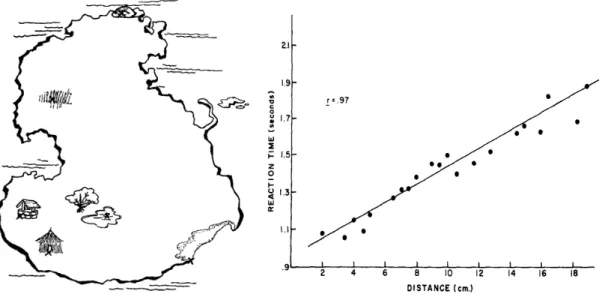
Further research on the topic of visual mental imagery was concerned with whether the spatial properties of visual perception and visual imagery were comparable. To this end Stephen Kosslyn, and colleagues devised a series of experiments in which participants were shown fictional maps or drawings to memorize (Figure 1) and where subsequently quizzed on the details of these maps. Specifically, participants memorized a map such as the one presented in Figure 1 and were then verbally given one object on the map, asked to close their eyes and form a mental image of the map, and then moments later verbally given a second item on the map. The participants were then asked to trace the distance between the first location and the second location in their mind.
Figure 1. How far from the hut to the well? Example map (left) and results (right) from Kosslyn,
Ball, & Reiser (1978). The plot on the right demonstrates the strong linear relationship between the distance between object pairs (e.g., the hut and the well) and their physical distance on the physical map that was memorized. This finding provided support for the notion that our visual mental imagery is picture-like by demonstrating that the time it took to scan between objects of a map in our mind scaled according to the true physical distance between the objects on that map.
The results from these experiments revealed a strong positive correlation between the time
it took to scan the distance between two objects in the participants’ minds and the physical distance on the maps they had memorized. Subsequent experiments demonstrated that the time it took to scan the distance between two objects also increased if the physical size of the same map was increased (in this case size does matter). That is, if the map in Figure 1 was printed on a poster rather than in this thesis and you memorized the poster-sized map, and I memorized the thesis-sized map, it would require more time for you to trace the distance
between the hut and the well in your mind than it would for me. Kosslyn concluded that, combined, these results demonstrated that the spatial relationship between objects in the physical world appeared to be preserved, suggesting that mental imagery is quasi-pictorial in nature rather than descriptive (Kosslyn, Ball, & Reiser, 1978; Kosslyn, 1973).
It was argued however, that rather than demonstrating the preservation of spatial content in visual mental imagery, these findings could merely represent the participants’ ability to simulate the physical laws of moving objects (Finke & Pinker, 1982) because the participants were specifically instructed to imagine an object moving from one location to another. And even more seriously, it was argued that these findings may merely be due to tacit knowledge about the purpose of the experiment in light of the fact that a similar relationship between the response times of participants and the physical distance between objects could be obtained when participants were asked to trace the distance in their mind between fictional locations for which they were only ever told the distances verbally (Mitchell & Richman, 1980; Richman, Mitchell, & Reznick, 1979). Follow-up experiments managed to provide support for Kosslyn’s original interpretation, however. By using simple random dot patterns, asking participants to remember the locations of the dots, then taking them away, and placing an arrow that either pointed to a location where a dot was or not. In these experiments, the participants were to indicate whether the arrow pointed to a location where a dot was previously. The key manipulation here, is that when the arrow did point to the location where a dot was, the distance between the arrow and a dot varied from 4-12cm. Just as in Kosslyn’s original study, and without the explicit instruction to imagine scanning the distance between the arrow and the dot, the participants’ reaction times to determine whether the arrow pointed in the location of a dot or not increased as the distance between the arrow and the previous location of the dot increased (Finke & Pinker, 1982). This suggests that when the participants were asked to determine whether there was a dot in a specific location they implicitly formed a mental image and mentally scanned the image to determine whether there was a dot in a particular location (with the arrow being their starting point)(Borst et al., 2006; Borst & Kosslyn, 2008; Finke & Pinker, 1982). These studies re-affirmed the notion that the spatial content of visual stimuli is preserved in visual perception.
Similarly, studies on auditory imagery investigated whether the temporal features of
auditory stimuli were preserved when imagining auditory stimuli and came to a similar conclusion. In experiments that I can only imagine must have felt to the participants a little like being a contestant on a game show, Andrea Halpern (1988) gave participants a task in which they were presented with two one-word song lyrics from familiar songs, and asked to determine whether the second song lyric was from the same song as the first (Halpern, 1988). The songs used were “Do re mi” (from The Sound of Music), “Hark the Herald Angles Sing”, and “The Star Spangled Banner” (the American National Anthem). These songs were selected based on specific features critical to the experiment such as familiarity, whether they had lyrics that started on the odd-numbered beats from 1-13, and had unique words in the first phrase. Halpern found that the participants’ reaction time increased as a function of the relative distance between the two lyrics within the song. This means that it took the same
amount of time for participants to determine whether the second lyric was in the song if the first lyric was form beat 1 and the second was from beat 5 (relative distance = 4 beats) as when the first lyric was form beat 7 and the second was from beat 11 (relative distance = 4 beats), but took less time when the first lyric was from beat 5 and the second was from beat 7 (relative distance = 2 beats). Moreover, this finding was consistent regardless of whether the participants were instructed to imagine hearing the lyrics in their mind or whether they were given notes instead of lyrics. The results form Halpern’s experiments are consistent with Kosslyn’s experiments on the preserved spatial content of visual images in visual imagery, and suggest that there is a preserved temporal format for auditory perception.
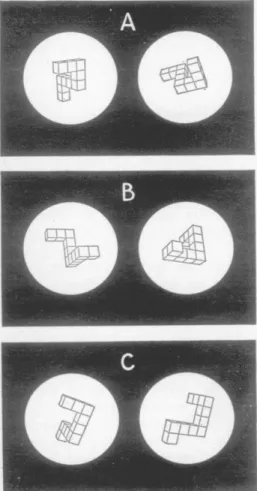
These findings from visual and auditory imagery are conceptually aligned with the widely popular and ingenious experiments conducted by Roger Shepard and Jacqueline Metzler at Stanford University around the same time. Shepard and Metzler (1971) came up with a unique experiment designed to measure how we solve a very simple problem that can be summarized in the following terms: Are these two objects the same or different? Shepard and Metzler asked participants to examine pictures of two three-dimensional objects side-by-side, and determine as quickly as possible whether they were the same of object or not (see Figure 2). The objects were either the same objects only rotated in the picture-plane (i.e., the picture of the object was rotated clockwise-or counterclockwise), the same objects only rotated in the depth-plane (i.e., rotated along their vertical axis within the picture), or they were different objects (see Figure 2 for examples)(Shepard & Metzler, 1971). Shepard and Metzler (1971) found that the time it took for participants to determine whether the objects were the same (when they were in fact the same, only rotated) was correlated with the extent that the second object was rotated. That is, it took participants longer to determine that two objects were the same if they were rotated 80° than if they were rotated 60°. This suggests that participants were creating an internal mental image of the object and then rotating it in their mind to try to match the other object. These findings, along with others that would serve to rule out alternative explanations for this effect such as response bias or differences in eye movements (Cooper & Shepard, 1973), were some of the first and most convincing that mental imagery is perceptual rather than propositional.
Figure 2. Are the two objects the same or different? Example stimuli used in experiments by Shepard & Metzler (1971) demonstrating the importance of mental imagery in human cognition. The objects were either the same objects rotated on their face (e.g., 80° clockwise) (A), the same objects rotated in depth (e.g., 80° in along the vertical axis)(B), or two objects that were different (C). Shepard and Metzler found that the time it took to determine whether the pairs were the same or not, was directly proportional to the degree of rotation no mater which plane the objects were rotated. These results suggest an important role of mental imagery in how we think2.
Further studies on mental imagery examined the interaction of mental imagery and perception ongoing perception. Many of these early behavioral studies were interested in examining the hypothesis that mental imagery and perception rely on overlapping neural representations, and therefore imagining sensory stimuli in a given sensory modality should inhibit or enhance sensory perception within that sensory modality. In this line, Segal and Fusella (1970) made use of psychophysics—an experimental technique in which some aspect of a stimulus is systematically varied as a participant makes perceptual judgments about that stimulus—in a modern version of the Perky effect described above. In this modernized version of the Perky effect, Segal and Fusella (1970) varied how visible visual stimuli were (from not visible at all to completely visible) while the participants indicated whether there was a visual stimulus present or not, and recorded on a trial-by-trial basis how well participants were able to detect the visual stimuli when they were imagining a visual stimulus
2 This figure was reproduced with permission from the American Association for the Advancement of Science
compared to when they were not imagining anything. Consistent with Perky’s original findings, they found that the participants were better at detecting the visual stimulus when they were not imagining anything compared to when they were imagining the visual stimulus. Moreover, they also found that participants were more likely to think they saw something that was not there when no visual stimulus was presented. However, Segal and Fusella (1970) also wanted to examine whether this hindrance of visual perception was merely the result of a shift in attention when imagining the visual stimuli or whether it was because the participants’ visual imagery was interfering with their ability to perceive real stimuli because of overlapping neural representations. They reasoned that imagining an auditory stimulus should hinder visual perception in the same manner as imagining a visual stimulus if the effect is due to changes in attention; however, if the effect reflects competition for neural resources, auditory imagery should not hinder visual perception as much as visual imagery, and visual imagery should not hinder auditory perception as much as auditory imagery. The results from these experiments were in support of this idea: the participants’ performances were worst when they imagined a stimulus in the same sensory modality. These results were strong behavioral evidence in support of the notion that imagery and perception rely on overlapping neural representations.
Another study conducted in the same vein by Martha Farah, from the Massachusetts Institute of Technology also made use of psychophysics to investigate the relationship of visual imagery and visual perception. Unlike the previous experiment, however, this study sought to examine whether there were instances in which visual imagery should facilitate visual perception, in order to try to dissociate purely propositional accounts of mental imagery (which purport that some independent non-perceptual process can explain effects found for mental imagery) from perception-based accounts of mental imagery (i.e., that the things we imagine are directly relatable to the things we perceive in the world). Farah argued that to make the case for the latter, it is important demonstrate that both the content and spatial format of the mental image were critical to alter visual perception. Thus, she examined whether imagining a specific letter facilitated visual processing of that letter, and when it was in the same location as the imagined letter. That is, if imagining the letter H facilitates processing of the letter H but not T, and this is due to a direct rather than abstract similarity in how we process imagined and perceived visual stimuli, it should only facilitate the perception of the letter H (and not T) and only when imagined in the same location. Indeed, this is exactly what Farah found: that if participants imagined a visual stimulus that was the same in content (e.g., imagine H; see H), and location, perception was significantly enhanced compared to when nothing was imagined or a different visual stimulus was imagined (Farah, 1985). Similar results were found for auditory imagery: Farah and Smith (1983) examined whether imagining hearing one tone facilitated perception of that tone vs. another tone in a detection task (a task in which participants judge whether a stimulus was present or not while the intensity of the stimuli are varied across trials). They found that imagining a tone selectively facilitated perceiving a tone of the same frequency, but not of a tone of a different frequency (Farah & Smith, 1983). Findings such these went a long way towards refuting
propositional theories of mental imagery, and provided strong behavioral evidence that certain aspects of mental imagery are perception-like rather than propositional.
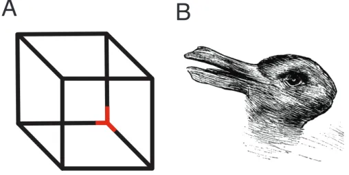
More recent behavioral experiments on the similarities between mental imagery and perception, however, have evolved along with our understanding of human perception. One technique that has been fruitful in furthering our understanding of visual perception is binocular rivalry. Binocular rivalry is a visual phenomenon in which when one object is presented to one eye and a completely different object is presented to the other eye, our brain fails to merge these two objects, and we are only consciously aware of one object at a time (Blake & Logothetis, 2002). This is conceptually similar to what happens when we look at ambiguous figures like the Necker cube and the duck/rabbit illusions (see Figure 3 for examples). Faced with conflicting pieces of information, our perception fluctuates between the two percepts. Unlike, in these ambiguous figures however, binocular rivalry has long been considered outside of our conscious control: we cannot consciously force ourselves to see one stimulus over the other (Blake & Logothetis, 2002). For example, in binocular rivalry, a house might be presented in one eye, and a face in the other eye, and rather than a fused combination of a house and a face, we fluctuate back and forth between seeing a house or a face without much control over it3. For these reasons, binocular rivalry is an ideal paradigm to study conscious visual perception and has been used extensively to examine which aspects of visual perception are related to early processing of the stimuli by the visual cortex, and which aspects are related to late processing by higher level visual cortices. For instance it turns out that certain illusions of visual perception that rely on early processing of stimuli are still present when the stimuli are suppressed from conscious awareness in a rivalry paradigm. For example, it has been found that the visual motion aftereffect (i.e., a visual illusion in which a static visual stimulus is perceived as moving in the opposite direction of a previously moving stimulus4) occurs for suppressed (i.e., not consciously perceived) moving stimuli (Lehmkuhle & Fox, 1975; O’Shea & Crassini, 1981). The effects of visual priming (i.e., the facilitation of visual perception via brief prior exposure to that visual stimulus) on the other hand, seem to be extinguished when the participant does not consciously perceive the visual stimuli, suggesting that priming is a process that requires conscious awareness of the stimuli and relies on a later stage of visual processing (Cave, Blake, & McNamara, 1998). These findings in the field of perception would be useful in disentangling the similarities and difference between mental imagery and perception.
3 However, it should be noted that visual attention can modulate the temporal dynamics (rate and duration of
switching) of rivaling stimuli with practice. If observers focus on one stimulus that is already perceived (i.e., dominant) carefully it can increase the duration that stimulus is consciously perceived (i.e., dominance) at the expense of the other, but switching still occurs none-the-less, outside of the observer’s control (Ooi & He, 1999). Furthermore, specific features of the rivaling stimuli (e.g., salience, contrast, context) will make one stimulus more dominant than the other (Blake & Logothetis, 2002).
4 A classic example of this illusion is the ‘waterfall effect’, whereby if an observer looks at a waterfall for a
period of time then looks to the trees or rocks to the side of the waterfall, they will appear to move upwards for a brief period.
One study made use of the motion aftereffect described above to examine whether visual mental imagery of motion would lead to a visual motion aftereffect (Winawer, Huk, & Boroditsky, 2010). In this study, Jonathan Winawer and colleagues (2010) investigated whether imagining a visual grating stimulus (i.e., a series of dark grey horizontal bars spanning the length of the visual display equally spaced from each other vertically) altered the perceived motion of moving dots presented afterwards. By psychophysically varying the extent that the moving dots were random, ranging from all the dots moving upwards to all the dots moving downwards, and completely random motion in the middle, Winawer et al. (2010) found that imagining a visual grating moving upwards caused the participants to see a given set of moving dots as moving downwards and imagining the visual grating moving downwards caused the participants to perceive the same set of dots as moving upwards. Interestingly, further experiments using this paradigm found that even listening to descriptions of literal visual motion (e.g., “the squirrels were teaming up the tree”) and metaphorical motion (e.g., “stock prices were falling), lead to this motion aftereffect suggesting an inherent link between language comprehension and visual imagery (Dils & Boroditsky, 2010). In light of the studies on visual perception described above, these findings provided strong evidence in favor of overlapping representations of mental imagery and visual perception at a very early stage of visual processing.
Another study made use of the binocular rivalry paradigm described above to examine the effects of visual imagery on visual perception (Pearson, Clifford, & Tong, 2008a). If you recall, from the section above, I described experiments which found seemingly conflicting results: the Perky-effect studies found that visual perception was hindered by visual imagery, whereas the studies by Farah (in which the participants imagined Hs & Ts) found that visual mental imagery of an object facilitated visual perception of that object. While both studies nicely demonstrate that visual mental imagery can alter perception within that sensory modality, they do so with opposite results; the problem being that there is no visual perception analog here, and so there is no way to compare these results with imagery to results with visual perception. It could simply be that imagining visual stimuli in these task changes the way in which the participants attend to the visual stimuli. By utilizing binocular rivalry, however, Pearson et al., (2008) were able to examine whether visual mental imagery facilitated or hindered subsequent visual perception. In this experiment, participants briefly (750 ms) viewed rivalry displays of a visual grating stimulus presented horizontally in one eye and a visual grating stimulus presented vertically in the other, then were either instructed to imagine the visual stimulus they just perceived (i.e., the dominant visual stimulus) or to imagine the visual stimulus they did not just perceive for 10 seconds. They found that when participants imagined the dominant visual stimulus, the stability of that visual stimulus was very high, whereas when they imagined the non-dominant visual stimulus, the stability of that visual stimulus dropped dramatically. That is, if the participant saw a vertical grating stimulus on the previous trial, and imagined seeing that stimulus for 10 seconds, they were more likely to see that stimulus again; however, if the participant saw a vertical grating stimulus and imagined a horizontal grating stimulus for 10 seconds, they were much less likely to see the
vertical stimulus again. A benefit of this paradigm, in contrast to earlier studies presented above on the effects of imagery on perception, is that real visual stimuli could also be presented in the periods between rivalry displays and the result on visual perception could be compared with those of mental imagery. In further experiments, Pearson et al. (2008) found that a weak visual stimulus presented in the 10 s periods between rivalry displays produced the same effect on visual perception stability suggesting that the observed changes in visual perception from visual imagery have the same underlying mechanisms as visual perception itself.
Another interesting experiment, which demonstrated a strong link between mental imagery and perception, measured pupil dilation in response to imagined visual stimuli (Laeng & Sulutvedt, 2013). In this study, the participants imagined the same visual stimulus (e.g., a triangle) at different levels of luminance while their pupil diameters were measured. Laeng and Sulutvedt (2013) found that the participants’ pupils constricted when imagining a bright visual stimulus, but dilated when imagining dark visual stimulus. These changes in pupil dilation were very closely related to those of perceiving the same stimuli, and suggest that when one imagines a visual stimulus, it engages processes of visual perception at an early enough stage in processing that the pupils constrict to protect the eyes sensitive light-absorbing cells (Laeng & Sulutvedt, 2013). One particularly striking aspect of this paradigm is that it runs counter to what one would expect if the results were due to cognitive effort, as previous studies have found that increasing cognitive effort is associated with increased pupil dilation (Hess E. & Polt J., 1964; Kahneman, Tursky, Shapiro, & Crider, 1969).
Despite the strong case made from behavioral evidence that mental imagery is
perception-like and relies on overlapping representations with perception, it has been argued that truly convincing evidence for either cannot come from behavioral experiments alone (Anderson, 1978, 1979). This is a ‘black box problem’ of sorts: when we can only control what goes in and what comes out of the black box, there will always be differing interpretations for what goes on inside (Anderson, 1978, 1979; Farah, 1989b). To truly determine how what goes in affects what comes out, we must look inside the black box. In this analogy, for our purposes, our brain is that black box, and what ‘goes in’ is a stimulus, real or imagined, and what comes out is some measurable change in behavior (e.g., response times) or perception, but to fully rule out whether these changes can be accounted for by perception-like representations, language-like propositional representations, both, or neither, we have to look inside the brain. In this next section, I will give a brief overview of the literature that has looked inside the brain and their findings concerning mental imagery and perception.
Figure 3.Front or back; duck or rabbit? Examples of the Necker cube (A) and duck/rabbit (B) ambiguous image illusions. The red corner in the Necker cube can either be perceived as being in the front of the cube (i.e., closest to you) or on the inside of the cube (i.e., furthest from you). The duck/rabbit ambiguous image drawing was originally used by Joseph Jastrow (1900) to illustrate the malleability of perception, and can either be perceived as, appropriately enough, a duck or a rabbit.
1.3 NEURAL FOUNDATIONS OF MENTAL IMAGERY
The neural basis for mental imagery is one of those things that are tricky to study in other animals. We cannot just ask a monkey or a mouse to imagine seeing or hearing something (even if we do so politely) while we record from electrodes in their brain5, and so the bulk of our knowledge on this subject comes from studies on humans. Some of the earliest evidence for the neural basis for mental imagery would rely on human lesion studies. It was discovered, for example, that brain damaged patients who lost the ability to perceive color, also lost the ability to imagine color (De Renzi & Spinnler, 1967), that patients with damage to their parietal lobe who lost the ability to perceive visual stimuli on one side of their body (i.e., hemispatial neglect) also lost the ability to imagine stimuli on that side of their body (Bisiach & Luzzatti, 1978; Farah, 1989b). One interesting case study in this regard found that if you had the patient imagine themself in a particular location and imagine looking at the scene in a very familiar town square (Piazza del Duomo, Milan, Italy), they would accurately describe from their visual imagery, all the buildings on their right side, but not those from the left. If you then immediately had them imagine standing at the opposite vantage point in the square and describe the scene, they could now imagine and describe all the buildings on their right side that they could not just moments before. Moreover, they could no longer imagine or describe the buildings they had just imagined and described moments before that were now on their left (previously on their right) (Bisiach & Luzzatti, 1978). In the case of auditory imagery, studies of patients with left or right temporal lobe excision (i.e., removal of their
5 Although a close approximation might be possible: some interesting work which has recently been taken
underway by researchers using associative learning paradigms in monkeys are making some strides in this direction; see (Albright, 2012) and Discussion (section 5) below for more on this.
temporal lobes)—found that patients performed equally poorly on perceptual and imagery versions of a pitch comparison task. The imagery pitch comparison task in this study was similar to the pitch comparison task described above that was used by Halpern (1988) to investigate whether the temporal properties of auditory stimuli were preserved in imagined auditory stimuli. Participants were given two song lyrics of familiar songs and asked to compare whether the pitch of the first one was higher than the second, forcing them to imagine hearing the song. In the perceptual version of the task, the participants performed the same task, however, this time they actually heard the song while making their comparison. Zatore and Halpern (1993) compared lesion patients’ and healthy controls’ perception of familiar songs, and found that temporal lobectomy patients performed equally poorly on the imagery and perceptual versions of the task, compared to healthy controls who performed well on both tasks (Zatorre & Halpern, 1993). These findings were consistent with the idea that if imagery and perception involve shared representations, then we should see visual imagery impairments where we see impairments in visual perception. However, brain lesions are always large, involving damage to multiple areas, and interrupt the underlying white matter fiber tracts. Therefore, to determine whether mental imagery and perception rely on exactly the same cortical areas, neuroimaging experiments with good anatomical resolution— functional magnetic resonance imaging in particular—would be needed. Neuroimaging experiments would expand upon the findings from lesion studies considerably, largely converging on evidence in favor of shared representations of mental imagery and perception. At the time of Stephen Kosslyn’s seminal review on the neural basis of mental imagery in 2001, over 50 neuroimaging studies had been conducted on visual imagery alone (Kosslyn et al., 2001). While I certainly will not cover all of these studies, I will try to survey some of the most influential neuroimaging studies on visual and auditory imagery that are relevant to the topic of this thesis.
Some of the earliest neuroimaging experiments examining the neural correlates of mental imagery made use of electroencephalography (EEG)—a neuroimaging technique in which electrodes placed on the scalp record fluctuations of electrical current generated by neurons in the brain (Nunez & Srinivasan, 2006). Using EEG, Martha Farah and her colleagues (1989) showed participants words, specifically either concrete nouns (e.g., surfboard) or abstract nouns6 (e.g., escapism), and asked them to imagine the concrete nouns as vividly as possible and to simply view the abstract nouns while recording event related potentials (ERPs) (i.e., the time-locked response of an electrode to a stimulus)(Farah et al., 1989). They found that imagining the concrete nouns led to a significant positive difference in ERPs compared to the abstract noun (i.e., no-imagery) control for electrodes placed over areas involved in visual perception (i.e., occipital lobe). They found the same effect of imagery vs. no-imagery trials when participants were given the words verbally. Further experiments using EEG, made use
6 Interestingly, abstract words were added to the experimental protocol as the no-imagery control because pilot
studies revealed that participants found it too difficult not to imagine anything when presented with concrete words. This is consistent with research discussed above demonstrating the involvement of visual imagery in language comprehension (Dils & Boroditsky, 2010; Farah, Péronnet, Gonon, & Giard, 1988).
of perceptual facilitation effect of mental imagery on perception described earlier, in which imagining an H facilitated perception of an H, whereas imagining a T did not (Farah et al., 1989; Farah, 1985). In this study, ERPs were recorded while the participants were imagining either an H or a T while detecting whether an H or T waspresented. They found that mental imagery of a letter facilitated visual detection of the same letter, as they found before (Farah, 1985), and that this perceptual facilitation was accompanied by changes in ERPs recorded from the occipital recording sites (i.e., over the visual cortex) compared to when they imagined a different letter than the one that was perceived (Farah et al., 1989). These early studies investigating the neural basis of mental imagery using EEG were consistent with the notion of shared representations of mental imagery and perception; however, the poor spatial resolution of EEG limits what can be said about how much perception and mental imagery truly involve overlapping mechanisms. Experiments using other neuroimaging techniques would prove more useful in this regard.
Researchers have also made use of other neuroimaging techniques such as single photon emission computed tomography (SPECT) and positron emission tomography study (PET) in the search for the neural correlates of mental imagery. SPECT and PET are neuroimaging techniques in which a radioactive tracer is injected into the bloodstream of the participant and then measured by the scanner as tracer-laced blood flows to different parts of the brain over the time course of the task (Gulyás & Sjöholm, 2007; Rahmim & Zaidi, 2008). The functional capacity of these techniques, from a neuroimaging standpoint, relies on the fact that increased blood flow to different parts of the brain is tightly coupled with neural activity (J. J. Chen, Wieckowska, Meyer, & Pike, 2008; Logothetis & Wandell, 2004). Goldenberg and colleagues (1988) conducted an experiment using (SPECT) that was conceptually similar to the EEG experiment described above in which participants were given lists of concrete words to imagine (Farah, 1989b; Goldenberg et al., 1989). Goldenberg et al. (1988) gave participants concrete words and asked participants to either imagine seeing the objects they referred to or to memorize the words while in the SPECT scanner and found increased blood flow (indicative of neural activity) to parts of the brain responsible for visual perception (i.e., occipital and temporal cortices). These findings were consistent with those from a (PET) study in which Roland and Friberg (1985) found increased blood flow to areas critical for visual processing (i.e., occipital, posterior parietal, and posterior inferior temporal areas) while participants imagined leaving their home and navigating through their neighborhood (Farah, 1989b; Roland & Friberg, 1985). They were also consistent with another PET study in which participants observed grids of different patterns of darkened and non-darkened squares, and were subsequently asked to indicate whether a darkened square was in a given location previously—a task which requires mental imagery (Kosslyn et al., 1994; Kosslyn, Thompson, & Alpert, 1997). This study found increased blood flow to areas of the visual cortex for this imagery task compare to control conditions in which the participants merely viewed the same stimuli (Kosslyn et al., 1997). In the case of auditory imagery, converging evidence for shared representations of imagery and perception came from another study conducted using PET which found that imagining familiar tunes was associated with
increased blood flow to areas of the brain (i.e., the auditory cortex) commonly associated with processing auditory stimuli (Halpern & Zatorre, 1999). Because SPECT and PET involve the injection of radioactive tracer, they are rather invasive techniques and so the vast majority of the neuroimaging research conducted on mental imagery would be done using less expensive and non-invasive techniques such as functional magnetic resonance imaging (fMRI) 7.
Like PET or SPECT, fMRI is a neuroimaging technique that makes use of the changes in blood flow related to neural activity, however, unlike PET/SPECT, fMRI does not require the use of radioactive tracers. Instead, fMRI makes use of the blood oxygen level dependent (BOLD) response; capitalizing on the fact that increased neural firing consumes oxygen (and glucose), which is then replenished by oxygen rich blood. Due to the difference in the magnetic properties of oxygen-rich blood (i.e., oxyhemoglobin is diamagnetic) and non-oxygen-rich blood (i.e., deoxyhemoglobin is paramagnetic), this causes distortions in the magnetic field that are then measured by a radiofrequency coil around the participants head. Early fMRI studies investigating the neural correlates of mental imagery largely sought to elaborate upon the findings from earlier lesion and PET studies with new experimental designs and questions concerning the extent of the involvement of early sensory cortices in mental imagery. The first fMRI study on mental imagery examined BOLD responses to imagining objects (e.g., a tree) compared to baseline BOLD activity, and perceiving those objects compared to baseline BOLD activity. This study revealed largely overlapping activations for visual imagery and visual perception in portions of the frontal, parietal, and occipital-temporal cortices (Ganis et al., 2004). In the first study on auditory imagery using fMRI, BOLD responses were compared between conditions in which the participants viewed silent videos of everyday scenarios and imagined hearing the associated sounds (e.g., someone blow-drying their hair) and a control condition in which the same videos were digitally scrambled. They also compared BOLD responses between viewing the same videos but while hearing the associated sounds and the scrambled-video control condition (Bunzeck, Wuestenberg, Lutz, Heinze, & Jancke, 2005). They found that the hearing the sounds led to increased activity in the primary auditory and secondary auditory cortices, and imagining the sounds lead to increased activity in the secondary auditory cortex (Bunzeck et al., 2005). These studies converged with those form lesion, EEG, and PET/SPECT studies, in showing that imagery and perception seemed to rely on mostly overlapping rather than distinct neural representations. So, it is settled then. Mental imagery and perception share overlapping and not distinct neural representations, right? Well, not quite. It remained to be gleaned from the studies presented above whether imagining sensory stimuli could activate primary sensory cortices. Such a finding would stand as a strong refutation of propositional theories of mental
7 It is important to note that as fMRI became more widely available, PET/SPECT neuroimaging studies
measuring regional blood flow alone have become largely obsolete due to their invasiveness and cost. Many of the current neuroimaging studies using these techniques today make use of different radioisotopes catered to questions concerning the underlying neurobiology of regional brain activation, which is something fMRI cannot assess (Small et al., 2008).
imagery (i.e., non-perception-based theories), and so many subsequent fMRI (and PET studies) would serve to establish whether mental imagery could activate early sensory cortices.
The vast majority of neuroimaging studies on visual imagery would come to affirm the notion that visual mental imagery can activate the primary visual cortex (Kosslyn et al., 2001); and although early studies on auditory imagery have been more mixed about the involvement of the primary auditory cortex when it comes to imaging auditory stimuli, recent evidence seems to converge on a consensus that auditory imagery also activates the primary auditory cortex (Oh, Kwon, Yang, & Jeong, 2013). Concerning visual imagery, some of the most convincing evidence of activation of the primary visual cortex comes from a study which made use of retinotopic mapping—mapping the visual input from the eyes on the early visual areas of the cortex (Van Essen, Newsome, & Maunsell, 2002)–to compare imagining and perceiving visual objects of different sizes and found that imagined visual stimuli activated the earliest portions of the visual cortex (i.e., area 17) and that the locus of activation in this area shifted with the size of the imagined visual stimulus in the same manner as real visual stimuli (Tootell, Hadjikhani, Mendola, Marrett, & Dale, 1998). Similarly, although studies have found activation of the primary auditory cortex for imagining non-speech auditory stimuli (e.g., simple tones or music without lyrics)(Kraemer, Macrae, Green, & Kelley, 2005; Yoo, Lee, & Choi, 2001), the most convincing evidence for shared representations of perceived and imagined auditory stimuli comes from a study which made use of tonotopic mapping—mapping the frequency of sounds presented to the ears to the early areas of the auditory cortex (Saenz & Langers, 2014)–and found that imagining tones of different frequencies selectively activates portions of the primary auditory cortex (i.e., Heschl’s gyrus) responsible for perceiving those frequencies (Oh et al., 2013). Together, these studies, and others like them, went a long way towards refuting propositional accounts of mental imagery, and suggested that early sensory cortices are involved in imagining the low-level features of sensory stimuli.
Further neuroimaging studies on mental imagery have approached the problem of whether imagery and perception involve overlapping neural representations by examining whether the content (rather than specific features such as the size of the stimuli) of imagined sensory stimuli led to same content specific brain activation of perceived stimuli. To get to this question, however, one must first have an understanding of which areas are responsible for content specific visual perception. Neuroimaging and electrophysiology studies making use of binocular rivalry (described in section 1.2 above) have pointed to a distinction between early visual areas (i.e., striate cortex) and later visual areas (i.e., extrastriate areas), with the former being involved in representing low-level spatial features of visual stimuli provided by the retina, and the latter being involved in the conscious perception of visual stimuli (Tong, Nakayama, Vaughan, & Kanwisher, 1998; Tootell et al., 1998). Although it was believed early on that conscious visual perception of stimuli is resolved by the primary visual cortex (Blake, 1989), electrophysiology recordings in monkeys found very little selectivity for the consciously perceived visual stimulus within the primary visual cortex during binocular
rivalry (Logothetis & Schall, 1989). This suggested that conscious visual perception is resolved at a later stage of visual processing. The question is where? Tong and colleagues (1998) addressed this question in a neuroimaging experiment using binocular rivalry on humans. They had participants view binocularly rivaling stimuli (i.e., a house and a face) and asked them to report their conscious visual perception while recording BOLD responses from regions just outside the primary visual cortex (i.e., extrastriate areas) which are known to respond selectively to house and face stimuli (Epstein & Kanwisher, 1998; Kanwisher, McDermott, & Chun, 1997), and found that the participants’ conscious visual percept selectively correlated with activity in these areas as if the visual stimuli were presented one at a time (i.e., monocularly) (Tong et al., 1998). This finding suggests that conscious visual perception of rivaling stimuli is still resolved in visual areas of the brain, but ones that lie outside the primary visual cortex. Thus, when it comes to examining the similarities between mental imagery and perception, perhaps it is more functionally relevant to consider the similarities between mental imagery and the perception in areas of the brain involved in the conscious perception of visual stimuli.
To this end, in one study, O’craven and Kanwisher (2000) investigated whether mental imagery of houses and faces involved content specific activation in extrastriate areas found to be selectively responsible for conscious perception of those stimuli. They found that imagining faces and houses lead to content specific activation within the ventral ocipito-temporal cortex called the fusiform face area and the ventromedial cortical region called the parahippocampal place area, respectively. These findings dovetailed nicely with the known selectivity for visual stimuli of these kinds within those regions, and served as strong evidence that mental imagery recruits the same neural machinery of conscious visual perception in a content specific manner. Another fMRI study conducted in the same vein utilized multi-voxel pattern analysis (MVPA) of the BOLD signal to examine whether the brain encodes the content and location of imagined visual stimuli in the same manner as perceived visual stimuli. Multi-voxel pattern analysis examines the fine-grain patterns of activation in the brain over time associated with a given stimulus or condition to create classifiers based on those stimuli or conditions (Norman, Polyn, Detre, & Haxby, 2006). These classifiers are then applied to new BOLD data from a different set of conditions to determine whether they can significantly predict the pattern of activation above chance. If they can, then the patterns of activation observed in the first set of data are reflected in the patterns of activation in the second (Norman et al., 2006). Thus, by having participants imagine seeing objects of different categories and in different spatial locations, and training classifiers on the brain activity from perceiving those objects from different spatial locations, Cichy et al. (2011) were able to determine whether the patterns of activity involved in the latter, could be used to predict brain activity of the former. They found that the classifiers trained on patterns of brain activity in response to visual stimuli of different categories (e.g., faces vs. houses) could significantly predict the patterns of activity in visual areas associated with perceiving those categories, and that the visual areas responsible for perceiving those categories maintained their category selectivity when imagining the stimuli (i.e., patterns of
activity in the occipital face are were significantly predicted by imagining faces, but not by imagining houses). Furthermore, they found that the patterns of activity associated with the spatial location of imagined visual stimuli could be predicted in early visual areas. Together, these neuroimaging experiments suggest that the content and spatial location of imagining visual stimuli and perceiving visual stimuli make use of overlapping neural mechanisms. In summary, the neuroimaging evidence seems to suggest that mental imagery and perception rely on largely overlapping neural mechanisms. Moreover, it appears that imagining sensory stimuli can activate early sensory cortices, refuting the notion that mental imagery is propositional in nature, and supporting perception-based theories of imagery. Interestingly, however, all the research on the similarities between imagery and perception thus far have relied on demonstrating similarities within a single sensory modality (i.e., similarities between visual imagery and visual perception, or between auditory imagery and auditory perception) despite the fact that perception routinely involves the integration of sensory stimuli from several sensory modalities (Ghazanfar & Schroeder, 2006; Stein & Stanford, 2008). Thus, to truly examine whether mental imagery is perception-like, one must consider its relation to converging sensory information from other sensory modalities. This is the aim of this thesis. In this next section I will discuss what is known about the multisensory nature of perception before outlining the aims of this thesis in more detail.
1.4 PERCEPTION AND MULTISENSORY INTEGRATION
As you walk the streets of a busy city during the day, you will see buildings, streets, cars, bicyclists, and fellow pedestrians; you will hear the cacophony of passing cars, bells of bicyclists, and the indistinct chatter of people around you; you will feel the wind, the sun, or the rain on your face, and perhaps the bump of a passing stranger. How is all of this information put together into a coherent perception of the world? How do we know which sights go with which sounds, or whether the bump we just felt was from a passing pedestrian, bicyclist, or car? Without some form of integration of these pieces of information, we would not be able to determine which sounds go with which objects we see, and the result would be, perceptually speaking, incomprehensible.
The proposed mechanism for how the brain solves this information-processing problem has been appropriately dubbed ‘multisensory integration’. Multisensory integration can be most generally defined as the effect that sensory stimuli from different sensory modalities have on an organism when they are combined rather than independent (Stein & Stanford, 2008). In the case of human perception, this can take the form of an enhancement or hindrance of perception in one or more sensory modalities, as is the case in multisensory interactions (i.e., when one sensory modality effects the processing of another sensory modality). Imagine the following: you are approached by a stranger on the bus while you are looking down at your feet (I don’t know why, maybe you just bought a pair of new shoes and you’re admiring how awesome your feet look in your new kicks—it’s just an example, so roll with it) minding your own business, when the stranger says something to you. You’re not quite sure what they said, but you think you heard them say, “are you going to take a dump?”, but that can’t be right. No. Even for public transportation, that is just too weird of a question (at least in most places: I’m looking at you San Francisco). So you politely look up from your amazing feet, and ask, “what’s that?” Then they begin to repeat themselves, but this time, as your gaze makes its way up to their face, a shiny button on their shirt distracts you, and you hear them repeat their question about your bowel movements. Still not convinced that a stranger could be asking you such a question, you look them directly in the face now and ask more directly, “what?!” They repeat the question, this time, with you looking directly at their lips as they speak, and now you hear it clearly: “Are you going to vote for Trump?” and all at once you realize that the button was a ‘Vote for Trump’ button, and you sincerely wish they asked you the other question. In this colorful example, your auditory perception was hindered every time you heard the question because of the noisy environment of the bus; however, once you looked at their mouth, violà! Your visual perception made what they said completely clear. In this case, your visual perception facilitated your auditory perception, but this could have been the other way around; your auditory perception could have facilitated your visual perception. Moreover, it could have been the case that your auditory perception was hindered by visual perception. Nevertheless, it is the interaction of the senses in this manner that we are discussing when we discuss multisensory interactions. In the case of multisensory integration, however, we are talking about the integration of sensory stimuli from two or more sensory modalities into a fused multisensory percept. That is, rather than