th
Abstracts and notes on CML presentations
1ASH 2012 Atlanta
Steve O’Brien, Newcastle University ________________________________________________________________________________________1
Highlights ... 1
2
Plenary session [3] ... 4
3
CML Therapy I [163-168] ... 4
4
CML Therapy II [913-918] ... 8
5
CML Therapy: Aiming for Deep Response [67-72] ... 12
6
CML Therapy: Pharmacodynamic Markers and Novel Treatment Approaches [691-696] ... 16
7
CML Biology and Pathophysiology, excluding Therapy I [907-912] ... 19
8
CML Biology and Pathophysiology, excluding Therapy II [31-36] ... 22
9
Biology and Pathophysiology, excluding Therapy: Poster I ... 25
10
Biology and Pathophysiology, excluding Therapy: Poster II ... 25
11
Biology and Pathophysiology, excluding Therapy: Poster III ... 26
12
Therapy: Poster I ... 26
13
Therapy: Poster II ... 27
14
Therapy: Poster III ... 27
15
All CML abstracts numbers ... 28
_________________________________________________________________________________________
1 Highlights
There were 159 abstracts about CML this year: 36 oral presentations and 123 posters. If you’re pushed for time read the first couple of pages and you’ll get the key points. I have not aimed to review all the abstracts, rather pick up and expand on some key (highly selected) themes. I have focused mainly on the oral presentations and clinically relevant studies. Complete abstracts are included for all oral presentations and just listings for posters. I’ve taken care to ensure the accuracy of the data but when furiously typing during sessions I can’t always guarantee complete precision!
Abstracts are available on line at: http://ash.confex.com/ash/2012/webprogram/meeting.html
If I had to highlight three things of most relevance to practising clinicians they would be: 1. The ‘10% thing’ is all over the place
2. Getting ‘even better’ responses and getting off treatment 3. Various drugs jostling for position – no clear winner
The ‘10% thing’ – again
…
This theme really took off this year. The story originated from Liverpool in 2003 but the large German study earlier this year from Hanfstein pushed things forward. I lost count of how many times I heard this presented at ASH. Put simply, failure to achieve a PCR level of less than 10% BCR-ABL/ABL after 3 months on a TKI (the story is expanding from just imatinib now [1678]) is associated with a less favourable outcome. Neelakantan looked at whether measuring 6 month PCR response on imatinib added anything to the <10% at 3 months story [68]. The answer seemed to be ‘no’, 3 months does the trick. The 10% story seems to hold true for dasatinib [1675] and for nilotinib [2797].
1 This review is provided free of charge as a service to the CML community and may be used, abused, distributed, shared or deleted without trace as you see fit without having to seek the permission of the author. Views expressed are those of the author alone; this work has not been commissioned/paid for by any organization, pharma company or otherwise and represents, for good or ill, the independent views of Prof O’Brien. The author is always keen to improve and any comments, good or bad, would be gratefully received (stephen.o’[email protected]). All cited data are freely available in the public domain.
There was an update on ENESTnd (nilotinib) [167, 1676] focusing on molecular response prediction. 33% of imatinib patients fail to get to less than 10% at 3 months whereas only 9.3% of nilotinib patients end up in the same boat. Switching to nilotinib from imatinb produces deeper molecular responses according to the ENESTcmr study [694] – 2 years follow up. Whether this is important in terms of survival or ability to stop remains to be seen. Peripheral arterial occlusive disease (PAOD) on nilotinib has been raised as a concern recently: abstracts 914 and 1679 say it’s a worry, 2757 says it isn’t. Hyperglycaemia is more common on nilotinib [1686] but it’s not clear what the mechanism of PAOD is. Make your own mind up but it seems to me that with all the second/third gen TKIs (is ponatinib ‘third’ gen?) we’re going to have look out for subtle but potentially important longer term side effects especially cardiovascular.
Tim Brummendorf gave an update on bosutinib in the BELA study, [69] again looking at the 10% at 3 months response question. Guess what, the same story comes out: getting below 10% at 3 months is important on this drug as well at least in terms of subsequent achievement of CCyR and MMR. Too early to see if survival affected.
Crucially what we don’t know, with our current state of knowledge, is whether switching at 3 months if the patient is over 10% can ‘rescue’ them. No studies have addressed this as yet and it will be a key aspect of SPIRIT 3 in the UK.
Getting ‘even better’ responses, getting patients off treatment
There were a lot of presentations about this. Various strategies to get PCR down to levels where patients can stop. Is low level important with respect to outcome, particulary survival? Does it allow more patients to stop? Is there more tox to put up with if you’re adding/switching drugs?
A retrospective survey from the MD Anderson [164] of patients treated with imatinib, nilotinib and dasatinib shed some light on the question of whether achievement of MR3 (MMR) predicts for survival. Although the differences are not large there’s a statistically significant benefit on OS in achieving MMR within 18-24 months. These data are retrospective and don’t of course address the question of whether switching drugs to obtain MR3 has a similar effect. Studies need to be done to investigate this further.
The Adelaide group added to this [165]: they suggest in imatinib-treated patients that 3 month PCR level, speed of response and gender are important. Women do better: maybe they’re better at taking the pills? OCT-1 activity may also be informative [693]. These factors might also contribute to our understanding of who might be able to stop treatment. Data along similar lines (although less mature) were presented from the ENESTnd study [167]. Whether the MMR ‘hares or tortoises’ differ in survival terms remains to be seen.
The German CML group have found that dose-optimised (between 400-800mg) imatinib produces more CMR4.5 responses and a landmark analysis at 48 month indicated better overall survival in these patients and of course these patients might be able to stop treatment [67].
Cytogenetics may still have a role to play in predicting outcome and a large study (1346 patients) from Germany [913] looked at this. Patients with major route additional chromosomal alterations (major ACA: +8, i(17)(q10), +19, +der(22)t(9;22)(q34;q11) have a worse outcome whereas patients with minor route ACA show no difference in overall survival (OS) and progression-free survival (PFS) compared to patients with the standard translocation, a variant translocation or the loss of the Y chromosome.
Next gen sequencing is coming to the fore in CML as everywhere else. The Italian group have used ‘ultra-deep’ sequencing to look at TK domain mutations as potential predictor of response to different drugs [692]. The answer? It seems to get more complicated (confusing?) with ‘better’ technology and it’s not clear to me that mutation analysis (perhaps apart from T315I) is that compelling when it comes to choosing TKI drugs. SMRT sequencing has also been used [917].
Lots of abstracts on stopping imatinib [e.g. 2788, 1684 – with maintenance IFN, 4274] including one looking at patient preferences [4274]. Nobody has yet explored dose reduction rather than stopping or the possibility of reducing dose from MR3: we’re planning to look at this in the DESTINY study in the UK. Delphine Rea updated the French experience of stopping 2nd generation TKIs [916]. This study, althought still early, seem to confirm what you might expect: higher rates of sustained negativity than with imatinib in the 39 patients with a minimum of 6 months follow up. 60% in the study were female and it was about 50:50 dasatinib:nilotinib although only 2 patients received these as first line therapy. 16 of 39 (41%) lost MMR (loss of MMR rather than CMR was used) most by 8 months but one at 24 months. All relapsers were fine when treatment restarted. Freedom from relapse was much better in those who were on 2nd gen first line or for imatinib intolerance rather than imatinib resistance: 68% vs 40% relapse free at 1 year. So, my simple take on the story so far: previous STIM study
th
(imatinib) ~40% can stay off treatment; on 2nd gen about 60% can stay off. Will be interesting to watch this story develop: will 2G drugs proved their worth in allowing more CML patients to stop treatment?
Jostling for position
There were updates on the primary clinical outcomes from some of the main second generation studies DASISION [1675, dasatinib], ENESTnd [1676, nilotinib], BELA [69, bosutinib] although emphasis was mainly on early response – I suspect we’ll get more comprehensive updates at ASCO in June 2013. I don’t think the story has changed since last year: undoubtedly better PCR responses but no difference in overall survival. I’m increasingly thinking there might be a trade off with the newer drugs: you will achieve a better response but this might be at the risk of other side effects, especially cardiovascular. We need to keep weighing these factors in the balance.
There was an update on the PACE study (ponatinib) with 12 months follow up [163]. This study enrolled 449 patients who had failed nilotinib or dasatinib; 234 were in chronic phase without T315I, 63 had T315I. CCR rate was 46% overall, MMR was 34% (91% remained in MMR). PFS at 1 year is 80%, OS is 94%. Pretty good in quite a difficult group of patients. Common side effects appeared to be rash, thrombocytopenia. Less commonly pancreatitis and hypertension. Phase 1 study (pre-PACE) was in NEJM on 29th Nov. Abstract [3763] described PCR response data and advanced phase use was also presented [915].
Unlike in the UK, our French colleagues continue to be interested in interferon alpha and as well as an update of the French SPIRIT study [168] data from an early phase nilotinib plus IFN study were presented [166]. A phase 3 trial (nilotinib +/- IFN) might follow. The French SPIRIT study has offered an interesting insight: the ‘10% at 3 months’ landmark that has received so much attention in the last year or two seems less relevant in predicting progression in patients treated with higher-dose imatinb or imatinib with interferon. Effects, if any, on overall survival are not know as yet – it’s too early. There was a 30 year follow up from the MD Anderson on patients treated with IFN [918].
A Japanese group have confirmed the observation that early lymphocytosis on dasatinib reliably predices for subsequent complete molecular response – mechanism not known [691].
Radotinib seems to have come out of the blue. Also called ‘Supect’ or IY5511 from Il-Yang Pharma in Korea [695] the dosing looks similar to nilotinib. There’s an ongoing phase 3 trial comaparing with imatinib. Interesting to see whether this one takes off elsewhere in the world. Could it compete on price?
An interesting study from France piloted the use of the PPAR-γ agonist pioglitazone (the diabetes drug) which can reduce activity of Stat5A/B. 27 patients with very good molecular response were given this drug in addition to imatinib to see if they could be rendered PCR-negative [696]. Early days but 3 patients so far have become PCR negative. Trouble is that pioglitazone appears to be associated with the development of bladder cancer. As imatinib goes off patent in 2016 in the UK (earlier in some countries e.g. Canada, New Zealand) it’s interesting to watch the development of (lots of) generics. So far there’s at least ten: Genfatinib, Imatinib Teva, Veenat, Celonib, Imatib, Mesylonib, Mitinab, Shantinib, Zoleta, Spotnib. What will happen in 2016 is anyones guess: there have been no set patterns of price reduction when other drugs have gone off patent but sometimes price has dropped by 90%.
A few other things caught my eye
KPT-330 looks interesting. A ’CRM1-inhibitor’, It’s the first selective inhibitor of nuclear export (SINE) and the group from Columbus, Ohio presented some initial in vitro data showing that it inhibits CML cells [35]. Different mechanism of action than TKIs and not yet in clinical trials. Targetting RAD52 might be a useful to eliminate CML stem cells [3, plenary session]. Lots more than I can summarise here of course – have a browse through the abstracts in the rest of the report. Hope you found my ramblings useful.
2 Plenary session [3]
[3] RAD52-Dependent Synthetic Lethality Eradicates Leukemia Stem Cells. Cramer. We showed that imatinib-naive and imatinib-treated BCR-ABL1 kinase-positive chronic myeloid leukemia in chronic phase (CML-CP) Lin-CD34+CD38- stem cells (LSCs) and Lin-CD34+CD38+ leukemia progenitor cells (LPCs) accumulate high numbers of the reactive oxygen species (ROS)-induced DNA double-strand breaks (DSBs) which, if not repaired, are lethal (Nieborowska-Skorska et al., Blood, 2012). Genome-wide microarray screen indicated that LSCs overexpress numerous genes responsible for homologous recombination repair (HRR) of DSBs. Direct targeting of RAD51, a key element in HRR, by small molecule inhibitor exerted anti-CML effect, but normal cells were also affected, indicating that RAD51 is not a preferable target. In normal cells HRR depends primarily on BRCA1/BRCA2-RAD51 pathway, while RAD52-RAD51 pathway may serve as a back-up. However BRCA1 protein is downregulated in CML, and RAD51(F259V) mutant (RAD52 binding-deficient) induced apoptosis and reduced cell growth in BCR-ABL1 –dependent manner, thus highlighting the potential role of alternative RAD52-driven HRR in CML. The absence of RAD52 (Rad52-/-) inhibited BCR-ABL1 –mediated cell cycle progression and clonogenic activity, induced apoptosis, reduced Lin-Kit+Sca1+CD34-Flt3- long-term LSCs and Lin-Kit+Sca1+CD34+Flt3- short-term LSCs, and abrogated leukemogenesis in murine model of CML. At the same time RAD52 was expendable in normal cells. In addition, RAD52 was essential to prevent accumulation of ROS-induced lethal DSBs in BCR-ABL1 –positive murine LSCs. Thus it appears that RAD52 is necessary to repair numerous ROS-induced DSBs in LSCs to promote leukemogenesis. BCR-ABL1 kinase interacts with and phosphorylates RAD52 on Y104, but phosphorylation-deficient RAD52(Y104F) mutant did not exert any negative effect on BCR-ABL1 kinase-driven leukemogenesis. This observation suggests that RAD52 activity is preserved in imatinib-treated cells. On the other hand, DNA binding-deficient RAD52(F79A) and RAD52(K102A) mutants (disrupt DNA binding domain I and II, respectively) inhibited clonogenic potential of Lin-CD34+ CML-CP cells and BCR-ABL1 –positive Lin-Kit+Sca1+ murine LSCs, but not normal counterparts. Therefore, RAD52-mediated DNA binding activity appears to be essential for BCR-ABL1 kinase-driven leukemogenesis, but not normal hematopoiesis. RAD52 DNA I binding groove containing F79 forms a pocket/niche. Peptide aptamer containing amino acid residues surrounding RAD52 F79, but not that with F79A substitution, inhibited RAD52 foci, RAD52-dependent RAD51 foci and HRR activity, resulting in elevation of the number of lethal DSBs and abrogation of clonogenic activity of BCR-ABL1-positive murine leukemia cells. Aptamer F79 reduced the growth of LSCs and LPCs from CML-CP and more aggressive CML-accelerated phase (CML-AP), depleted quiescent LSCs by attrition and eradicated BCR-ABL1 leukemia from SCID mice. Moreover, aptamer F79 enhanced the anti-CML effects of imatinib. The effect of aptamer F79 in BCR-ABL1 leukemia cells depended on downregulation of BRCA1 implicating “synthetic lethality”. At the same time the aptamer did not exert any measurable negative effects on normal cells and tissues. In conclusion, we postulate that CML-CP/AP cells are “addicted” to RAD52 and that targeting of DNA binding domain of RAD52 may induce “synthetic lethality” to eliminate proliferating LSCs/LPCs and to deplete quiescent LSCs. Moreover, anti-RAD52 F79 aptamer exerted anti-tumor activity in acute myeloid leukemia primary cells, and in cell lines derived from carcinomas of breast, ovary and pancreas displaying BRCA1 and/or BRCA2 deficiency. Thus, BRCA1/BRCA2ness-driven “addiction” to RAD52 may be selectively targeted to achieve “synthetic lethality” in wide-range of tumors while being harmless to normal cells.
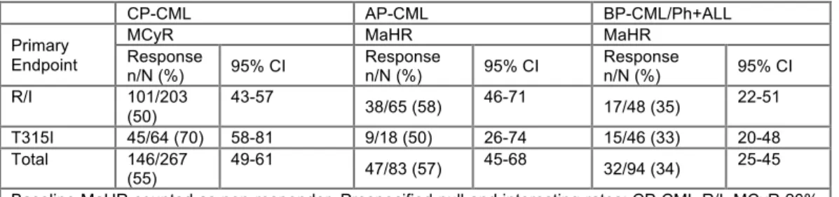
3 CML Therapy I [163-168]
[163] A Pivotal Phase 2 Trial of Ponatinib in Patients with Chronic Myeloid Leukemia (CML) and Philadelphia Chromosome-Positive Acute Lymphoblastic Leukemia (Ph+ALL) Resistant or Intolerant to Dasatinib or Nilotinib, or with the T315I BCR-ABL Mutation: 12-Month Follow-up of the PACE Trial. Cortes, Houston. Background: Despite progress in Ph+ leukemia therapy, patients who experience failure of tyrosine kinase inhibitors (TKIs) and those with the T315I BCR-ABL mutation have limited treatment options. Ponatinib is an oral TKI developed using computational and structure‑based design with optimal binding to the BCR‑ABL active site. At clinically achievable concentrations, ponatinib demonstrated potent in vitro activity against native BCR‑ABL and all BCR-ABL mutants tested, including T315I. The efficacy and safety of ponatinib (45 mg orally once daily) in patients with Ph+ leukemia were evaluated in a phase 2, international, open-label clinical trial. Methods: 449 patients resistant or intolerant (R/I) to dasatinib or nilotinib or with the T315I mutation confirmed at entry were enrolled and assigned to 1 of 6 cohorts: chronic phase (CP)-CML R/I (N=203), CP‑CML T315I (N=64), accelerated phase (AP)-CML R/I (N=65), AP-CML T315I (N=18), blast phase (BP)‑CML/Ph+ALL R/I (N=48), BP‑CML/Ph+ALL T315I (N=46). Five patients (3 CP‑CML, 2 AP-CML) without confirmed T315I and not R/I to dasatinib or nilotinib were treated, but not assigned to a cohort; they were included in safety analyses. The primary endpoint was major cytogenetic response (MCyR) at any time within 12 months for CP-CML and major hematologic response (MaHR) at any time within 6 months for advanced Ph+ leukemia. The trial is ongoing. Data as of 23 July 2012 are reported: median follow-up 11 (0.1 to 21) months; minimum follow-follow-up 9 months. Results: Median age was 59 (18-94) yrs; 53% were male. Median time from diagnosis to ponatinib was 6 (0.3‑28) yrs. Patients were heavily pretreated: 96% received prior imatinib, 84% dasatinib, 65% nilotinib; median number of prior TKIs was 3, with 53% exposed to all 3 approved TKIs. In patients previously treated with dasatinib or nilotinib (N=427), 88% had a history of resistance and 12% were purely intolerant to dasatinib or nilotinib. Best prior response to most recent dasatinib or nilotinib was 26% MCyR or better in CP-CML, and 23% MaHR or better in advanced Ph+ leukemia. Frequent BCR-ABL mutations confirmed at entry were: 29% T315I, 8% F317L, 4% E255K, 4% F359V, 3% G250E. No mutations were detected in 44%. The primary endpoint response rates (see Table) in each cohort exceeded the prespecified statistical criteria for success. In CP‑CML and AP‑CML R/I (the 3 largest cohorts), 95% CIs exceeded the prespecified response rate. Median time to response (for responders) was 84 days in CP‑CML, 112 days in AP-CML, 55 days in BP-CML/Ph+ALL. Responses were durable; the estimated (Kaplan‑Meier) probability of responders maintaining the primary endpoint at 1 yr was 91% in CP‑CML, 42% in AP‑CML, 35% in BP‑CML/Ph+ALL. In CP-CML, 46% had complete cytogenetic response and molecular response rates were 32% MMR, 20% MR4, and 12%
th
MR4.5. Response rates were higher in patients exposed to fewer prior TKIs and those with shorter disease duration. Similar response rates were observed in patients with and without BCR‑ABL mutations. In CP-CML, response rates were higher in those with T315I; however, a post hoc analysis found that presence of T315I was not a predictor of response. Instead, the difference in response rate was explained by T315I patients’ younger age, shorter duration of leukemia, and exposure to less prior therapy. At the time of analysis, 52% of patients remained on therapy (66% CP-CML). The most frequent reasons for discontinuation were progression (18%) and AEs (12%). The most common drug‑related AEs were thrombocytopenia (36%), rash (33%), and dry skin (31%). Pancreatitis was the most common drug‑related SAE (5%); however, it occurred early and was managed with dose modification (1 patient discontinued due to pancreatitis).Conclusions:Ponatinib has substantial activity and is generally well tolerated in these heavily pretreated Ph+ leukemia patients who have limited available treatment options. Data with a minimum follow-up of 12 months will be presented. NOTES: 23% any and 7% grade ¾ hypertension.
CP-CML AP-CML BP-CML/Ph+ALL
Primary Endpoint
MCyR MaHR MaHR
Response n/N (%) 95% CI Response n/N (%) 95% CI Response n/N (%) 95% CI R/I 101/203 (50) 43-57 38/65 (58) 46-71 17/48 (35) 22-51 T315I 45/64 (70) 58-81 9/18 (50) 26-74 15/46 (33) 20-48 Total 146/267 (55) 49-61 47/83 (57) 45-68 32/94 (34) 25-45
Baseline MaHR counted as non-responder. Prespecified null and interesting rates: CP-CML R/I, MCyR 20% and 35%; CP-CML T315I, MCyR 10% and 35%; advanced cohorts, MaHR 10% and 30%.
[164] Clinical Significance of Deeper Molecular Responses with Four Modalities of Tyrosine Kinase Inhibitors As Frontline Therapy for Chronic Myeloid Leukemia. Falchi, Houston. Background: The achievement of a major molecular remission (MMR) after imatinib therapy in pts with chronic myeloid leukemia (CML) in chronic phase (CP) predicts for decreased risk of events, but has little impact in overall survival (OS) among patients with complete cytogenetic response (CCyR). Deeper molecular responses (MR), including undetectable transcripts, are frequently sought in patients with CML treated with tyrosine kinase inhibitors (TKI), but the prognostic significance of these responses is not known. Objectives: To determine the long-term clinical significance of achieving deeper level of MR achieved after therapy with TKI for CML in CP. Methods: Pts were included in clinical trials for initial therapy for CML with one of the following modalities: imatinib 400mg/day (IM400), imatinib 800mg/day (IM800), nilotinib (NILO) and dasatinib (DASA). We defined the level of MR as MMR, MR4, MR4.5 and undetectable transcripts (UND), corresponding to an ABL/BCR-ABL ratio (International Scale) of ≤0.1%, ≤0.01%, ≤0.0032%, and undetectable transcripts (minimum sensitivity 4.5-log), respectively. Results: A total of 495 pts were treated: 83 pts with IM400, 204 with IM800, 106 with NILO and 102 with DASA. At presentation leukocyte counts were higher in the NILO group (41.5 vs 22.2, 27.5 and 27x109/L for IM400, IM800 and DASA pts). All other patient characteristics were equally distributed across the 4 treatment groups. After a median follow-up of 73 months (2 to 142), complete cytogenetic response (CCyR) was achieved in 88%. CCyR rates for IM400, IM800, NILO and DASA pts were 82%, 88%, 90% and 90%, respectively. Best level of MR for the entire population was: <MMR in 17% of pts, MMR in 13%, MR4 in 5%, MR4.5 in 19%, UND in 44%. In IM400 pts MR was <MMR in 28% of pts, MMR in 10%, MR4 in 8%, MR4.5 in 14%, UND in 40%. In IM800 pts MR was <MMR in 14% of pts, MMR in 8%, MR4 in 5%, MR4.5 in 19%, UND in 54%. In NILO pts MR was <MMR in 18% of pts, MMR in 20%, MR4 in 7%, MR4.5 in 22%, UND in 33%. In DASA pts MR was <MMR in 18% of pts, MMR in 18%, MR4 in 7%, MR4.5 in 23%, UND in 39%. There was a trend for earlier achievement of MR with NILO: median times to MMR, MR4, MR4.5 and UND were 12, 17.4, 17.9 and 25.1 months, respectively, for IM400 pts; 5.8, 8.7, 11.8 and 23.7 months, respectively, for IM800 pts; 5.7, 7, 8.3 and 16.4 months, respectively, for NILO pts; 5.7, 8.8, 17.4 and 27.2 months, respectively, for DASA pts. To analyze the relationship between the degree of MR and clinical outcome we excluded pts not achieving a CCyR as their best response since the clinical significance of CCyR is well known. For the remaining 438 pts, the depth of molecular remission was inversely correlated with the risk of losing CCyR (19%, 16%, 11%, 7%, 2% in pts with <MMR, MMR, MR4, MR4.5 and UND, respectively) or losing MMR (31%, 42%, 24%, 2%, respectively), as well as the risk of events (22%, 20%, 15%, 12%, 3%, respectively), transformation (3%, 5%, 0%, 1%, 0%, respectively), or death (25%, 11%, 8%, 6%, 4%, respectively). The 6-year OS for pts with <MMR, MMR, MR4, MR4.5 and UND is 74%, 84%, 95%, 96% and 99%, respectively (p<.0001); transformation-free survival (TFS) is 95%, 93%, 100% , 99% and 100% , respectively (p<.014); event-free survival (EFS) is 74%, 74%, 86%, 89% and 99%, respectively (p<.0001). To adjust for the lead-time to achieve deeper responses, we then calculated OS, TFS and EFS rates at 6 years according to the depth of molecular response at 18 or 24 months. Results are summarized in table 1.Conclusion:Most patients treated with TKI as initial therapy for early CP CML achieve a MR during the course of treatment. BCR-ABL transcripts become undetectable in a significant fraction of them. Achieving a MMR or better at 18 months or 24 months is associated with significantly superior 6-years OS, TFS and EFS. These result suggest that deeper molecular responses (MMR and beyond) are associated with clinical benefit, with a particularly good outcome for those achieving undetectable transcript levels.
Table 1. Survival at 6 years by level of molecular response at 18- and 24-month landmarks.
Outcome Molecular response at 18 months No MR MMR MR4 MR4.5 UND p-value
6-years OS 93% 98% 97% 95% 100% .153
6-years TFS 90% 100% 100% 100% 100% .03
6-years EFS 78% 94% 97% 93% 100% <.0001
Outcome Molecular response at 24 months
No MR MMR MR4 MR4.5 UND
6-years TFS 90% 100% 100% 100% 100% .057
6-years EFS 80% 90% 97% 95% 100% <.0001
[165] Early Molecular Response and Female Sex Strongly Predict Achievement of Stable Undetectable BCR-ABL1, a Criterion for Imatinib Discontinuation in Patients with CML. Branford, Adelaide. Introduction. The opportunity to discontinue kinase inhibitor therapy while maintaining a deep remission is desirable for many CML patients. Despite good responses to imatinib for most patients, treatment related side effects remain problematic and can affect quality of life. Studies have demonstrated that a proportion of carefully selected patients can sustain response after imatinib discontinuation. The first requirement for successful discontinuation is likely to be stable deep molecular response based on a sensitive RQ-PCR assay. The criteria for patient selection in the French Stop Imatinib (STIM) and Australian CML8 (TWISTER) imatinib discontinuation trials included stable undetectable BCR-ABL1 transcripts for at least 24 months with a PCR sensitivity of 5 and 4.5 log, respectively. The probability of continued remission after discontinuation for imatinib treated patients without prior interferon-α therapy was approximately 33%. It is not known how many imatinib treated patients will eventually meet these PCR criteria for a discontinuation trial. Aims. We aimed to determine 1) the cumulative probability of achieving the PCR criteria for imatinib discontinuation as defined in the CML8 study, and 2) factors that predicted its achievement. Method. The molecular response of 415 de-novo CML patients in chronic phase enrolled in consecutive clinical trials of imatinib since July 2000 was examined. The assigned daily imatinib dose was 400 mg for 90 patients, 600 mg for 202 patients and 800 mg for 123 patients. Molecular data were included until imatinib cessation or last follow-up. The minimum time since commencing imatinib was 30 months and the median time on imatinib was 45 months, range 3 to 136. The CML8 PCR criteria for imatinib discontinuation were confirmed undetectable BCR-ABL1 transcripts at a sensitivity of 4.5 log that remained undetectable on all PCR tests for at least 24 months while on imatinib therapy. In the current analysis the CML8 PCR discontinuation criteria are defined as ‘stable UMR4.5’. Results. At 8 years of imatinib therapy the cumulative incidence of stable UMR4.5 was 43%, Figure A. Patients were divided into groups according to the time to a confirmed major molecular response (MMR): by 3, 6, 12 or 18 months. There was a significant difference in stable UMR4.5, P<.001, Figure B. The cumulative incidence of stable UMR4.5 was more than 60% for all patients who achieved MMR by 12 months and only 16% for patients with MMR between 12 and 18 months. The time to a confirmed MMR influenced the time to reach a stable UMR4.5 after achieving MMR. Considering only patients who achieved stable UMR4.5, patients achieving MMR by 3 months took a further 39 months (median) to achieve stable UMR4.5. For those with MMR by 6 months and 12 months, the median month to a stable UMR4.5 was 50 and 76 months after MMR, P<.001. This suggests slower dynamics of BCR-ABL1 decline with delayed time to MMR. 52 patients achieved MMR after 18 months and none achieved a stable UMR4.5 by 8 years: median time to MMR was 27 months, range 21-87. Factors at the time of commencing imatinib (baseline) were examined for their association with stable UMR4.5; Sokal risk, age, sex, assigned imatinib dose and baseline BCR-ABL1 value, as well as the 3 month BCR-ABL1 value. Quantitative factors were categorized into groups, with cut-offs set at the median for age and quartiles for the baseline BCR-ABL1 value. By univariate analysis the only baseline factor that predicted for higher cumulative incidence of stable UMR4.5 at 8 years was female versus male, 68% versus 30%, P<.001, Figure C. During imatinib therapy females had significantly lower median BCR-ABL1 values at every assessment up to 42 months. The 3 month BCR-ABL1 value also predicted stable UMR4.5, P<.001, Figure D. Baseline and 3 month factors were entered into a multivariate analysis. The 3 month BCR-ABL1 value and sex were independent predictors of stable UMR4.5, P=.004 and P=.005, respectively. Conclusion. The time to achieve an MMR, sex and the 3 month BCR-ABL1 value predicted stable undetectable BCR-ABL1 while on imatinib. Lower BCR-ABL1 values and higher rates of stable UMR4.5 in females could be related to better drug adherence or biological differences. Further studies are indicated. Early MMR led to early achievement of the discontinuation criteria. The findings justify the focus on early achievement of MMR as a strategy to maximize recruitment to discontinuation studies.
[166] Pegylated Interferon-α 2a in Combination to Nilotinib As First Line Therapy in Newly Diagnosed Chronic Phase Chronic Myelogenous Leukemia Provides High Rates of MR4.5. Preliminary Results of a Phase II Study. Nicolini, Lyon. Background. Imatinib mesylate combined to pegylated interferon alfa 2a (Peg-IFN) has been reported to significantly enhance the molecular responses for de novo chronic phase chronic myeloid leukemia (CP-CML) patients compared to Imatinib alone in a Phase 3 study (Preudhomme et al. NEJM 2010). Second generation tyrosine kinase inhibitors (TKI2) such as nilotinib induce significantly higher levels of cytogenetic and molecular responses than imatinib as front line therapy for CP-CML (Saglio et al., NEJM 2010). Aims. Test the combination of nilotinib + Peg-IFN as front line therapy in CP-CML patients in order to check the safety and evaluate the molecular response rates (EudraCT 2010-019786-28). Methods. In this 2-step French national study, patients were assigned first to Peg-IFN (± HU) for a month at 90 mg/wk prior to a combination of nilotinib 300 mg BID + Peg-IFN 45 mg/wk for ≥ 1 year. The primary endpoint was the rate of confirmed (on 2 datapoints) molecular response 4.5 (MR4.5) by 1 year. Molecular assessments were centralised for all patients and expressed as BCR-ABLIS in %. Results. In the first cohort, 40+1 patients (1 screen failure) were enrolled and a second cohort of 20 patients was planned once the last patient of cohort 1 attained 1 year of treatment, if the primary endpoint would have not been reached. The current median follow-up is 13.6 (10.1–16.3) months. Sokal and Euro scores were high for 12% and 2%, intermediate for 49% and 55% and low for 39% and 43% of the patients respectively. Euro score was high for one patient. The median age was 53 (23-85) years. Two patients had a masked Philadelphia chromosome, 3 a variant form, and 1 had additional chromosomal abnormalities, all patients had a “major” BCR transcript. Five percent of patients were in CHR at 1 month of Peg-IFN and 100% at month (M) 2 (after 1 month of combination therapy). The rates of Complete Cytogenetic Responses (CCyR) at 3, 6, and 12 months of combination (i. e. at 2, 5, 8 and 11 months of TKI2) were 47%, 71%, 100% respectively on evaluable samples. The incidence of molecular responses are mentioned in figure 1 Figure 1: Incidence of molecular responses at definite time points.
Of note, 87% of the patients had a BCR-ABLIS ≤10% at M3. The rates of molecular responses broke down by major molecular response (MMR): 27%, 4 log reduction (MR4): 36%, and ≥4.5 log BCR-ABL reduction (MR4.5, MR5 and undetectable): 21% with a total number of 84% patients in ≥MMR and beyond (17.5% and 67.5% in intention-to-treat respectively) at 1 year. Confirmed molecular results at 1 year will be presented. Nilotinib trough levels centrally analysed at
th
M3, 6 and 12 for the vast majority of patients were ≥ 1000 ng/ml and Peg-IFN did not seem to impact on its pharmacokinetics. One patient went on unmutated myeloid blast crisis at M6 and is alive after allogeneic stem cell transplantation. Four additional patients were withdrawn from study: At M2 for non observance, at M6 for seizures related to an extra-dural hematoma, at M6 for recurrent grade 3 hepatic toxicity, at M9 for recurrent grade 3 pruritus. The median dose of Peg-IFN delivered to the patients during the first month was 90 (0-180) mg/wk, 45 mg/wk at M2, 3, 9, 12, and 33.75 mg/wk at M6. The median doses of nilotinib delivered to the patients were 600 mg daily at M2, 3, 6, 9, 12 and 15 as initially planned. The rate of grade 3-4 hematologic toxicities overall were anemia 2.5%, thrombocytopenia 41%, neutropenia 41% and pancytopenia 5%. These were observed mainly during M2 (16% neutropenia, 24% thrombocytopenia, 3% anemia), M3 (16% neutropenia, 13% thrombocytopenia, 3% pancytopenia) and M6 (12.5% neutropenia, 5% thrombocytopenia) and disappeared thereafter. Grade 3-4 toxicities occurred mostly during the first 3 months with 15% cholestatic episodes, 5% of ALAT elevation, 2.5% of lipase elevation, 2.5% arthro-myalgias, 2.5% abdominal pain without lipase elevation, 2.5% of depression. No PAO was observed and, to date, no dyslipidemia. Conclusion. The combination of nilotinib and Peg-IFN seems relatively well tolerated despite frequent initial and transient hematologic and hepatic toxicities, and provides very high rates of molecular responses at 1 year and beyond. According to the initial methodology of this trial, the second cohort of patients will not be enrolled as the MR4.5 rates at M12 are beyond the initial expectations. A randomised phase III study testing nilotinib versus nilotinib + Peg-IFN is warranted.
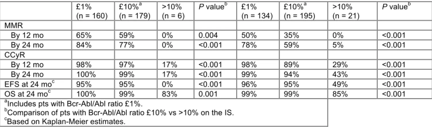
[167] Outcome of Patients with Chronic Myeloid Leukemia in Chronic Phase (CML-CP) Based On Early Molecular Response and Factors Associated with Early Response: 4-Year Follow-up Data From Enestnd (Evaluating Nilotinib Efficacy and Safety in Clinical Trials Newly Diagnosed Patients). Hochhaus, Jena. Background: In the ENESTnd study, nilotinib significantly reduced progression to accelerated phase/blast crisis (AP/BC) and demonstrated superior rates of deep molecular response vs imatinib. Data from ENESTnd demonstrated that significantly more patients achieved early molecular response of both < 10% and < 1% BCR-ABLIS at both 3 and 6 months on nilotinib vs imatinib. Here, we report landmark analyses based on BCR-ABL transcript levels at 3 and 6 months using data with a minimum follow-up of 3 years and also provide data on factors associated with poor early molecular response; data based on longer follow-up of 4 years will be presented. Methods: The nilotinib 300 mg twice daily (BID; n = 282) and imatinib 400 mg once daily (QD; n = 283) arms from ENESTnd were used for this analysis. Patients were grouped based on BCR-ABL transcript levels of ≤ 1%, > 1% to ≤ 10%, and > 10% at 3 months (n = 258 and n = 264 patients with available PCR samples at 3 months in the nilotinib and imatinib arms, respectively) and at 6 months (n = 257 and n = 256 patients with available PCR samples at 6 months in the nilotinib and imatinib arms, respectively). Rates of major molecular response (MMR; ≤ 0.1% BCR-ABLIS) and molecular response with a 4.5-log reduction in BCR-ABL transcript levels (MR4.5, ≤ 0.0032%IS) as well as rates of progression-free survival (PFS) and overall survival (OS) were evaluated among patients grouped according to their BCR-ABL transcript levels at 3 and 6 months. Data on selected baseline characteristics and dose intensity were also assessed. Results: Among evaluable patients at 3 months, 9% of patients (n = 24) in the nilotinib arm vs 33% (n = 88) in the imatinib arm had BCR-ABL transcript levels of > 10%; among evaluable patients at 6 months, 3% of patients (n = 7) in the nilotinib arm vs 16% (n = 40) in the imatinib arm had BCR-ABL transcript levels of > 10%. Patients with a BCR-ABL transcript level of > 10% had a lower probability of future MMR or MR4.5 as well as poorer PFS and OS compared with patients who had BCR-ABL transcript levels ≤ 10% at 3 months (Table). Results were similar based on 6-month landmark analyses. In patients with > 10% BCR-ABL transcript levels at 3 months, the average dose intensity of nilotinib within the first 3 months was 474 mg/day compared with 600 mg/day for patients with ≤ 10% BCR-ABL transcript levels; the average dose intensity of imatinib within the first 3 months was the same (400 mg/day) for patients with both ≤ 10% and > 10% BCR-ABL levels at 3 months (Table). Patients with > 10% BCR-ABL transcript levels at 3 months were also more likely to have high Sokal risk, larger spleen size, and additional chromosomal abnormalities compared with patients with ≤ 10% BCR-ABL transcript levels at 3 months. Other factors associated with early response and further data on long-term outcomes are being assessed and will be presented with a minimum follow-up of 4 years. Conclusions: Fewer patients in the nilotinib arm vs the imatinib arm had BCR-ABL transcript levels > 10% at 3 and 6 months. Reasons for poor early response appeared to be related, at least in part, to baseline factors and dose intensity. Early molecular response at 3 and 6 months correlated with future MMR and MR4.5as well as an increased probability of PFS and OS. Nilotinib frontline therapy allows more patients to achieve deeper responses earlier, associated with improved long-term outcomes vs imatinib.
Nilotinib 300 mg BID (N = 258)* Imatinib 400 mg QD (N = 264)* BCR-ABL at 3 months ≤ 10% n = 234 > 10% n = 24 ≤ 10% n = 176 > 10% n = 88 MMR n = 209 n = 24 n = 174 n = 88 by 1 year, % 61 4 40 2 by 2 years, % 80 29 58 21 MR4.5 n = 233 n = 24 n = 176 n = 88 by 2 years, % 29 4 14 0 by 3 years, % 38 4 24 1 Long-term outcomes n = 234 n = 24 n = 176 n = 88 PFS at 3 years, % 95.9 82.9 97.7 83.8 OS at 3 years, % 97.6 86.7 98.9 84.8
Factors associated with molecular
response at 3 months
Median dose intensity within the first
3 months, mg/day 600 474 400 400
High Sokal risk, % 25.6 41.7 17.6 44.3
Splenomegaly, % 38.9 75.0 26.7 59.1
Additional
chromosomal abnormalities, % 9.4 16.7 9.1 14.8
Response and outcomes were evaluated in patients who had available PCR samples at 3 or 6 months. Patients who achieved the target response within 3 or 6 months were excluded from the respective analysis of response outcomes; patients who had events or were censored within 3 or 6 months were excluded from the respective analysis of time-to-event outcomes.
[168] Relationship Between Molecular Responses and Disease Progression in Patients (Pts) Treated First Line with Imatinib (Im) Based Regimens: Impact of Treatment Arm within the French Spirit Trial From the French CML Group (FI LMC). Rousselot, Versailles. The SPIRIT phase III randomized multicenter open-label prospective trial was designed to compare 4-arms, IM-400 mg versus IM-600mg versus IM-400 mg + cytarabine at a dose of 20 mg / m² / day in cycles of 28 days, versus IM-400 mg + PegIFN at an initial dose of 90 µg per week. The planned molecular analysis after 1 year based on the outcome of 636 pts resulted in a highly significant superiority of superior molecular response (SMR) (0.01 % Bcr-Abl/Abl on IS) of the combination IM 400mg-PegIFN (N Engl J Med, 2010). As of December 31st 2010, date for closing accrual, 787 pts have been included. The current analysis provides update of the trial and additional information on relationship between molecular response and outcomes. Methods: Progression free survival (PFS) was defined by absence of accelerated phase (AP), blast crisis (BC), and death from any reasons, whichever came first. Rates were estimated by the Kaplan-Meier method and compared within groups by the log-rank test. In addition, time to progression (TTP) being defined as AP-BC only, was estimated by cumulative incidence function and compared within groups by the Gray test. Deaths unrelated with progression were then considered as competing events. Pts with available PCR samples at 3 months (N= 665 overall, 197 IM-400mg, 147 IM-600mg, 138 IM-AraC, 183 IM-PegIFN) were classified according to the BCR-ABL cut-off level of 10% IS at 3 and 6 months. Analyses of long term outcomes were then based on the kinetic of molecular response. Results. After a median follow-up of 68 months, out of the 787 pts, 59 PFS events (31 AP-BC; 28 deaths in CP) were recorded. Survival without progression at 60 months were for the IM-400mg, IM-600mg, IM-400mg + cytarabine and IM-400mg + PegIFN , 94% , 93%, 90% and 94% respectively (overall, p= 0.24). However we noticed that kinetic of molecular responses of pts who experienced AP-BC was very heterogeneous as showed in Fig 1. The accurate level of bcr-abl/abl transcript and the relevance of the IS conversion factor are questionable when values are above 10% or very low. Thus corresponding plots are shadowed in the Fig1. Then, when pts were stratified according to their molecular response at 3 months, 14 cases of AP-BC and 13 deaths without evidence of progression were recorded in the group of pts with a BCR-ABL ratio <=10% IS (n=522) whereas 14 cases of AP-BC and 9 deaths were recorded in the other group (n=143). Overall, PFS at 3 months was significantly better (p <0.0001) in pts with ratio ≤10% IS. Similarly, TTP was lower (p <0.0001). However, when same analyses were performed according to treatment arm, discrepancies were observed. The potential interest of the 10% BCR-ABL cut-off was still relevant in the IM-400 arm (p <0.0001) and IM-AraC arm (p=0.0199), but no statistical differences were observed in the IM-600 (p=0.6715) and in the IM-PegIFN (p=0.0887) arms respectively. Of interest, these results were confirmed when deaths with no evidence of AP-BC were considered as competing events. The TTP was still significant in the IM-400 arm (p=0.0002) and the IM-AraC arm (0.04). However, no TTP differences were observed in the IM-600 arm (p=0.38); moreover TTP is strictly similar in both molecular response rate groups at 3 months in the IM-PegIFN arm (p=0.96). We also analyzed the cumulative incidence of AP-BC within a period of 6 months according to the kinetic of molecular response (n=568 pts); 3 groups of pts were analyzed: ≤10% within 3 months, ≤10% within 6 months, still above 10% within 6 months. There is a significant advantage (p<0.0001) for early molecular response for all pts included in the trial except for those assigned to the IM-PegIFN arm (p=0.82). Of interest for pts assigned to the IM-PegIFN arm, rate of AP-BC were 3%, 4%, and 0% for the ≤10% within 3 months, ≤10% within 6 months, still above 10% within 6 months groups of pts, respectively. Conclusion: a 3-months BCR-ABL transcript below the level of 10% IS was associated with a PFS improvement. However, results which were observed with the addition of PegIFN or an increased dose of IM frontline do not confirm the relevance of the 10% BCR-ABL cut-off level as strong surrogate marker for progression to AP-BC. Pts assigned to the IM-PegIFN arm are at very low risk of progression to AP-BC even if their molecular response is delayed.
4 CML Therapy II [913-918]
[913] Impact of Balanced or Unbalanced Karyotype At Diagnosis On Prognosis of CML: Long-Term Observation From 1346 Patients of the Randomized CML Study IV. Fabarius, Mannheim. Introduction: Acquired genetic instability in chronic myeloid leukemia (CML) as a consequence of the translocation t(9;22)(q34;q11) and the resulting BCR-ABL fusion causes the continuous acquisition of additional chromosomal aberrations and mutations and thereby progression to accelerated phase (AP) and blast crisis (BC). At least 10% of patients in chronic phase (CP) CML show additional alterations at diagnosis. This proportion rises during the course of the disease up to 80% in BC. Acquisition of chromosomal changes during treatment is considered as a poor prognostic indicator, whereas the impact of chromosomal aberrations at diagnosis depends on their type. Patients with major route additional chromosomal alterations (major ACA: +8, i(17)(q10), +19, +der(22)t(9;22)(q34;q11) have a worse outcome whereas patients with minor route ACA show no difference in overall survival (OS) and progression-free survival (PFS) compared to patients with the standard translocation, a variant translocation or the loss of the Y chromosome (Fabarius et al., Blood 2011). However, the impact of balanced vs. unbalanced (gains or losses of chromosomes or chromosomal material) karyotypes at diagnosis on prognosis of CML is not clear yet. Patients and methods: Clinical and cytogenetic data of 1346 evaluable out of 1544 patients with Philadelphia and BCR-ABL positive CP CML randomized until December 2011 to the German CML-Study IV, a randomized 5-arm trial to optimize imatinib therapy by combination, or dose escalation and stem cell transplantation were investigated. There were 540 females (40%) and 806 males (60%). Median age was 53 years (range, 16-88). The impact of additional cytogenetic aberrations in combination with an unbalanced or balanced karyotype at diagnosis on time to complete cytogenetic and major molecular remission (CCR, MMR), PFS and OS was investigated. Results: At diagnosis 1174/1346 patients (87%) had the standard t(9;22)(q34;q11) only and 75 patients (6%) had a variant t(v;22). In 64 of 75 patients with t(v;22), only one further chromosome was involved in the translocation; In 8 patients two, in 2 patients three, and in one patient four further
th
chromosomes were involved. Ninety seven patients (7%) had additional cytogenetic aberrations. Of these, 44 patients (3%) lacked the Y chromosome (-Y) and 53 patients (4%) had major or minor ACA. Thirty six of the 53 patients (2.7%) had an unbalanced karyotype (including all patients with major route ACA and patients with other unbalanced alterations like -X, del(1)(q21), del(5)(q11q14), +10, t(15;17)(p10;p10), -21), and 17 (1.3%) a balanced karyotype with reciprocal translocations [e.g. t(1;21); t(2;16); t(3;12); t(4;6); t(5;8); t(15;20)]. After a median observation time of 5.6 years for patients with t(9;22), t(v;22), -Y, balanced and unbalanced karyotype with ACA median times to CCR were 1.05, 1.05, 1.03, 2.58 and 1.51 years, to MMR 1.31, 1.51, 1.65, 2.97 and 2.07 years. Time to CCR and MMR was longer in patients with balanced karyotypes (data statistically not significant). 5-year PFS was 89%, 78%, 87%, 94% and 69% and 5-year OS 91%, 87%, 89%, 100% and 73%, respectively. In CML patients with unbalanced karyotype PFS (p<0.001) and OS (p<0.001) were shorter than in patients with standard translocation (or balanced karyotype; p<0.04 and p<0.07, respectively). Conclusion: We conclude that the prognostic impact of additional cytogenetic alterations at diagnosis of CML is heterogeneous and consideration of their types may be important. Not only patients with major route ACA at diagnosis of CML but also patients with unbalanced karyotypes identify a group of patients with shorter PFS and OS as compared to all other patients. Therefore, different therapeutic options such as intensive therapy with the most potent tyrosine kinase inhibitors or stem cell transplantation are required.
914 Elevated Risk of Peripheral Artery Occlusive Disease (PAOD) in Nilotinib Treated Chronic Phase Chronic Myeloid Leukemia (CML) Patients Assessed by Ankle-Brachial-Index (ABI) and Duplex Ultrasonography
Michaela Schwarz, Berlin. In bcr-abl-positive CML first-line therapy with the second generation tyrosine kinase inhibitors (TKI) nilotinib or dasatinib results in superior response rates and prevention of transformation as compared to imatinib. Generally, these TKIs are associated with mild and reversible toxicities but recent reports have indicated an elevated risk of PAOD in patients (pts) treated with nilotinib (Aichberger et al. 2011, Tefferi et al. 2011, le Coutre et al. 2011, Quintás-Cardama et al. 2012). We therefore screened all 153 chronic phase CML pts at our center for PAOD by sequentially determining ABI, followed by duplex ultrasonography when indicated. Cardiovascular risk factors were assessed with a specific questionnaire and biochemical parameters associated with PAOD were analyzed. We here present a first interim analysis.
Overall, 146 of 153 pts were evaluable and categorized into five groups: (I) first-line imatinib (n = 53); (II) first-line nilotinib (n= 31); (III) second-line nilotinib (n = 32); (IV) previously exposed to nilotinib (n = 23) and (V) never treated with nilotinib and currently not on imatinib (n = 7).
A pathological ABI, defined as <0.9, occurred in 24/116 (21 %) of all examined patients, but was more frequent in pts on first-line (7/26; 27 %, p=0.0181) and second-line nilotinib (10/24; 42 %, p=0.0004) as compared to pts on first-line imatinib (3/45; 7 %) despite a significantly longer treatment on line imatinib (median 80.25 months, range 4 - 137) than on first-line nilotinib (median 25.48 months, range 5 - 49, p<0.001). Newly diagnosed PAOD defined as a peripheral vascular occlusive event or pathological duplex ultrasonography was observed in 13/146 (8.9 %) of all pts and was more frequent in pts on first-line nilotinib (3/31; 9.6%, p=0.1057), second-line nilotinib (5/32, 15.6 %, p=0.0166) or pts previously exposed to nilotinib (4/23; 17.4 %, p=0.0123) as compared to pts on first-line imatinib (1/53; 1.9 %). No substantial differences between the five groups with respect to clinical cardiovascular risk factors were observed. However, biochemical risk factor assessment showed significantly higher levels of cholesterol and LDL in pts receiving first-line (218.0 mg/dl, p< 0.001, and 135.5 mg/dl, p< 0.01) and second-line nilotinib (219.9 mg/dl, p < 0.001, and 138.9 mg/dl, p < 0.01) as compared to pts on first-line imatinib (167.2 mg/dl and 97.8 mg/dl).
Of 16 pts with PAOD under or after nilotinib treatment, including three additional patients from a second center, 4 pts received nilotinib as first-line, 10 pts as second-line and 2 pts as third-line treatment. Median age was 62.4 years (range, 38-76) and median duration of CML was 103.1 months (range: 22-209). Best responses observed in this group of patients were CMR or MMR in 10/16 (62.5 %) and CCyR or MCyR in 6/16 (37.5 %) of patients. Major cardiovascular risk factors such as hypertension (13/15; 87 %), diabetes mellitus (5/14; 36 %), nicotine abuse (11/15; 73 %), cholesterol (5/12; 42 %) or LDL (6/13; 46 %) elevation were detectable in the majority. Vascular lesions affected the lower limb in 15/16 (94 %) and the eye in 1/16 pts (6 %). Cardiovascular interventions included percutaneous transluminal angioplasty (PTA) (7/16; 44 %), stent-implantation (5/16; 31 %) and/or surgery, including amputation (5/16; 31 %). Nilotinib was discontinued in 10/16 pts (63 %), dose-reduced in 5/16 pts (32 %) and continued in 1/16 pts (6 %).
Conclusions: Prospective monitoring of pts with CML in chronic phase by sequential evaluation of ABI and duplex ultrasonography revealed a significantly higher frequency of PAOD in pts on nilotinib than in pts on imatinib. Mechanisms leading to the development of PAOD under nilotinib treatment remain unknown. Aside from elevation of cholesterol and glucose levels not yet fully understood mechanisms such as inhibition of targets involved in vascular cell homeostasis (i.e. DDR1, KIT or PDGFR) must be considered. More prospective data is needed to determine the cardiovascular risk attributable to nilotinib. But at present, caution is advised in pts with ≥ 2 major risk factors or a known history of arteriopathy. [915] Efficacy and Safety of Ponatinib in Patients with Accelerated Phase or Blast Phase Chronic Myeloid Leukemia (AP-CML or BP-CML) or Philadelphia Chromosome-Positive Acute Lymphoblastic Leukemia (Ph+ ALL): 12-Month Follow-up of the PACE Trial. Kantarjian, Texas Background: Many patients (pts) with advanced Ph+ leukemias experience failure of all currently available tyrosine kinase inhibitors (TKIs) targeting BCR-ABL and have limited treatment options. Ponatinib is a potent pan-BCR-ABL inhibitor that is active against native and mutated forms of BCR-ABL, including the TKI resistant T315I mutant. The efficacy and safety of ponatinib (45 mg orally once daily) in pts with AP-CML, BP-CML, or Ph+ ALL were evaluated in a phase 2, international, open-label clinical trial. Methods: The PACE trial enrolled 449 pts, including 85 AP-CML, 62 BP-CML, and 32 Ph+ ALL. Pts were resistant or intolerant (R/I) to dasatinib or nilotinib, or had the T315I mutation at baseline. AP-CML, BP-CML, and Ph+ ALL pts were assigned to 1 of 4 cohorts: AP-CML R/I, AP-CML T315I, BP-CML/Ph+ ALL R/I, BP-CML/Ph+ ALL T315I. Two AP-CML pts were not assigned to a cohort (post-imatinib, did not have T315I at baseline) and were excluded from efficacy analyses and included in safety analyses. The primary endpoint was major hematologic response (MaHR) at any time within 6 mos after treatment initiation. Data as of 23 July 2012 are reported, with a minimum follow-up of 9 mos (median 13 [4 to 21], 6 [0.1 to 18], and 6 [0.1 to 16] mos for AP-CML,
BP-CML, and Ph+ ALL, respectively). Results: The median age for AP-CML, BP-CML, and Ph+ ALL pts was 60, 53, and 62 yrs, respectively. Median time from initial disease diagnosis to start of ponatinib was 7, 4, and 1.5 yrs, respectively. Pts were heavily pretreated: 94% received prior imatinib, 88% dasatinib, 61% nilotinib; 8% received 1 prior approved TKI, 39% received 2, and 53% received 3. Sixteen percent had undergone prior stem cell transplant. In pts previously treated with dasatinib or nilotinib (N=171), 94% had a history of resistance to dasatinib or nilotinib, 6% were purely intolerant. Reported MaHR rates with the most recent dasatinib or nilotinib therapy were 35% AP-CML, 16% BP-CML, 43% Ph+ ALL.
At the time of analysis, 59% of AP-CML, 8% of BP-CML, and 9% of Ph+ ALL pts remained on study. Overall, the most common reasons for discontinuation were progressive disease (19%, 50%, and 53%, respectively) and adverse events (AEs; 11%, 16%, and 6%, respectively). Hematologic and cytogenetic response rates are shown in the table; MaHR and MCyR were observed across cohorts. MMR was achieved by 14% of AP-CML pts (14% R/I, 17% T315I). There was a trend for higher response rates among pts who received fewer prior approved TKIs. In AP-CML pts, the differences in MaHR rates by number of prior approved TKIs (1: 3/4 [75%]; 2: 20/33 [61%]; 3: 24/46 [52%]) were not significant (Fisher's Exact); differences in MCyR rates (1: 4/4 [100%]); 2: 13/33 [39%]; 3: 15/46 [33%]) were significant for pts treated with 1 vs 2 (p=0.0360) and 1 vs 3 prior approved TKIs (p=0.0168). Of pts achieving MaHR, 42% of AP-CML and 35% of BP-CML/Ph+ ALL pts were projected (Kaplan-Meier) to remain in MaHR at 1 yr. In AP-CML, the median progression-free survival (PFS) was estimated (Kaplan-Meier) as 80 (range 6 to 88) wks; the probability of maintaining PFS at 6 mos and 1 yr was estimated as 80% and 57%, respectively. Median overall survival (OS) had not yet been reached; the probability of OS at 6 mos and 1 yr was estimated (Kaplan-Meier) as 96% and 85%, respectively. In BP-CML/Ph+ ALL, median PFS was estimated as 18 (range 0.1 to 74) wks; the probability of maintaining PFS at 6 mos and 1 yr was estimated as 34% and 20%, respectively. Median OS was estimated as 30 (range 0.4 to 77) wks; the probability of OS at 6 mos and 1 yr was estimated as 54% and 34%, respectively. Ponatinib was generally well-tolerated; the most common treatment-related AEs were thrombocytopenia (29%), rash (25%), and neutropenia (22%). The most common serious treatment-related AEs were thrombocytopenia (3%) and pancreatitis (3%). Rash was generally grade 1 or 2 in severity. Thrombocytopenia, neutropenia, and pancreatitis were typically reported early in treatment and were manageable with dose modification.
Conclusions: Ponatinib was generally well-tolerated and had substantial activity in pts with AP-CML, BP-CML, or Ph+ ALL, regardless of mutation status or prior therapy. Data with a minimum follow-up of 12 mos will be presented.
AP-CML BP-CML Ph+ ALL R/I N=65 T315I N=18 R/I N=38 T315I N=24 R/I N=10 T315I N=22 MaHRa 58% 50% 32% 29% 50% 36% MCyR 34% 56% 18% 29% 60% 41% CCyR 22% 33% 16% 21% 50% 32%
aBaseline MaHR counted as non-responder
[916] Discontinuation of Second Generation (2G) Tyrosine Kinase Inhibitors (TKI) in Chronic Phase (CP)-Chronic Myeloid Leukemia (CML) Patients with Stable Undetectable BCR-ABL Transcripts Rea, Paris, Background: TKI have proven remarkably successful against CML. Despite this progress, current recommendation is to never stop therapy. However, prospective trials such as STIM and CML8 suggest that imatinib may be stopped in patients with deep and sustained molecular responses (Mahon et al, Lancet Oncol. 2010; Ross et al, Haematologica 2012). Here, we report on the feasibility of 2G-TKI discontinuation. Methods: Adult CP-CML patients on dasatinib or nilotinib without previous allogeneic transplantation or progression to advanced phase CML were proposed treatment discontinuation provided that (1) TKI treatment duration was at least 36 months (2) BCR-ABL transcripts were undetectable for at least 24 months, with at least 20 000 amplified copies of the control gene. The primary objective was treatment-free stable major molecular response (MMR: BCR-ABL/ABL internationally standardized (IS) ratio ≤ 0.1% IS). BCR-ABL transcripts were quantified in local laboratories monthly during the first 12 months, every 2-3 months during the 2nd year and every 3-6 months thereafter. Molecular relapse was defined by the loss of MMR and triggered therapy resumption. The study is ongoing and 42 patients currently agreed to stop dasatinib (53%) or nilotinib (47%). Data as of August 1, 2012 are reported, focusing on the subgroup of 34 patients with a minimum follow-up of 6 months (median 14, range 7-33). Results: Median age was 58 (34-81), 56% of patients were females (n=19). Sokal risk group was low in 56% (n=19), intermediate in 24% (n=8), high in 11% (n=4) and unknown in 9% (n=3). Twelve patients (35%) received interferon prior to TKI therapy, 25 (74%) were treated with 2G-TKI upfront (n=2) or after imatinib intolerance (n=25) and 9 (26%) received 2G-TKI due to suboptimal response/resistance. The median time since diagnosis was 87 months (31-218), the median time since TKI initiation was 79 months (30-133), the median duration of 2G-TKI therapy was 35 months (21-72) and the median duration of undetectable BCR-ABL transcripts was 27 months (21-64). At last follow-up, the 12-month probability to remain in stable MMR was 58.3% (95% CI, 41.5%-75%). The median time to MMR loss was 4 months (1-25). Importantly, no hematologic relapse was observed and none of the patients progressed to advanced phase CML. Since in imatinib discontinuation trials, different definitions for molecular relapse were used, we also calculated the rate of relapse according to STIM (detectable BCR-ABL on 2 consecutive tests with at least 1 log increase between the 2) and CML8 (detectable BCR-ABL on 2 consecutive tests at any level). The corresponding 12-month probabilities were 55.8% (95% CI, 39.2%-72.6%) according to STIM and 44.1% (95%CI, 27.4%-60.8%) according to CML8. We searched for factors possibly associated with treatment-free stable MMR. Patients treated with 2G-TKI first line or after imatinib intolerance had a significantly higher probability of stable MMR than those who were switched to 2G-TKI because of suboptimal response/resistance (12-month rate 67.3% (95% CI, 48.6%-86%) versus 33% (95% CI; 2.5%-64.1%), p=0.02). Gender, age, prior IFN exposure, 2G-TKI type and treatment duration were not found to have any impact but caution is needed due to the small size and heterogeneity of this series. Treatment was resumed in 15 patients after a median time of 5 months (2-29). The same 2G-TKI used prior to discontinuation was reintroduced in all but 1 patient, due to tolerance issues. The median follow-up since treatment resumption was 9 months
(0-th
27). At last follow-up, MMR was regained in all 13/15 evaluable patients and undetectable BCR-ABL in 10/13. Eighteen patients with stable MMR remained off-therapy at last follow-up (median 16 months, range: 7-33), among which 7 with stable undetectable BCR-ABL and 11 with weakly detectable BCR-ABL on 1 or more occasions. Conclusions: 2G-TKI may be safely discontinued in CP-CML patients with long-lasting undetectable BCR-ABL under strict molecular monitoring conditions, especially in patients with prior imatinib intolerance or treated with 2G-TKI as first line therapy. In patients with prior imatinib suboptimal response/resistance, strategies to improve outcome are needed. The recurrence of a low level of detectable residual disease below the MMR threshold after 2G-TKI withdrawal may not automatically herald CML relapse and may not preclude the possibility to remain treatment-free.
[917] Single Molecule Real Time (SMRT™) Sequencing Sensitively Detects the Evolution of Polyclonal and Compound BCR-ABL Mutations in Patients Who Relapse On Kinase Inhibitor Therapy Smith, San Francisco Background: Secondary kinase domain (KD) mutations represent the most well-documented mechanism of resistance to tyrosine kinase inhibitors (TKIs) in chronic myeloid leukemia (CML). In CML, multiple TKIs with different mutation profiles are approved and the ability to detect KD mutations at the time of disease progression can impact therapy choice. To optimize clinical impact, second generation TKI selection must consider the majority TKI-resistant mutant population as well as smaller mutant sub-populations that may be selected with subsequent treatment. Sequential TKI therapy is associated with additional complexity: multiple mutations can coexist separately in an individual patient (“polyclonality”) or can occur in tandem on a single allele (“compound mutations”). Multiple mutations are associated with poor clinical outcome (Parker et al., Blood 2012). Compound mutations can cause in vitro resistance to ponatinib, the only TKI clinically active against the highly resistant T315I mutation (Eide, et. al, ASH 2012 abstract #1416). Currently, no clinically adaptable technology can distinguish polyclonal from compound mutations. Due to the size of the BCR-ABL KD, most next-generation sequencing platforms cannot generate reads of sufficient length to determine if mutations separated by ≥500 nt reside on the same allele. Pacific Biosciences RS Single Molecule Real Time (SMRT) sequencing technology is a third generation deep sequencing technology capable of achieving average read lengths of ~1000bp and frequently >3000bp, enabling sensitive and accurate sequencing of the entire ABL KD on a single strand of DNA. Though allele-specific detection methods such as MassARRAY offer sensitivity as low as ~0.5%, these assays are designed to detect a limited number (~31) of mutations whereas SMRT sequencing offers an unbiased approach capable of detecting novel variants. We sought to (1) develop a potential clinically-applicable SMRT sequencing assay for the detection of BCR-ABLKD mutations capable of distinguishing polyclonal and compound mutations, and (2) compare the accuracy and sensitivity of this method to standard sequencing and MassARRAY. Results: We assessed 54 samples from 36 CML patients who had clinically relapsed on ABL kinase inhibitor therapy and were previously analyzed by standard sequencing, and in a subset, by MassARRAY. We amplified an 863bp area of the BCR-ABLKD from patient-derived cDNA with primers containing 5’ barcodes, enabling sequencing of 6 to 8 patient samples on a single SMRT cell on a single run. On average, 2519 reads were obtained for each sample per run (range 330 to 10,240). All of 131 known mutations detected by MassARRAY were identified by SMRT sequencing using a p-value threshold of 1.03e-03. SMRT sequencing also identified all 107 known mutations detected by direct sequencing with a p-value threshold of 6.0e-08. In addition to these known mutations, SMRT sequencing detected an additional 1320 non-silent mutations across all patient samples using a strict p-value threshold cut-off of 6e-08, ranging in abundance from 0.2% to 17% (median 0.75%). Among 47 samples where >1 mutation was detectable by direct sequencing or MassARRAY, SMRT sequencing revealed that 40 (85%) had compound mutations detectable at a frequency of ≥1%. In total, we detected 73 different compound mutations at a frequency of ≥1%. In all cases where compound mutations were detected and more than one treatment timepoint was available, at least one compound mutation clearly evolved from a mutation detectable at a prior timepoint. In the most complex case, 4 separate mutations yielded 8 different mutant alleles. Conclusions: Pacific Biosciences RS SMRT sequencing detects KD mutations in patient samples with sensitivity comparable to or better than MassARRAY and can distinguish compound from polyclonal mutant clones. Among patient samples with multiple mutations, compound mutations were detectable in the vast majority of samples by SMRT sequencing, revealing a complex mutational landscape not demonstrable by other clinically viable sequencing methods and previously unappreciated. Given the growing numbers of patients exposed to multiple TKIs in a sequential manner, the ability to accurately and sensitively characterize drug-resistant alleles by SMRT sequencing promises to further facilitate a personalized approach to patient management and inform models of disease evolution.
[918] Natural History and Potential for Cure of Patients with Chronic Myeloid Leukemia in Chronic Phase Receiving Frontline Therapy with Recombinant Interferon-Alfa: 30-Year Update From M.D. Anderson Cancer Center. Quintás-Cardama, Texas. Background: Prior to the advent of the tyrosine kinase inhibitor (TKI) imatinib, pioneering studies at our institution in the early 1980s established recombinant interferon-alfa (IFN-α) as standard therapy for chronic myeloid leukemia (CML). The use of IFN-α has come to the fore again given its therapeutic properties as an immunomodulatory agent and its putative activity against CML stem cells. We here provide an account of the natural history of patients with early chronic phase CML treated with IFN-α at our institution for the last 30 years. Methods: We analyzed 512 patients with early chronic-phase CML who were treated with IFN-α-based therapies between 1981-1995 for the rates of partial (PCyR) and complete cytogenetic response (CCyR), complete molecular response (CMR), major molecular response (MMR), overall survival (OS), transformation-free survival (TFS), and CML cure. Results: The median age of the cohort was 42 years (range, 15-76). The distribution of high, intermediate, and low risk patients by Sokal or Hasford was 21%/25%/36% and 10%/27%/44%, respectively. Of the 512 patients, 274 received IFN-α alone or in combination with hydroxyurea or high-dose chemotherapy, 148 received IFN-α and low-dose cytarabine, and 90 were treated with homoharringtonine followed by IFN-α as maintenance. After a median follow-up of 245 months (range, 4-360), the median OS was 82 months. The 5-, 10-, and 20-year survival was 62%, 41%, and 29%, respectively. Overall, 322 patients (63%) achieved a cytogenetic response, including CCyR in 140 (27%), which was obtained after a median of 16 months (range, 3–107 months), and PCyR in 72 (14%) for a major cytogenetic response rate of 41%. The median follow-up for patients who achieved CCyR was 252 months (range, 91-360). The 5-, 10-, and 20-year survival for patients who achieved CCyR was 90%, 79%, and 63%, respectively, with a 20-year TFS of 76%. Serial molecular monitoring by RT-PCR (at least 2 measurements) is available in 46 patients. Of them, 31 achieved CMR that lasted a median of 9 years (range, 1.5-17). Of them, 14 patients remain in CMR off therapy for
a median of 9.5 years (range, 1.5-17), 6 remain off therapy with detectable transcripts (5 in MMR) after a median of 10.5 years (range, 4.5-13), 9 remain in CMR after having relapsed and switched to other therapies (5 imatinib, 2 dasatinib, 1 allo-SCT, 1 chemotherapy), and 2 maintained MMR while receiving chemotherapy. Eight of the 31 patients relapsed (including 3 with sudden lymphoid BP). At the time of last follow-up, only 3 of the 31 patients who achieved CMR had died, one after , 1 lymphoid BP, 1 acute myeloid leukemia (with deletion 7), and 1 myeloproliferative disorder (with trisomy 8). All patients eventually discontinued IFN-α therapy (192 resistance, 92 toxicity, 40 resistance/toxicity, BP 37, loss of CCyR 12, 3 death in CCyR, 100 lost to follow-up/other) and received subsequent therapy with TKIs (n=52), allo-SCT (n=68), other therapies (n=74), or unknown (n=314). One hundred twenty-seven patients are still alive and have been followed in our clinics at least once in the last 24 months. Conclusion: While currently superseded by imatinib and other TKIs, IFN-α remains an active agent in CML, capable of inducing CCyR in approximately 25% and CMR in 5%-7% of patients in CP. Most patients achieving CMR on IFN-α can safely discontinue therapy and remain in remission with no evidence of residual disease for more than 10 years, suggesting the possibility of CML cure. Some patients relapse molecularly but remain in “non-interventional CCyR” (i.e no therapy and detectable BCR-ABL1 transcripts).
5 CML Therapy: Aiming for Deep Response [67-72]
[67] Complete Molecular Remission (CMR 4.5) of CML Is Induced Faster by Dose – Optimized Imatinib and Predicts Better Survival - Results From the Randomized CML-Study IV. Hehlmann, Germany. Dose optimized imatinib (IM) at doses of 400- 800mg has been show