The effect of ankle ligament damage and surgical reconstructions
on the mechanics of the ankle and subtalar joints revealed by
three-dimensional stress MRI
S.I. Ringleb
a, J.K. Udupa
b, S. Siegler
a,*, C.W. Imhauser
a, B.E. Hirsch
c,
J. Liu
b, D. Odhner
b, E. Okereke
d, N. Roach
ea
Department of Mechanical Engineering and Mechanics, Drexel University, 32nd and Chestnut Streets, Philadelphia, PA 19104, USA
b
Medical Image Processing Group, Department of Radiology, University of Pennsylvania, Philadelphia, PA 19104, USA
c
Department of Neurobiology and Anatomy, Drexel University College of Medicine, Philadelphia, PA 19129, USA
d
Department of Orthopaedic Surgery, University of Pennsylvania, Philadelphia, PA 19104, USA
e
Department of Radiology, University of Pennsylvania, Philadelphia, PA 1910, USA
Accepted 27 January 2005
Abstract
Common image-based diagnostic techniques used to detect ankle ligament injuries or the effects of those injuries (e.g., mechanical instability) include magnetic resonance imaging (MRI) and stress radiography. Each of these techniques has limitations. The inter-pretation of the results obtained through stress radiography, a two-dimensional technique, is highly controversial. MRI can facil-itate visualization of soft tissue, but three-dimensional visualization of the full length of the ligaments or detecting partial ligament damage is difficult. This work is part of a long-term study aimed at improving the diagnostic ability of MRI by utilizing it not only to visualize the ligaments but also to detect the mechanical instability produced at the ankle and subtalar joints due to ligament damage. The goal of the present study was to evaluate the ability of a previously developed technique called 3D stress MRI (sMRI) to detect in vitro the effect of damage to the lateral collateral ligaments and the stabilizing effect produced by two common surgical reconstruction techniques. MRI data were collected from eight cadaver limbs in a MR compatible ankle-loading device in neutral, inversion, and anterior drawer. Each specimen was tested intact, after cutting the anterior talo-fibular ligament followed by the calcaneo-fibular ligament and after applying two reconstructions. Ligament injuries produced significant changes in the response of the ankle and subtalar joints to load as detected by the 3D stress MRI technique. Both surgical procedures restored mechanical stability to the joints but they differed in the amount and type of stabilization achieved. We concluded that 3D sMRI can extend the diagnostic power of MRI from the current practice of slice-by-slice visualization to the assessment of mechanical function, the com-promise in this function due to injury, and the effects of surgery.
2005 Orthopaedic Research Society. Published by Elsevier Ltd. All rights reserved.
Keywords: Ankle; Ligaments; Surgical reconstructions; MRI
Introduction
Early diagnosis of an injury to the lateral collateral ankle ligaments and the selection of a proper treatment
procedure are believed to be essential for preventing long-term complications such as chronic instability
[31], pain, and osteoarthritis [9]. Existing diagnostic techniques can be classified into assessment of the mechanical response of the ankle complex to applied loads as in physical examination, stress radiography, and arthrometry, or visualization of the ligaments using imaging modalities such as magnetic resonance imaging
0736-0266/$ - see front matter 2005 Orthopaedic Research Society. Published by Elsevier Ltd. All rights reserved. doi:10.1016/j.orthres.2005.02.001
* Corresponding author. Tel.: +1 215 895 2316; fax: +1 215 895 1478.
E-mail address:[email protected](S. Siegler).
(MRI). Physical examination suffers from a lack of quantitative objectivity. Stress radiography is a common diagnostic technique[7], but both the method and inter-pretation of results are subject to criticism and contro-versy [12,16,20]. Furthermore, the 2D nature of this technique fails to account for the 3D behavior of the joints. Arthrometry is an experimental technique based on a device that quantifies externally the amount of ankle motion produced in response to applied loads
[14,18,19]. This technique is unable to differentiate be-tween ankle and subtalar motion, and the results are affected by soft tissue interference.
Image-based methods include arthrography [1], peroneal tenography [1], MRI [3,21,29] and MR arthrography [16]. MRI is able to image soft tissue, is non-invasive, and does not expose the body to harmful radiation. Limitations include difficulty in consistently imaging and visualizing the full length of the ligaments due to large variations in anatomical orientation and the concomitant difficulty in diagnosing partial ligament damage [4,28]. Further improvements are needed to accurately, reliably, and non-invasively detect ankle ligament damage.
While conservative treatment is common for acute sprains, surgery is generally recommended for athletes with chronic mechanical instability[1]. Among the large number of surgical techniques reported and clinically evaluated[15], the most popular is the Brostro¨m proce-dure [2]. However, some clinicians prefer the use of a tenodesis technique such as the Chrisman–Snook proce-dure for heavy athletes[6], because it is perceived to pro-vide higher stabilization. There are few in vitro studies
[11,21]that evaluated and compared the mechanical sta-bilization provided to the ankle complex by various sur-gical techniques. In these studies bone movements in response to applied loads were measured using kine-matic markers attached to pins fixed in the bones. These studies have limited clinical relevance to in vivo long-term effects and cannot be easily adopted for in vivo use. In the present study, a recently developed technique, called 3D stress MRI (3D sMRI)[24]was used to quan-tify the effects of lateral collateral ligament damage and the stabilization provided by surgical repair and recon-struction procedures in vitro. The technique combines the strength of ankle arthrometry [18,19,23] with that of image processing and visualization [8,10,26,27,29]. 3D sMRI uses a MR compatible, 3D, six-degrees-of-freedom positioning and loading device (the ankle load-ing device or ALD) to load the hindfoot with precise loads while it is imaged in an MRI scanner. Using an image processing and visualization software system
[27], the bony morphology, rear-foot architecture, and joint motion from the unloaded to loaded configuration can be calculated. 3D sMRI does not expose the body to harmful radiation, is non-invasive, and provides the ability to measure both ankle and subtalar joint motion.
A combination of these features is unavailable in any present clinical diagnostic procedure or experimental in vivo or in vitro technique. Our goals were to evaluate the ability of 3D sMRI to measure the effects of liga-ment damage on hindfoot kinematics in vitro and to evaluate and compare the mechanical stabilization pro-vided to the ligament deficient ankle by two common surgical reconstructive procedures. The evaluation of this research tool will provide the basis for future in vivo studies aimed both at establishing the stress MRI tech-nique as a clinical diagnostic tool and at evaluating effi-cacy of surgical procedures.
Methods
The study was conducted on eight unembalmed cadaver legs (mean age 80.8 ± 8.68 years, range 67–89) disarticulated at the knee and stored at 20C. The specimens were thawed to room temperature prior to the imaging process and examined for gross pathological con-ditions; specimens were excluded if evidence of surgery, tissue abnor-malities, or arthritis was found. By using a lateral skin incision and minimal surgical soft tissue removal, the anterior talo-fibular ligament (ATFL) and the calcaneo-fibular ligament (CFL) were identified in each specimen. The specimen was then secured in the ALD as previ-ously described (Fig. 1)[24].
Two 3-in. single-loop RF coils were placed on the medial and lateral sides of the ankle, and one 5-in. single-loop coil was placed underneath the heel, and configured in a multi coil receiver. Sixty 2.1 mm-thick contiguous slices were collected using a 3D Fast Gradi-ent Echo pulse sequence with a TR/TE/flip angle of 11.5 ms/2.4 ms/ 60, a 512·256 in-plane acquisition matrix, a ±31.2 receiver band-width, and a 180 mm field of view in a 1.5 Tesla GE Signa commercial MR scanner. Each volume required 3 min and 9 s to acquire.
The leg was placed in the ALD and configured in a neutral posi-tion, as defined by the International Society of Biomechanics[24,30]. Following an initial MR scan with the ankle locked in the neutral po-sition, the scanner table, with the specimen in place, was removed from the magnetic field. The ALD was unlocked, and a 3.4 N m inversion moment was applied. The ALD was locked in the loaded configura-tion, the amount of ALD rotation was recorded, and a second data set was acquired. Finally, the table was removed from the scanner, the ALD was returned to the neutral position, and a 150 N anterior drawer force was applied. The ALD was locked in this loaded config-uration, the ALD displacement was recorded, the table was returned to the scanner, and a final MRI data set was acquired. The specimen was kept moist by irrigating with saline solution and by covering the ex-posed ligament region with the dissected skin flap.
After testing the specimen with the ligaments intact, the procedure was repeated for the following situations: ATFL sectioned; ATFL and CFL sectioned; after application of a modified Brostro¨m procedure; and after application of a simulated Chrisman–Snook reconstruction (the sutures from the Brostro¨m procedure were removed before per-forming the Chrisman–Snook procedure). When perper-forming the mod-ified Brostro¨m reconstruction, the soft tissue dissection performed to create the ATFL deficient and ATFL + CFL deficient hindfoot were repaired by imbricating the ATFL, CFL and retinaculum. The Chris-man–Snook procedure was modified (Fig. 2) to approximate long-term in vivo fixation of the peroneus brevis tendon inside the fibular tunnel due to bone remodeling. This was completed by fixing a 1/4 in. threaded rod into the fibula above the lateral maleolus. The peroneus brevis tendon was attached to the anterior side of the rod using a cera-mic spiked washer and a nut. A piece of the tendon was then secured to the calcaneus with a plastic screw, and the other end was attached to the posterior part of the rod.
An image processing and visualization software system, 3DVIEW-NIX[27]was used to process the MR images. The methodology begins with the MR image data and produces a description of the morpholog-ical properties of the bones (tibia, fibula, talus, and calcaneus). From these data, the hindfoot bony architecture and the finite kinematics
describing relative bone motion from unloaded to loaded configuration were computed [24]. The changes in hindfoot architecture and the changes in the kinematic response to applied loads (either inversion or anterior drawer) were used to quantify the effect of ligament damage and of surgical reconstructions. Repeated measure ANOVA followed by Bonferoni post-hoc test (p> 0.05) were used to identify significant effects of ligament sectioning and surgical reconstruction.
Results
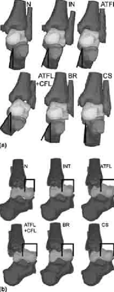
Qualitative examination of the bone surface rendition from one specimen (Fig. 3) shows that the 3.4 N m
inversion load caused negligible increased motion at the ankle and subtalar joint when the ATFL was sec-tioned compared to when all ligaments are intact. How-ever, sectioning of both the ATFL and CFL caused a large inversion instability (increased motion) (Fig. 3a). Both the Chrisman–Snook and the Brostro¨m proce-dures stabilized the ligament deficient ankle complex against an inversion load, but the Chrisman–Snook pro-vided higher stabilization (less motion) than the Bro-stro¨m. Qualitative examination of a sagittal view of the bone surface renditions from one specimen (Fig. 3b) shows that sectioning the ATFL created a noticeable instability in anterior drawer. Subsequent CFL section-ing further increased this instability.
External hindfoot response was defined as the dis-placement between the footplate and the tibial holder (Fig. 1) measured directly on the ALD in response to the applied loads. Sectioning the ATFL and CFL pro-duced a significant (p= 0.0003) increase in hindfoot rotation in inversion and an insignificant increase in anterior drawer displacement as measured on the ALD in this manner (Table 1).
Some of the architectural parameters changed signif-icantly under an inversion load when the ATFL and CFL were sectioned: L2, p= 0.0024; a1, p= 0.0014; a2,
p= 0.0021, a3, p= 0.0005; and b2, p= 0.001 (Fig. 4).
The Brostro¨m (L2, p= 0.0046, a3, p= 0.0013, and b3,
p= 0.0048) and the Chrisman–Snook (b3= 0.0021)
procedures returned these changes to near intact val-ues. When the joints were loaded in anterior drawer,
Fig. 1. The ankle loading device (ALD) used to maintain the ankle in various loaded and unloaded configurations in the MR scanner.
Fig. 2. Schematic diagram of the modified Chrisman–Snook surgical reconstruction procedure performed on the specimens following cutting of the ATFL and CFL.
significant changes were seen between ATFL sectioning in isolation and CFL sectioning; a2 (p= 0.004) and
(p= 0.0022) were affected. Although, both surgical pro-cedures returned these parameters towards intact values,
only the changes caused by the Chrisman–Snook proce-dure showed statistical significance (b3,p= 0.0042).
The applied inversion moment caused rotations at both the ankle and at the subtalar joint (Table 2). Sec-tioning the ATFL and CFL caused a significant increase in rotation at the ankle joint (p= 0.004) and at the ankle joint complex (p= 0.0003). The rotations at the subtalar joint were negligible. Both the Chrisman–Snook and the Brostro¨m procedures were effective in returning the
Fig. 3. Surface renditions showing qualitatively, on one specimen, the effect of sectioning the ATFL and CFL ligaments and of simulated surgical reconstruction on the mechanical response of the ankle complex to: (a) inversion (frontal plane) and (b) anterior drawer (sagittal plane) loads. The renditions are as follows. Top row (left to right): neutral (N), loaded intact (IN), loaded after sectioning ATFL (ATFL). Bottom row (left to right): loaded after cutting ATFL and CFL (ATFL + CFL), loaded after Brostro¨m procedure (BR), loaded after Chrisman–Snook procedure (CS).
Table 1
The externally measured motion produced at the ankle complex in response to applied inversion moment (3.4 N m) and anterior drawer force (150 N)
Intact ATFL CFL + ATFL BR CS
Inversion () 18.0 ± 6.8 20.0 ± 7.1 28.6 ± 11.3 20.6 ± 7.4 16.5 ± 6.6 Anterior Drawer (mm) 11.2 ± 3.1 12.8 ± 4.1 14.1 ± 4.1 12.7 ± 4.6 13.3 ± 4.8 Conditions include: ligaments intact, isolated ATFL cut (ATFL); both ATFL and CFL sectioned (CFL + ATFL); following surgical reconstruction using the Brostro¨m procedure (BR); and following a Chrisman–Snook procedure (CS). Sectioning the ATFL and CFL produced a significant increase in hindfoot rotation in inversion.
Fig. 4. Illustration of the definition of the architectural parameters of the hindfoot in a stylized exploded view of the ankle complex. Li
represents the distances between the geometric centroids of the bone surfaces,bidenotes the angle between the major principle axes of the bones, ai constitutes the internal angles of the triangle formed by connecting the centroids of the bones. See[24]for details. When the ATFL and CFL were sectioned L2, a1, a2, a3, and b2 changed
significantly. The Brostro¨m (L2,a3, andb3) and the Chrisman–Snook
procedures (b3) returned some of these changes to near intact values. When the joints were loaded in anterior drawer, significant changes were seen between ATFL sectioning in isolation and CFL sectioning, (a2anda3).
amount of rotation produced at the ankle and hindfoot in response to the inversion load to intact values.
With both ligaments intact, application of an anterior drawer force produced centroidal translations at both the ankle and the subtalar joints (Table 2). Sectioning the ATFL caused an insignificant increase in centroidal translation at the ankle and at the ankle complex with-out affecting the subtalar joint.
With the ligaments intact, a 3.4 N m inversion mo-ment caused inversion motion both at the ankle and subtalar joint (Table 3). After sectioning of the ATFL and CFL, the amount of rotation at the ankle complex more than doubled. This increase was associated with a corresponding large increase in inversion both at the ankle and at the subtalar joints. Both surgical proce-dures provided stabilization as is evident from the reduc-tion in the amount of inversion at the ankle complex, ankle, and subtalar joint. However, the amount of sta-bilization provided in this direction by the Chrisman– Snook procedure at the subtalar joint and at the ankle complex was larger than that provided by the Brostro¨m procedure.
Cutting the ATFL followed by the CFL caused pro-gressive increases in the amounts of anterior drawer mo-tion at the ankle and hindfoot. Both the Brostro¨m and the Chrisman–Snook procedures provided anterior drawer stabilization to the ligament deficient hindfoot. However, neither restored stability in this direction to pre-injury values. No significant changes were observed
when using the Grood and Suntay joint coordinate system.
Discussion
The traditional clinical description of hindfoot mo-tion suggests that inversion occurs mostly at the subtalar joint[25]. However, our results indicate that inversion of the ankle complex is associated with nearly equal contri-bution from the ankle joint (8.8) and the subtalar joint (7.6) (Tables 2 and 3), which is in agreement with other studies[5,22]. This discrepancy may be attributed to dif-ferences in experimental techniques. In the anterior drawer direction, the our results agree with the tradi-tional view that suggests that anterior drawer occurs al-most exclusively at the ankle joint with no subtalar joint displacement.
Sectioning the ATFL (in isolation or in combination with the CFL) did not produce instability at the subtalar joint in anterior drawer and inversion. This result is in agreement with previous investigations [21,22], which concluded that lateral collateral ligament injury does not compromise the mechanical stability of the subtalar joint, probably because the interosseous, talocalcaneal, and cervical ligaments are intact. If the anterior drawer test were to be examined in a weight bearing condition or under an axial load to simulate weight bearing, sub-talar joint instability might be identified. Isolated ATFL
Table 2
Equivalent axis rotation and centroidal translation produced in response to a 3.4 N m inversion moment and a 150 N anterior drawer force
Intact ATFL CFL + ATFL BR CS
Equivalent axis rotation () produced in response to a 3.4 N m inversion moment
Complex 11.6 ± 6.1 13.3 ± 6.0 23.4 ± 8.3(*) 13.0 ± 6.1 10.9 ± 5.5 Ankle 8.8 ± 4.1 12.6 ± 5.3 17.6 ± 8.5(*) 9.3 ± 5.4 9.8 ± 5.0 STJ 7.6 ± 4.2 6.8 ± 2.2 9.8 ± 8.4 8.5 ± 4.8 4.2 ± 1.8
Centroidal displacement (mm) produced in response to a 150 N anterior drawer force
Complex 7.3 ± 4.5 10.6 ± 6.8 9.8 ± 3.1 6.4 ± 2.1 8.2 ± 3.0 Ankle 3.5 ± 2.1 6.4 ± 4.8 7.1 ± 2.6 3.7 ± 1.4 4.9 ± 2.2 STJ 1.3 ± 1.0 1.1 ± 0.7 1.8 ± 2.3 1.0 ± 0.7 1.2 ± 0.6 Sectioning the ATFL and CFL caused a significant increase in rotation at the ankle joint and at the ankle joint complex.
*Statistically significant increase compared to the intact condition.
Table 3
Anatomical joint coordinates describing inversion and anterior drawer produced at the ankle, the subtalar joint, and the ankle complex in response to a 3.4 N m inversion moment and a 150 N anterior drawer force
Intact ATFL CFL + ATFL BR CS Ankle complex Inversion () 7.0 ± 5.4 7.9 ± 5.4 14.8 ± 4.4 9.3 ± 3.9 6.9 ± 3.5
Anterior drawer (mm) 2.9 ± 3.8 3.8 ± 2.8 4.9 ± 4.7 3.3 ± 1.9 3.2 ± 3.7 Ankle joint Inversion () 4.6 ± 10.4 4.0 ± 8.3 10.5 ± 6.2 3.1 ± 6.7 5.5 ± 3.4 Anterior drawer (mm) 1.8 ± 3.2 3.3 ± 3.9 6.4 ± 3.4 2.7 ± 2.3 3.8 ± 3.6 Subtalar joint Inversion () 4.0 ± 3.9 3.9 ± 2.8 7.9 ± 7.8 6.0 ± 4.5 2.9 ± 2.4 Anterior drawer (mm) 0.9 ± 1.0 0.6 ± 0.8 0.1 ± 1.2 0.1 ± 1.1 0.6 ± 1.5
sectioning produced no noticeable mechanical instability in inversion. A 30% increase in anterior drawer instabil-ity was created, but this increase was not statistically sig-nificant. This finding is consistent with the very large range of ‘‘normal’’ thresholds ranging from 4 mm to 10 mm reported in previous studies that used stress radiography[13,17]. Our results suggest that diagnosis of isolated ATFL injury based on inversion and anterior drawer tests is difficult and the development of diagnos-tic test for this injury may require further investigation. Another study found that internal rotation changed when the ATFL was sectioned [21], suggesting that internal rotation should be investigated using the sMRI technique to successfully detect isolated ATFL damage. Combined ATFL and CFL sectioning produced a sig-nificant mechanical instability in inversion, suggesting that inversion instability may provide a strong indica-tion that both ligaments are damaged.
The Chrisman–Snook procedure was more effective than the Brostro¨m in stabilizing the hindfoot in inver-sion. This result supports the clinical practice of using the Chrisman–Snook procedure for heavier patients where larger inversion stabilization may be required
[6]. Furthermore, the Chrisman–Snook procedure stabi-lized both the ankle and the subtalar joint, while the Brostro¨m procedure affected primarily the ankle joint. Considering the anatomy of these procedures, this observation is reasonable because the anterior portion of the Chrisman–Snook reconstruction spans both the ankle and the subtalar joint (Fig. 2) while the Brostro¨m procedure spans only the talo-crural joint. A contradic-tory study found that the effect of the Chrisman–Snook procedure was limited to the subtalar joint with little effect on the ankle joint [21]. The difference may be due to the modification of the traditional Chrisman– Snook procedure in that study by providing an anchor into the talus. In addition, motion of the tenodesis with-in the fibular tunnel was possible with-in that study while with-in our study no such motion could occur.
Under anterior drawer loading, both procedures in-creased the mechanical stability of the ankle and hind-foot towards pre-injury values, but neither procedure restored stability to intact values. The Brostro¨m proce-dure proved to be slightly more effective than the Chris-man–Snook procedure in stabilizing the ankle complex in anterior drawer. The difference may be explained by the difference in the anatomy of the two reconstructions
[24]. The Brostro¨m procedure attempts to restore ATFL support by creating a connection between the talus and the fibula. In contrast, the Chrisman–Snook procedure does not restore a talus-to-fibula connection. Therefore, in anterior drawer, as the talus is forced forward, the Brostro¨m procedure is expected to provide more sup-port than the Chrisman–Snook, as indeed was observed in the study. Improving the ability of the Chrisman– Snook procedure to stabilize the ankle in anterior
drawer may be achieved by adding a talar anchoring point for the peroneus brevis tendon.
The Grood and Suntay joint coordinate system was unable to detect significant changes between all condi-tions tested because of the small rotacondi-tions and transla-tions calculated. However, when the system is combined with the information from the helical axis rotations and centroidal translations, this method could further explain the instability. For example, when the hindfoot was in-verted with the ATFL and CFL sectioned, the helical axis rotations indicated instability at the ankle and ankle joint complex (Table 2). The Grood and Suntay data show that at the ankle, instability occurred only in the coronal plane. In the ankle joint complex there was an additional instability in the sagittal plane.
The 3D sMRI technique was used to evaluate the mechanical effects of lateral ligament injuries and the stabilizing function of two common surgical repair and reconstruction procedures in vitro. The study demon-strated how 3D sMRI presents an opportunity to extend the diagnostic power of MRI beyond mere geometrical, anatomical visualization to the assessment of mechani-cal function and the compromise in this function due to injury. Although this study focused on the ankle com-plex and its ligaments, a similar approach can be devel-oped and implemented to other joints such as the knee, the shoulder, and the spine. In the present study, the 3D sMRI technique was used in vitro since the specific dam-age could be carefully controlled. However, unlike most other experimental techniques reported in the past, the non-invasiveness of the 3D sMRI technique makes it equally effective in evaluating in vivo effects. With exten-sive in vivo testing and validation, this technique may be a powerful tool to assess the effects of injuries and surgi-cal reconstructions without being subjected to the seri-ous limitations present in in vitro studies. Such in vivo application will be the subject of our future studies.
Acknowledgment
The research described in this study was supported by US DHHS grant AR46902.
References
[1] Boruta PM, Bishop JO, Braly WG, Tullos HS. Acute lateral ankle ligament injuries: a literature review. Foot Ankle 1990;11:107–13. [2] Brostro¨m L. Sprain ankles: VI: surgical treatment of ‘‘chronic’’
ligament ruptures. Acta Chir Scand 1966;135:551–65.
[3] Cardone BW, Erickson SJ, Den Hartog BD, Carrera GF. MRI of injury to the lateral collateral ligamentous complex of the ankle. J Comput Assist Tomogr 1993:102–7.
[4] Cardone BW, Erickson SJ, Den Hartog BD, Carrera GF. MRI of injury to the lateral collateral ligamentous complex of the ankle. J Comput Assist Tomogr 1993;17:102–7.
[5] Chen J, Siegler S, Schneck CD. The three-dimensional kinematics and flexibility characteristics of the human ankle and subtalar joint—Part II: flexibility characteristics. J Biomech Eng 1988: 374–85.
[6] Chrisman OD, Snook GA. Reconstruction of lateral ligament tears of the ankle. An experimental study and clinical evaluation of seven patients treated by a new modification of the Elmslie procedure. J Bone Joint Surg Am 1969:904–12.
[7] Christensen J, Dockery G, schuberth J. Evaluation of ankle ligamentous insufficiency using the Telos ankle stress apparatus. J Am Podiat Med Assoc 1986:527–31.
[8] Falcao AX, Udupa JK, Samarasekera S, Sharma S. User-steered image segmentation paradigms: live wire and live lane. Graph Models Image Process 1998:233–60.
[9] Harrington KD. Degenerative arthritis of the ankle secondary to long-standing lateral ligament instability. J Bone Joint Stirg Am 1979:354–61.
[10] Hirsch BE, Udupa JK, Stindel E. Tarsal joint kinematics via 3D imaging. In: Herman GT, editor. 3D imaging in medicine. 2nd ed. Boca Raton, London, New York, Washington, DC: CRC Press; 2000. p. 329–59.
[11] Hollis JM, Blasier RD, Flahiff CM, Hofmann OE. Biomechan-ical comparison of reconstruction techniques in simulated lateral ankle ligament injury. Am J Sports Med 1995:678– 82.
[12] Karlsson J, Bergsten T, Lansinger O, Peterson L. Reconstruction of the lateral ligaments of the ankle for chronic lateral instability. J Bone Joint Surg Am 1988:581–8.
[13] Karlsson J, Lansinger O. Lateral instability of the ankle joint. Clin Orthop 1992:253–61.
[14] Kerkhoffs GMMJ, Blankevoort L, Schreurs AW, Jaspers JEN, van Dijk CN. An instrumented, dynamic test for anterior laxity of the ankle joint complex. J Biomech 2002;35:1665–70.
[15] Krips R, van Dijk CN, Halasi PT, Lehtonen H, Corradini C, Moyen B, et al. Long-term outcome of anatomical reconstruction versus tenodesis for the treatment of chronic anterolateral instability of the ankle joint: a multicenter study. Foot Ankle Int 2001;22:415–21.
[16] Lahde S, Putkonen M, Puranen J, Raatikainen T. Examination of the sprained ankle: anterior drawer test or arthrography? Eur J Radiol 1988:255–7.
[17] Lahde S, Putkonen M, Puranen J, Raatikainen T. Examination of the sprained ankle: anterior drawer test or arthrography. Eur J Radiol 1988;8:255–7.
[18] Lapointe SJ, Siegler S, Hillstrom H, Nobilini RR, Mlodzienski A, Techner L. Changes in the flexibility characteristics of the ankle
complex due to damage to the lateral collateral ligaments: an in vitro and in vivo study. J Orthop Res 1997:331–41.
[19] Liu W, Siegler S, Techner L. Quantitative measurement of ankle passive flexibility using an arthrometer on sprained ankles. Clin Biomech (Bristol, Avon) 2001:37–44.
[20] Martin DE, Kaplan PA, Kahler DM, Dussault R, Randolph BJ. Retrospective evaluation of graded stress examination of the ankle. Clin Orthop 1996:165–70.
[21] Rosenbaum D, Decker H, Wilke H, Claes L. Tenodeses destroy the kinematic coupling of the ankle joint complex. A three-dimensional in vitro analysis of joint movement. J Bone Joint Surg Br 1998;80:162–8.
[22] Siegler S, Chen J, Schneck CD. The three-dimensional kinematics and flexibility characteristics of the human ankle and subtalar joints—Part I: kinematics. J Biomech Eng 1988:364–73. [23] Siegler S, Lapointe S, Nobilini R, Berman AT. A
six-degrees-of-freedom instrumented linkage for measuring the flexibility char-acteristics of the ankle joint complex. J Biomech 1996:943–7. [24] Siegler S, Udupa JK, Ringleb SI, Imhauser CW, Hirsch BE,
Odhner D, et al. Mechanics of the ankle and subtalar joints through a three-dimensional quasi-static stress MRI technique. J Biomech 2005;38(3):567–78.
[25] Stiehl J. InmanÕs joints of the ankle. 2nd ed. Baltimore: Williams & Wilkins; 1991.
[26] Udupa JK, Hirsch BE, Hillstrom HJ, Bauer GR, Kneeland JB. Analysis of in vivo 3-D internal kinematics of the joints of the foot. IEEE Trans Biomed Eng 1998:1387–96.
[27] Udupa JK, Odhner D, Samarasekera S, Goncalves RJ, lyer K, Venugopal K, et al. 3DVIEWNDT: an open transportable, multidimensional, multiparametric, imaging software system. Proc SPIE 1994:58–73.
[28] Verhaven EF, Shahabpour M, Handelberg FW, Vaes PH, Opdecam PJ. The accuracy of three-dimensional magnetic reso-nance imaging in the diagnosis of ruptures of the lateral ligaments of the ankle. Am J Sports Med 1991;19:583–7.
[29] Verhaven EF, Shahabpour M, Handelberg FW, Vaes PH, Opdecam PJ. The accuracy of three-dimensional magnetic reso-nance imaging in the diagnosis of ruptures of the lateral ligaments of the ankle. Am J Sports Med 1991:583–7.
[30] Wu G, Siegler S, Allard P, Kirtley C, Leardini A, Rosenbaum D, et al. ISB recommendation on definitions of joint coordinate system of various joints for the reporting of human joint motion— Part I: ankle, hip, and spine. International Society of Biomechan-ics. J Biomech 2002:543–8.
[31] Yeung M, Chan K, So C, Yuan Y. An epidemiological survey on ankle sprains. Br J Sports Med 1994:112–6.