Soo Heui Baek,
1Hyun Kyung Jung,
1WooGyeong Kim,
2Suk Jung Kim,
1Hye Jin Baek,
1Seung Ho Kim,
1Yedaun Lee,
1and Young Mi Park
31Department of Radiology, Haeundae Paik Hospital, Inje University College of Medicine, Busan 612-896, Republic of Korea 2Department of Pathology, Haeundae Paik Hospital, Inje University College of Medicine, Busan 612-896, Republic of Korea 3Department of Radiology, Busan Paik Hospital, Inje University College of Medicine, Busan 614-735, Republic of Korea
Correspondence should be addressed to Hyun Kyung Jung; drsjung@gmail.com
Received 6 March 2015; Accepted 2 May 2015
Academic Editor: Jeffrey M. Weinberg
Copyright © 2015 Soo Heui Baek et al. This is an open access article distributed under the Creative Commons Attribution License, which permits unrestricted use, distribution, and reproduction in any medium, provided the original work is properly cited.
Merkel cell carcinoma (MCC) is a rare and aggressive neuroendocrine carcinoma of the skin. MCC is characterized by a high incidence of locoregional recurrence, and distant metastasis, and often requires short-term follow-up after treatment. In this present paper, we describe a rare case of MCC, which presented as a palpable axillary mass and an incidental adrenal mass, and report on the ultrasonography, computed tomography, and18F-fluorodeoxyglucose-positron emission tomography findings. The patient underwent surgery and adjuvant radiation therapy. Seven months after the initial diagnosis, distant metastasis was detected during a follow-up examination.
1. Introduction
Merkel cell carcinoma (MCC) is a rare and aggressive neuroendocrine carcinoma of the skin. MCC usually affects elderly patients, with a mean age of 70 years at the time of diagnosis [1]. It is usually detected on sun-exposed areas of the skin such as the head and neck region (47% of incidences), followed by the extremities (40%) and the trunk (8%) [2]. It is characterized by a high frequency of local recurrence, regional nodal metastasis, distant metastasis, and a low survival rate [2]. Agelli et al. reported that the age-adapted incidence rate of this disease has increased 3-fold, with an annual increase of 8% from 1986 to 2001 [3]. These statistics provide compelling reasons for early diagnosis and disease management for patients with MCC. Moreover, as MCC is a rare skin cancer, suitable imaging modalities have not been fully established [4–8]. In the present paper, we describe a unique case of MCC of the axilla and adrenal gland in a 53-year-old woman, report on the imaging findings, and review the relevant literatures regarding this disease.
2. Case Presentation
2 Case Reports in Medicine
(a) (b)
Figure 1: Transverse (a) and color Doppler (b) images of ultrasonography show an irregular mass with an indistinct margin, internal hypoechogenicity, and increased peripheral vascularity.
(a) (b)
Figure 2: Axial (a) and coronal (b) chest computed tomography images show a 12 cm mass with a lobulated contour and heterogeneous enhancement in the right axilla.
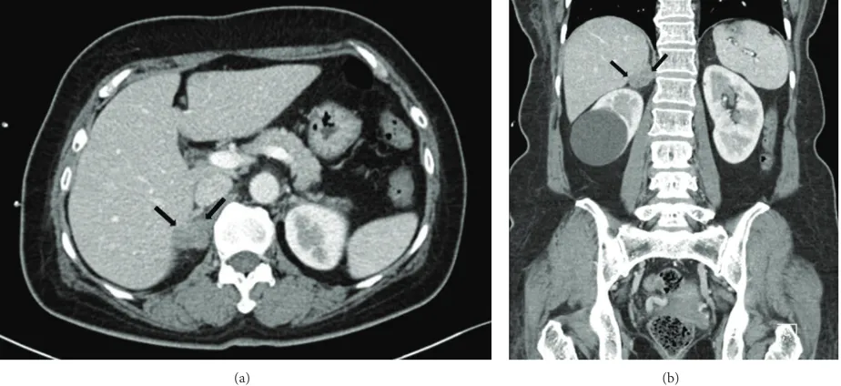
in the right axilla (Figure 2). Positron emission tomography with the glucose analog 2-[fluorine-18] fluoro-2-deoxy-D-glucose (18F-FDG-PET) for preoperative staging showed a focal FDG-avid uptake in the right axilla with a maximum standardized uptake value (SUV max) of 12.7 and also showed a focal FDG-avid uptake in the right adrenal gland with an SUV max of 4.7 (Figure 3). For further evaluation of the newly found mass in the adrenal gland, CT of the abdomen and pelvis was performed and a 1.5 cm mass with lobulated contours and mild enhancement was observed in the right adrenal gland, directly invading the liver (Figure 4). The patient underwent a wide local excision of the right axillary lesion and a right adrenalectomy with liver resection and cholecystectomy. Histologic examination confirmed that the right axillary mass was a neuroendocrine carcinoma,
Figure 3:18F-FDG-PET scan shows two focal FDG-avid uptakes in the right axilla (SUV max 12.7, black arrow) and right adrenal gland (SUV max 4.7, white arrow). SUV max: maximum standardized uptake value.18F-FDG-PET:18F-fluorodeoxyglucose-positron emission tomography.
(a) (b)
Figure 4: Axial (a) and coronal (b) abdominal computed tomography images during the late arterial phase show a 1.5 cm mass with a lobulated contour and mild enhancement in the right adrenal gland (black arrow).
small cell carcinoma of lung could have been ruled out with the lack of expression of markers including TTF-1, synaptophysin, and chromogranin. Based on the histopatho-logical and immunostaining findings, the two masses were diagnosed as MCC. Although the primary site of the tumor could not be clearly determined by histological examination, it is possible that the mass in the right axilla harbored a primary tumor that involved the dermis and subcutaneous fat. Nevertheless, the patient had no detectable primary skin lesion. After surgery, she received adjuvant radiation therapy. Seven months after the surgery, a follow-up 18 F-FDG-PET/CT was performed, which showed multiple FDG-avid
lymph node uptakes in the left external iliac chain, right aortocaval area, left axilla, left supraclavicular area, bilateral jugular chain, right retropharyngeal space, and right palatine tonsil. An endoscopic biopsy was performed on the right palatine tonsil and it was diagnosed as metastatic MCC in pathologic report. The patient subsequently underwent palliative radiation therapy.
3. Discussion
4 Case Reports in Medicine
(a) (b)
(c) (d)
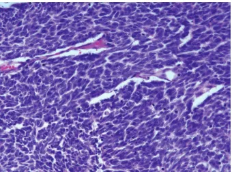
Figure 5: Microscopic findings of the right axillary mass showed diffuse sheets of basophilic tumor cells ((a) HE×200) with large and pale staining nucleus and tiny nucleoli ((b) HE×400). Lymphovascular invasion is an almost constant histological finding ((c) HE×400). Characteristic paranuclear dot-like staining of CK20 is specific for MCC ((d) CK20×400).
Figure 6: Histopathology of the adrenal gland mass showed similar findings observed in the axillary mass but had adopted more spindled cell morphology (HE×400).
by Toker as “trabecular carcinoma” [9]. Although the etiology and the mechanisms responsible for the regulation of its growth are currently unknown, exposure to sunlight and ultraviolet light, previous irradiation, infection with Merkel cell polyomavirus (MCV), and immunosuppression are likely to be significant risk factors [1,6]. Although the two masses were confirmed as MCC by surgery, we did not further perform an anti-MCV assay in our patient. MCC usually affects elderly patients, with a mean age of 70 years at the time of diagnosis, as well as immunosuppressed individuals such as organ transplant recipients and HIV infected people
[1, 10, 11]. It presents as a solitary, painless, pink to bluish papule or plaque on a sun-exposed area of the skin and grows rapidly [12]. Approximately 47% of these tumors occur in the head and neck, followed by the extremities (40%) and trunk (8%) [2]. Among the unusual extracutaneous primary sites, the parotid gland is the most common, followed by the submandibular gland, nasal cavity, mucosa of the lip, lymph nodes, and vulva/vagina [11]. In 14% of cases, the primary site is unknown and MCC presents at visceral or nodal sites [13]. Although MCC with an unknown primary site is unusual, no primary lesion can be identified in our patient after a thorough work-up. It is possible that the larger axillary mass was either a primary tumor involving the dermis and the subcutaneous fat or a metastatic MCC from an occult or regressed primary carcinoma [14]. Zhao and Meng reported the case of a MCC presenting as multiple lymph node metastases without a primary site [15].
To our knowledge, there have been a few cases of MCC involving the adrenal gland or the axilla [16,17] but this is the first reported case that involves both axilla and adrenal gland. Moreover, detection and analysis of MCC through imaging has not been widely reported.
by the neck (especially the parotid region), mediastinum, retroperitoneum, and groin [4,9]. The presence of stranding of cutaneous fat adjacent to the primary site of the MCC on CT scans may suggest the presence of edema from lymphatic invasion [5]. Furthermore, imaging work-up of soft tissue lesions is best performed with magnetic resonance imaging (MRI). However, there are only a few studies describing the MRI findings in such cases, which have reported the presence of inhomogeneous signal intensities on T1 and T2 weighted images [8].18F-FDG-PET has an important role in diagnostic imaging of MCC, and18F-FDG-PET/CT may also provide more accurate anatomic localization of tumors [4,6]. Peloschek et al. reported that18F-FDG-PET has a sensitivity of 85.7% and a specificity of 96.2% compared with those of 95.5% and 89.1% for conventional imaging modalities, respectively [18]. Based on the above mentioned findings,18 F-FDG-PET, US, CT, or MRI may be useful in patients with suspected metastatic MCC.
Although the treatment guidelines have not yet been defined, complete surgical excision is the best treatment option of MCC with a safety margin of 2–5 cm [19]. Histologi-cally, the tumor cells are characterized by a large, pale nucleus with a scanty cytoplasm [10]. Mitotic activity is often marked and lymphatic and vascular invasions are common and this is an important prognostic indicator [20]. Immunohistochem-istry has indicated that MCC expresses both epithelial (cytok-eratins and epithelial membrane antigen) and neuroen-docrine (neuron specific enolase, chromogranin, and synap-tophysin) markers. CK 20 is a sensitive and specific marker for MCC and is helpful in distinguishing between MCC and other malignant and benign neoplasms [2]. Staining for leukocyte common antigen (LCA), TTF-1, and vimentin usually yields negative results [10]. Although the differential diagnosis includes other neuroendocrine tumors, such as small cell lung cancer, melanoma, cutaneous lymphoma, Ewing sarcoma, neuroblastoma, rhabdomyosarcoma, and basal cell carcinoma, the two masses were diagnosed as MCC, based on the histopathology and immunohistochemistry, as mentioned earlier [15,21].
Previous studies demonstrated that male sex, tumor sizes larger than 2 cm, lymph node involvements, small cell pathology, lymphovascular invasions, and high mitotic rates are poor prognostic factors for MCC [2,20]. Most cases of recurrent disease appeared within the first 6 to 12 months after initial diagnosis [22]. Therefore, short-term follow-up is recommended.
In summary, MCC is a rare, aggressive skin tumor with a high rate of metastasis and mortality. In the present paper,
There is no conflict of interests regarding the publication of this paper.
References
[1] S. Vernadakis, D. Moris, A. Bankfalvi, N. Makris, and G. C. Sotiropoulos, “Metastatic Merkel Cell Carcinoma (MCC) of pancreas and breast: a unique case,”World Journal of Surgical Oncology, vol. 11, article 261, 2013.
[2] S. Akhtar, K. K. Oza, and J. Wright, “Merkel cell carcinoma: report of 10 cases and review of the literature,”Journal of the American Academy of Dermatology, vol. 43, no. 5, pp. 755–767, 2000.
[3] M. Agelli, L. X. Clegg, J. C. Becker, and D. E. Rollison, “The etiology and epidemiology of merkel cell carcinoma,”Current Problems in Cancer, vol. 34, no. 1, pp. 14–37, 2010.
[4] E. Enzenhofer, P. Ubl, C. Czerny, and B. M. Erovic, “Imaging in patients with merkel cell carcinoma,”Journal of Skin Cancer, vol. 2013, Article ID 973123, 6 pages, 2013.
[5] F. Eftekhari, S. Wallace, E. G. Silva, and R. Lenzi, “Merkel cell carcinoma of the skin: imaging and clinical features in 93 cases,”
British Journal of Radiology, vol. 69, no. 819, pp. 226–233, 1996. [6] B. D. Nguyen and A. E. McCullough, “Imaging of Merkel cell
carcinoma,”Radiographics, vol. 22, no. 2, pp. 367–376, 2002. [7] M. J. Gollub, D. R. Gruen, and D. D. Dershaw, “Merkel cell
carcinoma: CT findings in 12 patients,”American Journal of Roentgenology, vol. 167, no. 3, pp. 617–620, 1996.
[8] S. E. Anderson, K. T. Beer, A. Banic et al., “MRI of Merkel cell carcinoma: histologic correlation and review of the literature,”
The American Journal of Roentgenology, vol. 185, no. 6, pp. 1441– 1448, 2005.
[9] C. Toker, “Trabecular carcinoma of the skin,” Archives of Dermatology, vol. 105, no. 1, pp. 107–110, 1972.
[10] J. C. Becker, “Merkel cell carcinoma,”Annals of Oncology, vol. 21, no. 7, pp. vii81–vii85, 2010.
[11] J. Albores-Saavedra, K. Batich, F. Chable-Montero, N. Sagy, A. M. Schwartz, and D. E. Henson, “Merkel cell carcinoma demographics, morphology, and survival based on 3870 cases: a population based study,”Journal of Cutaneous Pathology, vol. 37, no. 1, pp. 20–27, 2010.
[12] A. Alzaraa, G. D. H. Thomas, A. Vodovnik, and V. K. Modgill, “Merkel cell carcinoma in a male breast: a case report,”Breast Journal, vol. 13, no. 5, pp. 517–519, 2007.
[13] M. L. Heath, N. Jaimes, B. Lemos et al., “Clinical characteristics of Merkel cell carcinoma at diagnosis in 195 patients: the AEIOU features,”Journal of the American Academy of Dermatology, vol. 58, no. 3, pp. 375–381, 2008.
[14] E. J. Kim, H. S. Kim, H. O. Kim et al., “Merkel cell carcinoma of the inguinal lymph node with an unknown primary site,”
6 Case Reports in Medicine
[15] M. Zhao and M.-B. Meng, “Merkel cell carcinoma with lymph node metastasis in the absence of a primary site: case report and literature review,”Oncology Letters, vol. 4, no. 6, pp. 1329–1334, 2012.
[16] Z. Alguraan, O. Agcaoglu, S. Aliyev, and E. Berber, “A rare case of merkel cell carcinoma metastasis to the adrenal resected robotically,”Surgical Laparoscopy, Endoscopy and Percutaneous Techniques, vol. 23, no. 1, pp. e35–e37, 2013.
[17] T. Schnabel and M. Glag, “Breast metastasis of Merkel cell carcinoma,”European Journal of Cancer, vol. 32, pp. 1617–1618, 1996.
[18] P. Peloschek, C. Novotny, C. Mueller-Mang et al., “Diagnostic imaging in Merkel cell carcinoma: lessons to learn from 16 cases with correlation of sonography, CT, MRI and PET,”European Journal of Radiology, vol. 73, no. 2, pp. 317–323, 2010.
[19] V. Koljonen, “Merkel cell carcinoma,”World Journal of Surgical Oncology, vol. 4, article 7, 2006.
[20] P. McKee, E. Calonje, T. Brenn, and A. Lazar, “Neroendocrine carcinoma of the skin,” inMcKee’s Pathology of the Skin, vol. 2, pp. 1141–1147, 4th edition, 2011.
[21] T. S. Wang, P. J. Byrne, L. K. Jacobs, and J. M. Taube, “Merkel cell carcinoma: update and review,”Seminars in Cutaneous Medicine and Surgery, vol. 30, no. 1, pp. 48–56, 2011.
Submit your manuscripts at
http://www.hindawi.com
Stem Cells
International
Hindawi Publishing Corporationhttp://www.hindawi.com Volume 2014
Hindawi Publishing Corporation
http://www.hindawi.com Volume 2014
Behavioural
Neurology
Endocrinology
International Journal of Hindawi Publishing Corporationhttp://www.hindawi.com Volume 2014
Hindawi Publishing Corporation
http://www.hindawi.com Volume 2014
BioMed
Research International
Oncology
Journal of Hindawi Publishing Corporationhttp://www.hindawi.com Volume 2014
Hindawi Publishing Corporation
http://www.hindawi.com Volume 2014
Oxidative Medicine and Cellular Longevity
Hindawi Publishing Corporation
http://www.hindawi.com Volume 2014
PPAR Research
Immunology Research
Hindawi Publishing Corporation
http://www.hindawi.com Volume 2014
Journal of
Obesity
Journal ofHindawi Publishing Corporation
http://www.hindawi.com Volume 2014
Hindawi Publishing Corporation
http://www.hindawi.com Volume 2014
Computational and Mathematical Methods in Medicine
Ophthalmology
Journal ofHindawi Publishing Corporation
http://www.hindawi.com Volume 2014
Hindawi Publishing Corporation
http://www.hindawi.com Volume 2014
Research and Treatment
AIDS
Hindawi Publishing Corporation
http://www.hindawi.com Volume 2014
Parkinson’s
Disease
Evidence-Based Complementary and Alternative Medicine
Volume 2014 Hindawi Publishing Corporation