an updated systematic review of the literature
Lesley Belanger, BA, DC
Dawn Burt, BSc (Hons), DC
Julia Callaghan, BSc (Hons), DC
Sheena Clifton, BSc, DC
Brian J. Gleberzon, DC, MHSc*
Objectives: The purpose of this study was to conduct a
systematic review regarding the purported differences in
anterior cruciate ligament (ACL) laxity throughout the
course of the menstrual cycle.
Methods: A systematic review was performed by
searching electronic databases, along with
hand-searching of journals and reference tracking for any
study that assessed ACL integrity throughout the
menstrual cycle from 1998 until 2011. Studies that
met the pre-defined inclusion criteria were evaluated
using the Modified Sackett Score (MSS) instrument that
assessed their methodological quality.
Results: Thirteen articles out of a possible 28 met the
inclusion criteria.
Conclusions: This systematic review found 13 clinical
trials investigating the effect of the menstrual cycle on
ACL laxity. There is evidence to support the hypothesis
that the ACL changes throughout the menstrual cycle,
with it becoming more lax during the pre-ovulatory
(luteal) phase. Overall, these reviews found statistically
significant differences for variation in ACL laxity and
injury throughout the menstrual cycle, especially during
the pre-ovulatory phase. Female athletes may need to
take precautions in order to reduce the likelihood of ACL
injury. However, the quality of the assessments was low
and the evidence is still very limited. More and better
quality research is needed in this area.
k e y w o r d s :
ligament, laxity, menstrual
*Professor & Chair, Department of Chiropractic Therapeutics, CMCC 6100 Leslie St, Toronto, Ontario. M2H 3J1
©JCCA 2013
Objectifs : Le but de cette étude était de procéder à
un examen systématique concernant les prétendues
différences dans le laxisme du ligament croisé antérieur
(LCA) tout au long du cycle menstruel.
Méthodologie : Un examen systématique a été effectué
en recherchant des bases de données électroniques, ainsi
qu’en effectuant une recherche manuelle des revues et
un suivi de références pour toute étude de 1998 jusqu’en
2011 qui a évalué l’intégrité du ligament croisé antérieur
LCA tout au long du cycle menstruel. Les études qui
répondaient aux critères d’inclusion prédéfinis ont été
évaluées en utilisant le score modifié de Sackett (MSS)
qui a évalué la qualité de leur méthodologie.
Résultats : Treize articles, sur un total possible de 28,
répondaient aux critères d’inclusion.
Conclusions : Cet examen systématique a découvert
13 essais cliniques portant sur l’effet du cycle menstruel
sur le laxisme du LCA. Il existe des preuves pour étayer
l’hypothèse que le LCA change tout au long du cycle
menstruel, devenant plus relâché lors de la phase
pré-ovulatoire (lutéale). Dans l’ensemble, ces examens ont
montré des différences statistiquement significatives
entre la variation de laxisme et de blessures du LCA
tout au long du cycle menstruel, en particulier pendant
la phase pré-ovulatoire. Les athlètes de sexe féminin
devraient peut-être prendre des précautions pour
réduire le risque de blessures du LCA. Cependant,
les évaluations qualitatives étaient insuffisantes et les
preuves sont encore très limitées. Donc, il faut effectuer
plus de recherches, et de meilleure qualité, dans ce
domaine.
Introduction
The physical disability and long rehabilitation process
as-sociated with anterior cruciate ligament (ACL) injury can
be both psychologically and financially devastating to the
individual, ultimately resulting in a decreased quality of
life.
1Female athletes have a higher rate of ACL injury
than do men, and many of these injuries require extensive
surgical and rehabilitative interventions, with a financial
burden to the American healthcare system estimated to
approach $650 million annually.
1Bearing that in mind,
it is imperative to understand the mechanisms leading to
such an injury in an effort to prevent its occurrence and its
subsequent sequelae. Although both men and women are
susceptible, the literature states that women have a 4 to 6
fold increased incidence of ACL injury.
2,3Notwithstand-ing the fact that a definitive etiology for this discrepancy
between the sexes has not been established, proposed
theories to account for it include: neuromuscular and
bio-mechanical factors (differences in pelvis width/increased
Q-angles in females, smaller femoral notch widths in
fe-males, increased female hamstring flexibility, and
imbal-anced hamstrings-to-quadriceps strength leading to
differ-ences in landing patterns); psychological factors (women
may be more prone to maladaptive perfectionism leading
to overtraining and burnout) and nutritional differences
(higher frequency of food restriction and decreased
cal-cium intake among females compared to males).
1,3,4An
additional theory posits increased ligament laxity is
relat-ed to hormonal fluctuations during the menstrual cycle.
1The menstrual cycle is controlled by the
pituitary-hypothalamic-ovarian axis and involves the complex
interaction of estrogen, progesterone, relaxin and
testos-terone.
1Typically, each menstrual cycle spans 28 days,
beginning with the follicular phase from days 1-9 during
which estrogen predominates, followed by the ovulatory
phase spanning days 10-14, where estrogen continues to
prevail and reaches its peak.
1The cycle ends with the
lu-teal phase extending from days 15-28 during which time
progesterone levels surpass that of estrogen levels.
1Re-laxin is secreted during the follicular and luteal phases,
reaching its peak during the luteal phase.
5Lastly,
testos-terone fluctuates throughout the cycle, and functions to
contribute to the formation of estrogen.
6Although the
hormones that predominate during each phase are
con-sistent among all women with normal functioning cycles,
the levels of each hormone varies among individuals.
3The hormones controlling the menstrual cycle are
thought to affect the overall integrity of the ACL by
al-tering its structure.
7In general, these hormones decrease
the tensile properties of the ACL by binding to specific
receptors on it.
7Specifically estrogen, when bound to
re-ceptors on the ACL, has been shown to decrease
fibro-blast proliferation, subsequently decreasing collagen
production.
7This could theoretically result in a greater
incidence of ACL injuries during the pre-ovulatory phase
spanning days 1-14 of the menstrual cycle, when
estro-gen predominates. This theory has been supported by a
case-control study in which female recreational skiers
who sustained a non-contact ACL injury demonstrated a
two-fold increase in injury rates during the pre-ovulatory
phase compared to the uninjured controls.
8However,
other studies have reported contradictory results that
re-fute the theory that hormonal variations during the
men-strual cycle contribute to ligament laxity. For example,
Van Luren et al
9reported arthrometric analysis of ACL
laxity that failed to demonstrate any variation in ACL
lax-ity throughout the menstrual cycle. In addition, Belanger
and colleagues
10examined 18 female subjects and were
unable to establish an association between increased ACL
laxity and the menstrual phase.
The objective of this article is to review the literature
regarding changes to anterior cruciate ligament laxity
dur-ing the menstrual cycle, builddur-ing on previous reviews by
Zazulac et al
1and Hewett et al
2. A better understanding of
the mechanism of injury may allow clinicians to identify
females who are at greatest risk of ACL injury and
subse-quently contribute to injury prevention in female athletes.
Methods
searched independently through selected databases
fol-lowed by hand searching throughout the periodicals.
Ref-erence tracking yielded one article. Periodical searching
yielded no eligible articles.
Inclusion/Exclusion Criteria
Inclusion criteria were as follows: female subjects of
re-productive age; published between 1998 and August 2011;
papers written in the English language and studies using
human subjects only. Articles that focused on therapy for
ACL injuries were excluded. Papers were also excluded
if they had been reviewed in the most recent literature
re-view by Hewett et al in 2007.
2Using these
inclusion/ex-clusion criteria 13 articles were selected for review.
Quality Appraisal
The methodological quality of the studies that met the
se-lection criteria was assessed using a modified version of
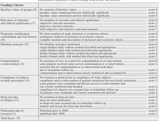
an instrument developed by Sackett (see Table 1).
11Since
the majority of research on the topic of ACL laxity and
menstrual hormonal fluctuations is limited to
observa-tional study designs rather than randomized clinical trials,
Table 1:
Instrument Categories Used to Grade Articles for this Review
Grading Criteria:
Baseline values of groups (/8) No mention of baseline values ... score 0;
baseline values mentioned but not statistically significant ... score 4;
baseline values mentioned and not statistically significant ... score 8.
Relevance of outcomes
and clinical significance (/7)
No mention of outcomes and clinical significance ... score 0;
subjective outcome measures ... score 3;
objective outcome measures ... score 5;
both subjective and objective outcome measures ... score 7.
Prognostic stratification
(comorbidity and risk factors)
(/6)
No clear mention of study inclusion or exclusion criteria ... score 0;
inadequate mention of inclusion or exclusion criteria ... score 3;
complete mention and description of inclusion and exclusion criteria ... score 6.
Blinding strategies (/5)
No blinding strategies mentioned ... score 0;
single blinded study without method described and appropriate ... score 2;
single blinded study with method described and appropriate ... score 3;
double blinded study without method described and appropriate ... score 4;
double blinded study with method described and appropriate ... score 5.
Contamination/
co-intervention (/4)
No mention of ways to control for contamination or co-intervention ... score 0;
some patients received some sort of contamination or co-intervention ... score 2;
assumed that no contamination or co-intervention took place
due to immediate follow-up ... score 3;
contamination and co-intervention closely monitored and accounted for ... score 4.
Compliance of subjects
to study procedures (/4)
No mention or detail given to compliance of study subjects ... score 0;
compliance and co-intervention of patients monitored but not closely monitored ... score 1;
some patients were compliant and did not receive co-interventions and
was closely monitored and detailed ... score 2;
compliance of subjects was assumed due to immediate follow-up ... score 3;
all patients were compliant and closely monitored and detailed ... score 4.
Drop-out rates
of subjects (/3)
No mention of drop-out rates ... score 0;
drop-out rates mentioned ... score 1;
no drop-out rates assumed due to immediate follow-up ... score 2;
number and reason for drop-outs described ... score 3.
Publication date of
the ‘assignment of patients’ and ‘follow-up levels’ criteria
were not included in our grading as they were deemed
incompatible with the majority of the research designs.
As a result, the instrument was modified and scored out
of 38 rather than 50.
The eligible articles were randomly assigned to four
authors (LB, DB, JC, SC). Each accepted article was
re-viewed by two authors independently. The data from all
accepted articles were recorded onto a data extraction
sheet by the authors as part of their review. The
auth-ors checked and edited all entries for accuracy and
con-sistency. Recorded data included study authors and quality
score, details of the study design, sample, interventions,
outcome measures, and main results/conclusions of the
study. Any discrepancies of scores between the authors
were settled via discussion until consensus was reached.
Results
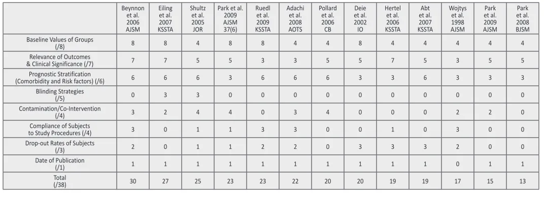
Thirteen articles met the inclusion criteria (see Table 2).
12-24After methodological quality assessment of each article
using the modified Sackett grading instrument, papers
were allocated scores out of a possible 38 points (Table
3). Of the 13 articles, 9 articles investigated ACL injuries
throughout the menstrual cycle and 4 articles investigated
the issue of ACL laxity throughout the menstrual cycle
Table 2:
Flow chart of retrieved articles used in this Review.
Records identified through database searching
(n = 27)
Additional records identified through other sources
(n =1)
Records after duplicates removed (n = 28)
Records screened (n =28)
Full-text articles assessed for eligibility
(n =28)
Studies included in qualitative synthesis
(n = 13)
Studies included in final qualitative synthesis
(n = 13)
Records excluded (n =0)
Full-text articles excluded because they focussed
on ACL rehabilitation or had been included in
the 2007 review (n =15)
From: Moher D, Liberati A, Tetzlaff J, Altman DG, The PRISMA
Group (2009). Preferred Reporting Items for Systematic Reviews and
Meta-Analyses: The PRISMA Statement. PLoS Med 6(6): e1000097.
doi:10.1371/journal.pmed1000097
Table 3:
Beynnonet al. 2006 AJSM
Eiling et al. 2007 KSSTA
Shultz et al. 2005 JOR
Park et al. 2009 AJSM 37(6)
Ruedl et al. 2009 KSSTA
Adachi et al. 2008 AOTS
Pollard et al. 2006 CB
Deie et al. 2002 IO
Hertel et al. 2006 KSSTA
Abt et al. 2007 KSSTA
Wojtys et al. 1998 AJSM
Park et al. 2009 AJSM
Park et al. 2008 BJSM Baseline Values of Groups
(/8) 8 8 4 8 8 4 4 8 4 4 4 4 4
Relevance of Outcomes
& Clinical Significance (/7) 7 7 5 5 3 3 5 5 7 5 3 5 5
Prognostic Stratification
(Comorbidity and Risk factors) (/6) 6 6 6 3 6 6 6 3 3 6 3 3 3
Blinding Strategies
(/5) 0 3 3 0 0 0 0 0 0 0 0 0 0
Contamination/Co-Intervention
(/4) 3 2 4 4 0 3 4 0 0 0 2 2 0
Compliance of Subjects
to Study Procedures (/4) 3 0 1 1 3 3 0 0 1 0 3 0 0
Drop-out Rates of Subjects
(/3) 2 0 1 1 2 2 0 3 3 3 2 0 0
Date of Publication
(/1) 1 1 1 1 1 1 1 1 1 1 0 1 1
Total
(/38) 30 27 25 23 23 22 20 20 19 19 17 15 13
(see Tables 4a and 4b respectively). Articles are listed in
descending order of their score. In the event two or more
articles had the same score, they were arranged
alphabetic-ally. A brief summary of each of the 13 articles graded in
this study is provided in Table 4a and 4b.
The accepted studies, determined by Sackett et al,
scored between 30 and 13 out of a possible 38 points on
the Modified Sackett Score (MSS) instrument (Table 2,3).
Eight
12-19of the thirteen studies reported that knee ligament
laxity changes throughout the menstrual cycle, although
the phase during which this ligamentous laxity occurred
varies throughout the cycle. Ruedl et al (MSS=23/39)
12,
Adachi et al (MSS=22)
13, Wojtys et al (MSS=17)
14and
Park-b et al (MSS=13)
15all reported increased knee laxity
during the ovulatory phase and Beynnon et al (MSS=30)
16reported increased knee laxity during the pre-ovulatory
phase (compared to post-ovulatory phase). However,
Schult et al (MSS=25)
17and Deie et al (MSS=20)
18re-ported increased knee laxity during the follicular phase
and Parka (MSS=15)
19reported increases knee laxity
dur-ing the luteal phase of the menstrual cycle.
On the other hand, five studies
20-24did not report any
statistically significant changes in knee laxity during the
menstrual cycle. Eiling et al (MSS=27)
20reported that
there was no statistically significant effect on anterior
knee ligament laxity throughout the menstrual cycle and
that ‘musculoskeletal stiffness’ was lower during the
ovu-latory phase of the menstrual cycle as compared to day
one of menstruation and the mid-follicular phase. Two
studies compared men to women with respect to knee
lax-ity. The study by Deie et al
18reported there was no
statis-tical difference in the anterior knee movement of 8 men
assessed during the same three week period as 16 women
and the study by Pollard et al
21that compared 12 men to
12 women reported that, while knee laxity increased
fol-lowing exercise, there was no difference across the sexes.
Discussions
Zazulak et al conducted a systematic review similar to
ours in 2006.
1Those researchers were able to retrieve
nine studies. Subjects included collegiate athletes,
high-school athletes, recreational athletes, non-athletes and
‘unspecified’ sport participants. Cohort sizes ranged from
7 to 41. Anterior tibiofemoral movement was measured in
all studies using KT 1000 or KT 2000 arthrometers.
1In that review, six of the nine reviewed studies
re-ported no statistically significant effect of the menstrual
cycle on ACL laxity. However, the reviewers reminded
the reader that the majority of these six studies based
their observations on a single sampled day of the cycle,
or randomly sampled across the cycle without hormonal
or cycle landmark confirmation.
1Of the three studies that
did report increased laxity of ACL during the menstrual
cycle, all three reported it occurred during the ovulatory
or post-ovulatory (luteal) phase
1, a finding similar to what
we found among the 13 articles we reviewed. Despite
di-versity in the literature, Zazulak et al
1suggested that the
three studies which found a positive association between
the menstrual cycle and ligament laxity were superior in
study design, methodology and consistency compared to
the 6 studies which failed to show any association,
there-by concluding that the menstrual cycle may have a
signifi-cant effect on anterior knee laxity.
Hewett et al
2performed a similar systematic review to
the one by Zazulak et al
1, with the primary difference
be-ing that Hewett et al reviewed articles that investigated
the effects of the menstrual cycle on anterior cruciate
liga-ment injury risk among high-risk female athletes,
where-as Zazulek et al investigated the effect of the menstrual
cycle on anterior knee laxity. In the Hewett et al
2review,
seven studies met the study’s inclusion criteria. Hewett
et al
2reported that all seven articles favoured an effect of
the first half of the menstrual cycle for the increased ACL
injuries, most commonly during the pre-ovulatory phase.
These authors also reported that the use of oral
contracep-tives in combination with neuromuscular training may
in-crease the stability of the knee joint and dein-crease the risk
of injury to female athletes.
2Hewett et al suggested that
disproportionate or isolated quadriceps recruitment can
create forces higher than those required for ACL failure.
3Therefore, neuromuscular training should focus on
bal-ancing hamstrings-to-quadriceps strength and recruitment
in order to increase stability of the knee.
While Zazulak et al
2focused on knee laxity and Hewett
et al
1focused on injury, this most recent review looked at
a combination of both laxity and injury. The results of our
review are in agreeance with Zazulak et al
2and Hewett
et al
1, supporting an effect of menstrual cycle on anterior
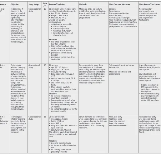
Table 4a:
Studies Investigating ACL Laxity
Reference Objective Study Design Score /38 Patients/Conditions Methods Main Outcome Measures Main Results/Conclusions
Eiling et al
2007 1. To examine changes in lower limb musculo-tendinous stiffness (MTS) over the course of the menstrual cycle 2. Investigate the
interaction of warm-up on MTS
Cross-sectional
Study 27 11 adolescent females.Played netball for minimum 5 yrs. • eight A-grade players and state
representatives
• two B-grade players and two C-grade players
The average age, height and weight of the subjects was: • 16.3 ± 0.65 years • 164.12 ± 6.2 cm and 60.72 ±
6.3 kg
Trained min 2 hrs per week. Consistent menstrual cycles for 3 mths.
Menarche >1 yr ago.
No use of contraceptives or other hormones for 3 mths. No history of serious lower limb injury.
Normal joint ROM.
Subjects documented menstrual history for 3 months prior and post testing,
Each subject tested at each of the 4 phases of the cycle: – blood levels for LH, FSH,
estradiol and progesterone. – ACL laxity using KT-2000. – MTS assessed before and after
5 min cycling warm up using unilateral hopping on force plate.
1. Blood levels LH, FSH, estradiol and progesterone. • The levels were analysed
which allowed the levels to be matched with the testing date • If the values of the hormone
analysis were not within the documented ranges for the specific phase, it was assumed that either the test date was miscalculated or that the cycle was anovulatory
• In both cases, the subject was re-tested for that particular phase in the subsequent cycle.
2. KT-2000
• The knee was placed in 30 deg. of flexion as the subject lay supine on a bench 3. Force plate
• Following a warm-up of 5 min of cycling at 50 W together with ten run-ups and netball landings subjects were instructed to perform a unilateral hop on a force plate in time with a metronome at a frequency of 2.2 Hz
No statistically significant effect of the menstrual cycle on anterior knee laxity. MTS significantly decreased following warm up. • Repeated measures ANOVA revealed significant (P < 0.05) main effects of test-session and warm-up on MTS for the dominant leg.
• MTS was found to significantly decrease by 4.2% following the warm-up intervention It was significantly lower during the ovulatory phase compared to day one of menstruation and mid-follicular phase, 8.7 and 4.5%.
Schultz et al
2005 To investigate if hormone levels across the menstrual cycle can affect anterior knee laxity
Cross-sectional
study 25 22 females with normal self-reported menstrual history in the previous 6 months
Between the ages of 18 and 30, with a body mass index (BMI = weight/height2) less than or equal to 30
Inclusion:
• no history of pregnancy • no use of oral contraceptives
or other hormone-stimulating medications for 6 months • non-smoking behavior • two healthy knees with no prior
history of joint injury or surgery, no medical conditions affecting the connective tissue • physical activity limited to 7 h or
less per week. Exclusion:
• experienced an anovulatory cycle or missed three or more consecutive days of testing
Measured blood levels of estradiol, progesterone and testosterone. Then measured knee joint laxity with an arthrometer
Minimum and peak levels of blood estradiol, progesterone, and testosterone.
Knee laxity using an arthrometer
The minimum concentrations of estradiol and progesterone in the early follicular phase are important factors in determining sensitivity of the knee joint’s response to changing hormone levels.
When minimum progesterone concentrations were higher and minimum estradiol concentrations were lower during the early follicular phase, females experienced greater increases in knee laxity across the menstrual cycle with attainment of peak estradiol and testosterone levels post ovulation.
Park et al 2009 (Alterations in knee joint…) To investigate whether the hormonal cycle has an influence on knee joint mechanism and whether increased knee joint loading during the menstrual cycle affects knee joint mechanics.
Controlled laboratory study.
23 26 healthy women: • age 22.7 ± 3.3 years • height, 170.1 ± 7.1 cm • mass, 65.0 ± 9.3 kg • body mass index (BMI), 22.4 ± 2.5 • average menstrual cycle, 28.9 ± 2.7 days • activity level, 8.7 ± 4.4 h/wk Inclusion: • required that the subject have a normal menstrual cycle • no history of oral contraceptive
use, and no previous knee injury Refrain from exercise 6 hrs prior to testing.
Blood samples drawn at 3 different phases of the menstrual cycle in each subject.
Knee joint loading was then measured during each phase using the KT-2000 arthrometer. Motion analysis testing of the knee was then performed.
Blood serum estradiol and progesterone. KT-2000
No significant difference in knee joint mechanics between phases. However, increased knee joint laxity was associated with higher knee joint loads during movements.
Table 4a:
Studies Investigating ACL Laxity (continued)
Reference Objective Study Design Score /38 Patients/Conditions Methods Main Outcome Measures Main Results/Conclusions
Pollard et al
2006 To investigate the collective effects of gender, estrogen and exercise on anterior knee laxity in active individuals
Observational
Study 20 12 women: age 24.8 years12 men: age 24.3 years – All 24 men and women had a
history of participating in high school and/or recreational cutting and landing sports which included basketball, volleyball, field hockey and soccer. – Inclusion criteria: subjects had
to have performed moderate exercise at least 4 times a week for at least 45 mins in duration for 2 months prior to participation in the study, had to have no history of significant lower extremity injury, were injury-free at the time of data collection, females had to have not taken oral contraceptives for the past 6 months and had experienced a normal 27-31 day cycle for the past 3 months. – Exclusion: if they had
participated in collegiate level athletics at any time
All subjects came to the lab prior to data collection for a pre-collection session to familiarize them with the KT-1000. Female subjects were given ovulation kits that detect the surge of LH immediately preceding ovulation to determine the time of ovulation. Each completed an informed consent and was instructed to refrain from exercise prior to data collection on that day. Females assigned to start data collection at the onset of menses or the onset of ovulation and completed 5 day data collections following the same protocol each which occurred at a specific time to correlate with different phases of the menstrual cycle – onset menses, 10 and 12 days post onset, 7 and 9 days post ovulation. Male subjects started collection on a day of convenience and completed 3 data collections following the same protocol as females, 10-12 days apart
Exercise:
• Subjects ran on a treadmill for 15 min at a self-selected pace. • The subject was asked to set
the pace to correspond to what they would consider ‘‘moderately hard’’. Once this pace was established, it was used throughout the rest of the data collections.
• For each subsequent treadmill run, the subject was instructed to warm up during the first three minutes and to reach the predetermined pace by the end of 3 min.
• immediately following the treadmill run, subjects were instructed to perform three dynamic lower extremity tasks consisting of the following: two minutes of weaving (grapevine) along a 20 m long runway; two minutes of left and right cutting along 2 m wide runway; and, 25 jump downs from a 46 cm step.
KT-1000 arthrometer.
Blood samples: looking at estrogen levels across the menstrual cycles
Knee laxity increased following exercise but did not differ across genders.
Deie et al
2002 To determine whether ACL laxity in women changes significantly during their menstrual cycles
Case-Control
study. 20 16 women, aged 21-23 (average age of 21.6 years) 8 men
No BCP
Regular menses (28±4 days) No previous knee injury
Measurements of their knees using KT-2000 arthrometer were performed 2-3 times every week over 4 consecutive weeks. Women measured their basal body temp daily for 4 weeks and estradiol and progesterone levels in their blood weekly. From their BBT or estradiol and progesterone levels the follicular, ovulatory, and luteal phases were delineated. 342 measurements were made. 158 measurements= follicular phase, 56=ovulatory, 128=luteal phase – Men’s measurements of their
knees using KT-2000 were performed 3 times a week over a 3 week period. 144 measurements were taken with 48 measurements in each of the first, second and third phases (based on when the measurement was taken in what week)
Arthrometer Basal body temp Blood samples
In men, no statistical significance with anterior movement through the 3 week period. In women, anterior or terminal stiffness was higher in the follicular phase than the ovulatory phase, which was in turn higher than the luteal phase.
Hertel et al
2006 To investigate changes in neuromuscular control and laxity at the knee across the menstrual cycle
Cross-sectional
study. 19 – 14 female collegiate athletes • age 19.3 ± 1.3 years • height 163.6 ± 8.5 cm • mass 59.4 ± 6.8 kg. – normal ovulatory menstrual
cycles (28-35 day cycles) with confirmed ovulation – not taking oral contraceptives – no history of serious knee injury – Subjects participated in either
competitive soccer or stunt cheerleading
Urine hormone levels and ovulation measured.
Neuromuscular performance and laxity of knee were measured in each phase of the cycle.
Hormone levels.
Peak flexion and extension torque. Hamstring: quadriceps strength. Joint position sense. Centre of pressure velocity. Anterior knee laxity.
Neuromuscular control and knee joint laxity do not change substantially across the menstrual cycle despite varying estrogen and progesterone levels.
Table 4a:
Studies Investigating ACL Laxity (continued)
Reference Objective Study Design Score /38 Patients/Conditions Methods Main Outcome Measures Main Results/Conclusions
Abt et al
2007 To determine if changes in the levels of estradiol and progesterone significantly alter fine motor coordination, postural stability knee strength and knee joint kinematics and kinetics between the menses, post-ovulatory, and mid-luteal phases of the menstrual cycle.
Cross-sectional
study. 19 10 physically active females were recruited from the local university • Age: 21.4 ± 1.4 years • Height: 1.67 ± 0.06 m • Mass: 59.9 ± 7.4 kg who do not use oral contraceptives.
– subjects were screened for: • history of injury • nutritional practices • menstrual dysfunction • thyroid dysfunction, and
physical activity.
Exclusion:
• mid-luteal progesterone level less than 10 ng/ml • history of serious knee injury
or other lower extremity injury within the prior 6 months • previous or current eating
disorder
• previous or current menstrual dysfunction
Measured single leg postural stability, fine motor coordination, knee strength, knee biomechanics, and serum estradiol and progesterone.
Estradiol Progesterone Fine motor coordination Postural stability Hamstring: quad strength Knee flexion and valgus excursion Peak proximal ant tibial shear force Flexion and valgus moments at peak proximal ant tibial shear force
Neuromuscular and biomechanical characteristics are not influenced by estradiol and progesterone fluctuations
Park et al
2009 To determine whether changing hormone levels influence joint laxity and stiffness of a non-contractile knee joint and knee joint structures using a new analysis technique. To determine whether subsets of women exist who demonstrate or do not demonstrate changes in knee laxity in response to circulating hormone levels throughout their menstrual cycle.
Observational
Study 15 26 women• age, 22.7 ± 3.3 years • height, 170.1 ± 7.1 cm • mass, 65.0 ± 9.3 kg • body mass index (BMI), 22.4
± 2.5
• average menstrual cycle, 28.9 ± 2.7 days
• and activity level, 8.7 ± 4.4 h/wk.
• Most subjects regularly participated in a sports activity at a recreational level. Inclusion:
• no previous knee injuries • never been pregnant • have regular menstrual cycles
(approximately 28 days) with no missed cycles over the previous 24 months
• no oral contraceptive use for the previous 6 months
Each completed a blood draw and laxity tests at 3 different times during her menstrual cycle. Blood samples were collected to determine the levels of estradiol and progesterone, indicating an appropriate phase of testing. Passive laxity and stiffness were measured using arthrometer
Self reported menstrual history Arthrometer
Measured for estradiol and progesterone
Lowest hormones in follicular phase, highest in luteal phase
Lowest estradiol and progesterone were in follicular and highest were in luteal phase
– Greater knee laxity at 89N was recorded in ovulation compared to luteal phase – Max knee laxity during
ovulation significantly exceeded max laxity during follicular phase
Park et al 2009 (Relationship between…)
To investigate whether changing knee laxity during the menstrual cycle correlates with changing knee joint loads in a cutting maneuver
Cross-sectional
study 13 25 healthy women:• mean age 22.7 years • height 170.2 cm • mass 64.7 kg
• body mass index 22.3 menstrual cycle 28.9 days
• activity levels 8.7 h/week The subjects regularly participated in sports activity at a recreational level.
Inclusion:
• a normal menstrual cycle • no history of oral contraceptive
use
• no knee injury within the preceding 6 months
Serum hormone concentrations were assessed and knee joint laxity was measured during the follicular, ovulation and luteal phases. Performed 10 trials of a cutting maneuver.
Knee joint laxity Peak knee angle Knee joint moment Knee joint impulse Blood hormone levels
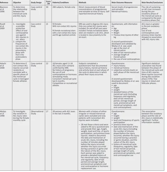
Table 4b:
Studies Investigating ACL Injury
Reference Objective Study Design Score /38 Patients/Conditions Methods Main Outcome Measures Main Results/Conclusions
Beynnon et al 2006 To determine the relationship between the menstrual cycle and ACL injury
Case-control
study 30 200 subjects, female only Direct measurement of blood concentrations of progesterone and estradiol at time of injury. Self reported menstrual history.
Serum levels of progesterone and estradiol.
Menstrual history.
The risk of sustaining an ACL tear increases during the pre-ovulatory phase of the menstrual cycle as compared to the post-ovulatory phase (3x) Ruedl
et al 2009
1. investigate a possible protective effect of oral contraceptive use against ACL injuries in rec. skiers 2. compare the
frequencies of non-contact ACL injuries in the preovulatory phase with that in the postovulatory phase
Case-control
Study 23 93 femalesWith non-contact ACL injuries
93 female recreational skiers with a non-contact ACL injury and 93 age matched controls
MRI was used to diagnose ACL injury. Only non-contact ACL injuries were included. On and off pill users were included. Female recreational alpine skiers are treated in a ski clinic, which is located in close proximity to the ski resort.
Questionnaire, with information on: • Age • Height • Weight • Previous knee injuries of either leg
A second questionnaire developed and validated by Wojtys et al. was used: • age at the start of
menstruation • date of last menstruation • average length of
menstruation
• the use of oral contraceptives
ACL injury is greater in the pre-ovulatory phase.
Use of oral contraceptives and previous knee injuries showed no association with ACL injury rate.
Adachi et al 2008
To determine if non-contact ACL injuries occurred randomly or correlate with a specific phase of the menstrual cycle in teenaged female athletes
Case-control
Study 22 18 females aged 11-18ACL injury (non-contact) confirmed by MRI. No history of pregnancy. No use of oral
contraceptives or hormone stimulating meds. Consistent menstrual cycle last 6 months.
Competitive or recreational athlete.
Subjects completed a questionnaire that documented injury history, menstrual history and activity level at each phase of the cycle to determine in which phase their injury occurred.
Questionnaire: • injury history • menstrual history • subjective activity levels on
each phase of the menstrual cycle
A second questionnaire developed by Wojtys et al. was used to document:
• age • height • weight
• detailed history of the menstrual cycle (including frequency and regularity, date of last menstrual period, average length of cycle, premenstrual and menstrual symptoms, and oral contraceptives)
Significant statistical association was found between the phase of the menstrual cycle and time of ACL injuries. More injuries occurred during the ovulation phase (72%). Few injuries in luteal and follicular phases.
Wojtys et al 1998
To investigate the variation in ACL injury rates during the female monthly cycle
Observational
Study 17 28 women with ACL tears in the last 3 months. Women with a history of either irregular or missed menstrual cycles were excluded and only patients with noncontact ACL injuries were included.
– 28 met these criteria and were asked to fill out a questionnaire and provide their age, height, weight, level and freq. of sports participation, and previous knee injuries. Asked to document the date and mechanism of acute ACL injury, including the number of minutes played before the injury occurred, whether the injury occurred during a practice or game and the nature of the ACL injury. Each woman was asked to provide a detailed history of her menstrual cycle, frequency and regularity, date of last period and average length, premenstrual symptoms and oral contraceptive or hormone replacement use.
Questionnaire: • age • height • weight
• level and frequency of sports participation
• previous knee injuries • date and mechanism of the
acute ACL injury (including the number of minutes played before the injury occurred, whether the injury occurred during a practice or game situation, and the nature of the ACL injury • history of menstrual cycle
(frequency, regularity, date of last menstrual period, average length of cycle, premenstrual symptoms, and oral contraceptive use
is associated with greater ligament laxity. The current
re-view found that the majority of studies (4 studies out of
8) that reported a positive association between increased
laxity, injury and the menstrual cycle implicated the
ovu-latory phase as the most significant time for laxity to
occur. These findings are somewhat in concordance with
the conclusions of Zazulak et al
2, who identified the
great-est laxity during the ovulatory and post-ovulatory phases.
In contrast, Hewett et al
1identified that the greatest injury
risk occurred during the pre-ovulatory phase.
Overall, limited evidence from the three reviews
sup-ports the theory that ACL ligament laxity varies with the
fluctuations of the hormonal cycle, thus predisposing
fe-male athletes to ACL injury. What remains to be clarified
is what phase of the cycle females are most at risk. Future
research should aim to clarify whether this fluctuation
in ligament laxity is consistent amongst all women with
hormonal fluctuations throughout their cycle, or whether
ligament laxity is dependent on the absolute or relative
hormonal level changes throughout a woman’s cycle.
Future studies can address this issue by focusing more
stringently on measuring hormone levels and by
examin-ing women over a longer period of time (more than one
cycle) to try and establish whether a trend in hormonal
levels and ligament laxity can be established and a phase
of increased risk identified.
Limitations
Many limitations were encountered throughout the review
of the recent literature. Limitations included: the majority
of the research was conducted during only one menstrual
cycle per participant, which does not account for variation
from cycle to cycle; there was no standardized definition
of the phases of the menstrual cycle, resulting in variation
of the phases from paper to paper; typically only one knee
was assessed per participant and therefore the results
can-not be confidently distinguished from conditions that may
have been pre-existent in that knee; the majority of studies
were conducted exclusively on women with normal
28-day cycles and; women who were on oral contraceptives
were often excluded by design. The average woman, and
the elite athlete, are not so easily categorized- especially
since menstruation may cease among some high
perform-ance athletes with low body mass indexes, and thus the
results reported in these studies may not be extrapolated
to the female population most at-risk of ACL injury.
Other limitations of this review are that we only
searched for articles in English and did not go further
back than 1998, since that was where other similar
re-viewers ended. Another limitation was our use of an
adapted Sacket instrument for the purposes of this review.
Although it had face validity to do so, to the best of our
knowledge there is no evidence that specifically supports
the validity of the modifications we made to Sackett’s
ori-ginal instrument. Furthermore, this tool does not assess
important aspects including confounding factors,
partici-pation rates, study population consistency or selection
bias. Lastly, 5 of the 13 studies accepted in this review are
cross-sectional and therefore cannot be used to determine
any cause and effect relationship between menstrual cycle
and knee ligament laxity.
Conclusions
There is preliminary evidence to suggest that
ligament-ous laxity of the knee changes throughout the course of
a women’s menstrual cycle, with the majority of
stud-ies reporting the greatest change is during the ovulatory
phase. However it is important to note that the evidence
remains inconsistent and is based predominantly on
stud-ies of low methodological quality. Certainly better
clin-ical trials need to be conducted that follow women over
several menstrual cycles and that include women not on
a standard 28-day cycle. Moreover, clinical trials
inves-tigating changes to ACL laxity should assess both knees.
That said, this review, as well as the previously published
study by Hewitt et al
2,3, suggest that healthcare
References
1. Zazulak B, Paterno M, Myer G et al. The effects of the
menstrual cycle on anterior knee laxity: a systematic
review. Sports Med. 2006; 36(10):847-862.
2. Hewett T, Zazulak B, Myer G. Effects of the menstrual
cycle on anterior cruciate ligament injury risk: a systematic
review. Am J Sports Med. 2007; 35(4): 659-668.
3. Hewett T, Myer G, Ford K. Anterior cruciate ligament
injuries in female athletes: Part 1, mechanisms and risk
factors. Am J Sports Med. 2006; 34(2):299-311.
4. Elliot D, Goldberg L, Kuehl K. Young women’s anterior
cruciate ligament injuries: an expanded model and
prevention paradigm. Sports Med. 2010; 40(5):367-376.
5. Wreje U, Kristiansson P, Aberg H et al. Serum levels of
relaxin during the menstrual cycle and oral contraceptive
use. GynecolObstert Invest. 1995; 39(3): 197-200.
6. Mathor M, Achado S, Wajchenberg B et al. Free plasma
testosterone levels during the normal menstrual cycle. J
Endocrinol Invest. 1985; 8(5), 437-441.
7. Yu W, Liu S, Hatch J et al. Effect of estrogen on cellular
metabolism of the human anterior cruciate ligament. Clin
Orthop 1999; 366: 229-238.
8. Ruedl G, Ploner P, Linortner I et al. Are oral contraceptive
use and menstrual cycle phase related to anterior cruciate
ligament injury risk in female recreational skiers? Knee
Surg Sport Tr A. 2009; 17(9):1065-1069.
9. Van Lunen B, Roberts J, Branch J et al. Association of
menstrual-cycle hormone changes with anterior cruciate
ligament laxity measurements. J Athlet Train. 2003; 38(4):
298.
10. Belanger M, Moore D, Crisco J et al. Knee laxity does not
vary with the menstrual cycle, before or after exercise. Am
J Sports Med. 2004; 32(5):1150-1157.
11. Sackett DC, Willams MC, Rosenbery JA. Evidence base
Medicine: What is it and what it isn’t. BMJ. 1996;
312:71-72.
12. Ruedl G, Ploner P, Linortner I et al. Are oral contraceptive
use and menstrual cycle phase related to anterior cruciate
ligament injury risk in female recreational skiers? Knee
Surg Sports Tr A. 2009; 29(17):1065-1069.
13. Adachi N, Nawata K, Maeta M et al. Relationship of the
menstrual cycle phase to anterior cruciate ligament injuries
in teenaged female athletes. Arch Orthop Trauma Surg.
2008; 128:473-478.
14. Wojtys E, Huston L, Lindenfeld T et al. Association
between the menstrual cycle and anterior cruciate ligament
injuries in female athletes. Am J Sport Med. 1998; 26:
614-619.
15. Park SK, Stefanyshyn D, Loitz-Ramage B et al. Changing
hormone levels during the menstrual cycle affect knee
laxity and stiffness in healthy female subjects. Am J Sport
Med. 2009; 37: 588-598.
16. Beynnon B, Johnson R, Braun S et al. The relationship
between menstrual cycle phase and anterior cruciate
ligament injury: A case-control study of recreational alpine
skiers. Am J Sport Med. 2006; 34:757-764.
17. Shultz S, Gansneder B, Sander T et al. Absolute serum
hormone levels predict the magnitude of change in anterior
knee laxity across the menstrual cycle. J Ortho Res. 2006;
24:124–131.
18. Deie M, Sakamaki Y, Sumen Y et al. Anterior knee laxity
in young women varies with their menstrual cycle. Int
Orthop. 2002; 5(26):154–156.
19. Park S, Stefanyshyn D, Ramage B et al. Relationship
between knee joint laxity and knee joint mechanics during
the menstrual cycle. Br J Sports Med. 2009; 43: 174-179.
20. Eiling E, Bryant A, Petersen W et al. Effects of
menstrual-cycle hormone fluctuations on musculotendinous stiffness
and knee joint laxity. Knee Surg Sport Tr A. 2007; 15:126–
132.
21. Pollard C, Braun B, Hamill J. Influence of gender, estrogen
and exercise on anterior knee laxity. Clin Biomech. 2006;
21:1060–1066.
22. Abt J, Sell T, Laudner K et al. Neuromuscular and
biomechanical characteristics do not vary across the
menstrual cycle. Knee Surg Sports Tr A. 2007; 15:901–
907.
23. Hertel J, Williams N, Olmsted-Kramer L et al.
Neuromuscular performance and knee laxity do not change
across the menstrual cycle in female athletes. Knee Surg
Sports Tr A. 2006; 14: 817–822.