Observational Study Design in Veterinary Pathology. Part 2: Methodology.
Jeff L Caswell, Laura L Bassel, Jamie L. Rothenburger, Andrea Gröne, Jan M. Sargeant, Amanda P. Beck, Stina Ekman, Katherine N. Gibson-Corley, Thijs Kuiken, Elise E.B. LaDouceur, David K. Meyerholz, Francesco C Origgi, Horst Posthaus, Simon L Priestnall, Lorenzo Ressel, Leslie Sharkey, Leandro B.C. Teixeira, Kazuyuki Uchida, Jerrold M Ward, Joshua D. Webster, Jyoji Yamate.
Jeff L Caswell, [email protected], Department of Pathobiology, University of Guelph, Guelph, ON, Canada N1G 2W1
Laura L Bassel, [email protected], Department of Pathobiology, University of Guelph, Guelph, ON, Canada N1G 2W1
Jamie L. Rothenburger, [email protected], Department of Ecosystem and Public Health; Canadian Wildlife Health Cooperative (Alberta), Faculty of Veterinary Medicine, University of Calgary, 3280 Hospital Dr. NW, Calgary, AB, Canada T2N 4Z6
Andrea Gröne, [email protected], Department of Pathobiology, Faculty of Veterinary Medicine, Utrecht University, Utrecht, the Netherlands
Jan M. Sargeant, [email protected], Department of Population Medicine and Centre for Public Health and Zoonoses, University of Guelph, Guelph, ON, Canada N1G 2W1.
Amanda P. Beck, [email protected], Albert Einstein College of Medicine, 1301 Morris Park Ave, Bronx, NY 10461
Stina Ekman, [email protected], Department of Biomedicine and Veterinary Public Health, Swedish University of Agricultural Sciences, Box 7028, 75007 Uppsala, Sweden
Katherine N. Gibson-Corley, [email protected], Department of Pathology, Roy J. and Lucille A. Carver College of Medicine, University of Iowa, Iowa City, IA 52242 Thijs Kuiken, [email protected], Department of Viroscience, Erasmus University Medical Centre, Rotterdam, The Netherlands.
Elise E.B. LaDouceur, [email protected], Joint Pathology Center, 606 Stephen Sitter Ave, Silver Spring, MD 20910
David K. Meyerholz, [email protected], University of Iowa Carver College of Medicine; 1165 Medical Laboratories, University of Iowa Carver College of Medicine, Iowa City, Iowa, 52242
Francesco C. Origgi, [email protected], Centre for Fish and Wildlife Health, Vetsuisse Faculty, University of Bern, Switzerland
Horst Posthaus, [email protected], Institute of Animal Pathology, Vetsuisse-Faculty, University of Bern, Switzerland
Lorenzo Ressel, Department of Veterinary Pathology and Public Health, Institute of Veterinary Science, University of Liverpool, Liverpool, United Kingdom.
Leslie Sharkey, [email protected], Department of Clinical Sciences, Cummings School of Veterinary Medicine, Tufts University, 200 Westboro Rd., N. Grafton, MA 01536
Leandro B.C. Teixeira, [email protected], Department of Pathobiological Sciences, University of Wisconsin-Madison, 2015 Linden Drive, Madison, WI 53706, USA (608) 262-8089
Kazuyuki Uchida, [email protected], Department of Veterinary Pathology, The University of Tokyo, Tokyo 113-8657, Japan
Jerrold M Ward, [email protected], GlobalVetPathology, Montgomery Village, Maryland
Joshua D. Webster, [email protected], Genentech, 1 DNA Way, South San Francisco, CA 94080
Jyoji Yamate, [email protected], Laboratory of Veterinary Pathology, Graduate School of Life and Environmental Sciences, Osaka Prefecture University, 1-58 Rinku-Ourai-Kita, Izumisano City, Osaka 598-8531, Japan.
Corresponding author:
Jeff L Caswell [email protected]
Abstract
Observational studies are a basis for much of our knowledge of veterinary pathology, yet considerations for conducting pathology-based observational studies are not readily available. In part 1 of this series, we offered advice on planning and carrying out an observational study. Part 2 of the series focuses on methodology. General recommendations are to consider using already-validated methods, published guidelines, data from a primary source, and quantitative analysis. We discuss 3 common methods in pathology research—histopathologic scoring, immunohistochemistry, and polymerase chain reaction—to illustrate principles of method validation. Some aspects of quality control include use of clear objective grading criteria, validation of key reagents, assessing sample quality, determining specificity and sensitivity, use of technical and biologic negative and positive controls, blinding of investigators,
approaches to minimizing operator-dependent variation, measuring technical variation, and consistency in analysis of the different study groups. We close by discussing approaches to increasing the rigor of observational studies by corroborating results with complementary methods, comparing results among many different animals, consideration of similar published studies, replicating the results in a second study population, and critical analysis of the study findings.
Keywords
Observational studies are the most frequent study type published in the pages of Veterinary Pathology and fundamentally guide the daily practice of veterinary pathologists. Although advice is available from other disciplines, it may be difficult for veterinary pathologists to apply these guidelines to the types of observational studies they typically conduct. The first article of this series focused on design and development of observational studies.6 In the current article, we—editors and editorial board members of Veterinary Pathology and their colleagues—focus on principles of method validation and quality control. These are discussed in the context of 3 methods commonly used by veterinary pathologists: histopathologic scoring,
immunohistochemistry, and polymerase chain reaction (PCR). We emphasize that this article is neither intended as a ‘cookbook’ for validating these methods nor as a list of requirements for publishing in Veterinary Pathology. Instead, we focus on using these examples to illustrate considerations and principles that are relevant to any investigative method (Table 1).
Innovative methods can provide unexpected answers to longstanding and important questions. However, the methods should not be the basis for the study, but instead are used to address a hypothesis, question or objective backed up by a clear rationale. If the study objective is this journey’s roadmap, then the methodology is the vehicle’s engine. Studies with a clear objective can cruise toward their destination while still having resources to spare for stops at points of interest along the way. Alternatively, investigations built only on the novelty of a method tend to pleasantly ramble through an interesting landscape but never find a destination.
Investigators are also directed to the reporting guidelines available for many analytic methods, which are also relevant to study design and method validation (Minimum Information for
Biological and Biomedical Investigations; https://fairsharing.org/collection/MIBBI). Of particular relevance are those for veterinary observational studies (STROBE-Vet36,44), animal studies (ARRIVE: https://www.nc3rs.org.uk/arrive-guidelines), diagnostic test accuracy (STARD9,15), immunohistochemistry and in situ hybridization (http://mged.sourceforge.net/misfishie/), flow cytometry (http://flowcyt.sourceforge.net/miflowcyt/), and reverse transcription-quantitative PCR (RT-qPCR; http://www.rdml.org/miqe.php).
Microscopic assessment
Diagnosis, classification, and scoring or grading of microscopic lesions are mainstays of pathology research. Because these methods are subjective and sometimes challenging to quantify, effective study design and validation of methods are particularly critical. Errors in measuring lesions result in imprecision and variability of the data, thereby reducing the
likelihood of detecting a difference between study groups. Furthermore, systematic errors that differ between the study groups may cause differential information bias that could lead to a spurious outcome of the study. Guidelines for histologic scoring and grading have been published16,23,30,32,45 including a series in Veterinary Pathology on scoring histologic lesions in comparative pathology research,22,24,25,31,51 and guidelines and insights on cancer grading studies are available.29,54
Validate information obtained from others
At the beginning of a new study, at least one of the investigators with appropriate training and experience should validate all of the original pathologic diagnoses by re-evaluating the
error into the study.
Obtain data from the primary source, if the pathology records are likely to contain errors. We all recognize the frequency of omissions and mistakes in the clinical histories submitted to pathology laboratories. Thus, as an example, the clinical diagnoses and clinical findings should be obtained directly from the clinical case records and not based only on the pathology record. Even then, it should be recognized that variation among clinicians or inconsistency in recording clinical findings may affect the quality of the data.
Definition of the outcome
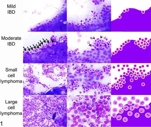
Clear, objective and reproducible criteria are essential for defining the outcome. For example, studies of equine sarcoma and canine liposarcoma provide examples of clearly defined characteristics for each neoplasm.1,12 Another study included a schematic, microscopic images, and detailed description of cytologic grading criteria (Figure 1).27 Visual guides such as these are highly useful to clearly define the cut points between grades and thus reduce both intra- and inter-observer variation.
Diagnostic criteria are more clear and reproducible if they are based on objectively observed lesions instead of the inferred morphologic diagnosis. This is particularly important when the diagnostic terms have subtly different meanings among pathologists. For example, “interstitial pneumonia” is used differently among pathologists, whereas the descriptive term “the
presence of lymphocytes within pulmonary alveolar septa” should be uniformly understood. A clear, objective, and precise definition of the outcome is expected to reduce variability of the data, improve understanding of the study methods and results, and make it easier for readers to apply the study findings to their own caseload.29 Some of our current cancer grading
schemes lack clarity or require subjective assessments or interpretations. “Severity of nuclear pleomorphism” and “degree of differentiation” are examples of criteria that cannot be
consistently applied to tumor grading by different individuals. Even “percentage of necrosis” is of dubious consistency because the amount of necrosis depends on which parts of the tumor were examined.29 For studies of new grading schemes, investigators should measure the inter-observer variability, ideally using experienced collaborating pathologists who are provided with no additional training other than the grading criteria used in the study. This tests whether the criteria are sufficiently clear, and reflects the likelihood that a grading scheme can be reliably applied to daily practice.46 If collaborators cannot use the published methods to replicate the results, then it is unlikely that others will be able to validly apply the grading scheme to their clinical case material.29,46
Defining the scope of analysis
subjects. The balance between these two approaches depends on the number of available cases and the study objectives. If there are few cases then intensive analysis of each of them will improve the data, whereas if numerous cases are available then a more focused analysis of all of them will improve the statistical power. A broad-based investigation might be
appropriate in an exploratory or descriptive study. However, a focused analysis allows for a more directed interrogation of a specific question. This can allow the investigators to
triangulate on testing a specific hypothesis, spend more time and energy on validating the results, and streamline the story that will become the manuscript. Intensive investigation of a single case or very few cases might reveal something new, but analysis of a large number of cases is generally needed to ensure the findings are consistent and applicable to the larger animal population.
Validation of the methods
Repeated analysis of all or a subset of the same samples (separated by a “wash-out” period to reduce recall) is an important validation of the histopathology data and measures the technical variability or the consistency of scores assigned by a single individual.16
If assessment of inter-observer variation is an objective, each parameter should be independently evaluated by 3 or more of the authors. The variability among observers is quantified by kappa analysis for binary data, or by other approaches for continuous data. For example, canine mast cell tumors were independently graded by 3 anatomic pathologists and 3 clinical pathologists, with 72-77% agreement for the various grading schemes.4 In contrast to some diagnostic tests, there may not be another “gold standard” for validating histopathologic data, but measuring the consistency of diagnoses among different pathologists can serve a similar purpose.
Avoiding technical error, bias and confounding
Pathology data can have poor intra-observer and inter-observer consistency. As discussed above, variability is minimized if grading criteria are precisely defined, based on observed lesions instead of inferred diagnoses, and illustrated by examples or a visual grading key. Ensure that the individual who makes the assessments or carries out the methods has
sufficient training, experience and knowledge, as the results are expected to be more uniform and more accurate than those assigned by non-experts.42 Conversely, inadequate training and experience with the grading method may reduce the ability to detect a difference between groups.
The different study groups must be analyzed in the same way. All samples should ideally be analyzed by the same individual. Alternatively, diagnoses or scores can be assigned by
consensus among a team that evaluates each sample in a standardized way. If it is necessary that different cases are evaluated by different individuals, be sure the assignments are
randomized, and incorporate a statistical comparison of the different individuals into the study design. Histologic sections from the study groups should be examined concurrently in random order, instead of sequentially analyzing the different study groups. Evaluation of all subjects should be done within a narrow time period to avoid “diagnostic drift”.45
without prior knowledge of either the exposure or the outcome. Most observational studies employ a 2-step process. First, tissues are examined in a non-blinded manner to identify the important criteria to be measured and to establish cut-points between the different histologic grades, and then those few specific lesions that are most relevant to the study objectives are scored in a blinded formal analysis. Finally, recognize that blinding may be impossible in pathology-based studies if the observations defining the exposure and the outcome cannot be made independently. For example, immunohistochemistry findings cannot be assessed
independently of tumor subtype, if the morphology of the tumor is evident on the
immunolabelled glass slide. In such instances, consider how this might cause a differential information bias, and how the impact can be minimized by ensuring the data are collected as objectively as possible.
It is critical to ensure that the study groups are comparable in order to avoid selection bias, information bias or confounding. This issue is particularly relevant for studies of archived samples or data not acquired for the purpose of the study. Analysis of the pathology records may not be sufficient to ensure that the 2 study groups were drawn from similar populations; information from clinical hospital records, owner interviews, or knowledge of laboratory
protocols may be required. Further, consider if there might there be differences between cases and controls in the details of how samples were obtained, processed or analyzed. Failure to ensure that the study groups are comparable and investigated in a consistent manner may result in spurious findings, as discussed in the first article of this series.6
These factors argue in favor of systematic methodology for routine diagnostic investigations, especially for those involving case material that is likely to be used for future observational studies. This approach has been particularly useful in investigations of wildlife disease. For example, routine mortalities of harbor seals had been investigated in a standardized manner including recording of life history data, necropsy methods, and sampling of tissues and sera. These provided an effective control group for comparison to harbor seals that died during a phocine distemper epidemic, with respect to analysis of antibody titers, morbillivirus infection, gross and microscopic lesions, and bacterial isolates from lung and other organs.41 Similarly, consistent collection of demographic data (sex, body weight, geographic location, etc) and standardized necropsy methods including evaluation of the vascular system and collection of muscle samples, were key to analytic studies of muscle pathology and verminous arteritis in stranded cetaceans.10,48
Immunohistochemistry
Immunohistochemistry is a cardinal method in veterinary pathology, yet it is a technically complex procedure. The many variables in the method allow the possibility of technical error unless assays are optimized and validated for the species, tissue and application of interest. Once validated, quality control procedures are necessary for reliable analysis of samples used in the study. The details of immunohistochemistry validation and quality control are described elsewhere.17,20,30,38,52,53
Antibody validation
The sensitivity and specificity of the primary antibody is fundamental to the
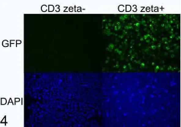
react with the expected antigen in a non-target species. Conversely, analytic specificity reflects whether the antibody reacts only with the intended target antigen. Cross-reactivity with a non-target antigen can lead to false-positive labelling that may be difficult to recognize (Figures 2-3). Note that these terms—analytic sensitivity and specificity—are related to but different from diagnostic sensitivity and specificity as used in the context of epidemiology studies (i.e. the proportion of truly positive and truly negative animals that are correctly classified by the test, respectively).
Where possible, veterinary pathologists should use antibodies that have been previously validated for immunohistochemistry in the species of interest. For example, an antibody raised in rabbits against a human protein may not react with the corresponding canine protein, and may react with other unrelated canine antigens. It has been estimated that 75% of
commercially available antibodies are non-specific.55 When it is claimed that antibodies have been validated in the dog, consider if the supporting data are available and if they are
compelling. If not, investigators must be aware that antibody validation is not trivial, yet it is essential to the credibility and truthfulness of the results.
There are several approaches to validating the analytic sensitivity and specificity of an
antibody.52 One approach is to show immunolabelling in cells or anatomic locations expected to express the antigen, and absence of immunolabelling in cells expected to not express it. For example, immunolabelling is compared in epithelial vs connective tissues of the skin, or in T- vs B-cell areas of lymph node, or in cortex vs medulla of the adrenal gland. However, this is of limited value for antigens that do not have cell- or tissue-specific expression patterns. The subcellular pattern of immunolabelling may also reveal a problem with antibody specificity, if this does not match the expected membranous, cytoplasmic or nuclear location of the protein. A second approach is western blot analysis of the tissue or tumor being studied (eg. canine lymphoma). An alternative is to use a normal tissue from this species that is known to express the target protein (eg. canine lymph node). In the western blot, the antibody should label a band of the expected molecular weight for the target protein. If there are additional bands, these could be analyzed by mass spectrometry to determine if they are a non-target protein, or variants or isoforms of the target protein. These additional bands are highly relevant: while target and non-target antigens can be differentiated on western blots based on molecular weight of the protein bands, these signals are indistinguishable by immunohistochemistry. An important caveat that limits the value of this approach is that the labelling pattern in western blot analysis of fresh tissue may differ from that in formalin-fixed tissues. The proteins in a western blot are denatured, and the process of formalin fixation and paraffin embedding may alter the protein conformation and thus affect the epitope recognized by the antibody.
Consequently, immunolabelling of the target antigen or of a cross-reacting antigen in the western blot does not necessarily mean the same reaction would occur in fixed tissues, and vice versa.38
mass spectrometry showed that these bands did not contain HER2. This cross-reactivity may have been responsible for the unexpected cytoplasmic immunolabelling of neoplastic cells and labelling of non-neoplastic cells, which were observed when canine tissues were probed with this antibody.3,40 These are disruptive findings: they challenge the validity of previously
published studies, and question the use of canine mammary tumors as a model of HER2 over-expressing breast cancer.3
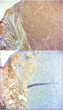
A third approach to validating antibody specificity is to compare cells that are engineered to express or not express the target protein. One method is to transfect non-expressing cells with the gene of interest (Figure 4). A second method is knockout or knockdown of the gene of interest in cells that naturally express it. Expressing and non-expressing cells can be fixed in formalin, and immunoreactivity is compared in histologic sections of the different cell pellets. Note that this approach may not detect immunoreactivity against cross-reacting antigens, because complex tissues include a greater diversity of proteins than are expressed by the negative control cell line.
A fourth approach is competitive inhibition or adsorption. In this procedure, antibody is pre-incubated with an excess of the purified antigen (or adsorbed using antigen-bound beads) before applying it to the tissue section, and the resulting neutralization of the antibody’s binding site is expected to abrogate immunolabeling. 2,47 However, this method may not rule out cross-reactivity with proteins containing an epitope similar to that of the target protein, or if the antibody has greater affinity for the target antigen compared to the non-target antigen.17,52 A fifth approach is in silico comparison of the amino acid sequence of the target protein among species. This can be used to infer cross-reactivity, particularly when the epitope targeted by the antibody is known, although it does not directly confirm antibody specificity.
Additional approaches include a comparison of immunolabelling patterns using 2 or more antibodies that bind different epitopes of the target protein, a correlation of
immunohistochemistry findings with those of another analytical method such as in situ hybridization, ELISA or RT-qPCR, and immunocapture followed by mass spectrometry.17,52 Assay validation
In addition to validating the primary antibody, it is necessary to validate the assay as a whole: the combined effectiveness of sample preparation, reagents, buffers, antigen retrieval,
blocking, antibody avidity and specificity, batch-to-batch variability of antibodies, incubation conditions, detection platforms, and the ability of individual observers to interpret the findings. This is done by demonstrating positive reactivity in tissue known to express the protein of interest, and negative reactivity in tissue known to not express the protein of interest. Here we take a qualitative approach to assay validation; quantitative measurement of test accuracy, sensitivity and specificity is described elsewhere.9,13,15
the tissue, considering the cell types that are labelled and whether the labelling is nuclear, cytoplasmic or membranous.
Negative controls identify non-specific labelling and thereby identify false-positives. The first negative control—the technical negative control—uses the positive control tissue but replaces the primary antibody with a non-antigen-specific substitute. If the primary antibody is a
monoclonal antibody, the negative control should be an irrelevant monoclonal antibody of the same concentration and isotype. In the same way, purified immunoglobulin is a technical negative control for purified polyclonal antibodies, and pre-immune serum or normal serum would be appropriate if immune serum were used as the primary antibody. Omission of the primary antibody is often used as a negative control for diagnostic case material once the method has been well-validated and is in routine use. However, in our opinion, this is
insufficient for assay validation or in assays that have some nonspecific background staining. It is important to note that the technical negative control does not assess nonspecific labeling with the primary antibody; if the antibody cross-reacted with an unrelated antigen, the staining would be abrogated by replacement or omission of the primary antibody. Additional technical negative controls may be useful in troubleshooting problems when validating a new assay. The second negative control—the biologic negative control—should normally include both lesional tissue samples as well as normal tissues. Sometimes, a single organ may contain different tissues that can serve as positive and negative control (such as epidermis adjacent to a tumor). Tissues from 10 positive and 10 negative individuals have been suggested as a minimum for analytic validation and can be inexpensively analyzed in tissue microarrays or multi-tissue blocks.13 Tissues used for validation must be processed in the same way as would be done for clinical specimens, including fixation and decalcification.
These guidelines apply to qualitative analysis of the presence or absence of immunolabeling, and do not measure test sensitivity and specificity in an epidemiologic context. Furthermore, additional validation is needed for quantitative immunohistochemical analysis, if the goal is to compare the immunolabelling scores with pathogen load, or to compare the findings of two different antibodies.
Quality control
When using a validated assay to analyze samples from the study, one aspect of quality control is to ensure the immunohistochemistry procedures remain effective. These use the same approaches as described above and can be considered as external positive and negative controls.
The second aspect of quality control is to provide sample-specific validation of every
Conversely, false positive findings represent non-specific staining and are detected by an internal negative control (labelling of an area of tissue not expected to contain the antigen; Figure 5), and secondly by applying an isotype-matched irrelevant antibody to a different section of the same sample.
Immunohistochemistry is a wonderful tool for co-localization of lesions and antigens, but careful attention to method validation is essential to avoid misleading results. As discussed below, confirmation of immunohistochemistry findings by a second analytical method can provide additional rigor.
Polymerase chain reaction
Assay validation
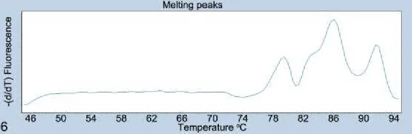
While it is true that PCR assays provide more objective data than the subjective interpretations sometimes required for histopathology or immunohistochemistry, effort is nonetheless required to ensure PCR data are valid. A first step it to ensure that the assay is measuring the intended target. For either conventional or quantitative PCR (qPCR), the primer and probe specificity should be confirmed by BLAST search against the target and non-target species, and by sequencing the amplified products. For quantitative PCR, additional confirmation is provided by analysis of the amplification curve, product melting curve and melting peaks for each reaction (Figure 6). Use of labeled probes can be valuable to provide additional specificity. False-positive outcomes are further avoided by use of negative controls, and these may include 3 forms that address different potential problems. First, using water in place of DNA template from the sample distinguishes positive from negative results. Second, for reverse transcription PCR (RT-PCR), a positive sample processed without reverse transcriptase would detect false positive results caused by sample contamination. Third, cross-reactions should be further ruled out with samples representing alternative disease conditions and using the same sample matrix as clinical samples.49 Examples include testing an assay for Mannheimia haemolytica against a panel of other bacteria,18 testing an assay for detection of ovine herpesvirus-2 infection in cattle and sheep against other species of animals infected with closely related herpesviruses,37 and measuring the sensitivity and specificity of RT-qPCR for bovine respiratory syncytial virus compared to an established assay.50
Quality control
Following validation of the method, quality control is required in each run of the analysis to ensure valid results. Negative controls may include water in place of DNA template, as well as a positive sample processed without reverse transcriptase as described above. False-positive results due to cross-contamination were prevented in 1 investigation by using a new
microtome blade for each sample, and forceps cleaned with detergent and alcohol.12 The positive control—analysis of a known positive sample or a synthesized nucleic acid target—would detect a general failure of the assay. However, this would not detect sample-specific false negative results such as those caused by sample degradation, poor yield or poor quality of nucleic acid, inadequate deparaffinization, the presence of PCR inhibitors, or
samples were spiked with positive control DNA to investigate the possibility of PCR inhibitors.28 For quantitative PCR, additional standard quality control procedures include a dilution series (standard curve) of the positive control for each plate of reactions, establishing the crossing point at which samples are considered reliably positive, documentation that values for samples fall within the dynamic range of the assay, analysis of amplification efficiency for each gene targeted, consideration of the method used to normalize data obtained from different runs, establishing the repeatability and reproducibility of the assay, and validation of reference
(“housekeeping”) genes including changes induced by the stimulus being studied. Readers are further directed to technical guidelines for analysis of gene expression
(miqe.gene-quantification.info).
Quantitative assessment and statistical analysis
In general, for analytic studies, quantitative or semiquantitative assessment of pathologic findings is preferred over a descriptive narrative, because it allows statistical analysis of the data and formal comparison of study groups. As an example, cluster analysis of multiple quantified parameters was used for unbiased analysis of glomerular diseases in dogs, which defined and validated their morphologic categorization as membranoproliferative and
membranous patterns of immune complex–mediated glomerulonephritis, glomerular amyloidosis, glomerulosclerosis, and normal.8 When a difference between two groups is identified, the level of certainty beyond chance is estimated by the P value. However, the statistical analysis should not end with the P value. As important, the magnitude of the difference between study groups is summarized by the risk ratio or odds ratio for categorical data or the mean difference between groups for continuous data. Finally, the study data are only an estimate of the truth: the 95% confidence interval considers the variability of the data and the sample size to estimate the range in which the actual (true) parameter likely lies, and is therefore essential for valid interpretation of the results.
It is a rare veterinary pathologist who has deep knowledge of statistical analysis. Given the effort put into designing and carrying out an observational study, why risk a deceiving outcome caused by inappropriate statistical analysis? Seek professional help in the planning stage to ensure an effective study design, and after the study is completed to have confidence in the analysis. Common pitfalls in statistical analysis are discussed in an upcoming commentary in Veterinary Pathology.31 Readers are also directed to the “Statistics simplified” series published in 2011 in the Journal of the American Veterinary Medical Association.
Approaches to increased rigor
Rigor involves an approach to conducting research that ensures the findings reflect the truth. Casadevall described 5 pillars of scientific rigor: redundancy in experimental design, sound statistical analysis, recognition of error, avoidance of logical traps, and intellectual honesty.5 How can we achieve rigor? First, examine your motivations for conducting the study. Surely, a passion for finding the truth motivates most of us in these endeavors. How far are you willing to go to bring rigor to your investigations and ensure the study outcomes are valid?
One way is to replicate the technical analyses on different occasions. Especially for
Another way is to use different methods to show the same finding, an approach known as triangulation.34 Particularly for unexpected outcomes, choose complementary methods that address a different aspect of biology and therefore greatly increase the confidence in the findings. Probe the research question from different angles for “validation of tissue
pathobiology”16 by using combinations of morphologic and molecular pathology, clinical biochemistry, clinical observations, diagnostic imaging, epidemiologic analyses, and microbiology assays, as examples. For example, a study used immunohistochemistry, polymerase chain reaction and in situ hybridization as redundant methods to demonstrate canine papillomavirus within pigmented plaques that progressed to squamous cell
carcinoma.26 Imaging can be used to corroborate the histopathologic findings. For example, X-ray fluorescence microscopy and Raman spectroscopy were used to characterize metal alloy and oxalate crystals within tissues, respectively.11,33 Bone metastases were verified by both histopathology and computed tomography.7 Just as you wouldn’t invest retirement savings in a single stock, we shouldn’t base our understanding of a disease on a single methodology. Critically consider how the study findings concur and differ from those of other investigators. Often this is done superficially with the simple goal of justifying the current results. A more enlightened approach, in those situations where the prior study design is indeed comparable to the present one, is a detailed analysis filled with sparkling insights on what the discrepancies suggest about the true biology of the disease, with additional analyses to investigate these novel possibilities.
The same investigators or ideally an independent group should confirm the results on a second population of study subjects. This is essential for major findings that change routine practice. Cancer grading schemes are an example: a new grading scheme is not rock solid until it has been validated in a second population of study subjects (see Hill’s criterion of consistency6). We have illustrative examples of this in veterinary pathology.4,21,35,39,43,46
Generally, we as journal editors expect that such confirmatory studies not only corroborate or refute the outcome of the original study, but also include additional results that add novelty to the new investigation.
Finally, develop a mindset of critical analysis and skeptical interpretation of your own findings. Carefully probe a variety of alternative explanations. Consider the possibility of bias and how it might impact the findings. Don’t ignore troublesome details of the data that seem not to make sense. Sometimes, niggling problems are the window that opens to reveal a new landscape of truth.
Conclusions
The methods that veterinary pathologists use in observational studies are key to advancing knowledge of animal disease. Yet, the most-used methods in pathology are somewhat subjective, inherently variable, not easily quantitated, and susceptible to technical error. Careful attention to validation, quality control, methodologic rigor, and critical analysis of the results are required for advancing knowledge in veterinary pathology.
Acknowledgements
Declaration of conflicting interests
Some authors of this commentary are editors of Veterinary Pathology. This editorial commentary was not peer-reviewed.
Funding
References
1. Avallone G, Roccabianca P, Crippa L, et al. Histological Classification and
Immunohistochemical Evaluation of MDM2 and CDK4 Expression in Canine Liposarcoma. Vet Pathol. 2016;53(4):773-780.
2. Bland SK, Schmiedt CW, Clark ME, DeLay J, Bienzle D. Expression of Kidney Injury Molecule-1 in Healthy and Diseased Feline Kidney Tissue. Vet Pathol. 2017;54(3):490-510.
3. Burrai GP, Tanca A, De Miglio MR, et al. Investigation of HER2 expression in canine mammary tumors by antibody-based, transcriptomic and mass spectrometry analysis: is the dog a suitable animal model for human breast cancer? Tumour Biol.
2015;36(11):9083-9091.
4. Camus MS, Priest HL, Koehler JW, et al. Cytologic Criteria for Mast Cell Tumor Grading in Dogs With Evaluation of Clinical Outcome. Vet Pathol. 2016;53(6):1117-1123.
5. Casadevall A, Fang FC. Rigorous Science: a How-To Guide. MBio. 2016;7(6).
6. Caswell JL. Observational Study Design in Veterinary Pathology. Part 1: Study Design and development. Veterinary Pathology. 2018;55(5):XXX-XXX.
7. Charney VA, Miller MA, Heng HG, Weng HY, Knapp DW. Skeletal Metastasis of Canine Urothelial Carcinoma: Pathologic and Computed Tomographic Features. Vet Pathol. 2017;54(3):380-386.
8. Cianciolo RE, Mohr FC, Aresu L, et al. World Small Animal Veterinary Association Renal Pathology Initiative: Classification of Glomerular Diseases in Dogs. Vet Pathol.
2016;53(1):113-135.
9. Cohen JF, Korevaar DA, Altman DG, et al. STARD 2015 guidelines for reporting diagnostic accuracy studies: explanation and elaboration. BMJ Open.
2016;6(11):e012799.
10. Díaz-Delgado J, Fernández A, Xuriach A, et al. Verminous Arteritis Due to Crassicauda sp. in Cuvier’s Beaked Whales (Ziphius Cavirostris). Vet Pathol. 2016;53(6):1233-1240.
11. DiVincenzo MJ, Frydman GH, Kowaleski MP, et al. Metallosis in a Dog as a Long-Term Complication Following Total Hip Arthroplasty. Vet Pathol. 2017;54(5):828-831.
12. Epperson ED, Castleman WL. Bovine Papillomavirus DNA and S100 Profiles in Sarcoids and Other Cutaneous Spindle Cell Tumors in Horses. Vet Pathol. 2017;54(1):44-52.
13. Fitzgibbons PL, Bradley LA, Fatheree LA, et al. Principles of analytic validation of immunohistochemical assays: Guideline from the College of American Pathologists
14. Flatland B, Friedrichs KR, Klenner S. Differentiating between analytical and diagnostic performance evaluation with a focus on the method comparison study and identification of bias. Vet Clin Pathol. 2014;43(4):475-486.
15. Gardner IA, Nielsen SS, Whittington RJ, et al. Consensus-based reporting standards for diagnostic test accuracy studies for paratuberculosis in ruminants. Prev Vet Med.
2011;101(1-2):18-34.
16. Gibson-Corley KN, Olivier AK, Meyerholz DK. Principles for valid histopathologic scoring in research. Vet Pathol. 2013;50(6):1007-1015.
17. Goodwin PC, Johnson B, Frevert CW. Microscopy, immunohistochemistry, digital imaging, and quantitative microscopy. In: Treuting PM and Dintzis SM (Editors): Comparative Anatomy and Histology, a Mouse, Rat and Human Atlas. 2nd ed. Academic Press; 2018:53-66. https://doi.org/10.1016/B978-0-12-802900-8.00004-X.
18. Guenther S, Schierack P, Grobbel M, Lübke-Becker A, Wieler LH, Ewers C. Real-time PCR assay for the detection of species of the genus Mannheimia. J Microbiol Methods. 2008;75(1):75-80.
19. Halsey CHC, Thamm DH, Weishaar KM, et al. Expression of Phosphorylated KIT in Canine Mast Cell Tumor. Vet Pathol. 2017;54(3):387-394.
20. Hewitt SM, Baskin DG, Frevert CW, Stahl WL, Rosa-Molinar E. Controls for
immunohistochemistry: the Histochemical Society’s standards of practice for validation of immunohistochemical assays. J Histochem Cytochem. 2014;62(10):693-697.
21. Horta RS, Lavalle GE, Monteiro LN, Souza MCC, Cassali GD, Araújo RB. Assessment of Canine Mast Cell Tumor Mortality Risk Based on Clinical, Histologic,
Immunohistochemical, and Molecular Features. Vet Pathol. 2018;55(2):212-223.
22. Janke, L.J., Ward, J.M., Vogel, P. Classification, scoring, and quantification of cell death in tissue sections. Vet Pathol. 2018;55:XXX-XXX.
23. Klopfleisch R. Multiparametric and semiquantitative scoring systems for the evaluation of mouse model histopathology--a systematic review. BMC Vet Res. 2013;9:123.
24. Knoblaugh, S.E., Himmel, L.E. Keeping score: semiquantitative and quantitative scoring approaches to genetically engineered and xenograft mouse models of cancer. Vet Pathol. 2018;55:XXX-XXX.
25. La Perle, K.M.D. Comparative Pathologists: Ultimate Control Freaks Seeking Validation! Vet Pathol. 2018;55:XXX-XXX.
27. Maeda S, Tsuboi M, Sakai K, et al. Endoscopic Cytology for the Diagnosis of Chronic Enteritis and Intestinal Lymphoma in Dogs. Vet Pathol. 2017;54(4):595-604.
28. Meason-Smith C, Edwards EE, Older CE, et al. Panfungal Polymerase Chain Reaction for Identification of Fungal Pathogens in Formalin-Fixed Animal Tissues. Vet Pathol.
2017;54(4):640-648.
29. Meuten D, Munday JS, Hauck M. Time to Standardize? Time to Validate? Vet Pathol. 2018;55(2):195-199.
30. Meyerholz DK, Beck AP. Principles and approaches for reproducible scoring of tissue stains in research. Lab Invest. May 2018.
31. Meyerholz, D.K., Beck, A.P. Recognizing common pitfalls in analysis of tissue scores. Vet Pathol. 2018;55:XXX-XXX.
32. Meyerholz DK, Sieren JC, Beck AP, Flaherty HA. Approaches to Evaluate Lung Inflammation in Translational Research. Vet Pathol. 2018;55(1):42-52.
33. Mitchell EP, Church ME, Nemser SM, et al. Pathology and Epidemiology of Oxalate Nephrosis in Cheetahs. Vet Pathol. 2017;54(6):977-985.
34. Munafò MR, Davey Smith G. Robust research needs many lines of evidence. Nature. 2018;553(7689):399-401.
35. Nagamine E, Hirayama K, Matsuda K, et al. Diversity of Histologic Patterns and Expression of Cytoskeletal Proteins in Canine Skeletal Osteosarcoma. Vet Pathol. 2015;52(5):977-984.
36. O’Connor AM, Sargeant JM, Dohoo IR, et al. Explanation and Elaboration Document for the STROBE-Vet Statement: Strengthening the Reporting of Observational Studies in Epidemiology-Veterinary Extension. J Vet Intern Med. 2016;30(6):1896-1928.
37. Pesavento, P., Cunha, C., Li, H., Jackson, K., O’Toole D. Demonstration of ovine
herpesvirus 2, the agent of sheep-associated malignant catarrhal fever, in formalin-fixed tissues. Vet Pathol. 2018;55:XXX-XXX.
38. Ramos-Vara JA, Miller MA. When tissue antigens and antibodies get along: revisiting the technical aspects of immunohistochemistry--the red, brown, and blue technique. Vet Pathol. 2014;51(1):42-87.
39. Rasotto R, Berlato D, Goldschmidt MH, Zappulli V. Prognostic Significance of Canine Mammary Tumor Histologic Subtypes: An Observational Cohort Study of 229 Cases. Vet Pathol. 2017;54(4):571-578.
41. Rijks JM, Read FL, van de Bildt MWG, et al. Quantitative analysis of the 2002 phocine distemper epidemic in the Netherlands. Vet Pathol. 2008;45(4):516-530.
42. Rousselet M-C, Michalak S, Dupré F, et al. Sources of variability in histological scoring of chronic viral hepatitis. Hepatology. 2005;41(2):257-264.
43. Sabattini S, Scarpa F, Berlato D, Bettini G. Histologic grading of canine mast cell tumor: is 2 better than 3? Vet Pathol. 2015;52(1):70-73.
44. Sargeant JM, O’Connor AM, Dohoo IR, et al. Methods and processes of developing the strengthening the reporting of observational studies in epidemiology - veterinary
(STROBE-Vet) statement. Prev Vet Med. 2016;134:188-196.
45. Schafer KA, Eighmy J, Fikes JD, et al. Use of Severity Grades to Characterize Histopathologic Changes. Toxicol Pathol. January 2018:192623318761348.
46. Schott CR, Tatiersky LJ, Foster RA, Wood GA. Histologic Grade Does Not Predict Outcome in Dogs with Appendicular Osteosarcoma Receiving the Standard of Care. Vet Pathol. 2018;55(2):202-211.
47. Senthilkumaran C, Hewson J, Ollivett TL, et al. Localization of annexins A1 and A2 in the respiratory tract of healthy calves and those experimentally infected with Mannheimia haemolytica. Vet Res. 2015;46:6.
48. Sierra E, Espinosa de Los Monteros A, Fernández A, et al. Muscle Pathology in Free-Ranging Stranded Cetaceans. Vet Pathol. 2017;54(2):298-311.
49. Ståhl M, Kokotovic B, Hjulsager CK, Breum SØ, Angen Ø. The use of quantitative PCR for identification and quantification of Brachyspira pilosicoli, Lawsonia intracellularis and Escherichia coli fimbrial types F4 and F18 in pig feces. Vet Microbiol. 2011;151(3-4):307-314.
50. Timsit E, Maingourd C, Le Dréan E, et al. Evaluation of a commercial real-time reverse transcription polymerase chain reaction kit for the diagnosis of Bovine respiratory syncytial virus infection. J Vet Diagn Invest. 2010;22(2):238-241.
51. Treuting, P., Boyd, K. Histopathological Scoring. Vet Pathol. 2018;55:XXX-XXX.
52. Uhlen M, Bandrowski A, Carr S, et al. A proposal for validation of antibodies. Nat Methods. 2016;13(10):823-827.
53. Ward JM, Rehg JE. Rodent immunohistochemistry: pitfalls and troubleshooting. Vet Pathol. 2014;51(1):88-101.
54. Webster JD, Dennis MM, Dervisis N, et al. Recommended guidelines for the conduct and evaluation of prognostic studies in veterinary oncology. Vet Pathol. 2011;48(1):7-18.
Table
Table 1. General principles for valid research methods. Many of these principles apply to subjective analyses such as histopathologic grading or immunohistochemistry scoring as well as to objective analyses such as machine-based measurements of analytes. Validation and quality control of the latter are beyond the scope of this article and reviewed elsewhere.9,14
General considerations
o Use methodology appropriate to the objectives of the study
o Use approaches that increase rigor, including quantitative measurements
o Seek the advice of a statistician
o Analyze magnitude of differences between study groups, confidence intervals of the estimates, and likelihood of statistical significance
Validate novel or newly implemented assays; ensure quality control for established methods
o Use previously validated and published methods, and consult published guidelines for specific assays
o Validate the method using technical and biologic controls (positive and negative)
o Ensure that the assay measures the intended target and does not measure other targets (analytic specificity)
o Determine the lower limit of detection (analytic sensitivity), and the range of valid measurements, when relevant
o Where appropriate, measure diagnostic test sensitivity and specificity in a population of animals
Minimize and measure technical error (non-differential information bias)
o Use data obtained from the primary source, not a secondary record
o Validate information provided by others
o Minimize variability by training and experience of personnel, validation and optimization of methods, standardized protocols, clear and reproducible
methods, objective and uniformly understood criteria, and visual examples of cut-points or diagnostic criteria
o Assess sample quality, to avoid false negative tests
o Positive and negative controls for the general method, and for each sample
o Replicate unexpected or important results on a different day with different samples
o Ensure one operator makes all subjective assessments, or statistically analyze differences between operators
o Measure intra-observer and inter-observer variation for subjective assessments; measure technical variation for objective or machine-based measurements. Avoid errors that differ between study groups (differential information bias)
o Use the same methods for both groups: acquisition of case information, and sample collection, storage, preparation and measurement
o Analyze the different study groups concurrently in randomized order over a short time period
o Analyze differences between study groups in lost samples or study subjects lost to follow-up
Critically validate the study findings
o Corroborate the results using complimentary analytic methods
o Compare the results in many different animals
o Carefully consider similarities and discrepancies with prior studies
o Replicate the findings in a second population of animals
Figure legends
Figures 2-3. Mannheimia haemolytica pneumonia, lung, steer. Fig 2A. Strong immunolabeling of leukocytes and plasma within the lesions. Immunohistochemistry using a rabbit antibody to malondialdehyde (a marker of lipid peroxidation). Fig 2B. Lack of immunolabelling in the same tissue, probed with normal rabbit serum. Fig 3. In a section of histologically normal lung from the same steer probed with the antibody to malondialdehyde, there is an absence of immunolabelling except for plasma within a blood vessel (arrow). Later analysis
Figure 5. Sarcoma, skin, rabbit. A. Immunohistochemistry using a mouse monoclonal antibody to α-smooth muscle actin labels both the tumor and the adjacent dermis. B. With omission of the primary antibody, labeling of the dermis remains strong, while labeling of the tumor is abrogated. It was later identified that the detection system (secondary antibody) labels both mouse and rabbit antibodies, and the immunolabelling presumably represents rabbit