University of Pennsylvania
ScholarlyCommons
Publicly Accessible Penn Dissertations
1-1-2015
Ex Vivo Gene Therapy for Lysosomal Storage
Disease Using Ipsc-Derived Neural Stem Cells
Tagan Aaron Griffin
University of Pennsylvania, origamimarmot@gmail.com
Follow this and additional works at:
http://repository.upenn.edu/edissertations
Part of the
Cell Biology Commons
,
Neuroscience and Neurobiology Commons
, and the
Pathology Commons
This paper is posted at ScholarlyCommons.http://repository.upenn.edu/edissertations/1746
For more information, please contactlibraryrepository@pobox.upenn.edu.
Recommended Citation
Griffin, Tagan Aaron, "Ex Vivo Gene Therapy for Lysosomal Storage Disease Using Ipsc-Derived Neural Stem Cells" (2015).Publicly Accessible Penn Dissertations. 1746.
Ex Vivo Gene Therapy for Lysosomal Storage Disease Using Ipsc-Derived
Neural Stem Cells
Abstract
Diseases affecting the central nervous system (CNS) pose a formidable obstacle to the delivery of effective
therapeutics. A tight-knit collection of cells and macromolecules known as the blood-brain-barrier (BBB)
prevents most substances from entering the brain. One intriguing approach to overcoming this obstacle
involves transplanting neural stem cells (NSCs), the precursor cells to neurons and glia in the brain, as
vehicles for the delivery of therapeutic proteins in their native environment. Notably, this strategy has already
been successfully applied to several lysosomal storage diseases caused by genetic deficiencies in one of the
many lysosomal hydrolases expressed throughout the body. A major drawback to this approach is that foreign
NSCs, e.g. immortalized cell lines and primary fetal NSCs can be tumorigenic and immunogenic. Recently
developed induced pluripotent stem cell (iPSC) technologies, combined with pluripotent stem cell
differentiation techniques, have the potential to overcome these obstacles. This approach was evaluated using
a comprehensive strategy targeting a prototypical lysosomal storage disease, Sly disease (MPS VII). MPS VII
patient fibroblasts were transduced with retroviral vectors expressing the transcription factors Oct4, Sox2,
Klf4, and c-Myc. Patient fibroblasts were reprogrammed into embryonic stem cell-like iPSCs that
demonstrated hallmarks of pluripotency. Patient iPSCs, alongside iPSCs derived from an unaffected
individual, were subjected to a stepwise differentiation protocol, yielding a relatively homogenous population
of NSCs. Following in vitro characterization, patient iPSCs were genetically corrected using a DNA
transposon-based vector. Transplantation of NSCs into neonatal MPS VII mice revealed that these cells could
migrate long distances and survive for several months. However, corrected grafts expressing physiological
levels of the missing enzyme, β-glucuronidase, were too sparse to significantly ameliorate pathology. In
contrast, the same cells transplanted into the post-symptomatic adult MPS VII striatum were restricted to the
injection site. Corrected, but not uncorrected patient iPSC-NSCs, were able to restore pathologically activated
microglia to a normal quiescent state in a zone surrounding the graft. Together, these results provide evidence
that ex vivo NSC gene therapy may be a viable option for many lysosomal storage diseases using easily
attainable, non-neural patient tissue.
Degree Type
Dissertation
Degree Name
Doctor of Philosophy (PhD)
Graduate Group
Cell & Molecular Biology
First Advisor
John H. Wolfe
Second Advisor
Paul J. Gadue
Keywords
beta-Glucuronidase, Cell Transplantation, Gene Therapy, Induced Pluripotent Stem Cells, Lysosomal Storage
DIsease, Neural Stem Cells
Subject Categories
Cell Biology | Neuroscience and Neurobiology | Pathology
EX VIVO GENE THERAPY FOR LYSOSOMAL STORAGE DISEASE USING
IPSC-DERIVED NEURAL STEM CELLS
Tagan Aaron Griffin
A DISSERTATION
in
Cell and Molecular Biology
Presented to the Faculties of the University of Pennsylvania
in
Partial Fulfillment of the Requirements for the
Degree of Doctor of Philosophy
2015
Supervisor of Dissertation:
_________________________
John H. Wolfe, V.M.D, Ph.D.
Professor of Pathology and Medical Genetics
Graduate Group Chairperson:
_________________________
Daniel S. Kessler, Ph.D.
Chair, Cell and Molecular Biology Graduate Group
Dissertation Committee:
Paul J. Gadue, Ph.D., (Chair) Professor of Pathology and Laboratory Medicine
Virginia M.Y. Lee, Ph.D., Professor of Pathology and Laboratory Medicine
Jonathan A. Raper, Ph.D., Professor of Neuroscience
EX VIVO GENE THERAPY FOR LYSOSOMAL STORAGE DISEASE USING IPSC-DERIVED
NEURAL STEM CELLS
COPYRIGHT
2015
iii
ACKNOWLEDGMENTS
I feel a deep sense of gratitude and appreciation for the opportunities that I have been
afforded these last few years. To be a part of such a vibrant and stimulating intellectual
community has been an honor. I feel especially indebted to a number of people who made this
possible. Without the support from my mentor, family, and friends, I would not have had the
fortitude to carry on with such a demanding project.
First and foremost, I would like to thank my family. The smartest man I know (my father),
William Griffin has been a constant source of advice and encouragement. Having gone through
the Ph.D. thing himself, his advice was invaluable; I never once went wrong when I actually took
it. My mother and biggest fan, Nancy Stuart, has been there every step of the way offering
encouragement, love, and a helping hand when I needed it most. Both my parents have been
extremely lucky to find amazing spouses who seem too good to be true. They went far and
beyond to care for me and to give support. I love them for that but mostly because they are just
wonderful people. Thank you so much Allan Ware and Judy Griffin!
My grandparents deserve a special mention. Both maternal grandparents (Gaggy and
Dedaddy) have been huge supporters, and have wanted nothing more than for me to be happy. I
was lucky to be at my grandfather’s bedside as he passed away, and I know how proud he would
be now. My grandmother is an amazing woman, an invaluable assistant and good friend to Dr.
Garret FitzGerald and his family at Vanderbilt and later here at UPenn for over 20 years; she
inspired me to come to this great school in the first place. Her tenacious spirit is truly inspiring.
Thanks Gaggy! My paternal grandparents have shown me that no matter what hardships we
face, life can be simple and life can be good. Not much makes me happier than when my
grandmother says in her wonderful voice, “Hey darlin’, how your little cell-babies doin’? Thank
you Bill and Grannie Ray!
I am extremely grateful for my undergraduate training in the lab of Dr.Tsafrir Mor, under
whom I was able to lean many techniques that have served me well over the years (if you can
iv
Dr. Nobuyuki Matoba, who was an amazing postdoc at the time, now a professor at the University
of Louisville, who trained me as an apprentice and ingrained within me the value of proper
technique (it’s simple really, just do it perfect every time). Meticulous attention to detail and good
experimental design are lessons I will never forget.
I owe an extra special thanks to my friends and colleagues who offered advice, comfort,
and camaraderie throughout the past several years and I absolutely could not have gotten to this
point without them. Emma Reuchel, Ruchira Wanaweera, Juliana Small, Senthil Kannan, Lauren
Bane, and Lara Abramowitz come to mind, and to the many others I have omitted, you know who
you are. A special mention goes out to Rajiv Sharma and Rachel Lynn to whom I owe a huge
debt of gratitude – I would not be graduating (not an exaggeration) if it were not for their love and
assistance over the years, they are a special pair and nothing puts more ideas in my brain than
hanging out with them. Chris Romano has been my best friend since middle school, thousands
of miles apart but we are still in contact almost every day. I’m proud of you and I love you! My
favorite lab mate and companion over the last several years has been Tanya Weerakkody. Her
entire family supported me and took care of me. I will never forget that. With such a wise father
and loving mother it’s no surprise that their kids turned out so great.
Perhaps most importantly, I must thank my mentor Dr. John H. Wolfe. He has supported
me graciously over the years. When my project stalled, he was there to support me and give
encouragement. He has a keen understanding of the difficult and sometimes frustrating task of
developing new techniques in the lab and gave me the resources and flexibility to pull it off. I will
always appreciate everything he has done for me and I hope I can fully repay him someday. The
other members of the Wolfe lab are what ultimately made my decision to join; they are a blast to
hang out with and usually made coming into lab a joy. Trena Clarke, Michael Parente, Ara
Polesky, Erlinda Cabacungan, Michael Castle, Faez Siddiqi, Hayley Anderson, Alexandria
Trakimas, Sea Young-Yoon, Carlos Gay-Antaki, you all contributed both technically and
intellectually - Thank you.
Finally, I would like to thank the excellent thesis committee I was lucky to assemble.
v
room at the same time was a challenge to say the least, but when they were, great ideas seemed
to appear out of thin air. Having such diverse expertise was invaluable in suggesting vital
experiments and kindly reminding me to stay focused when I was going off on an experimental
tangent, as I am sometimes prone to do. I would also like to acknowledge the funding sources
that made this work possible: These studies were funded in part by grants to Dr. Wolfe from the
National Institutes of Neurological Diseases and Stroke (1R01NS088667-01A1) and I was
vi
ABSTRACT
EX VIVO GENE THERAPY FOR LYSOSOMAL STORAGE DISEASE USING IPSC-DERIVED
NEURAL STEM CELLS
Tagan Aaron Griffin
John H. Wolfe, V.M.D., Ph.D.
Diseases affecting the central nervous system (CNS) pose a formidable obstacle to the delivery
of effective therapeutics. A tight-knit collection of cells and macromolecules known as the
blood-brain-barrier (BBB) prevents most substances from entering the brain. One intriguing approach
to overcoming this obstacle involves transplanting neural stem cells (NSCs), the precursor cells to
neurons and glia in the brain, as vehicles for the delivery of therapeutic proteins in their native
environment. Notably, this strategy has already been successfully applied to several lysosomal
storage diseases caused by genetic deficiencies in one of the many lysosomal hydrolases
expressed throughout the body. A major drawback to this approach is that foreign NSCs, e.g.
immortalized cell lines and primary fetal NSCs can be tumorigenic and immunogenic. Recently
developed induced pluripotent stem cell (iPSC) technologies, combined with pluripotent stem cell
differentiation techniques, have the potential to overcome these obstacles. This approach was
evaluated using a comprehensive strategy targeting a prototypical lysosomal storage disease, Sly
disease (MPS VII). MPS VII patient fibroblasts were transduced with retroviral vectors expressing
the transcription factors Oct4, Sox2, Klf4, and c-Myc. Patient fibroblasts were reprogrammed into
embryonic stem cell-like iPSCs that demonstrated hallmarks of pluripotency. Patient iPSCs,
alongside iPSCs derived from an unaffected individual, were subjected to a stepwise
differentiation protocol, yielding a relatively homogenous population of NSCs. Following in vitro
characterization, patient iPSCs were genetically corrected using a DNA transposon-based vector.
Transplantation of NSCs into neonatal MPS VII mice revealed that these cells could migrate long
vii
levels of the missing enzyme, β-glucuronidase, were too sparse to significantly ameliorate
pathology. In contrast, the same cells transplanted into the post-symptomatic adult MPS VII
striatum were restricted to the injection site. Corrected, but not uncorrected patient iPSC-NSCs,
were able to restore pathologically activated microglia to a normal quiescent state in a zone
surrounding the graft. Together, these results provide evidence that ex vivo NSC gene therapy
may be a viable option for many lysosomal storage diseases using easily attainable, non-neural
viii
TABLE OF CONTENTS
ACKNOWLEDGMENTS
...iiiABSTRACT
... viLIST OF TABLES
... xLIST OF FIGURES
... xCHAPTER 1: General Introduction
... 1I. Lysosomal Storage Disease and Mucopolysaccharidosis ... 2
II. Neuropathology of Mucopolysaccharidosis ... 5
III. Induced Pluripotent Stem Cells ... 9
IV. Generating and Utilizing Neural Lineages from Pluripotent Cells ... 11
V. Rationale ... 15
CHAPTER 2: Genetically Corrected Neural Stem Cell Generated from MPS VII
Patient Fibroblasts
... 16I. Introduction ... 17
II. Results ... 18
III. Discussion ... 28
CHAPTER 3: MPS VII iPSC-Derived Neural Stem Cells Engraft Widely in Neonates
and Correct Microglial Pathology in Adult MPS VII Mice
... 30I. Introduction ... 31
II. Results ... 32
ix
CHAPTER 4: Conclusions and Future Directions
... 53I. Conclusions………..….55
II. Issues Related to Measuring Neuropathology in MPS VII Mice ... 58
III. Future Aims ... 59
IV. Future CNS Cell Therapy ... 62
CHAPTER 5: Materials and Methods
... 66x
LIST OF TABLES
Table 5.1 ... 70
LIST OF FIGURES
CHAPTER 2
Figure 2.1 ... 19Figure 2.2 ... 21
Figure 2.3 ... 22
Figure 2.4 ... 25
Figure 2.5 ... 26
CHAPTER 3
Figure 3.1 ... 33Figure 3.2 ... 34
Figure 3.3 ... 35
Figure 3.4 ... 36
Figure 3.5 ... 37
Figure 3.6 ... 39
Figure 3.7 ... 40
Figure 3.8 ... 42
Figure 3.9 ... 44
Figure 3.10 ... 46
xi
CHAPTER 4
Figure 4.1 ... 54
1
CHAPTER 1
2
I. Lysosomal Storage Disease and Mucopolysaccharidosis
Optimal cellular health requires a balance between the metabolism and catabolism of
macromolecules within the cell. In concert with the ubiquitin-proteasome system, the lysosome is
responsible for the breakdown and recycling of macromolecules directly from the cytoplasm and
via fusion with vesicles such as phagosomes, autophagosomes, and endosomes. The discovery
of the lysosome was a seminal event in the history of cell biology, winning Christian de Duve the
Nobel Prize in Medicine or Physiology in 1974. It is now increasingly recognized that the
autophagosome-lysosome system acts as a central point of convergence for many cellular
processes, reacting dynamically to the environment and contributing to overall cellular
homeostasis (Behrends et al. 2010). The lysosome consists of many soluble acid hydrolases
(~50) along with dozens of membrane bound proteins (~25 discovered so far) responsible for
trafficking, nutrient sensing, membrane fusion, cytoplasmic import/export, and generation of an
immense pH gradient (Saftig et al. 2009).
Loss-of-function mutations in lysosomal genes are responsible for many diseases related
to defective catabolism within the cell, the lysosomal storage diseases (LSDs). Individually, these
diseases are rare, but as a group they represent one of the most common causes of monogenetic
disease affecting humans today (~1:7500 live births) (Meikle et al. 2004). The majority of LSDs
are the result of genetic mutations that negatively impact the enzymatic function of a soluble
lysosomal hydrolase. Depending on which hydrolase is affected, the consequent accumulation of
the primary substrate/s involved can result in myriad indirect pathologies which are incompletely
understood (Walkley et al. 1998, Settembre et al. 2008, Ohmi et al. 2009).
While many of the pathological cascades accompanying lysosomal dysfunction remain a
mystery, there remains tremendous hope for treating soluble hydrolase deficiencies based on
some of their unique physiological properties. Lysosomal hydrolases are translated in the
endoplasmic reticulum and processed in the golgi to include a terminal mannose-6-phosphate
(M6P) moiety on specific asparagine residues (Brown et al. 1984). M6P receptors concentrated
in the cis-golgi direct bound enzymes through pre-lysosomal compartments towards the lysosome
3
lysosome but rather escape the cell via endosomal fusion with the cell membrane. Now in the
extracellular space, soluble enzymes can bind M6P receptors on the surface of neighboring cells,
triggering endocytosis and trafficking towards the lysosome (Coutinho et al. 2012). The sensitive
pH dependence of lysosomal hydrolases (~4.5) prevents premature activation prior to reaching
the lysosome. (Roederer et al. 1987). Importantly, this pathway can be exploited to treat the
majority of lysosomal storage diseases.
Definitive evidence for the inter-cellular trafficking of lysosomal hydrolases came from a
series of landmark studies, led by Elizabeth Neufeld’s lab, using mixed fibroblast cultures derived
from two patients with different lysosomal sorage diseases. The cells from both patients
contained pathologically high levels of mucopolysaccharides (known today as
glycosaminoglycans, or GAGs) (Fratantoni et al. 1968). One patient had Hurler’s syndrome
(MPS I), while the other had Hunter’s syndrome (MPS II). The two diseases are clinically related
and both phenotypes exhibit elevated levels of the GAGs heparan sulfate and dermatan sulfate
throughout the body (Wraith 1995). Remarkably, co-culturing MPS I and MPS II fibroblasts
reduced the GAG burden in cells from both patients, a process now referred to as
cross-correction. Neufeld’s group went on to demonstrate that the corrective factors were functional
soluble hydrolases (α-L-iduronidase and iduronidate-2-sulfatase, respectively) absent in diseased
cells (Fratantoni et al. 1968, Fratantoni et al. 1969).
The mucopolysaccharidoses (MPSs) are an important group of LSDs caused by
mutations affecting one of 11 enzymes responsible for the step-wise degradation of GAGs (Platt
et al. 2004). All but one of these diseases are autosomal recessive (MPS II is X-linked) (Berg et
al. 1968). GAGs are a heterogenous group of linear polysaccharides broadly categorized into
groups defined by one of the following component disaccharide units: heparin/heparan sulfate,
chondroitin/dermatan sulfate, keratan sulfate, or hyaluronan. Depending on which enzyme is
defective, patients can accumulate one or more classes of GAG (Esko et al. 2009).
Since the early days of Neufeld’s pioneering studies, the MPSs have been studied
extensively, yielding diverse insights into lysosome biogenesis, autophagy, intracellular
4
McGlynn et al. 2004, Settembre et al. 2008, Cox et al. 2012). MPS diseases are utilized not only
for their contributions to basic cell biology, but also to evaluate novel treatment strategies, in part
because of a well-understood therapeutic mechanism (the M6P pathway) and numerous animal
models that recapitulate human LSDs.
Enzyme replacement therapy (ERT) was first developed for Type I Gaucher disease, the
most common LSD (Barton et al. 1990). Unique among enzyme therapeutics, currently approved
formulations of β-glucocerebrosidase for Gaucher disease are modified such that enzyme
glycans terminate in mannose residues. The rationale for this approach was to increase uptake
in macrophages, preferentialy affected in Type I Gaucher disease, via the mannose receptor
rather than the M6P receptor (Brady 2006). Although the benefit of this particular approach is
unproven, ERT is nevertheless effective in treating nonneuronopathic forms of Gaucher disease.
ERT is now available for several LSDs including MPS I, MPSII, and MPS IVA. However, there
remains a fundamental obstacle to treating LSD pathology in the CNS: the blood-brain-barrier.
Gaucher disease patients with the acute neuronopathic form of the disease (L444P) showed no
improvement when treated with recombinant enzyme even very early in life (Prows et al. 1997).
Most LSDs, including the MPSs, have some level of CNS involvement (Platt et al. 2004).
While it has been reported that extremely high levels of IV recombinant enzyme
(20/mg/kg/week) can have modest success in clearing storage lesions in the brains of MPS VII
mice, it is clear that additional strategies are needed (Vogler et al. 2005). Some groups have
reported enzyme activity in the brain following bone marrow transplantation, presumably
mediated by bone marrow derived cells entering the brain (Zheng et al. 2003, Zheng et al. 2004).
However, success requires lethal irradiation or intense chemotherapy, creating a niche into which
bone marrow derived monocytes can enter the brain to replace resident microglia (Morganti et al.
2014). It has since been demonstrated that the yolk-sac, and not the bone marrow, is the normal
developmental origin of microglia and other tissue resident macrophages (Perdiguero et al.
2014). Microglia are normally self-renewing, they are replaced by bone marrow derived
5
whole-body irradiation (Derecki et al. 2012). These studies demonstrate an unmet need for
effective strategies targeting LSD pathogy in the CNS.
II. Neuropathology of Mucopolysaccharidosis
In order to develop effective CNS therapies, well characterized animal models are crucial.
Naturally occurring and genetically modified animals with MPS have served as some of the best
models for developing therapies targeting the CNS. True homologues of human disease, MPS
animal models have been extensively characterized (Levy et al. 1996, Haskins et al. 2002).
Large animal models of MPS are particularly important for evaluating CNS-targeted therapies.
When compared to mice, animals such as cats and dogs have very large brains, more accurately
representing the challenge of achieving widespread enzyme delivery in humans (Wolfe et al.
2000). Local treatments that work well in mice, such as the injection of viral vectors encoding
lysosomal enzymes, are generally ineffective when it comes to treating global CNS pathology in
large animals and LSD patients (Heuer et al. 2002, Cearley et al. 2007, Ponder et al. 2007,
Worgall et al. 2008).
In order to effectively evaluate corrective therapies, it is important to understand the
complicated pathological sequelae of primary substrate accumulation. As long lived non-dividing
cells, neurons are particularly susceptible to the buildup of GAGs. Consequently, MPS diseases
often involve neurological impairment. Patterns of neuropathology, common to a wide range of
LSDs, provide insights into disease mechanisms and may help guide treatment strategies.
While the primary feature of all MPSs is the intracellular accumulation of undegraded
GAGs, there is a secondary accumulation of products for which GAG degrading enzymes are not
required (Walkley 2004). Primary GAG storage affects the expression and distribution of many
lysosomal enzymes in MPS patients (Kint et al. 1973, Hollak et al. 1994). The GAGs heparan
sulfate and chondroitin sulfate can bind many lysosomal enzymes, decreasing their activity in
vitro (Avila et al. 1975). Interestingly, secondary storage products often do not co-localize with
6
The exact composition of secondary storage products depends on the particular disease.
The brains of mice with MPS I, IIIA, IIIB, and VII were all found to accumulate the gangliosides
GM2 and GM3 in varying amounts. Cholesterol storage was shown in MPS I, IIIA, IIIB, but not in
MPS VII (McGlynn et al. 2004). Specific morphological alterations can be observed in cells
accumulating certain primary or secondary storage products. Alterations include the enlargement
of axon hillocks (meganeurites) and the formation of ectopic dendrites, observed in cortical
pyramidal neurons subsequent to GM2 accumulation (Walkley 2004). Meganeurites are found in
many LSDs including Tay-Sachs, α-mannosidosis, fucosidosis, Batten disease, Niemann-Pick
type C, MPS I, and MPS VI.
GM2 ganglioside is thought to be particularly important in the neuropathology of many
LSDs. Ectopic dendrites, often protruding from the meganeurite, are found exclusively in neurons
accumulating GM2 ganglioside (Goodman et al. 1991). High levels of GM2 ganglioside are not
commonly seen in healthy mature neurons, but they are present in developing neurons
undergoing normal dendritogenesis. One theory is that GM2 may be inappropriately activating
dendritogenesis in susceptible neuronal subtypes, thus contributing to neurological dysfunction
(Goodman et al. 1996).
Another common feature of LSD neurons is the presence of axonal spheroids. These
large swellings, distal to the cell body, are common to a wide range of neurodegenerative
diseases. They contain a dense collection of autophagosomal-like and multivesicular-type bodies
(Platt et al. 2004). Axonal spheroids are most common in GABAergic neurons, especially
purkinje cells in the cerebellum, which are particularly vulnerable to cell death in several LSDs
(March et al. 1997). A characteristic of MPS neurons are “zebra bodies”, so-called for their
multilamellar striped appearance (Bhaumik et al. 1999).
Inflammation is a significant component of many LSDs, including MPS diseases.
Gene expression profiling of the MPS VII mouse brain revealed that many inflammatory-related
transcripts are significantly altered relative to unaffected littermate controls (Parente et al. 2012).
There is evidence that GAGs may directly contribute to this pro-inflammatory state. Heparan
7
activation correlates with disease progression in MPS mice (Ohmi et al. 2003, Ausseil et al.
2008). However, deletion of TLR4 or the adapter protein MyD88 did not decrease other markers
of disease progression in MPS IIIB mice, indicating that microglial activation is not a major
determinant of neurodegeneration in this disease (Ausseil et al. 2008). Recently, an MPS
VII/TLR4/complement component 3 (C3) (MPS VII-/-/TLR4-/-/C3-/-) mouse was reported, but the
CNS phenotype was not described (Xing et al. 2015). Neurological function and lifespan in mice
with the LSD Sandhoff disease (GM2 gangliosidosis) are improved when crossed to mice lacking
Macrophage-inflammatory protein 1 alpha (MIP-1α) (Wu et al. 2004). GAGs can bind and
enhance the acitivity of several pro-inflammatory cytokines, including MIP-1α (Ali et al. 2000).
MPS IIIB mice have high levels of perforin and granzyme B transcripts in the brain, suggesting
inappropriate NK or CD8 T cell activity (Villani et al. 2009).
Autophagy is a process involving the selective and non-selective degradation of cytosolic
proteins and entire organelles. Autophagy becomes grossly dysregulated in many LSDs because
lysosomal hydrolases are ultimately responsible for much of the actual degradation following
autophagosome-lysosome fusion (Settembre et al. 2008). Mutant mice lacking key autophagic
proteins gradually accumulate undegraded proteins within large poly-ubiquitinated inclusions,
leading to cell death and severe neurodegeneration (Komatsu et al. 2006). Interestingly,
poly-ubiquitinated inclusions and other hallmarks of defective autophagy are common to many
neurodegenerative diseases including dementia, Parkinson’s, Huntington’s, and Alzheimer’s
(Damme et al. 2014). All the evidence is consistent with a central role for autophagy in the
neurodegenerative process, suggesting that insights gleaned from genetically tractable LSDs
may also apply to more common, idiopathic forms of neurodegenerative disease.
Autophagy and lysosomal biogenesis are tightly regulated at the transcriptional level by
the transcription factor TFEB (Sardiello et al. 2009). mTORC1-dependent phosphorylation of
TFEB results in cytoplasmic sequestration, whereas dephosphorylated TFEB translocates to the
nucleus, greatly enhancing lysosome biogenesis (Roczniak-Ferguson et al. 2012, Settembre et
8
may explain why some pathological features (e.g., secondary enzyme elevation) are common to
many or all LSDs.
Much of the data regarding neuropathology in the MPS VII mouse brain comes from
electron microscopy and immunohistochemical studies (Levy et al. 1996, Heuer et al. 2002).
Electron micrographs show that lysosomal storage is established by three weeks of age and
gradually increases thereafter. Storage is distributed uniformly in most non-neuronal cells, and
varies among neurons depending on subtype and location. Neurons in the CA2, CA3, and CA4
regions of the hippocampus are heavily distended by cytoplasmic vesicles whereas neurons in
CA1 appear relatively unaffected. Brain regions with a high storage burden correspond to regions
of weak β-glucuronidase activity in normal mice (Levy et al. 1996). Immunohistochemical
staining showed a significant difference in the number of ubiquitin-positive inclusions in MPS VII
and normal mice at 3 months of age (Heuer et al. 2002). By 5 months, additional markers of
neuropathology are present including neurofilament inclusions, Fluoro-Jade positive cells, and
astrogliosis. Following administration of an adeno-associated viral (AAV) vector expressing
β-glucuronidase, markers of neuropathology were restored to normal levels in the transduced
region (Heuer et al. 2002). Currently, viral vectors are limited in their ability to transduce cells
throughout the brain. This limitation might be overcome, perhaps in the near future, through a
strategic choice of vector serotypes and optimized routes of administration (Cearley et al. 2008,
Hinderer et al. 2014).
Another promising approach to achieving widespread enzyme delivery throughout the
brain is neural stem cell (NSC) transplantation. Over 20 years ago, a mouse NSC line was
shown to migrate widely and secrete therapeutic levels of β-glucuronidase in MPS VII mice
(Snyder et al. 1995). Since that time, key discoveries have made it possible to derive NSCs from
easily accessible patient tissue, but ex vivo gene therapy using this approach has yet to be
9
III. Induced Pluripotent Stem Cells
Experiments by Briggs and King in the 1950s demonstrated that nuclei from frog
blastocysts could be transferred to enucleated oocytes, giving rise to a healthy adult frogs (Briggs
et al. 1952). John Gurdon later showed that nuclei from fully differentiated skin cells could
accomplish the same feat (Gurdon et al. 1975). The clear implication of these studies was that
nuclei removed from fully differentiated cells retained the epigenetic plasticity necessary to derive
the entire embryo. Dolly the sheep famously demonstrated that this phenomenon was not just a
quirk of frog biology (Wilmut 2003). Other milestones of reprogramming include the development
of mouse and human embryonic stem cells (ESCs), derived from the inner cell mass of mice
(Evans et al. 1981, Martin 1981) and men (Thomson et al. 1998).
Less than a decade ago, Shinya Yamanaka demonstrated that retroviral expression of 4
genes (the transcription factors Oct4, Sox2, Klf4, and c-Myc) was sufficient to reprogram somatic
cells to a pluripotent state (Takahashi et al. 2006). When these induced pluripotent stem cells
(iPSCs) were transplanted into a mouse blastocyst they were capable of forming chimeras and
contributing to every cell type in an adult mouse, including germ cells (Wernig et al. 2007).
Shortly thereafter, this same strategy was successfully applied to human cells using the same set
of factors (Takahashi et al. 2007) or a different set (Oct4, Sox2, Nanog, and Lin28) (Yu et al.
2007). With these developments, the field of transcription factor-based nuclear reprogramming
was born.
iPSCs can be generated using relatively simple procedures, leading to the rapid adoption
of this technology as a means to study embryogenesis, create in vitro models of disease, and
develop novel stem cell therapeutics. For extensive reviews on the rapid advances following
Yamanaka’s original discovery, see (Robinton et al. 2012) and (Stadtfeld et al. 2010). There has
been debate over exactly how faithful these iPSCs are to blastocyst-derived ESCs, with some
groups showing that the cell-of-origin biases iPSC gene expression, leaving behind an
“epigenetic memory” (Mikkelsen et al. 2008, Kim et al. 2010, Sullivan et al. 2010). However, most
of these studies used early passage iPSCs, and a preponderance of evidence has shown that
10
conditions and cultured in the same lab, iPSCs fall well within the normal variation of ESC lines in
terms of DNA methylation status, histone modifications, and differentiation capacity (Bammler et
al. 2005, Guenther et al. 2010), While some debate remains (Chin et al. 2010), iPSCs and ESCs
can, for most practical purposes, be considered epigenetically and functionally equivalent (Smith
et al. 2009, Zhao et al. 2009, Guenther et al. 2010).
Most labs currently working with iPSCs have been interested in one of three primary
areas of research: basic mechanisms of reprogramming and the epigenetic basis of cell fate,
disease modeling using patient-derived iPSCs, and cell therapy using patient-derived iPSCs
(Kanawaty et al. 2009, Okano et al. 2014, Theunissen et al. 2014). The first labs to isolate iPSCs
were focused on the mechanisms of how the pluripotent state arises and is maintained. iPSCs
are valuable tools for addressing these questions and much has been learned regarding the
molecular mechanisms of reprogramming. How exactly can so few TFs induce a genome-wide
pluripotent state? Can small molecules speed up the process or make it more efficient? How
closely related are pluripotent cells in vitro to their inner cell mass counterparts? Can any
differentiated cell type be converted directly into any other through the proper combination of TF
overexpression? Some of these questions have been answered using iPSC technology, and
many questions remain (Hanna et al. 2010).
Application of high-throughput sequencing techniques yields insights related to global
transcriptional and epigenetic networks and how these change during reprogramming (Kim et al.
2008, Ang et al. 2011). Studies comparing ESCs and blastocysts demonstrated that transcription
factors involved in the reprogramming process, such as Oct4 and Nanog, are the same factors
responsible for maintaining pluripotency in the early blastocyst (Marson et al. 2008). Global
reorganization of gene expression, histone status, and DNA methylation has been found to be a
highly coordinated process. Many factors are involved including polycomb repressor complexes
(Boyer et al. 2006), DNA methyltransferases (Li et al. 2007, Deng et al. 2009), histone
modification complexes (Lessard et al. 2010), microRNAs (Wang et al. 2013) and gene regulatory
networks (Muraro et al. 2013). Using techniques such as ChIPseq, high-throughput DNA and
11
ESCs and iPSCs, we understand much more about the pluripotent state and cell differentiation
than we did only a decade ago (Robinton et al. 2012).
Since the first derivation of human ESCs (Thomson et al. 1998), many labs have focused
on developing protocols to generate specific cell types from pluripotent cells (Odorico et al. 2001).
The advent of iPSC technology allows for the derivation of patient-specific cell types, useful for
disease modeling and cell therapy, which would have been difficult or impossible to obtain
otherwise.
IV. Generating and Utilizing Neural Lineages from Pluripotent Stem Cells
By the early 1990’s, methods were established for the identification of NSCs, as well as
the culture conditions and growth factors required to isolate and maintain primary fetal and adult
NSCs in vitro (Cattaneo et al. 1990, Reynolds et al. 1992, Kilpatrick et al. 1993, Davis et al. 1994,
Palmer et al. 1995). Shortly thereafter, the first therapeutic studies were carried out, using the
MPS VII mouse as a disease model (Snyder et al. 1995). NSCs isolated from the cerebellum of
neonatal mice, immortalized with the oncogene v-Myc (C17.2 cell line) (Ryder et al. 1990, Snyder
et al. 1992), were successfully transplanted into the ventricles of neonatal MPS VII mice (Snyder
et al. 1995). Not only did the cells survive but they migrated widely, delivering sufficient levels of
the missing enzyme, β-glucuronidase (GUSB), to prevent storage lesions throughout the brain
(Snyder et al. 1995). Unfortunately, the use of immortalized cell lines for transplantation has clear
disadvantages, namely the strong likelihood of tumorigenesis or immune rejection in the host.
The MPS VII mouse has served as a useful model for developing new, more clinically relevant
gene and cell therapies in the brain thanks to its well characterized pathology, sensitive and
quantitative enzyme assays, and a viable mechanism for correcting every cell in the brain
(cross-correction) (Wolfe et al. 1990).
Tissue isolated from fetal brains has been proposed as an alternate source of NSCs and
several small trials have evaluated this strategy, with mostly disappointing results (Bjorklund et al.
2000, Aboody et al. 2011, Thomsen et al. 2014). A rare example of success was recently
12
ventral mesencephalic tissue over 15 years ago. Both patients showed moderate motor
improvement over time and were able to discontinue dopaminergic therapy (Kefalopoulou et al.
2014). Many similar trials involving fetal grafts for Parkinson’s disease over the past 30 years
have ended in failure, either because the grafted cells quickly took on the Parkinsonian
phenotype of the surrounding cells (Kordower et al. 2008, Li et al. 2008) or because the grafted
cells actually induced additional dyskinesias (Aboody et al. 2011, Lindvall 2013).
Clearly there is not enough basic knowledge of NSCs, regarding their diversity and their
interactions with diseased environments, to proceed with large-scale clinical trials. More animal
studies are needed to determine the optimal source of transplantable NSCs. If the fetal brain is
not an optimal source, than what is? Pluripotent stem cells have the ability to differentiate into
any cell type in the body, and can now be derived from a patient’s own cells, making them
particularly attractive candidates for NSC therapy.
Many protocols have been developed for the differentiation of pluripotent stem cells
(primarily ESCs) into multipotent neural progenitors as well as mature neurons and glia. While
specific protocols come with advantages and disadvantages, they all rely on similar mechanisms
to recapitulate early neural development (Lanza et al. 2004, Abranches et al. 2009). Nearly every
protocol that successfully induces neural lineage differentiation relies on a stepwise series of
culture conditions, utilizing combinations of signaling molecules at specific dosages and time
points to mimic in utero development. Confirming their functionally similar nature, ESCs and
iPSCs respond the same way to various neuralization protocols (Hu et al. 2010).
Neuralization procedures first recapitulate early neurogenic signaling events involving
molecules such as FGFs, Wnts, and BMPs (Wilson et al. 2001). The first example of directed
neural differentiation from ES cells comes from (Bain et al. 1995). ESCs were detached from
their mouse embryonic fibroblast (MEF) feeder layer to generate spherical aggregates, or
“embryoid bodies” (EBs), which were then exposed to retinoic acid. Many cells within the EBs
differentiated into neuron-like cells characterized by the firing of action potentials and expression
of tetrodotoxin (TTX)-sensitive sodium channels, voltage-gated potassium channels, and calcium
13
increases the efficiency of EB conversion towards neural lineages (Rathjen et al. 2003). EBs can
be dissociated and grown as a monolayer of neural progenitors, which led to the co-culture of
ESCs with various cells lines in an attempt to convert ESCs even more efficiently. It was found
that certain mouse stromal lines, e.g. PA6 (Kawasaki et al. 2000) or MS5 (Barberi et al. 2003),
support neural differentiation from ESCs quite efficiently.
A crucial refinement to the basic EB protocol by (Okabe et al. 1996) involved plating 4
day-old EBs in fetal calf serum to promote attachment, followed by growth in a minimal neural
induction media. Further passaging of these cells in an optimized NSC media, plus the addition
of cytosine arabinose to inhibit astrogliogenesis, yields relatively pure populations of
transplantable NSCs (Okabe et al. 1996).
One of the easiest and most efficient neural differentiation protocols is known as “dual
SMAD inhibition” (Chambers et al. 2009), which involves the application of only two crucial
morphogens (Noggin, an inhibitor of TGF-β proteins including BMP-4, and the small molecule
SB431542, an inhibitor of several activin receptor-like kinases [AKTs]) (Lamb et al. 1993, Laping
et al. 2002). Following the application of these 2 morphogens, over 80% of ESC or iPSCs
become PAX6+ early neural progenitor cells that can be further differentiated towards various
mature neuronal subtypes (Chambers et al. 2009).
Another important development in the generation of transplantable NSCs does not
involve pluripotent cells at all. Combinations of transcription factors were shown by several
groups to directly generate neural progenitors, thus bypassing the pluripotent stage (Han et al.
2012, Lujan et al. 2012, Sheng et al. 2012). This method has several advantages, specifically a
lower risk of tumorigenesis following transplantation, as well as the speed at which these cells
can be generated and characterized. It remains to be seen how faithful these cells are to
endogenous and pluripotent cell-derived NSCs, but so far the data seem promising (Lujan et al.
2012).
One particularly interesting feature of neural differentiation is that it seems to be the
default pathway for differentiating pluripotent cells (Munoz-Sanjuan et al. 2002). This indirectly
14
human ESC cultures, in the absence of exogenous factors, remain in the same culture dish for
several weeks, neural cells begin to emerge (Reubinoff et al. 2001). The addition of FGF and
EGF enhance this induction, allowing NSCs to be cultured as a relatively homogenous population
expressing the neural markers nestin, vimentin, and PAX6 (Reubinoff et al. 2001).
While there are differences in neurodevelopmental signaling pathways between mice and
humans, they appear to be more alike than different (Moon et al. 2006). Similar protocols yield
remarkably similar results and human pluripotent stem cell-derived neural progenitors can
functionally integrate into cortical circuits in mice, although this process can take several months
(Espuny-Camacho et al. 2013). One significant example of a species-specific difference is that
Sox1 is the first neural-associated gene to be expressed in mice while Pax6 (expressed before
Sox1) is both necessary and sufficient for neurectodermal formation in humans (Zhang et al.
2010).
There are several different protocols that give rise to early neural progenitors, but every
method has drawbacks, depending on the application in question. Most protocols generate
neural lineages by recapitulating early neural development. In doing so, they suffer the same fate
as endogenous neural progenitors, demonstrating a shift from a neuronogenic to a gliogenic bias
after many cell divisions, mimicking the situation seen in normal brain development (Abranches et
al. 2009, Edri et al. 2015). Several groups have tried to overcome this bias by selectively
culturing cells at an early “rosette” stage of neural development, reminiscent of the early neural
tube. It has been shown that such cells can retain a broad and consistent differentiation potential
over many passages (Elkabetz et al. 2008, Koch et al. 2009).
Large scale clinical application of ESC/iPSC-derived NSCs will require a level of
consistency that is difficult to obtain using cells that change significantly with passage number. It
would require redifferentiating each batch of cells and extensively testing them for batch-to-batch
variability. So far, a protocol developed by Koch et al. is unique in that it produces cells that
maintain a consistent gene expression profile and differentiation capacity over 100 passages at
an NSC stage (Koch et al. 2009). This method was developed using ESC lines but also works
15
studies or future clinical use. These “long-term self-renewing NSCs” do show a bias towards
generating GABAergic neurons of the hindbrain upon passive withdrawal of growth factors, but
the cells remained responsive to regionalization cues across time, generating both dopaminergic
and motor neurons in appropriate culture conditions (Koch et al. 2009).
V. Rationale
We have devised and evaluated a comprehensive strategy for the treatment of lysosomal
enzyme deficiencies in the brain involving the reprogramming, differentiation, and genetic
correction of diseased somatic cells. We chose MPS VII as a model system because it has been
well characterized and for its long history as a platform for the development of novel therapeutics,
particularly in the brain (Snyder et al. 1995). We used recently developed techniques to
reprogram somatic tissue into pluripotent cells with the rationale that patients would be less likely
to mount a counter-productive immune response against autologous cells. We decided that
NSCs would be most appropriate for cell-based therapy in the brain based on previous studies
demonstrating that NSC cell lines and primary NSCs can respond appropriately to developmental
cues and migrate widely (Flax et al. 1998, Gage et al. 2013). Importantly, immortalized NSCs
have already proven to be effective following transplantation into neonatal and adult MPS VII
mice (Snyder et al. 1995, Buchet et al. 2002).
We hypothesized that a comprehensive strategy, beginning with diseased MPS VII
patient fibroblasts and ending with corrected patient NSCs, would effectively treat an
immunodeficient mouse model of MPS VII. To test this, we derived patient iPSCs from frozen
fibroblasts using a modification of the Park et al. protocol (Park et al. 2008), omitting 2 of the 6
reprogramming factors (hTERT and the SV40 large T antigen) to reduce the risk of tumogenesis.
We then used a protocol previously shown to convert ESC/iPSCs into a stable long-term
self-renewing population of NSCs (Koch et al. 2009). We reasoned that such a population of
patient-derived iPSC-NSCs would be ideal for expansion, genetic correction, and transplantation. This
combination of cellular reprogramming and genetic engineering techniques is a logical step
16
CHAPTER 2
Genetically Corrected Neural Stem Cells
Generated from MPS VII Patient Fibroblasts
This chapter is adapted from Griffin TA, Anderson HC, and Wolfe JH. Ex Vivo Gene Therapy
Using Patient iPSC-Derived NSCs Reverses Pathology in the Brain of a Homologous Mouse
17
I. Introduction
Induced pluripotent stem cells are promising candidates for treating many diseases in the
brain. Unfortunately, disease-related mutations can impede this process. It may not be possible
to generate iPSCs from all patients, and differentiation towards therapeutic cell types can be
impeded as well (Hamasaki et al. 2012, Ogawa et al. 2013). It has been reported that iPSCs
from mouse models of some LSDs (Sandhoff and MPS VII) are defective in their ability to
differentiate towards neural lineages, which may severely hamper efforts to generate patient
specific iPSC-derived NSCs suitable for transplantation (Meng et al. 2010, Ogawa et al. 2013).
We hypothesized that β-glucuronidase (GUSB) deficiency should not be an obstacle to the
reprogramming of MPS VII patient cells because of the cross-correction process. Exogenous
GUSB derived from the mouse embryonic feeder (MEF) layer upon which iPSCs are generated
should serve as an adequate source of enzyme for deficient cells (Fratantoni et al. 1968).
However, differentiation requires that iPSCs be removed from the MEF feeder layer, and it was
unclear whether differentiation towards neural lineages would be impaired relative to control
iPSCs.
The current study demonstrates that fibroblasts from a female patient with MPS VII,
frozen for ~30 years, can be reprogrammed through retroviral overexpression of Oct4, Sox2, Klf4,
and c-Myc. The GUSB mutations in these fibroblasts and consequent MPS VII phenotype has
been extensively described elsewhere (Wu et al. 1994). We show that these MPS VII iPSCs
display stringent correlates of pluripotency including teratoma formation. MPS VII iPSCs were
successfully differentiated alongside control iPSCs into long-term self-renewing NSCs using a
protocol developed for ESCs described previously (Koch et al. 2009). Finally, a functional version
of the GUSB gene was introduced into MPS VII iPSC-derived NSCs using a DNA
transposon-based vector (PiggyBac), demonstrating a comprehensive strategy that may be useful for
18
II. Results
Generation and characterization of MPS VII iPSCs
Frozen dermal fibroblasts from a patient with MPS VII (GM02784, Coriell Institute) were
thawed, expanded, and transduced with VSV-G pseudotyped retroviral vectors expressing OCT4,
SOX2, KLF-4, and c-MYC to initiate reprogramming. This patient was previously reported to
have 1.4% of normal β-glucuronidase activity in cultured fibroblasts (Wu et al. 1994). Despite the
fact that these fibroblasts had been frozen for ~30 years, colonies of ESC-like cells emerged
alongside aggregates of partially reprogrammed cells, consistent with previous reports (Chan et
al. 2009). One line was selected for further characterization.
To confirm the pluripotency of MPS VII iPSCs, standard in vitro and in vivo assays were
performed. The putative MPS VII iPSC line displayed typical ESC/iPSC-like colony morphology
(Fig. 2.1a) and expressed multiple markers of pluripotency including SSEA4, 1-60, and
Tra-1-81 (Fig. 2.1b-d). Cells injected into immunodeficient mice formed teratomas containing the
three primary germ layers. Hematoxylin and eosin stained teratoma sections revealed structures
typical of endoderm, ectoderm, and mesoderm (Fig. 1e-h), and immunostained teratoma sections
were positive for markers of each lineage (Fig. 2.1i-k). iPSCs were passaged >40 times with no
19
Fig. 2.1 Generation of iPSCs from an MPS VII patient
.
Fibroblasts from a female patientwith MPS VII (GM02784) were transduced with retroviral vectors expressing the reprogramming
factors Oct4, Sox2, Klf4, and c-Myc. A putative iPSC line was isolated and expanded under
serum-free conditions on a feeder layer of mouse embryonic fibroblasts. MPS VII iPSC colonies,
seen in the phase contrast image (a), expressed markers of pluripotency (b-d). After injection
into immunodeficient mice, MPS VII iPSCs formed teratomas (e) that differentiated into the three
primary germ layers as shown by H&E stained sections (e-h) and germ layer specific marker
expression (i-k). α-FP = α-fetoprotein, SMA = smooth muscle actin, Tuj1 = βIII-tubulin, scale bars
20
Differentiation of MPS VII iPSC-NSCs
MPS VII and control iPSCs were passaged at least 20 times before being subjected to an
adapted NSC differentiation protocol (Koch et al. 2009) (Fig. 2). After iPSCs were removed from
the MEF feeder layer and grown in suspension culture containing FBS, they formed large
spherical aggregates. These “embryoid bodies” were plated and grown in a minimal neural
induction medium. A variety of cell types grew outward from the plated aggregates, including
many with neurite-like extensions. After approximately two weeks, neural tube-like structures
began to form on some cell aggregates, consisting of a raised ring surrounding a central lumen
(Elkabetz et al. 2008). These rosette structures were isolated and grown as neurospheres for two
days, after which they were dissociated and plated, yielding an adherent monolayer. No obvious
differences were observed between normal and MPS VII iPSCs during the differentiation
procedure at any point and both yielded a relatively homogenous population of putative neural
stem cells (Fig. 2.2).
Characterization of MPS VII iPSC-NSCs
The majority of the iPSC-NSCs (89.9 ± 2.9%) retained expression of the neural stem cell
marker nestin (Fig. 3a). The rate of spontaneous differentiation into MAP2-positive neurons (9.5 ±
0.8%) and GFAP-positive astrocytes (0.4 ± 0.6%) was low (Fig. 2.3a,b). Importantly, no cells
expressed the pluripotency marker Tra-1-60 or the reprogramming factor Oct4 (Fig. 2.3a,b). The
generation and culture of iPSCs from frozen MPS VII fibroblasts and the subsequent
differentiation and propagation of iPSC-NSCs did not introduce any gross chromosomal
abnormalities, as shown by a normal 46,XX karyotype (Fig. 2.3c). To test the differentiation
capacity of these cells, they were grown in terminal differentiation medium without growth factors
for one month. Differentiating conditions yielded neurons and astrocytes (Fig. 2.3d), as
measured by MAP2 (86.4 ± 1.6%) and GFAP (13.9 ± 8.2%) expression, respectively (Fig. 2.3e).
The majority of cells (74.8 ± 5.2%) were positive for the inhibitory neurotransmitter GABA, while
21
Figure 2.2 Differentiation of NSCs from MPS VII and normal iPSCs
.
Control and MPS VII iPSCs were removed from their MEFfeeder layer and grown in suspension culture with FBS for five days to induce embryoid body formation. The cell clusters were plated onto
poly-ornithine coated dished and grown in neural induction media. Neural tube-like structures (white arrows) began to appear and were
manually isolated at day 20 and cultured in suspension in neurosphere media for two days. Neurospheres were trypsinized and plated
22
23
Figure 2.3 Characterization and terminal differentiation of MPS VII iPSC-NSCs.
(a,b) MPS VII NSC cultures expressed nestin (89.9 ± 1.5%) and showed low levels of
spontaneous differentiation towards neurons (MAP2, 9.5 ± 0.4%) and astrocytes (GFAP, 0.4% ±
0.3%). No cells expressed the reprogramming factor Oct4 or the pluripotency marker Tra-1-60.
(c) MPS VII iPSC-NSCs had a normal 46,XX karyotype. (d,e) NSCs were subjected to terminal
differentiation by the removal of growth factors and the addition of cAMP for one month.
MAP2-positive neurons comprised 86.4% ± 0.8% of all differentiated cells. The majority of those cells
(74.8% ± 2.6%) were GABA-positive and none of the cells stained positive for the dopaminergic
marker tyrosine hydroxylase (TH). GFAP-positive astrocytes comprised 13.9% ± 4.1% of all
differentiated cells. Data are represented as the mean ± SEM, n=4 independent cultures. Scale
bars = 200µm
Generation of GFP labeled and GUSB expressing MPS VII iPSCs
To introduce GFP into NSCs for the purpose of tracking their engraftment following
transplantation, we first evaluated previously optimized transduction conditions using VSV-G
pseudotyped lentiviral vector preps that had been extensively validated both in vitro and in vivo.
Surprising, iPSC-derived NSCs proved exquisitely sensitive to lentiviral vectors. Multiple
attempts with different pseudotypes consistently resulted in cell death at MOIs >10 and were
ineffective at transducing cells when the MOI < 10. We therefore decided to switch to a PiggyBac
transposon-based system consisting of 2 plasmids. The first plasmid contains terminal repeats
flanking two promoter units in tandem. The 5’ promoter (CMV) drives expression of GUSB,
followed by the EF1α promoter, which drives expression of both GFP and a puromycin resistance
gene via a 2A polypeptide linker. The second plasmid transiently expresses a transposase
capable of integrating plasmid sequence bounded by terminal repeats into the genome at TTAA
sites. The use of a Lonza nucleofector and proprietary transfection reagents was very efficient at
introducing vectors into iPSC-derived NSCs (fig..2.4a). However, when we placed a gene
downstream of the CMV promoter (or CAG, switched to avoid promoter shutdown in the brain
24
promoter, but expression of GFP and the puro resistance gene driven by EF1α was barely
detectable.
The iPSC-NSCs were electroporated with a PiggyBac vector containing GUSB cDNA
driven by the CAG promoter. A separate culture of the same passage of NSCs was prepared as
a negative control by electroporating them with a mock-correction vector containing GUSB cDNA
in the reverse orientation (revGUSB). Following puromycin selection, mock-corrected MPS VII
iPSC-NSCs had negligible GUSB activity (1.3 ± 1.3 nmol 4-MU/µg protein/ hr) while the corrected
cells showed strong GUSB activity (116.8 ± 2.7 nmol 4-MU/µg protein/hr, p<0.01), comparable to
an iPSC-NSC line derived from a healthy control (112.3 ± 3.1 nmol 4-MU/µg protein/hr, p >0.05)
(Fig. 2d).
To evaluate the issue of low expression from the downstream promoter, several
transgenes or non-coding sequences were placed under the control of the upstream CAG
promoter (GUSB, revGUSB, sialidase, EGFRVII, reprog), all of which had the same effect on
GFP expression. Expression of the puromycin resistance gene was good enough to select for
transduced cells (fig. 2.4d) thanks in part to the sensitivity of iPSC-NSCs to puromycin, but no
GFP could be visualized, even after immunohistochemistry for GFP protein. We therefore used
three populations of cells for future transplantation experiments: uncorrected/GFP-positive NSCs,
corrected/GFP-negativeNSCs, and mock-corrected/GFP-negative cells. Further experiments
using 293t cells demonstrated that replacing CAG with the minimal GUSB promoter results in
much higher levels of GUSB as well as restoring strong expression of GFP and puromycin
25
Figure 2.4 A PigyBac transposon vector efficiently introduces GFP or GUSB into
MPS VII iPSC-derived NSCs.
(a) A PiggyBac transposon-based plasmid encoding GFP anda puromycin resistance gene was electroporated into MPS VII iPSC-NSCs. (b) Puromycin was
applied for 7 days in order to select for NSCs that had been stably transduced. (c) MPS VII
iPSC-NSCs retained their nestin expression and growth capacity after electroporation and
puromycin selection. (d) Levels of β-glucuronidase expression in iPSC-derived NSCs from a
normal control compared to MPS VII iPSC-derived NSCs receiving PiggyBac vector containing
GUSB cDNA in the reverse orientation (mock-corrected) or GUSBin the forward orientation
26
27
Figure 2.5 The Effect of Promoter Choice on GUSB and GFP levels.
The dualpromoter PiggyBac system (Systems Biosciences) vector suffers from a major flaw. When a
coding sequence (or non-coding sequence [revGUSB]) (b,c) is inserted into the multiple cloning
site downstream of the CMV or CAG promoter, GFP expression from the EF1α promoter is
severely reduced. This is consistent with either promoter-shutdown or frank toxicity, both of
which have been described previously (van den Pol et al. 1998, Papadakis et al. 2004). (e) Map
of the dual expression plasmid vector. (f) Transient transfection of 293t cells with
PB.GUSB.GUSB shows improved GFP expression from the second promoter and much higher
levels of GUSB expression. PB.CAG.MCS = PiggyBac driven by the CAG promoter with nothing
inserted into the multiple cloning site. PB.CAG.GUSB = PiggyBac driven by the CAG promoter
with GUSB cDNA inserted into the multiple cloning site. PB.CMV.Reprog = PiggyBac driven by
the CMV promoter with a quadrivalent set of reprogramming cDNAs inserted into the multiple
cloning site. PB.GUSB.GUSB = PiggyBac driven by the minimal GUSB promoter (~500 bp
28
III. Discussion
First and foremost, these results demonstrate that a deficiency in GUSB does not
preclude the generation of iPSCs or neural stem cells. Given that deficient fibroblasts have
access to exogenous enzyme from the MEF feeder layer during the reprogramming process, we
predicted that GUSB deficiency would not be an obstacle to reprogramming. It would have been
preferable to use fresh patient cells, but the scarcity of MPS VII patient samples precluded this
possibility. Because our patient sample had been frozen so long ago, and given how extensively
it has been utilized, it’s unclear how many divisions these cells have gone through. Both of these
factors (long-term freeze and high passage number) are known to reduce the efficiency of
reprogramming. As somatic cells divide, several factors decrease the efficiency of
reprogramming, notably telomerase length (Huang et al. 2011). We modified the iPSC
generation protocol developed by Park et al. through the omission of the reprogramming factors
telomerase (hTERT) and the SV40 large T antigen (Park et al. 2008). Potential consequences of
including these factors include tumorigenesis and chromosomal instability (Ouellette et al. 2000,
Ali et al. 2001). Many groups have derived iPSCs without these factors, and we decided to omit
them at the price of reprogramming efficiency.
Subsequent to the generation of our MPS VII iPSCs, an article was published reporting
the generation of iPSCs from three mouse models of LSD, including MPS VII (Meng et al. 2010).
Of the LSDs examined, MPS VII iPSCs displayed defects in their differentiation capacity as
measured by a significant decrease in the number and size of embryoid bodies generated when
iPSCs were removed from their MEF feeder layer. Exogenous application of recombinant
β-glucuronidase partially corrected this defect (Meng et al. 2010). Another paper using a mouse
model of the LSD Sandhoff disease showed specific defects in neural differentiation, raising
further doubts about the ability to generate neural lineages from our human MPS VII iPSCs
(Ogawa et al. 2013).
In seeming contradiction to the Meng et al. study, when our MPS VII iPSCs were
29
generate neural lineages or long-term self-renewing NSCs. Many factors could explain this
discrepancy, including subtle differences in differentiation protocols, species-specific differences,
or low levels of GUSB activity in the human iPSCs. While undetectable using fluorometric
methods in our lab, the patient fibroblasts were previously reported to express 1.4% normal levels
of β-glucuronidase (Wu et al. 1994). Human GUSB mutations resulting in the complete absence
of β-glucuronidase activity usually result in pre-term or neonatal mortality (Vervoort et al. 1997,
Vervoort et al. 1997, Tomatsu et al. 2009). In contrast, MPS VII mice can be viable in the
complete absence of detectable β-glucuronidase activity, indicating the possibility of
species-specific differences (Sands et al. 1993). Whatever the reason, our results clearly show that MPS
VII patient fibroblasts can be reprogrammed. Furthermore, they can be removed from all
potential sources of exogenous enzyme, and differentiated into long-term self-renewing NSCs
30
CHAPTER 3
MPS VII iPSC-Derived Neural Stem Cells Engraft Widely in Neonates
and Correct Microglial Pathology in Adult MPS VII Mice
This chapter is adapted from Griffin TA, Anderson HC, and Wolfe JH. Ex Vivo Gene Therapy
Using Patient iPSC-Derived NSCs Reverses Pathology in the Brain of a Homologous Mouse
31
I. Introduction
A combination of iPSC generation from easily accessible patient tissue, NSC
differentiation protocols, ex vivo gene therapy, and transplantation directly back into the patient,
may be an effective strategy for treating many LSDs. In this chapter we transplant MPS VII
iPSC-derived NSCs into the brains of neonatal and adult mice with MPS VII. To prevent immune
rejection of human cells we used the immunodeficient NOD/SCID/MPS VII model previously
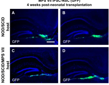
described (Hofling et al. 2003). GFP-labeled MPS VII iPSC-derived NSCs were transplanted in
the ventricles of neonatal NOD/SCID and NOD/SCID/MPS VII littermates to evaluate the patterns
and persistence of engraftment over the course of 4 months. Corrected or mock-corrected MPS
VII NSCs were also injected into the post-symptomatic striatum of 2 month old mice to evaluate
the extent of disease correction. We identified CD68-positive, activated microglia as an easily
quantifiable biomarker of CNS pathology in these animals. This strategy permitted us to visualize
engrafted cells and neuropathology in the same samples. We further validated this method using
an adeno-associated viral vector expressing β-glucuronidase in MPS VII mice.
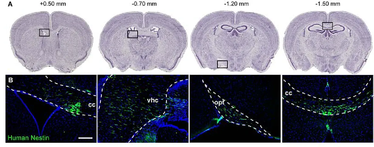
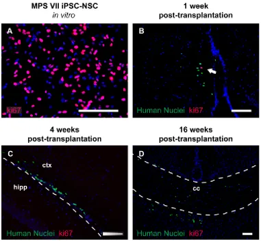
Following neonatal intraventricular transplantation, iPSC-NSCs engraft throughout the
rostrocaudal axis of the CNS, primarily within white matter tracts, and survive for at least four
months with little evidence of differentiation. Genetically corrected and mock-corrected cells,
while dispersed widely, were unable to correct microglial pathology. Transplanted cells exited the
cell-cycle rapidly and no differences were found between MPS VII and normal iPSC-NSCs
engrafted in normal and MPS VII mice. Genetically corrected MPS VII iPSC-NSCs transplanted
post-symptomatically into the striatum of adult NOD/SCID/MPS VII mice reversed neuropathology
in a zone surrounding the grafts, while control mock-corrected grafts did not. MPS VII and control
iPSC-NSCS did not migrate from the injection site, more consistent with data from primary NSCs
than immortalized NSC lines. The results suggest that improvements are needed to increase
survival, enzyme expression levels, and migration, especially in adult animals. However, the