Original Research Article.
626 |P a g e Int J Med Res Prof.2018 Jan; 4(1); 626-31. www.ijmrp.com
Nephrolithiasis Manage by Percutaneous Nephrolithotomy in Patients with
Renal Insufficiency
Shailesh P. Bajaniya
M.Ch. (Urology), Associate Professor, Department of Urology, B. J. Medical College and Civil Hospital, Ahmedabad, Gujarat, India.
ABSTRACT
Introduction: The prevalence of nephrolithiasis are depends on multiple factors. Commonly renal stone disease depends on climate, environment, genetic factor, geographical distribution, ethnic factor, diet. Renal stones may lead to varying degrees of renal insufficiency by due to urinary stasis and chronic infections. According to the European association of urology guideline 2015, percutaneous nephrolithotomy (PCNL) is the standard surgical treatment for large renal stones. We aimed to determine the impact of PCNL management on renal functions in renal stone patients with renal insufficiency.
Objective: To study and compare the parameters associated
with PCNL management for nephrolithisis in patients with renal insufficiency and evaluate the post-operative outcomes based on differences in preoperative renal function test values in patients who underwent percutaneous nephrolithotomy. Methods: From July 2014 to December 2016, in the present study, in a series of continuous 640 patients who were operated by PCNL, 65 patients with altered serum creatinine more than 1.5 were studied, observed and followed. All data were collected from medical records, which contained the clinical, laboratory evaluation and diagnostic imaging.
Results: Average age among the study group was 47.75 years being youngest 28 yrs and the eldest 70 yrs. M: F ratio was 44 (67.69%):21 (32.30%). In present study pre operatively 32, 24 patients out of total 65 patients were inserted PCN tube, D J stent and in remaining 9 patients were without PCN or D J stent insertion. Pre-operative hemodialysis was done in 39 out of 65 patients. Mean operative time 127 min (30-180 min). In
the study 7 (10.76%) patients out of 65 total patients required blood transfusion. In study group mean haematuria time was 15.98 hrs (6- 33 hrs). Average drop in hemoglobin percentage was 1.6. Mean hospital stay was 3.6 days.
Conclusions: The patients of renal stones with compromised renal insufficiency benefit from the elimination of calculi from the urinary tract, which may lead to improved renal function and avoidance or postponement of dialysis. On the other hand, there is concern for the detrimental effect of surgical and endourological procedures on kidney function and the possibility of increased complications in patients with kidney failure.
Keywords: Nephrolithisis, Renal Insufficiency, Renal Stone With Altered Renal Function Test.
*Correspondence to:
Dr. Shailesh P. Bajaniya, M.Ch. (Urology), Associate Professor,
Department of Urology,
B. J. Medical College and Civil Hospital, Ahmedabad, Gujarat, India.
Article History:
Received: 30-11-2017, Revised: 27-12-2017, Accepted: 23-01-2018 Access this article online
Website:
www.ijmrp.com
Quick Response code
DOI:
10.21276/ijmrp.2018.4.1.137
INTRODUCTION
The prevalence of nephrolithisis are depends on multiple factors. Commonly renal stone disease depends on climate, environment, genetic factor, geographical distribution, ethnic factor, diet. In general population prevalence of renal stone has been rising with average between 1.7-14.8%.1 Renal stones may lead to varying
degrees of renal insufficiency by due to urinary stasis and chronic infections.2 The rate of end-stage renal disease development is
between 0.2- 3.2%.3 Although it is not clearly demonstrated, it is
believed that there is a strong relation between renal stone disease and renal failure.4 According to the European association
of urology guideline 2015, percutaneous nephrolithotomy (PCNL) is the standard surgical treatment for large renal stones.5
Information on the clinical course of renal stone with renal insufficiency is inadequate.2 Renal parenchyma is damaged while
accessing the stone during PCNL. In addition, anesthetic agents may produce nephrotoxic effect. It may cause progression of renal failure. However, elimination of obstruction and eradication of chronic infection by removal of renal stones may lead to regression of RF.
management of renal stones with Percutaneous Nephrolithotomy (PCNL) alone or in combination with Extracorporeal Shock Wave Lithotripsy (ESWL) yet the issue of management of renal stones with renal failure has not been addressed adequately till date. Nowadays the role of open surgery in the management of patients with renal and ureteral calculi has diminished due to availability of excellent expertise in endourological procedures and advances in armamentarium. The indications for open surgery in the 1990s include complex stone burden, failure of ESWL or endourological treatment, anatomic abnormalities such as infundibular stenosis, renal calyceal diverticulum or concomitant ureteropelvic junction obstruction requiring surgery and morbid obesity.1 As compared to
renal stones with normal renal function, patients with associated altered renal function are logically more prone to bleeding diathesis and septicemia due to infected stones. Fluid overload is another potential problem restricting the intraoperative irrigation time during PCNL. This leads to more number of sittings for PCNL and increases the morbidity and the overall cost of treatment significantly.
We aimed to determine the impact of PCNL management on renal functions in renal stone patients with renal insufficiency.
AIMS AND OBJECTIVES Aim
To evaluate the outcome and impact of PCNL management in renal stone patients with renal insufficiency.
Objectives
1. To study and compare parameters associated with PCNL in patients with renal insufficiency
2. To evaluate post-operative outcomes based on differences in preoperative renal function in patients who underwent percutaneous nephrolithotomy (PCNL).
MATERIALS AND METHODS
In this prospective study, nephrolithisis patients with renal insufficiency were included using criteria mentioned below, were investigated using various urine, blood biochemistry and radiological tests. Renal stone were managed with PCNL procedure and Patients were followed up in post-operative period and evaluated for outcome and impact of PCNL in patients of renal stone with renal insufficiency from July 2014 to December 2016.
Inclusion Criteria
Patients of renal stone having S.creatinine level > 1.5 mg/dL. Exclusion Criteria
▪ Uncorrected urinary tract infection
▪ Bleeding disorders
▪ An acute rise of creatinine
▪ Aspirin or anticoagulant use
▪ Pregnancy
All patients were investigated on OPD basis in form of Complete haemograme, renal and liver functions tests, RBS, BT, CT, PT apTT, Urine R/M, C/S, X RAY KUB, USG KUB, IVP and Pre-operative medical fitness metabolic works up in patients with bilateral stones and recurrent stones in form of serum calcium, phosphate and uric acid and 24 hr urinary calcium, phosphate and uric acid and oxalate.
The calculus burden, anatomy of the renal collecting system, and the degree of hydronephrosis were evaluated using plain
radiography; renal ultrasonography; and non–contrast-enhanced spiral computed tomography.
Antimicrobial therapy was administered for patients with a positive urine culture for microorganisms.
METHODS
PCNL procedure was performed in all the eligible cases with strict aseptic precautions and prophylactic antibiotic cover.
Pre-operative advice included
▪ Nil by mouth 10:00 Pm night before surgery
▪ Detailed explained informed consent
▪ Part clean & shave
▪ Tab Ciprofloxacin(500mg) night before surgery
▪ Tab dulcolax (2 tabs) night before surgery
▪ Soap-water enema on coming morning after admission to hospital
▪ X-ray KUB on the morning of surgery
Under general anesthesia with the patient intubated, eye pads were put on. Patient was placed in a lithotomy position, the parts were cleaned and draped, cystoscopic examination was done and placement of ureteral access catheter (5F) under fluoroscopic
guidance was done. This was fastened to a Foley’s catheter (14 F
or 16 F) So as to prevent its subsequent accidental dislodgement. The patient is placed in prone position. Delineation of pelvicalyceal system was done by injecting contrast agent through ureteral access catheter.
Standard Puncture was made with needle and help of "C" Arm and a J tipped guide wire is negotiated into the pelvicalyceal system. Tract dilatation done under fluoroscopy over the guide wire with the help of coaxial alkane metallic dilators .At the end Amplatz sheath is positioned over the last dilator to the appropriate site as this will be the conduit for further instrumentation.
Stone Removal
Pneumatic lithotripsy was required to break the larger stones into manageable fragments while Small stones were removed intact with forceps.
In some cases where the stone is large or has extension into different calyces than additional tract required in some cases to achieve stone free Kidney.
By fluoroscopy and nephroscopy the pelvicalyceal system is checked for residual fragments and once all the stones are removed, a nephrostomy tube 24 F or 28 F passed through Amplatz sheath.
In post-operative period X ray KUB and chest X- ray PA view done in all cases. Nephrostomy tube removed after 24 hrs if no significant residual stone burden on post-operative X- ray KUB necessitating relook surgery or post op CXR showing no hemo /pneumothorax and a repeat X- ray chest 2-3 hrs after removal of nephrostomy tube to rule out hemo /pneumothorax (which may necessitates intercostals chest tube drainage) and patient is discharged on next morning after removal of ureteric catheter along with per urethral catheter and checking no soakage from track.
OBSERVATIONS
In the present study, in a series of continuous 640 patients who were operated by PCNL, 65 patients with altered serum creatinine more than 1.5 were studied, observed and followed.
A. Preoperative Parameters
I. Age Distribution: In PCNL with renal insufficiency group, average age among the group was 47.75 years. The youngest being 28 and the eldest being 70 years of age. II. Sex Distribution: In the study group, out of total 65 patients
44 are male and 21 patients are female.
III. Stone Parameters (Stone burden, size & Location): In this study, 21 patients had stone burden of less than 500 mm2,
18 had 501 to 1000 mm2, 8 patients had 1001 to 1500
mm2, 7 patients had 1501 to 2000 mm2 and 11 patients
had stone burden more than 2000 mm2.
IV. Location: In the study group, there was stone in renal pelvis in 24 patients, 19 patients were had stones in middle calyx, 24 patients having stone in the inferior calyx and 13 patients in the superior calyx. Total 25 (38.46%) out of 65 patients included in the study had staghorn stones.
V. Number of patients with pre-operative PCN / DJ stent - In present study pre operatively PCN inserted in 32 patient out of 65, in 24 patients D J stent were kept and in remaining 9 patients were without PCN Or D J stent insertion.
VI. Solitary kidney: In our study no patient of renal insufficiency with solitary unit underwent PCNL.
VII. Laterality: In this study 32(49.23%) out of 65 patients had bilateral renal stones.
VIII. Renal parenchymal thickness on USG: In this study preoperative renal parenchymal thickness of the disease kidney units were less than 4 mm in 11 patients, between 5-7 mm in 26 patients, between 8-10 mm in 19 patients, and more than > 10 mm in 9 patients.
IX. Serum creatinine: The serum creatinine value distribution among the patient enrolled was 1.5-2.0 mg% 12 patients, 2.1-3.0 mg% 26 patients, 3.1-4.0 mg% 16 patients and > 4.0 mg% 11 patients.
X. Pre-operative hemodialysis was done in 39 (60%) out of 65 patients.
B. Operative Parameters
I. Operating Time (Time from needle puncture up to putting off nephrostomy): In study the mean operative time 127 min (30-180 min).
Table 1: Mean operative time Mean operative time
Mean operative time (min) 127
Max. operative time (min) 180
Min. operative time (min) 30
II. Number of tracts used: In this study single tract was used in all cases except 13 patients in whom with double/more punctures and track were made.
III. Number of supracostal/subcostal punctures: Out of 65
patients’ 10 required supracostal puncture while in 55
patient’s subcostal puncture was done.
IV. Postoperative analgesic requirement: In the study the mean analgesic requirement (Inj Tramadol) during hospital stay was 215 mg.
V. Drainage: In the study in all the patients D J stent was kept.
VI. Blood transfusion: In the study 7 (10.76%) patients out of 65 total patients required blood transfusion.
VII. Bleeding from nephrostomy site after nephrostomy removal: In the study 2 cases had oozing from nephrostomy site after nephrostomy removal which subsided by pressure over puncture site.
VIII. Postoperative haematuria: In this study mean time 15.98 hrs (6- 33 hrs).
IX. UTI/urosepsis: In the study group, 2 patients had postoperative fever for which they were admitted and antibiotics given according to urine culture/sensitivity. X. Hospital stay: In this study mean hospital stay was 3.6
days.
XI. Postoperative urinary leak from nephrostomy site: In this study average duration of post-operative urinary leak from nephrostomy site was 18.76 hrs (4- 78 hrs).
XII. Drop in haemoglobin in post-operative period: In this study average drop in haemoglobin percentage was 1.6.
C. Follow Up
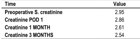
I. S. creatinine value in preoperative and post-operative measurements:
Table 2: S. creatinine value in preoperative and Post-operative measurements
Time Value
Preoperative S. creatinine 2.95
Creatinine POD 1 2.86
Creatinine 1 MONTH 2.61
Creatinine 3 MONTHS 2.54
II. Parenchymal thickness on USG: At 1 month follow up the parenchymal thickness on sonography were less than 4 mm in 11 patients, 5-7 mm in 17 patients, 8-10 mm in 27 patients, more than > 17 mm 10 patients.
III. Parenchymal thickness on USG: At 3 months follow up measurements of the renal parenchymal thickness in patients were less than 4 mm in 7 patients, 5-7 mm 12 patients, 8-10 mm 25 patients, > 10 mm 21 patients. IV. Hemodialysis: Post-operative hemodialysis was done in
11 (16.92%) patients and 7 (10.76%) patients became dialysis dependent as compare to 39 (60%) were need hemodialysis preoperatively.
DISCUSSION
The incidence of renal insufficiency in patients treated for urinary stone disease shows variations between centers and probably depends on the socioeconomic characteristics as well as the referral patterns of the region.
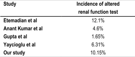
a. Incidence of renal insufficiency: In the 640 consecutive patients who underwent PCNL at our institution, 65 patients (10.15%) had impaired renal function defined as serum creatinine level greater than 1.5 mg/dl.
Table 3: Incidence of altered renal function test
Study Incidence of altered
renal function test
Etemadian et al 12.1%
Anant Kumar et al 4.6%
Gupta et al 1.65%
Yaycioglu et al 6.31%
Our study 10.15%
b. Age distribution: Mean age in our study was 47.75 years which is comparable to other studies with altered renal function.
Table 4: Age distribution
Study Average age
Etemadian et al 51.7 years
Majid yousefi et al 45.1 years
Our study 47.75 years
c. Sex distribution: In the study, out of total 65 patients 44(67.69%) are male and 21 (32.30%) patients are female which suggests male had more incidence of renal stone disease with altered renal function compared to female in this study.
Table 5: Sex distribution
Study Male Female
Etemadian et al 56.7% 43.3%
Majid yousefi et al 66 % 34%
Our study 67.69% 32.30%
d. Duration of surgery: Duration of surgery in our study is approximately 127 minutes
Table 6: Duration of surgery
Study Duration of surgery
Yaycioglu et al 175 minutes
Synder et al 155.1 minutes
Anant Kumar et al 152.5 minutes
Our study 127 minutes
e. Drop in hemoglobin: The drop in hemoglobin in our study is 1.6 gm/dl which is comparable to other studies.
Table 7: Drop in hemoglobin
Study Drop in haemoglobin
Yaycioglu et al 1.7
Anant Kumar et al 1.8
Our study 1.6
f. Nephrostomy time: The mean nephrostomy time was 9.6 hours which is significantly lower than than other studies.
Table 8: Nephrostomy time
Study Nephrostomy time
Yaycioglu et al 93.6 hours
Our study 9.6 hours
g. Hospitalisation: Mean hospitalization time in our study is 2.6 days compared to 6.6 days reported in yaycioglu et al.
Table 9: Hospitalisation
Study Nephrostomy time
Yaycioglu et al 6.6 days
Anant Kumar et al 12.9 days
Our study 3.6 days
h. Stone free rate: Stone free rate in our study is 78.94% which are comparable to other studies.
Table 10: Stone free rate
Study Stone free rate
Etemadian et al 83.7%
Anant Kumar et al 56.23%
Kuzgunbay et al 50%
Our study 78.94 %%
i. Mortality: Agrawal et al.3 reported 3.8% mortality and 17.3%
morbidity rates for PCNL in 78 patients with advanced renal failure. In our study none of the patient expired.
j. Renal function: In the era before PCNL, Witherow and Wickham had reported that mean creatinine clearances increased significantly after nephrolithotomy in patients with severely decreased renal function due to stone disease.7
After PCNL became a viable method of stone-treatment, laboratory and clinical studies showed that percutaneous procedures cause no significant damage to functional Nephron.8-10 As PCNL became the method of choice for
patients with bulky stones, several authors reported the beneficial effect of PCNL on kidney function in patients with renal failure.
Gupta et al.1 treated 33 patients with serum creatinine levels of 2
mg/dl or greater at presentation. The patients underwent multiple procedures including PCNL, ESWL, Ureteroscopy stone extraction, alkalinisation and open surgery. In 32 of 33 patients post treatment serum creatinine value was lower than the pre-treatment value (mean 2 vs. 3.2 mg/dl, P < 0.001). Of the 13 patients with longer than 1-year follow-up, 4 had progression of intrinsic renal disease and elevation in serum creatinine level to greater than the pre-treatment value. Three of these patients had subsequently end stage renal disease.
Agrawal et al.3 performed PCNL in 78 patients with calculus
follow-up showed a significant improvement over those before treatment. Sixty-four patients had improvement of renal function
and in 11 patients’s renal function remained unchanged or
deteriorated during follow-up.
Kukreja et al.2 analyzed data from 84 patients with baseline serum
creatinine of >1.5 mg/dl treated for calculus disease. The mean baseline serum creatinine concentration was 2.87 mg/dl. Twelve patients had bilateral stone disease while another 12 patients had a solitary functioning kidney. Primary surgical treatment was PCNL for most of the patients but some underwent nephrolithotomy and nephrectomy as well. Overall renal functions improved in 33 patients (39.3%), stabilized in 24 patients (28.6%), and deteriorated in 27patients (32.2%). The baseline serum creatinine concentration correlated well with the postoperative renal function. Renal function stabilized or improved in nearly all patients with baseline serum creatinine less than 2 mg/dl and deteriorated in all patients with baseline serum creatinine higher than 6 mg/dl. All patients with creatinine higher than 6 mg/dl, progressed to end-stage renal disease.
Goel et al.11 reported on the role of intervention in 20 patients with
a solitary kidney, nephrolithisis and chronic renal insufficiency. In this group, 15 patients underwent PCNL, 2 ESWL and 3 open surgeries. The mean glomerular filtration rate improved significantly in renal failure patients after treatment of stone disease. Improvement in glomerular filtration rate was greater in mild to moderate renal failure. GFR deteriorated in one patient with baseline serum creatinine higher than 4 mg/dl. Patients with residual disease had longer mean hospital stay, more repeat anesthesia, blood transfusions and total cost.
Chandhoke et al.12 found no significant deterioration in renal
function after PCNL in patients with moderate renal insufficiency. Jones et al.13 reported on the results of PCNL on 14 patients with
solitary kidney and abnormal renal function at presentation. After PCNL, serum creatinine increased in 2 and decreased in 12 patients.
In our patients there was a slight but insignificant decrease in serum creatinine values at the end of the follow- up period. Serum creatinine values decreased to normal range in 15 patients but in most of these patients pre-operative baseline Serum creatinine was less than 2 mg/dl. Creatinine values did not decrease to the normal range in any of the patients with a baseline creatinine level higher than 2 mg/dl. In our study out of 65 patients 37 patients after PCNL had improved renal function, in 16 patients there were stable renal function and in 12 patients renal function was worsened. Our results and the results of previous reports indicate that most patients presenting with kidney-stone disease and renal insufficiency, experience improvement or stabilization of renal function with early aggressive intervention aimed at complete stone clearance and prevention of urinary infection. Improvement in renal function is greater in mild to moderate renal failure. Patients with severe kidney-failure, reduced parenchymal thickness and pus in the collecting system are less likely to show a significant improvement in renal function. However, even these patients will enjoy the benefits of improved quality of life and postponement of replacement therapy if their stones and infection can be cleared [1–3, 11]. Therefore, the statement “one should
temper obsessive attempts at clearance of small fragments with
caution in the kidney with severely damaged function” is equally
true for PCNL as it is for open-surgery.7,13
Table 11: Comparison with other studies (Creatinine values)
Study Pre op
mean creatinine
Follow up mean creatinine
Kuzgunbay et al 2.30 2.67
Gupta et al 2.22 1.89
Iqbal singh et al 4.79 1.53
Paryani et al 6.27 4.08
Yaycioglu et al 2.8 2.6
Majid Yousefi et al 5.52 2.03
Our study 2.95 2.54
Table 12: Comparison with other studies (Outcome)
Study No of
pts
improved Stable Worsened
Gupta et al 33 29 1 3
Agrawal et al 78 64 11
Kukreja et al 84 33 24 27
Jones et al 14 12 2
Goel et al 20 1
Chandhoke et al
No significant deterioration in renal function
Our study 57 37 16 12
CONCLUSION
Renal failure is frequently a progressive condition. The presence of stones in the urinary tract may accelerate the course of the disease. Presence of the stones deteriorates renal functions mainly by causing obstruction and infection.6 The changes in the
kidney parenchyma caused by infection are more pronounced if there is concomitant obstruction. Duration of the stone disease, multiple procedures, and stone recurrence also have negative influence on renal function.4 Therefore, patients with compromised
renal function benefit from the elimination of calculi from the urinary tract, which may lead to improved renal function and avoidance or postponement of dialysis. On the other hand, there is concern for the detrimental effect of surgical and endourological procedures on kidney function and the possibility of increased complications in patients with kidney failure.
LIMITATIONS
Evaluation of renal function by serum creatinine level has some disadvantages, especially in patients with two functioning kidneys. The deterioration in one kidney is compensated by the contra lateral kidney. Thus, the change in serum creatinine does not accurately reflect the change in the function of the concerned kidney. Unfortunately, differential renal function and creatinine clearance measurements were not available for most of the patients in this study. However, we feel that in the with proper follow-up, serum creatinine provides valid information on the overall renal function that reflects the effect of PCNL.
REFERENCES
2. Kukreja R, Desai M, Patel SH, Desai MR (2003) Nephrolithiasis associated with renal insuYciency: factors predicting outcome. J Endourol 17:875–879.
3. Agrawal MS, Aron M, Asopa HS (1999) Endourological renal salvage in patients with calculus nephropathy and advanced uremia. BJU Int 84:252–256.
4. Marangella M, Bruno M, Cosseddu D, Manganaro M, Tricerri A, Vitale C, Linari F (1990) Prevalence of chronic renal insuYciency in the course of idiopathic recurrent calcium stone disease: risk factors and patterns of progression. Nephron 54:302–306. 5. Gupta NP, Kochar GS, Wadhwa SN, Singh SM (1985) Management of patients with renal and ureteric calculi presenting with chronic renal insuYciency. Br J Urol 57:130–132.
6. Gambaro G, Favaro S, D’Angelo A (2001) Risk of renal failure
in nephrolithiasis. Am J Kidney Dis 37:233– 243.
7. Witherow RO, Wickham JE (1980) Nephrolithotomy in chronic renal failure saved from dialysis! Br J Urol 52:419– 421.
8. Mayo ME, Krieger JN, Rudd TG (1985) EVect of percutaneous nephrostolithotomy on renal function. J Urol 133:167– 169. 9. Trinchieri A, Mandressi A et al. Renal tubular damage after renal stone treatment. Urol Res 1988; 16:101–104.
10. Wilson WT, Husmann DA, Morris JS, Miller GL, Alexander M, Preminger GM (1993) A comparison of the bioeVects of four diVerent modes of stone therapy on renal function and morphology. J Urol 150:1267– 1270.
11. Goel MC, Ahlawat R, Kumar M, Kapoor R (1997) Chronic renal failure and nephrolithiasis in a solitary kidney: role of intervention. J Urol 157:1574–1577.
12. Chandhoke PS, Albala DM, Clayman RV (1992) Long-term comparison of renal function in patients with solitary kidneys and/or moderate renal insuYciency undergoing extracorporeal shock wave lithotripsy or percutaneous nephrolithotomy. J Urol 147:1226–1230.
13. Jones DJ, Kellett MJ, Wickham JEA (1991) Percutaneous nephrolithotomy and the solitary kidney. J Urol 145:477– 480. 14. Majid Yousefi Moghaddam, Muhammad Naeem Outcome of Surgical Clearance of Renal and Ureteric Calculi on Improvement in Renal Parameters in Patients with Renal Insufficiency. Ann. Pak. Inst. Med. Sci. 2015; 11(1): 47-50.
15. Kumar A, Verma BS, Gogoi S, Kapoor R, Srivastava A, Mandhani A. A prospective randomized trial of open surgery versus endourological stone removal in patients of staghorn stones with chronic renal failure. Indian J Urol [serial online] 2001 [cited 2017 Apr 5];18:14-9. Available from: http://www.indianjurol.com/text.asp?2001/18/1/14/37373
16. Snyder JA, Smith AD. Staghorn calculi: percutaneous extraction versus anatrophic nephrolithotomy. J Urol 1986; 136: 351-354.
[
Source of Support: Nil.
Conflict of Interest: None Declared.
Copyright: © the author(s) and publisher. IJMRP is an official publication of Ibn Sina Academy of Medieval Medicine & Sciences, registered in 2001 under Indian Trusts Act, 1882. This is an open access article distributed under the terms of the Creative Commons Attribution Non-commercial License, which permits unrestricted non-commercial use, distribution, and reproduction in any medium, provided the original work is properly cited.